Dermatology · Medicine
Laser therapies
Also known as Laser therapy · Light therapy · Selective photothermolysis · Laser resurfacing · Laser hair removal
Laser therapies in dermatology rest on selective photothermolysis (Anderson and Parrish, 1983): a monochromatic, coherent beam at a wavelength preferentially absorbed by a target chromophore — melanin, oxyhaemoglobin, water, or exogenous tattoo pigment — deposits heat in that target while a pulse duration shorter than the target's thermal relaxation time confines injury to it and spares surrounding tissue. The principal chromophore–laser pairings are: oxyhaemoglobin for pulsed-dye (PDL 585-595 nm), KTP (532 nm) and long-pulsed Nd:YAG (1064 nm) — vascular lesions; melanin for Q-switched ruby (694 nm), alexandrite (755 nm), diode (800-810 nm) and Nd:YAG — pigmented lesions and hair; water for CO2 (10600 nm) and Er:YAG (2940 nm) — resurfacing; and tattoo ink for Q-switched and picosecond lasers. Fractional photothermolysis creates microscopic treatment zones for safer resurfacing; the 308-nm excimer targets vitiligo and localised psoriasis; IPL is a broadband non-laser alternative. Safety demands wavelength-specific eye protection for everyone in the room, epidermal cooling, Fitzpatrick-tailored fluence, test patches in skin of colour, and deferral of resurfacing for at least 6 months after isotretinoin (scarring risk).
On this page & tools
Your progress
Saved locally on this device.
Exam tags
Red flags
Overview & Definition
The word LASER is an acronym: Light Amplification by Stimulated Emission of Radiation. Unlike ordinary white light, a laser beam is monochromatic (a single wavelength), coherent (waves in phase), and collimated (parallel, low divergence). These three properties allow a large amount of energy to be delivered to a microscopic target in skin with minimal scatter — the physical basis of every laser procedure in dermatology.[1]
What transformed lasers from blunt thermal instruments into precise surgical tools was the principle of selective photothermolysis, articulated by Anderson and Parrish in Science in 1983.[1] They showed that if three conditions are met — a wavelength preferentially absorbed by the target, sufficient fluence to damage it, and a pulse duration no longer than the time heat takes to diffuse out of it — the target is destroyed while the surrounding tissue is preserved. That single insight made it possible to ablate a 100-micrometre blood vessel, shatter a tattoo particle, or coagulate a hair follicle without burning the overlying epidermis, and it underpins the entire modern speciality of laser medicine.
Epidemiology & Risk Factors
Laser and energy-based procedures are among the most commonly performed cosmetic interventions worldwide, and their volume has grown relentlessly for two decades. Laser hair removal is consistently the single most-performed non-surgical aesthetic procedure globally, with demand extending across all Fitzpatrick types as device technology — particularly the long-pulsed Nd:YAG — has made treatment of dark skin safer.[2]
Several epidemiological currents drive the broader field. Tattoo prevalence has risen sharply in young adults; community surveys report that up to a third of adults under forty in Western populations now have at least one tattoo, and regret — and therefore the demand for removal — is reported by as many as half of those tattooed.[5] Port-wine stain, the capillary malformation that is the paradigmatic PDL indication, is present at birth in roughly 0.3 percent of newborns; when it lies in the V1 (ophthalmic) trigeminal dermatome, roughly 8-15 percent of affected infants have or will develop Sturge-Weber syndrome (leptomeningeal angiomatosis with seizures, intellectual disability and glaucoma), which makes dermatological diagnosis a neuro-ophthalmological sentinel.[9]
Risk factors for an adverse outcome cluster around three axes. The first is skin type: Fitzpatrick IV-VI skin carries far higher rates of post-inflammatory hyper- and hypopigmentation because epidermal melanin competes with the target chromophore.[10] The second is anatomical site: the chest, shoulders, jawline, neck and lower legs are prone to hypertrophic scarring and delayed healing, particularly after resurfacing. The third is patient factors: a recent tan, isotretinoin in the preceding six months, a history of herpes simplex or keloids, pregnancy, and the use of photosensitising drugs each raise the complication rate. Recognising these risk factors at consultation — and deferring or modifying treatment accordingly — prevents the majority of serious adverse events.
The Four Chromophores and Their Targets
Every clinical laser decision reduces to the question: which chromophore am I trying to hit, and what wavelength and pulse duration does it demand? Skin contains four clinically relevant chromophores, each with a characteristic absorption spectrum and thermal relaxation time.[1]
Oxyhaemoglobin
Melanin
Water
Exogenous pigment (tattoo ink)

Laser Physics: Why Wavelength, Fluence and Pulse Duration Matter
Stimulated emission is the quantum event at the heart of the laser. An atom whose electron sits in an excited state is struck by a photon whose energy exactly matches the gap to a lower state; the encounter triggers release of a second photon identical to the first in wavelength, phase and direction. Cascaded through an optical cavity bounded by mirrors, this amplification produces the intense, coherent beam. The lasing medium — a gas (CO2), crystal (Nd:YAG, ruby, alexandrite, Er:YAG), semiconductor (diode), or liquid dye — determines the emitted wavelength, and each medium gives the laser its name.[1]
Once the beam enters skin, three interactions determine the clinical effect: [1]
Absorption by the target chromophore is what we want. The absorption coefficient of the chromophore at the chosen wavelength dictates how much energy is deposited per unit depth. Scattering by collagen deflects photons and is wavelength-dependent — longer wavelengths penetrate deeper with less scatter, which is why Nd:YAG 1064 nm reaches deep reticular dermis and leg veins while KTP 532 nm treats only superficial vessels. Reflection at the skin surface (4-7%) is reduced by skin coupling gels and by matching the handpiece.[1]
The pulse duration is the single most controllable variable for safety. Heat generated in a target diffuses outward at a finite rate; the thermal relaxation time (TRT) is the time for the target to cool to half its peak temperature. If the laser pulse is shorter than the TRT, heat stays confined and only the target is injured; if longer, heat spreads into surrounding tissue and causes collateral damage. Small targets have short TRTs:[1]
This is why a Q-switched nanosecond ruby pulse shatters a melanosome without burning the epidermis, while a long-pulsed millisecond alexandrite heats the whole hair follicle to coagulation — the same wavelength, different pulse duration, different target. [1]
Depth of penetration rises with wavelength within the optical window: KTP 532 nm is superficial; PDL 585-595 nm reaches the papillary and superficial reticular dermis (ideal for port-wine stain capillaries); alexandrite 755 nm, diode 800 nm and Nd:YAG 1064 nm penetrate progressively deeper. In the mid-infrared, water absorption dominates: Er:YAG 2940 nm is absorbed so strongly (roughly sixteen times the water absorption of CO2) that each pulse removes only a few micrometres of tissue with negligible residual thermal damage, whereas CO2 10600 nm ablates and leaves a zone of coagulation that contracts dermal collagen — the basis of its tightening effect.[4]
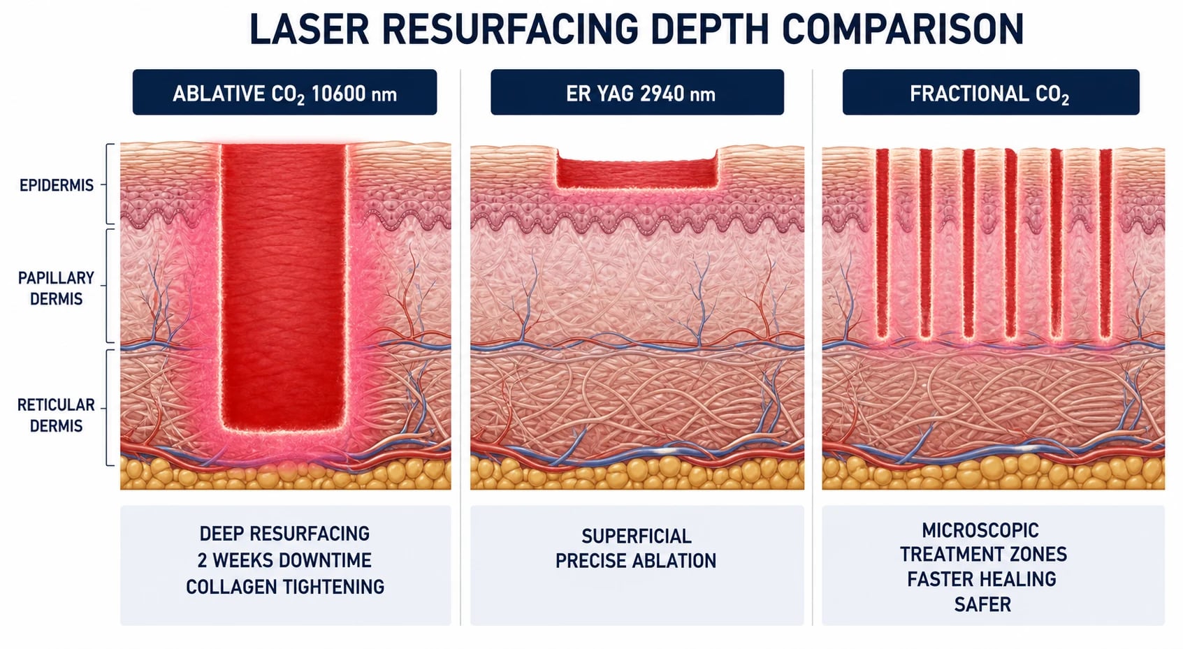
The clinical implication of penetration depth is captured in the table below: longer visible and near-infrared wavelengths reach deeper dermal targets, which is why device choice is dictated as much by lesion depth as by chromophore.
Epidermal cooling is the second great enabler of safe laser practice. Because the epidermis is rich in melanin and water, it absorbs a fraction of every vascular, hair and pigment pulse; without protection this produces a burn and, weeks later, pigmentary change. Cooling lowers the basal epidermal temperature so it tolerates the brief thermal pulse while the deeper target is still heated above its injury threshold. Three methods are in routine use: contact cooling (a chilled sapphire or copper window pressed to the skin, applied before, during and after the pulse); dynamic cryogen spray (a timed burst of tetrafluoroethane delivered 10-100 ms before the laser pulse, evaporating on the surface); and forced refrigerated air (a continuous stream of cold air at minus 30 degrees). Cooling is mandatory for hair-removal and vascular lasers in all but the fairest skin, and is adjusted with fluence as skin type darkens. [4]
Classification of Lasers by Chromophore and Use
A working classification groups devices by the chromophore they target and, in turn, by the indications they treat. The categories below cover virtually every laser an examiner will name.[1]
Vascular lasers (oxyhaemoglobin)
- Pulsed-dye laser (PDL), 585-595 nm — the gold standard for vascular lesions. The flashlamp-pumped dye emits a yellow light matched to an oxyhaemoglobin absorption peak. The original 585 nm, 0.45 ms pulse produced brisk purpura; modern 595 nm systems with longer pulse durations (1.5-40 ms, stacked sub-purpuric pulses) deliver erythema control with less downtime. Used for port-wine stain, infantile haemangioma, facial telangiectasia, rosacea erythema, hypertrophic scar redness, and stretch marks.[9][14]
- KTP (frequency-doubled Nd:YAG), 532 nm — green light; excellent for fine facial telangiectasia, spider naevi, cherry angiomas. Less purpura than PDL, but more melanin absorption so use with caution in darker skin.[9]
- Long-pulsed Nd:YAG, 1064 nm — deeper penetration for reticular leg veins, larger facial and nasal vessels, and venous lakes; lower melanin absorption makes it the vascular laser of choice in Fitzpatrick V-VI.[9]
- Intense pulsed light (IPL) — broadband 500-1200 nm with cut-off filters; not a laser, but used for diffuse erythema, photorejuvenation, and pigment.[2]
Pigment and hair-removal lasers (melanin)
- Q-switched ruby, 694 nm — high melanin absorption; tattoo removal (black, blue, green ink) and pigmented lesions (lentigo, café-au-lait, naevus of Ota). High PIH risk in dark skin.[5]
- Q-switched alexandrite, 755 nm — tattoo removal (black, blue, green) and pigmented lesions; slightly safer than ruby in darker skin.[5]
- Q-switched Nd:YAG, 1064 nm (532 nm doubled) — the 1064 nm treats black/blue tattoo ink, dermal pigmentation (naevus of Ota, Hori naevus) and is the safest Q-switched wavelength in dark skin; the 532 nm frequency-doubled output treats red, orange and purple ink and epidermal lentigines.[5]
- Long-pulsed alexandrite 755 nm, diode 800-810 nm, long-pulsed Nd:YAG 1064 nm — hair removal. The alexandrite is effective in fair skin (I-III), the diode suits olive skin (III-IV), and the long-pulsed Nd:YAG is the safest in dark skin (V-VI) because its longer wavelength minimises competing epidermal melanin absorption.[2]
- Intense pulsed light — broadband hair removal and pigment.[2]
Resurfacing lasers (water)
- CO2, 10600 nm — continuous or pulsed ablative resurfacing; removes epidermis and induces a coagulation zone in the papillary-reticular dermis, producing collagen contraction and remodelling. Best single-session result for wrinkles, acne scars, photoaging, warts and surgical scars, at the cost of two weeks of downtime and significant complication risk.[4]
- Er:YAG, 2940 nm — very high water absorption gives precise shallow ablation with minimal residual thermal damage; good for fine wrinkles and epidermal lesions but less collagen tightening than CO2.
- Fractional lasers — ablative (fractional CO2, fractional Er:YAG) and non-ablative (1550, 1927 nm). Rather than removing the whole surface, they create a grid of microscopic treatment zones (MTZs) — columns of thermal injury 100-400 micrometres wide surrounded by viable tissue, so re-epithelialisation is rapid (days not weeks) and scarring risk is far lower than fully ablative resurfacing.[3]
Picosecond lasers (tattoo and acne scars)
Picosecond lasers deliver pulses of one-trillionth of a second (a thousand times shorter than Q-switched nanosecond pulses). At these durations the dominant effect is photoacoustic — a pressure wave that fractures ink particles into fragments fine enough for macrophage clearance, with less photothermal damage to surrounding dermis. They are superior to Q-switched for multicolour and resistant tattoos, brightly coloured inks, and — with diffractive lens arrays — for acne scarring.[11]
Excimer 308 nm
The xenon-chloride excimer laser emits monochromatic 308 nm ultraviolet B, the same most-effective wavelength as narrowband UVB phototherapy but delivered to a discrete lesion. It repigments stable vitiligo and clears localised plaque psoriasis with a fraction of the cumulative UV dose of whole-body phototherapy, sparing uninvolved skin.[6][7]
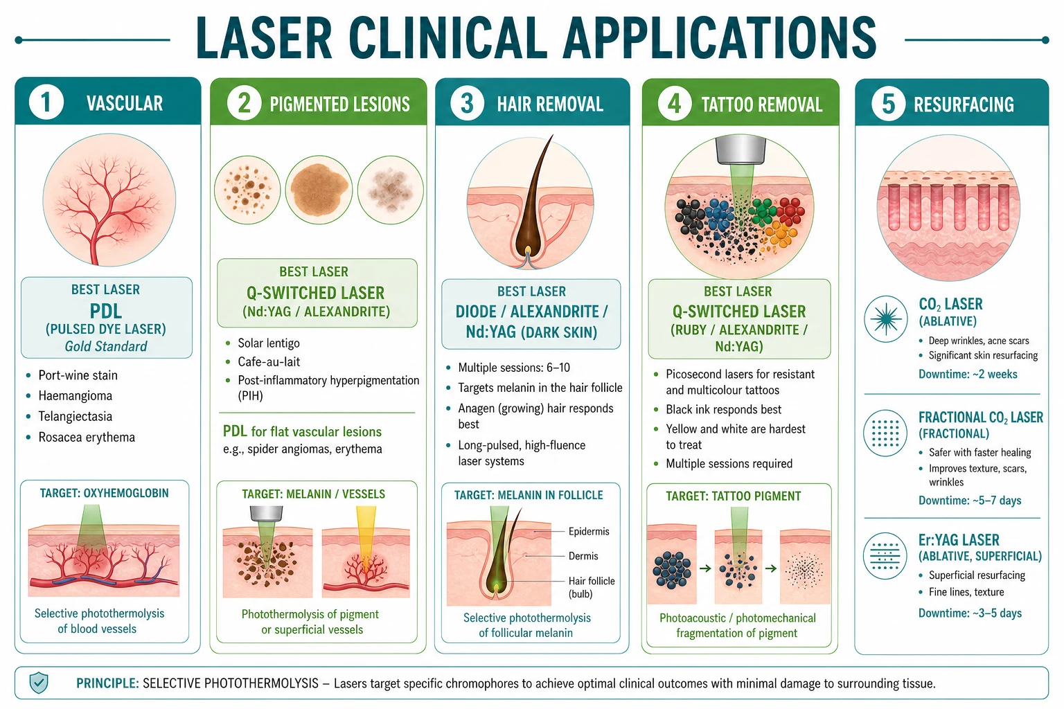
Clinical Applications by Indication
Vascular lesions
Port-wine stain (capillary malformation) is the classic indication for PDL. These congenital, pink-to-dark-red patches in the superficial vascular plexus grow darker and thicker with age; early treatment in infancy takes advantage of thinner skin and smaller vessels. Six to twenty or more sessions are typical, yielding roughly 50-70 percent lightening with complete clearance in only 10-20 percent of patients.[9] PDL is also first-line for superficial infantile haemangioma, especially ulcerated or superficial lesions, where early treatment limits proliferation; deep or segmental haemangiomas still require oral propranolol.[14] Diffuse facial telangiectasia and the background erythema of rosacea respond to sub-purpuric PDL or KTP. Hypertrophic and burn scars treated early with PDL show reduced erythema and improved pliability.[13]
Pigmented lesions
Epidermal pigmentation — solar lentigines, café-au-lait macules, naevus spilus, and epidermal melasma — responds to Q-switched (532 nm for superficial lentigines; 1064 nm for dermal naevus of Ota) or picosecond lasers. Melasma is treated cautiously: dermal melasma in particular may paradoxically darken, so low fluence, test patches, and concurrent hydroquinone are essential.[10] Post-inflammatory hyperpigmentation in skin of colour demands conservative settings, aggressive cooling, and pre-treatment with a tyrosinase inhibitor; multiple sessions with low fluence and long intervals outperform aggressive single treatments.[10]
Tattoo removal
Q-switched ruby (694 nm), alexandrite (755 nm) and Nd:YAG (1064 nm plus the 532 nm doubled beam for red-orange ink) remain the workhorses; picosecond lasers improve clearance of multicolour, brightly coloured and resistant tattoos and reduce the total number of sessions.[11][5] Black and dark-blue ink clears fastest; green, yellow and white respond poorly; flesh-toned cosmetic ink (titanium dioxide) may undergo paradoxical darkening to grey-black when reduced by the laser and must always be test-treated. Amateur tattoos generally clear in fewer sessions than professional tattoos. Sessions are spaced six to eight weeks apart to allow macrophage clearance of fragmented pigment; typically five to fifteen sessions are needed.[5]
Hair removal
Long-pulsed lasers target melanin in the anagen hair bulb and bulge. Best results are in patients with dark, coarse hair on light skin, where the chromophore contrast is maximal; blonde, grey, red and white hair respond poorly because the follicle lacks pigment. Six to ten sessions at four-to-eight-week intervals achieve durable 70-90 percent hair reduction rather than permanent eradication; maintenance treatments are expected.[2] Device selection is driven by Fitzpatrick skin type.
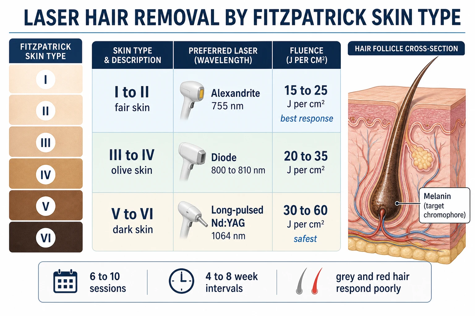
Resurfacing and rejuvenation
Ablative CO2 resurfacing remains the benchmark for deep rhytides, atrophic acne scars, surgical and burn scars, actinic cheilitis and diffuse photoaging, delivering a single-session result with five-to-ten-year durability.[4] Fractional CO2 and Er:YAG deliver comparable improvement over three to five sessions with a fraction of the downtime, and have become the default for most scar and rejuvenation work because of their markedly better safety profile.[3] Burn scars benefit from fractional CO2 or PDL, which improve texture, pliability and erythema even in mature scars.[13]
Targeted phototherapy: vitiligo and psoriasis
The 308-nm excimer laser is used to repigment stable, localised vitiligo (face and neck respond best, extremities and bony prominences poorly) and to clear localised plaque, scalp and palmoplantar psoriasis with fewer treatments and a lower cumulative UV dose than whole-body narrowband UVB. Protocols use twice- or thrice-weekly sessions with incremental fluence; combination with topical calcineurin inhibitors or vitamin D analogues enhances efficacy.[6][7]
Clinical Presentation
Laser therapy is indication-driven rather than disease-driven, so the "presentation" is the lesion or concern the patient brings to clinic. The common presenting scenarios, and the questions each raises, are:[1]
- A flat pink, red or purple vascular patch at birth — port-wine stain (capillary malformation). Distinguish from a salmon patch (stork bite, fades) and an early infantile haemangioma (proliferates after birth). Ask about V1 distribution and Sturge-Weber.
- A red, proliferating plaque in an infant — infantile haemangioma; ask about ulceration, airway (beard distribution), periocular location (amblyopia risk) and segmental PHACE syndrome.
- Diffuse facial redness with papules and papulopustules — rosacea erythema and telangiectasia.
- Discrete facial or leg veins — telangiectasia, reticular veins, spider naevi, venous lakes.
- Brown macules and patches — solar lentigines, café-au-lait, melasma (symmetrical, photo-distributed), naevus of Ota (unilateral dermal grey-blue), post-inflammatory hyperpigmentation.
- A decorative or traumatic tattoo the patient wishes removed — note ink colours, density, age, amateur vs professional.
- Unwanted hair — note skin type, hair colour and calibre, hormonal pattern (polycystic ovary syndrome if hirsutism).
- Photoaging — fine and coarse rhytides, dyschromia, solar elastosis, actinic damage.
- Atrophic, hypertrophic or erythematous scars — acne, surgical, traumatic or burn scars.[13]
Atypical presentations examiners test deliberately: a pigmented lesion that is new, changing, or atypical within a tattoo or naevus (must exclude melanoma); a "tattoo allergy" with scaly, nodular or ulcerated change (sarcoidal or lichenoid reaction — biopsy before laser); rapidly enlarging or painful vascular lesion (consider pyogenic granuloma, not a simple haemangioma); melasma that has been repeatedly lasered and is worsening (post-inflammatory darkening — stop and reassess); and a port-wine stain that thickens, darkens or develops pyogenic granulomas in adulthood (treated with PDL or Nd:YAG).[1]
Investigations
Most laser indications are diagnosed clinically and require no investigation, but several targeted work-ups are essential. Wood's lamp examination distinguishes epidermal from dermal pigmentation in melasma (epidermal pigment brightens under Wood's light; dermal does not), guides prognosis and treatment intensity, and characterises the depth of vitiligo. Dermoscopy is performed on every pigmented lesion before any laser — atypical network, blue-white veil, regression, streaks or pseudopods mandate biopsy rather than laser. Biopsy is required for any lesion with clinical or dermoscopic features of melanoma, for a nodular or ulcerated change within a tattoo, and for a vascular lesion of uncertain nature.[1]
For a port-wine stain in the V1 distribution, the work-up for Sturge-Weber syndrome includes an ophthalmology review (tonometry for glaucoma, which can develop at any age) and, if neurological signs or a large forehead PWS are present, MRI with contrast to detect leptomeningeal angiomatosis. A test patch in an inconspicuous area, read at 24-48 hours, is the functional investigation that determines fluence tolerance before full treatment, particularly in Fitzpatrick IV-VI. Standardised baseline photography documents the starting point for objective response assessment at each subsequent visit. Where hirsutism is marked, consider screening for polycystic ovary syndrome (free testosterone, LH:FSH ratio, DHEAS) before committing to hair removal, as treating the hair without the underlying driver is suboptimal.[9]
Differential Diagnosis and Pre-Treatment Assessment
Because lasers treat manifestations rather than diseases, the diagnostic question is always: is this lesion what I think it is, and is it safe to laser? The single most important rule — and a frequent examination stem — is that no undiagnosed or atypical pigmented lesion should be lasered. A melanoma misdiagnosed as a lentigo and treated with a Q-switched laser will be masked, biopsied late, and prove fatal. Any lesion with asymmetry, border irregularity, colour variegation, diameter over 6 mm or evolution (the ABCDE criteria), or with atypical dermoscopy, must be biopsied first.[1]
Before any vascular treatment, distinguish a port-wine stain (congenital capillary malformation, present at birth, grows darker and thicker, never involutes) from an infantile haemangioma (appears in the first weeks of life, proliferates, then involutes), because management diverges — propranolol dominates the latter. A port-wine stain in the V1 (ophthalmic) dermatomal distribution mandates evaluation for Sturge-Weber syndrome (leptomeningeal angiomatosis, seizures, glaucoma): ophthalmology review for glaucoma and MRI if neurological signs are present. A pigmented lesion that has appeared within a tattoo, or an allergic, scaly or nodular reaction within a tattoo, should be biopsied (sarcoidal, lichenoid or pseudolymphomatous reactions occur) before laser removal is attempted. [14]
A focused pre-treatment assessment documents: Fitzpatrick skin type (the master determinant of fluence and pigmentary risk); the colour, depth, calibre and density of the target (vessel, hair, ink, scar); sun-exposure and tan status; a full medication history concentrating on isotretinoin in the preceding six months, photosensitisers (tetracyclines, retinoids, NSAIDs, St John's wort), anticoagulants; a history of herpes simplex, keloids or hypertrophic scarring, and prior aesthetic procedures; and pregnancy status. A test patch is performed in skin of colour (Fitzpatrick IV-VI) or in any high-risk lesion 24-48 hours before full treatment, and the response — erythema, purpura, blanching, blistering, pigment change — is read before committing to a full session. [1]
Management — Resuscitation and Acute Emergencies
Laser treatment is an elective, ambulatory procedure, and the indications themselves are not life-threatening — but two genuine emergencies can arise in or around a laser session, and the operator must be prepared for both. [1]
Acute thermal injury (over-treatment). Excessive fluence, inadequate cooling, or treatment of tanned or dark skin can produce a partial- or full-thickness burn within minutes, with erythema, blistering and pain. Immediate management is that of any thermal burn: stop further treatment, cool the area with running cool water or compresses for 15-20 minutes, apply a topical corticosteroid to blunt inflammation, cover with a non-adherent dressing, and give oral analgesia. Assess burn depth; deep partial- or full-thickness injuries, especially on the face, warrant referral to a plastic or burns service. Counsel the patient about the near-certain post-inflammatory pigment change and arrange early follow-up at 48-72 hours.[10]
Ocular exposure. A reflected or direct laser beam reaching the eye can cause instant, permanent retinal, corneal or iris injury — most feared with invisible near-infrared beams (Nd:YAG, diode, alexandrite) where there is no blink reflex. Any suspected intraocular exposure is an ophthalmological emergency: stop the procedure, keep the patient still, cover both eyes, and arrange immediate ophthalmology assessment. Prevention is paramount and entirely reliable when wavelength-specific goggles and intraocular metal shields are used correctly.[1]
Acute laser burn — immediate steps
Stop the laser; assess airway/breathing/circulation if the injury is extensive or periorbital
Cool the area with running cool water or compresses for 15-20 minutes
Apply a potent topical corticosteroid and a non-adherent dressing; give oral analgesia
Document depth (superficial / deep partial / full thickness) and photograph
Review at 48-72 hours; refer to plastics/burns for deep or full-thickness injury
Plan for post-inflammatory pigmentation: hydroquinone and strict photoprotection once healed
Technique and Treatment Parameters
Every laser session is governed by four interdependent settings: spot size (handpiece diameter, typically 2-10 mm — larger spots penetrate deeper by reducing peripheral scatter), fluence (energy density in J/cm2), pulse duration, and the number of passes. Epidermal cooling — contact sapphire window, cryogen spray, or forced cold air — protects the epidermis by lowering its temperature while the deeper chromophore is heated; it is essential for hair-removal and vascular work in all but the fairest skin. A small degree of overlap between spots ensures uniform coverage without ridging.[2]
The clinical endpoints differ by target: for vascular work, a transient grey-blue or mild purpura indicates adequate vessel injury; for hair removal, perifollicular erythema and oedema around treated hairs confirm follicle uptake; for tattoo removal, immediate whitening (steam cavitation) followed by pinpoint bleeding at higher fluences; for resurfacing, a uniform pink frost and contraction of tissue.[4]
Representative starting parameters by indication: [2]
Fluence is titrated to the lowest effective setting: start conservative, increase at the next session if tolerated without adverse effect, and never exceed the manufacturer's specification for a given spot size and pulse duration.[2]
Safety: Protecting Patient, Operator and Bystanders
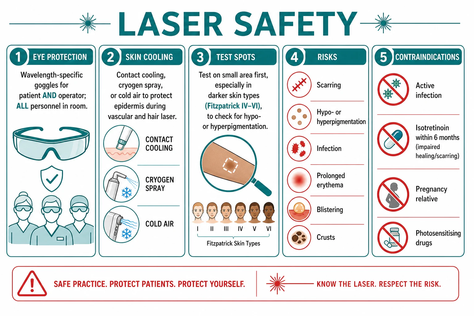
Laser safety rests on protecting the eye, protecting the skin, and excluding patients in whom the procedure is contraindicated. The beam is intense enough to cause permanent retinal, corneal or iris injury in a fraction of a second, and reflected (specular) beams carry the same hazard as the primary beam. Wavelength-specific protective goggles — matched to the laser's wavelength and optical density — must be worn by the patient, the operator and every person in the treatment room; generic sunglasses are not acceptable. For periorbital work, internal metal eye shields are placed after topical anaesthesia. Windows are covered, warning signs are displayed, and the door is locked during firing. The laser plume (vaporised tissue smoke) contains viable viral particles and combustible material; a dedicated smoke evacuator held at the handpiece is mandatory, and masks with high filtration are worn. [1]
Epidermal cooling — contact cooling (sapphire tip chilled to a few degrees), dynamic cryogen spray (tetrafluoroethane delivered milliseconds before the pulse), or forced refrigerated air — protects the basal melanocytes and lowers the pigmentary and burn risk, and is essential whenever melanin competes with the deeper target. A test patch in an inconspicuous area is read at 24-48 hours for blistering or pigment change before a full first treatment, particularly in Fitzpatrick IV-VI and for any new device setting. [10]
LASER SAFETY
Contraindications
Exam application bank (NEET-PG / INICET)
One-line answer
Laser therapies in dermatology rest on selective photothermolysis (Anderson and Parrish, 1983): a monochromatic, coherent beam at a wavelength preferentially absorbed by a target chromophore — melanin, oxyhaemoglobin, water, or exogenous tattoo pigment — deposits heat in that target while a pulse duration shorter than the target's thermal relaxation time confines injury to it and spares surrounding tissue. The principal chromophore–laser pairings are: oxyhaemoglobin for pulsed-dye (PDL 585-595 nm), KTP (532 nm) and long-pulsed Nd:YAG (1064 nm) — vascular lesions; melanin for Q-switched ruby (694 nm), alexandrite (755 nm), diode (800-810 nm) and Nd:YAG — pigmented lesions and hair; water for CO2 (10600 nm) and Er:YAG (2940 nm) — resurfacing; and tattoo ink for Q-switched and picosecond lasers. Fractional photothermolysis creates microscopic treatment zones for safer resurfacing; the 308-n
Worked stems (answer without another resource)
Stem 1 — Classic presentation. Map symptoms to mechanism; name the first investigation and first treatment step with dose/route if drug therapy is standard. [1]
Stem 2 — Unstable / complicated. List red flags that force immediate resuscitation, theatre, ICU, antidote, or reperfusion — and what you do in the first 15 minutes. [1]
Stem 3 — Atypical group. Elderly, pregnancy, child, or immunocompromised: how presentation and thresholds change. [1]
Stem 4 — Differential trap. Name the three closest mimics and one discriminator for each. [1]
Stem 5 — Disposition. Who goes home with safety-netting, who is admitted, who needs HDU/ICU/theatre, and what follow-up is mandatory. [1]
Rapid viva checklist
- Definition + classification
- Pathophysiology chain
- Bedside signs / criteria
- Score with exact components (if any)
- Emergency bundle
- Definitive therapy with doses
- Complications of disease and of treatment
- Special populations
- Guideline/trial name if classic
- Three exam traps
Coverage self-check
If you cannot answer any stem above from this page alone, re-read the matching section — the page is intended to be self-sufficient for final-prof and NEET-PG/INICET questions on Laser therapies.
Pre- and Post-Treatment Care
Careful preparation and aftercare determine the outcome as much as the laser itself. Four weeks before treatment the patient avoids sun exposure and tanning beds and uses a broad-spectrum sunscreen daily; in Fitzpatrick IV-VI and for pigment work, a topical tyrosinase inhibitor such as hydroquinone 4 percent (or azelaic acid, kojic acid, cysteamine, or tranexamic acid for melasma) is applied for four to six weeks beforehand to minimise epidermal melanin competition and the risk of post-inflammatory hyperpigmentation.[10] Patients with a history of herpes simplex undergoing perioral or facial resurfacing receive antiviral prophylaxis — valaciclovir 500 mg twice daily or aciclovir 400 mg three times daily — beginning the day before treatment and continuing for seven to ten days until re-epithelialisation is complete. Cosmetics, retinoids and exfoliants are stopped three to five days before. Hair to be treated is shaved (not waxed or plucked — the follicle must be present) the day before.
Immediately after ablative resurfacing the wound is an open, weeping surface managed with occlusive dressings (petrolatum or Vaseline, or a hydrogel/silicone sheet) and frequent emollient; non-ablative and fractional patients need only gentle cleansing, moisturiser and sunscreen. Sun avoidance and SPF 50 sunscreen are non-negotiable for at least four to six weeks (longer in dark skin) to prevent pigmentary change. Patients are reviewed at 24-48 hours after the first session and at each subsequent visit to titrate fluence and detect early complications.[4]
Complications and Pitfalls
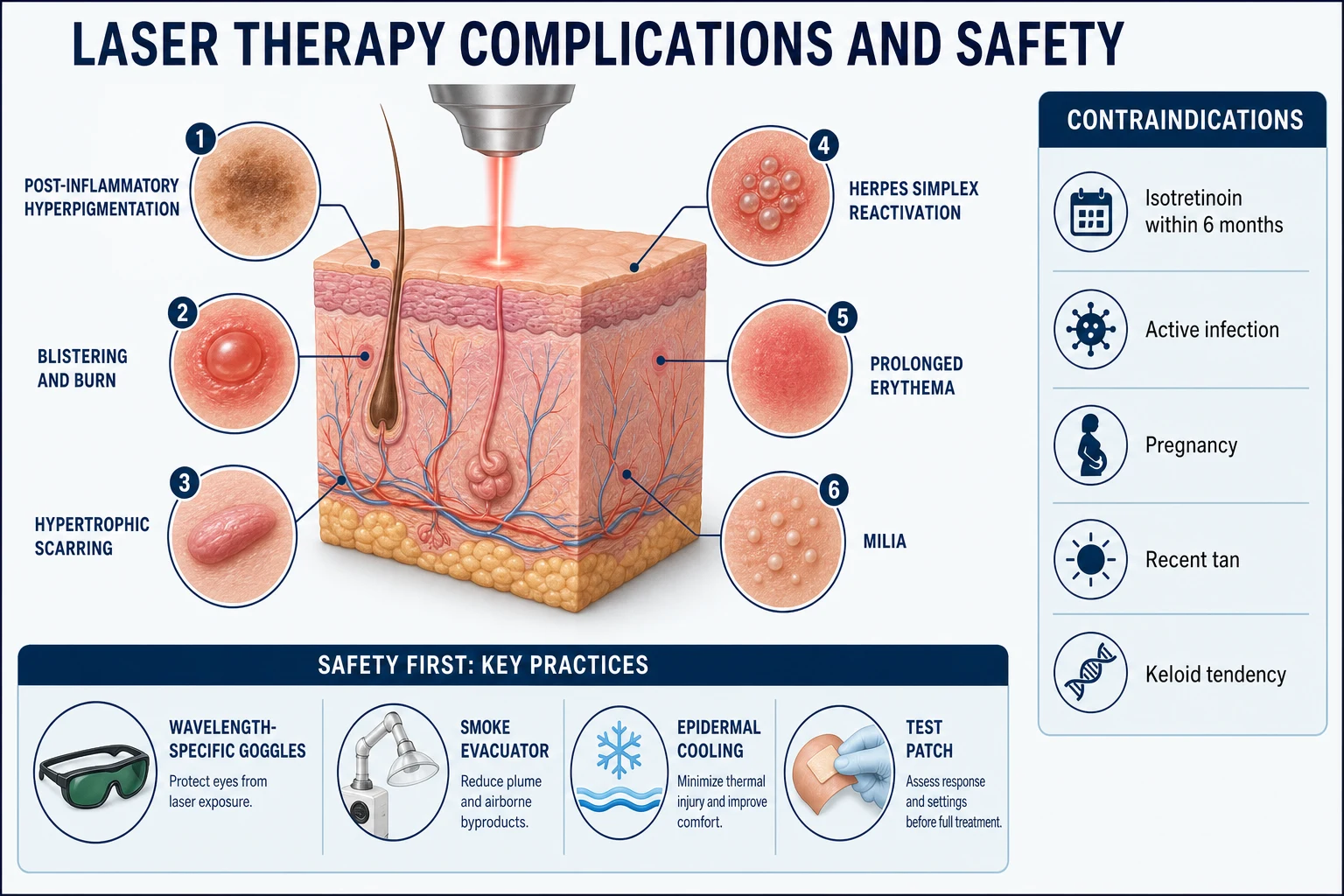
Complications fall into pigmentary, thermal, infectious, scarring and cosmetic categories, and their incidence rises with darker skin type, higher fluence, inadequate cooling, recent tan and isotretinoin exposure.[9]
Pigmentary changes are the commonest adverse event in Fitzpatrick IV-VI. Post-inflammatory hyperpigmentation appears three to four weeks after treatment and may persist for months; it is minimised by conservative fluence, aggressive cooling, hydroquinone pre-treatment and sun protection, and treated with a tyrosinase inhibitor, mild topical steroid and time. Hypopigmentation is less common but more recalcitrant, resulting from melanocyte destruction, and is a particular risk of overly aggressive hair removal and Q-switched treatment in dark skin.[10]
Thermal burns, blistering and crusting result from excessive fluence, inadequate cooling or a recent tan; they are managed as thermal injuries with cool compresses, a topical corticosteroid, non-adherent dressings and analgesia, with a low threshold for review if the burn is deep. Infection — reactivated herpes simplex, bacterial folliculitis or candidiasis — is prevented by antiviral prophylaxis in at-risk patients and by aseptic technique; it is treated promptly with the appropriate antimicrobial. Hypertrophic scarring and keloids are rare but devastating, concentrated on the chest, jawline and neck and after resurfacing during or soon after isotretinoin; the dictum is to defer resurfacing for at least six months after isotretinoin cessation.[8]
Prolonged erythema (beyond one month), acne and milia flares, contact dermatitis to dressings or topical agents, and textural change complete the list. Ocular injury — corneal burn, retinal photocoagulation, iris depigmentation — is prevented entirely by correct eye protection and is a sentinel never-event. Paradoxical darkening of cosmetic tattoo ink (titanium dioxide reduction to grey-black) is avoided by a mandatory test patch on flesh-toned or white ink before full treatment. Purpura after PDL is expected and resolves over one to two weeks; modern long-pulsed sub-purpuric protocols reduce it. A systematic review of PDL for port-wine stain confirms the procedure is safe overall, with transient pigmentary change and blistering the most frequent events and scarring rare.[9]
Special Populations
Children. Port-wine stains are best treated in infancy, when the skin is thinner, the vessels smaller and the stain lighter, and before psychosocial consequences accrue. Infantile haemangiomas are treated with PDL when superficial or ulcerated. Topical anaesthesia, distraction and, for extensive work, sedation or general anaesthesia address the practical barriers of treating a child.[14]
Skin of colour (Fitzpatrick IV-VI). The competing epidermal melanin absorbs strongly at shorter wavelengths, raising the risk of pigmentary change and burns. Strategy: choose the longest effective wavelength (long-pulsed Nd:YAG 1064 nm for hair and deeper vessels; Q-switched 1064 nm for pigment and tattoo), use lower fluence, longer pulse durations (which may be stacked), aggressive cooling, hydroquinone pre-treatment, a mandatory test patch, and longer intervals between sessions.[10]
Pregnancy. Elective laser is deferred. Hormone-driven melasma and spider naevi may worsen, and there are no safety data on fetal exposure to scattered laser light or to topical anaesthesia and antivirals.[2]
Immunocompromised patients have slower healing and higher infection risk; conservative settings, antiviral prophylaxis and a low threshold for antibiotic cover are appropriate.[1]
Patients on isotretinoin must defer ablative and fractional resurfacing for at least six months after cessation because of the markedly increased risk of hypertrophic scarring from retinoid-impaired wound healing.[8]
Evidence, Guidelines and Controversies
The landmark Anderson and Parrish 1983 paper in Science defined selective photothermolysis and remains the single most cited reference in laser dermatology.[1] The introduction of fractional photothermolysis (Manstein and colleagues, 2004) was the next paradigm shift, converting resurfacing from an all-or-nothing ablative procedure into a safer, fractionated one whose role and indications are still being defined.[3] Picosecond lasers, with a thousand-fold shorter pulse duration than Q-switched devices, have displaced Q-switched as the preferred modality for multicolour and resistant tattoos and, with diffractive optics, for acne scarring; a 2021 systematic review and a 2022 comparative review confirm their advantage for brightly coloured ink.[11][5]
For 308-nm excimer, systematic reviews of randomised trials in vitiligo and protocol reviews in psoriasis confirm efficacy with a lower cumulative UV burden than whole-body phototherapy.[6][7] A 2023 systematic review and meta-analysis of PDL for port-wine stain quantifies the safety profile: transient pigmentary change and blistering are common, scarring rare, supporting early and repeated treatment.[9] Reviews of PIH management in skin of colour emphasise prevention — hydroquinone pre-treatment, conservative fluence, cooling and test patches — over rescue.[10]
A brief history of laser dermatology
Theodore Maiman demonstrates the ruby laser — the first operable laser, built around a synthetic ruby crystal.
Anderson and Parrish publish the foundational Science paper defining selective photothermolysis, transforming lasers from non-selective thermal tools into chromophore-targeted instruments.[1]
The flashlamp-pumped pulsed-dye laser revolutionises port-wine stain treatment; Q-switched ruby, alexandrite and Nd:YAG become the standard for tattoos; long-pulsed ruby and then alexandrite, diode and Nd:YAG launch laser hair removal.[2]
CO2 and Er:YAG resurfacing become widespread for photoaging and scars, delivering dramatic results but with significant downtime and risk.
Manstein and colleagues introduce fractional photothermolysis — microscopic treatment zones — making resurfacing safer and expanding its indications.[3]
Picosecond devices enter practice; their photoacoustic effect improves multicolour and resistant tattoo clearance and, with diffractive optics, treats acne scars.[11]
The chief controversy concerns the mandatory interval after isotretinoin. Traditional teaching — and most guideline bodies — defer ablative and fractional resurfacing for six to twelve months. Khatri and colleagues challenged this in a small study of patients resurfaced during isotretinoin without excess scarring, but the numbers are too small to overturn conservative practice, and most practitioners still defer for at least six months.[8] Other controversies include the optimal PDL pulse parameters for sub-purpuric versus purpuric treatment of port-wine stains, the role of laser-assisted drug delivery (fractional ablation to enhance topical agents), and whether picosecond devices justify their cost over Q-switched for routine black-ink tattoos.
Adjunctive and Combination Therapies
Laser results are often enhanced by combination. Microneedling — mechanical creation of micro-wounds that trigger collagen remodelling — treats acne scars, fine wrinkles and stretch marks and can be combined with topical agents (platelet-rich plasma, vitamin C, growth factors) for enhanced penetration and synergy with fractional laser.[12] Platelet-rich plasma is used adjunctively for rejuvenation and hair loss. Laser-assisted drug delivery — using fractional ablative laser to create channels for topical agents — is an emerging application for vitiligo, alopecia areata and scar modulation. For burn and surgical scars, PDL plus fractional CO2 outperforms either alone.[13]
Prognosis and Disposition
Outcomes are indication- and technique-dependent and are set realistically at consent. Port-wine stains lighten by roughly 50-70 percent with complete clearance in only 10-20 percent; recurrence and darkening occur over years and require maintenance. Hair removal achieves durable 70-90 percent reduction rather than permanent eradication, with maintenance sessions and a poorer response in hormonal areas. Tattoo removal is incremental — amateur tattoos clear faster than professional, black ink faster than colour — and complete clearance is often unattainable. A single ablative CO2 resurfacing session delivers results durable for five to ten years; fractional resurfacing requires three to five sessions. Melasma recurs with sun and hormones and requires ongoing tyrosinase inhibitors and photoprotection. Patients are counselled, consented for the expected number of sessions and interval, and reviewed at each visit to titrate parameters and detect complications early. [2]
Exam Pearls
Quick self-test: which laser for each scenario?
- Newborn with a pink V1 port-wine stain on the forehead — PDL 585-595 nm, start in infancy; assess for Sturge-Weber.
- Fair-skinned woman, unwanted axillary hair — alexandrite 755 nm (skin I-III).
- South-Asian man, beard pseudofolliculitis and unwanted facial hair — long-pulsed Nd:YAG 1064 nm (safest in Fitzpatrick V-VI).
- Professional multicolour tattoo, removal desired — picosecond laser (532/1064/755), 6-8 week intervals.
- Deep perioral rhytides and acne scarring — ablative or fractional CO2 10600 nm (defer if isotretinoin within 6 months).
- Localised plaque psoriasis on the elbow — 308-nm excimer laser.
- Naevus of Ota (dermal grey-blue pigmentation) — Q-switched Nd:YAG 1064 nm. [5]
References
- [1]Anderson RR, Parrish JA. Selective photothermolysis: precise microsurgery by selective absorption of pulsed radiation Science, 1983.PMID 6836297
- [2]Gan SD, Graber EM. Laser hair removal: a review Dermatol Surg, 2013.PMID 23332016
- [3]Bogdan Allemann I, Kaufman J. Fractional photothermolysis--an update Lasers Med Sci, 2010.PMID 19787413
- [4]Janik JP, Markus JL, Al-Dujaili Z, et al. Laser resurfacing Semin Plast Surg, 2007.PMID 20567665
- [5]Gurnani P, Williams N, Al-Hetheli G, et al. Comparing the efficacy and safety of laser treatments in tattoo removal: A systematic review J Am Acad Dermatol, 2022.PMID 32763326
- [6]Sun Y, Wu Y, Xiao B, et al. Treatment of 308-nm excimer laser on vitiligo: A systemic review of randomized controlled trials J Dermatolog Treat, 2015.PMID 25428573
- [7]Mudigonda T, Dabade TS, Feldman SR. A review of protocols for 308 nm excimer laser phototherapy in psoriasis J Drugs Dermatol, 2012.PMID 22206083
- [8]Khatri KA, Iqbal N, Bhawan J. Laser skin resurfacing during isotretinoin therapy Dermatol Surg, 2015.PMID 25993614
- [9]Shi MD, Yang K, Li SB, et al. Complication rates and safety of pulsed dye laser treatment for port-wine stain: a systematic review and meta-analysis Lasers Med Sci, 2023.PMID 38141129
- [10]Sowash M, Alster T. Review of Laser Treatments for Post-Inflammatory Hyperpigmentation in Skin of Color Am J Clin Dermatol, 2023.PMID 36781686
- [11]Wu DC, Goldman MP, Wat H, et al. A Systematic Review of Picosecond Laser in Dermatology: Evidence and Recommendations Lasers Surg Med, 2021.PMID 32282094
- [12]Alster TS, Graham PM. Microneedling: A Review and Practical Guide Dermatol Surg, 2018.PMID 28796657
- [13]Altemir A, Boixeda P. Laser Treatment of Burn Scars Actas Dermosifiliogr, 2022.PMID 35963335
- [14]Sebaratnam DF, Rodríguez Bandera AL, Wong LF, et al. Infantile hemangioma. Part 2: Management J Am Acad Dermatol, 2021.PMID 34419523