Dermatology · Medicine
Leishmaniasis (cutaneous)
Also known as Cutaneous leishmaniasis · Mucosal (mucocutaneous) leishmaniasis · Visceral leishmaniasis (kala-azar) · Post-kala-azar dermal leishmaniasis (PKDL) · New World / Old World leishmaniasis · Espundia · Chiclero ulcer
Leishmaniasis is a protozoal parasitic disease caused by Leishmania species (kinetoplastid flagellates) transmitted by the female phlebotomine sandfly, producing a spectrum from self-healing cutaneous ulcers (the chronic painless ulcer with a raised indurated border, the 'volcano sign') through destructive mucosal disease (espundia, L. braziliensis) to fatal visceral leishmaniasis (kala-azar, L. donovani/infantum/chagasi) with fever, massive splenomegaly, and pancytopenia, and post-kala-azar dermal leishmaniasis after treatment. Old World (L. major, L. tropica, L. aethiopica, L. infantum) and New World (L. mexicana and L. braziliensis complexes) species determine the syndrome and therapy. Diagnosis rests on Giemsa smear/biopsy showing intracellular amastigotes (Leishman-Donovan bodies), culture on NNN medium, PCR for speciation, and the rK39 rapid test for visceral disease. Treatment spans watchful waiting and local physical/topical therapy for simple Old World cutaneous disease to systemic pentavalent antimonials, oral miltefosine, paromomycin, and liposomal amphotericin B for New World, mucosal, and visceral disease.
On this page & tools
Your progress
Saved locally on this device.
Exam tags
Red flags
Overview & Definition
Leishmaniasis is a protozoal parasitic disease caused by obligate intracellular kinetoplastid flagellates of the genus Leishmania, transmitted to humans and other mammals by the bite of the female phlebotomine sandfly. It is a disease of striking clinical diversity: the same genus of organism can produce a minor, self-healing skin ulcer, a relentlessly mutilating destruction of the face, or a uniformly fatal systemic illness — and which of these unfolds is determined almost entirely by the species of parasite, the immune response of the host, and the geography of exposure.[1][4]
Three major clinical syndromes are recognised. Cutaneous leishmaniasis (CL) is the commonest, presenting as a chronic papule that evolves into a painless ulcer with a raised indurated border on an exposed site, healing over months to years with a scar. Mucosal (mucocutaneous) leishmaniasis (MCL), or espundia, is the destructive destruction of the nasal and oropharyngeal mucosa that follows cutaneous infection with L. braziliensis years later. Visceral leishmaniasis (VL), or kala-azar (Hindi for 'black disease'), is a systemic reticuloendothelial infection with fever, massive splenomegaly, pancytopenia, and cachexia that is over 95 percent fatal if untreated.[1][5] Two further forms complete the spectrum: diffuse cutaneous leishmaniasis (DCL), an anergic non-ulcerative nodular form, and post-kala-azar dermal leishmaniasis (PKDL), a maculopapular eruption that appears after treated visceral disease.[7]
Fellowship-level competence requires the candidate to hold the Old World species (L. major, L. tropica, L. aethiopica, L. infantum, transmitted by Phlebotomus sandflies across the Mediterranean, Middle East, and Africa) and the New World species (the L. mexicana complex and the L. braziliensis complex including L. panamensis, L. guyanensis, and L. chagasi, transmitted by Lutzomyia sandflies across Central and South America) in mind at once, because the species dictates the syndrome, the reservoir, the risk of mucosal spread, and the choice of therapy.[2][9]
Classification
Leishmaniasis is classified along two axes that together predict the clinical course: the clinical syndrome (cutaneous, mucosal, visceral, and the variants) and the parasite species complex and its geography (Old World versus New World), which in turn determines the reservoir (zoonotic versus anthroponotic) and therefore the public-health strategy.[1][9]

By clinical syndrome
- Cutaneous leishmaniasis (CL) — the commonest form, ~1 million cases annually worldwide. Subdivided into localised CL (the classic ulcer), diffuse cutaneous leishmaniasis (DCL, anergic nodular disease), leishmaniasis recidivans (chronic relapsing lupus-like form), and the New World chiclero ulcer of the ear.
- Mucosal (mucocutaneous) leishmaniasis (MCL / espundia) — destructive naso-oropharyngeal disease, classically from L. braziliensis, appearing years after cutaneous infection.
- Visceral leishmaniasis (VL / kala-azar) — systemic reticuloendothelial infection with fever, splenomegaly, pancytopenia; ~50,000 to 90,000 cases annually.
- Post-kala-azar dermal leishmaniasis (PKDL) — a dermal eruption following treated VL, mainly in South Asia and East Africa, acting as a reservoir for anthroponotic transmission.[7][8]
By species and geography
The species, its vector, its reservoir host, and the syndrome it tends to produce are inseparable. Old World leishmaniasis spans the Mediterranean basin, the Middle East, North and sub-Saharan Africa, and Central Asia; New World leishmaniasis spans Mexico through to northern Argentina. The distinction matters because L. braziliensis-complex cutaneous disease carries the risk of later mucosal spread and therefore warrants systemic therapy even when the skin lesion looks trivial.[3][9]
Epidemiology & Risk Factors
Leishmaniasis is endemic in 98 countries across the tropics and subtropics, with an estimated 1.3 million new cases each year and a global prevalence of 12 million infected people. It ranks second only to malaria among protozoal causes of death, and visceral leishmaniasis alone kills an estimated 20,000 to 40,000 people annually despite effective therapy existing — a stark reminder that mortality here is a disease of poverty and access, not of biology.[1][6]
The geographic burden is uneven. Cutaneous leishmaniasis is concentrated in the Middle East (Afghanistan, Iran, Saudi Arabia, Syria — L. major, L. tropica), North Africa, and Latin America (Brazil, Colombia, Peru — L. braziliensis, L. panamensis, L. mexicana). Visceral leishmaniasis is concentrated in India (Bihar, Jharkhand, West Bengal — L. donovani, anthroponotic), East Africa (Sudan, Ethiopia, South Sudan — L. donovani), and Brazil (L. infantum chagasi, zoonotic from dogs), with smaller foci in the Mediterranean (L. infantum, zoonotic from dogs and foxes). Post-kala-azar dermal leishmaniasis follows VL principally in India and Sudan.[5][7]
The risk factors cluster around exposure to the sandfly and impairment of the cellular immune response. Poverty, malnutrition, and overcrowding raise both exposure and susceptibility; deforestation, urbanisation, and conflict-driven displacement bring humans into sandfly habitat and have produced major epidemics (the Sudanese VL epidemic of the 1980s to 1990s killed an estimated 100,000 people). Climate change is expanding the sandfly's range northward and to higher altitudes. HIV co-infection is the most important biological amplifier: VL is an AIDS-defining illness, HIV-VL co-infection carries a far higher relapse rate, and the two diseases fuel one another in a vicious cycle. Occupational and recreational exposure matters — soldiers deployed to endemic regions, farmers, forest workers (chicleros in Mexico harvesting chicle from forest trees), and eco-tourists — and male sex is over-represented purely because of exposure patterns.[4][6]
Pathophysiology
Understanding leishmaniasis begins with the parasite's dimorphic life cycle, because the two morphological forms map exactly onto the two hosts and onto every diagnostic test the candidate will be asked about.[2][9]
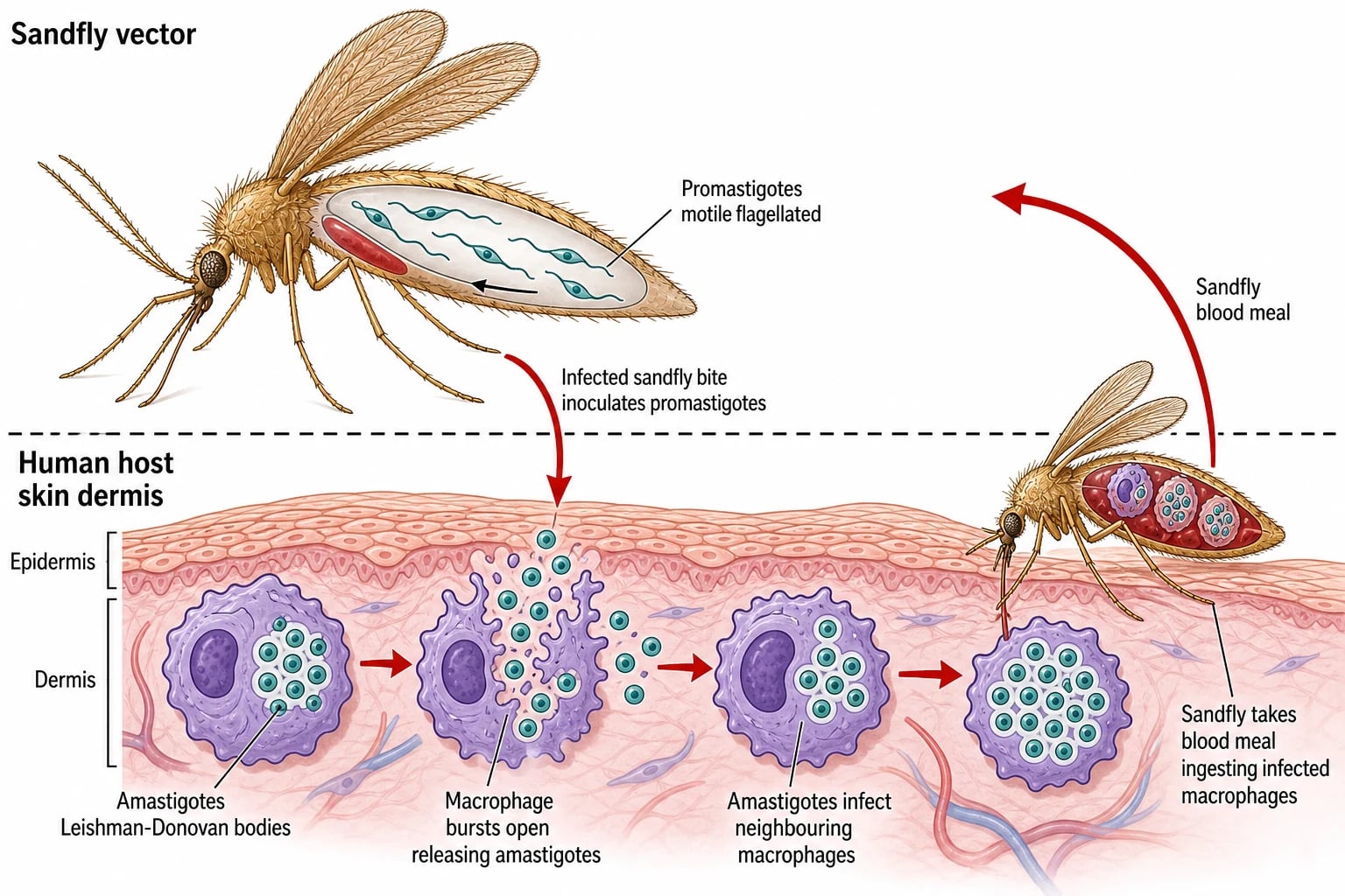
The two forms. In the sandfly midgut, Leishmania lives as the promastigote — an elongated, motile, flagellated form (10 to 20 micrometres) that attaches to the gut wall and, in the infectious metacyclic stage, migrates to the proboscis. In the mammalian host, it lives as the amastigote — a small (2 to 5 micrometres), round, non-motile, intracellular form inside macrophages, recognised on Giemsa stain by its nucleus and the rod-shaped kinetoplast (the signature DNA-containing mitochondrial structure that gives the order Kinetoplastida its name). These amastigotes inside macrophages are the Leishman-Donovan (LD) bodies named for the discoverers of visceral leishmaniasis.[9][10]
Transmission and the intracellular cascade. When an infected female sandfly takes a blood meal, she inoculates metacyclic promastigotes into the dermis along with sandfly saliva (which itself has immunosuppressive properties that aid establishment). Dermal macrophages rapidly phagocytose the promastigotes. The parasite survives and indeed thrives inside the macrophage's phagolysosome — a normally hostile environment of low pH (4.5 to 5.0) and oxidative burst — by deploying its surface lipophosphoglycan (LPG) to resist complement lysis in the extracellular phase and to deflect the respiratory burst intracellularly. Inside the phagolysosome the promastigote transforms into the amastigote and begins to divide by binary fission. The engorged macrophage eventually ruptures, releasing amastigotes that infect neighbouring macrophages and, in viscerotropic species, are carried by lymphatics and blood to the reticuloendothelial system — spleen, liver, bone marrow, and lymph nodes. When another sandfly feeds on an infected host, it ingests amastigote-laden macrophages, in which the parasite transforms back into promastigotes in the fly's midgut, completing the cycle.[2][9]
The immunity spectrum and tissue tropism. The clinical face of leishmaniasis is the visible product of the host's cellular immune response. A brisk Th1 (cell-mediated) response — driven by interferon-gamma and interleukin-12 from CD4+ T cells and natural killer cells — activates macrophages to kill intracellular amastigotes via nitric oxide and reactive oxygen species, and localises the infection to a contained cutaneous lesion that eventually heals. A deficient or anergic cellular response fails to contain the parasite and produces either diffuse cutaneous leishmaniasis (in which parasites proliferate unchecked in skin macrophages without a granulomatous response) or visceral dissemination. The striking tissue tropism of different species — why L. donovani seeds the spleen and bone marrow while L. major stays in the skin — is determined by parasite surface molecules (notably the LPG and gp63 protease), by the temperature preference of each species, and by host genetic factors such as the SLC11A1 (NRAMP1) polymorphism.[2][4]
The reservoir. Whether a species is zoonotic (animal reservoir — rodents, dogs, foxes, sloths) or anthroponotic (human reservoir) dictates the public-health strategy: zoonotic disease is controlled by vector control and animal reservoir management, while anthroponotic disease (notably L. donovani in India and L. tropica) is controlled by finding and treating human cases — and it is precisely why PKDL, a human-only post-treatment eruption, matters as a transmission reservoir.[1][7]
Clinical Presentation
Each clinical syndrome of leishmaniasis has a recognisable morphology and tempo, and examiners reward a precise description anchored to the species and the geography.[1][4]
Cutaneous leishmaniasis (CL) — the volcano sign
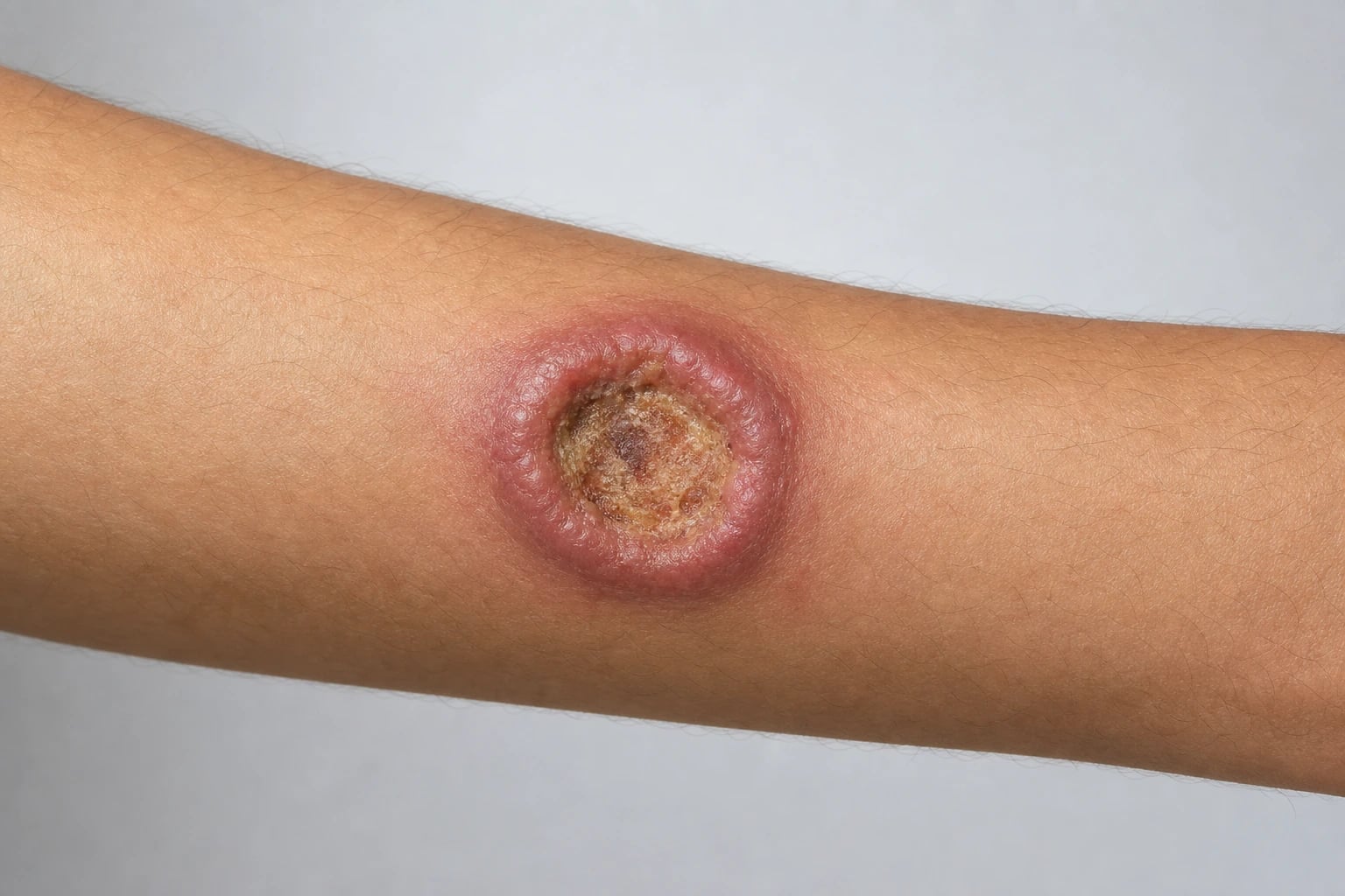
The classic lesion begins, weeks to months after an infected sandfly bite, as a small erythematous papule at an exposed site — the face, arms, or legs. The papule enlarges into a nodule and then ulcerates, producing the signature lesion: a chronic, painless ulcer with a raised, indurated border and a crusted base, the 'volcano sign' — so named because the firm, heaped-up inflammatory rim resembles the cone of a volcano surrounding the central crater. The ulcer is usually single but may be multiple, and regional lymphadenopathy is common. Untreated, the lesion persists for months to years, eventually healing spontaneously with a depressed, often disfiguring scar — particularly reliably in L. major and L. mexicana (which self-heal in 70 percent or more of cases within 6 to 18 months), and less reliably in L. tropica and L. braziliensis, which tend to persist, recur, or, in the case of L. braziliensis, herald later mucosal disease.[3][11]
Atypical cutaneous variants are high-yield: [1]
- Diffuse cutaneous leishmaniasis (DCL) — a rare, anergic, non-ulcerative form seen with L. aethiopica in Ethiopia and L. amazonensis in the Americas, in which the host mounts no cellular response. Widespread nodules and plaques teeming with parasites accumulate over the skin, mimicking lepromatous leprosy, and the disease is refractory to standard therapy.[1]
- Leishmaniasis recidivans — a chronic, relapsing, lupus-like form of L. tropica infection in which new scaly papules appear at the edge of an old scar years after apparent cure, driven by an exuberant but ineffective cell-mediated response; it is paucibacillary and the leishmanin skin test is strongly positive.[3]
- Chiclero ulcer — chronic destructive ulcers of the ear pinna caused by L. mexicana in forest workers (chicleros) of Central America; the cool temperature of the ear cartilage suits this species, and the lesion can destroy the auricular cartilage over years.[9]
Mucosal (mucocutaneous) leishmaniasis — espundia
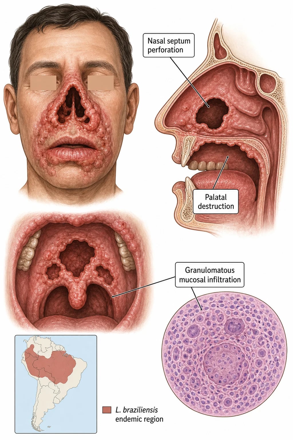
Mucosal leishmaniasis is the most feared cutaneous complication. It arises, usually from cutaneous L. braziliensis (or L. panamensis) infection, years to decades after the original skin lesion healed — the parasite having persisted silently in mucosal tissue. It begins with nasal stuffiness and epistaxis, then progresses to destructive granulomatous inflammation of the nasal and oropharyngeal mucosa: perforation of the nasal septum, erosion of the nasal alae, perforation of the palate, and destruction of the lips, uvula, and larynx. The resulting mutilation — the 'tapir nose' deformity — is profoundly disfiguring and was named espundia in South America. Mucosal disease is refractory and recurrent, threatens the airway and swallowing, and always requires systemic therapy plus ENT and maxillofacial surgical input for reconstruction.[3][4]
Visceral leishmaniasis (VL) — kala-azar
Visceral leishmaniasis is a systemic infection of the reticuloendothelial system that develops weeks to months (occasionally years) after exposure. The classic tetrad is irregular (often biphasic) fever, marked splenomegaly (with or without hepatomegaly), pancytopenia, and progressive cachexia with weight loss. The spleen is typically massive, soft, and non-tender. Secondary features include hypergammaglobulinaemia (a polyclonal B-cell activation that produces a falsely positive rheumatoid factor and a high erythrocyte sedimentation rate), hypoalbuminaemia, oedema, diarrhoea, bleeding from thrombocytopenia, and the greyish skin hyperpigmentation that gives the disease its Hindi name kala-azar ('black disease'). In endemic children, VL may present simply as fever, failure to thrive, and splenomegaly. Untreated, VL is progressive and over 95 percent fatal — death comes from secondary bacterial infection (pneumonia, tuberculosis), bleeding, or overwhelming parasitaemia.[1][5]
Post-kala-azar dermal leishmaniasis (PKDL)
PKDL is a dermatological eruption that appears after a patient has been treated for visceral leishmaniasis — typically 6 months to 3 years later in India and during or soon after treatment in Sudan. It presents as hypopigmented or erythematous macules, papules, and nodules on the face, trunk, and extremities, which can mimic leprosy, melasma, or a drug eruption. Crucially, PKDL patients carry parasites in their skin and act as a reservoir for anthroponotic transmission of L. donovani — a fact that is central to the Indian and Sudanese elimination programmes, because PKDL is the bridge between one epidemic and the next.[7][8]
Atypical presentations
HIV co-infection produces atypical, refractory, and relapsing leishmaniasis: VL in HIV-positive patients may present without massive splenomegaly, may seed atypical sites (the gastrointestinal tract, the skin), responds poorly to therapy, and relapses repeatedly, necessitating secondary prophylaxis. The immunosuppressed transplant recipient or patient on anti-TNF therapy may suffer reactivation of latent infection. In malnutrition, VL runs a more fulminant course with higher mortality.[4][6]
Differential Diagnosis
The differential of leishmaniasis is syndrome-specific. The discipline is to pair each syndrome with its mimics and to distinguish them by the travel and exposure history, the Giemsa smear and PCR, and the systemic context.[3][10]
The can't-miss considerations are squamous cell carcinoma arising in a chronic non-healing ulcer (biopsy any change), visceral leishmaniasis in a febrile traveller with splenomegaly (a medical emergency), mucosal leishmaniasis evolving insidiously from a healed New World skin lesion, and granulomatosis with polyangiitis (GPA) mimicking mucosal leishmaniasis in a returned traveller (distinguished by ANCA, renal involvement, and tissue histology).[10]
Clinical & Bedside Assessment
The focused assessment serves four goals: characterise the lesion, screen for mucosal and visceral involvement, establish the exposure, and assess the immune status.[1][4]
Characterise the lesion. Inspect every chronic ulcer for the volcano sign — a raised, indurated border with a crusted base on an exposed site (the sandfly cannot bite through clothing). Note the site, size, number, and chronicity (months rather than days), and the characteristically painless nature. Examine for regional lymphadenopathy and for satellite papules. A non-ulcerative nodular eruption on a patient from Ethiopia or the Amazon should raise diffuse cutaneous leishmaniasis. [1]
Screen for mucosal involvement in EVERY New World case. Inspect the nasal mucosa (septal perforation, crusting), the oropharynx and palate (perforation, ulceration), and ask about epistaxis and nasal stuffiness — these are the earliest signs of espundia and may appear years after the skin lesion healed. Neglecting this screen is a classic pitfall. [1]
Screen for visceral disease in any febrile patient from an endemic region: examine the abdomen for splenomegaly and hepatomegaly, look for cachexia, pallor, petechiae, and oedema, and assess for lymphadenopathy. Visceral leishmaniasis must not be missed — it is uniformly fatal untreated. [1]
Establish the exposure. Take a meticulous travel, residence, and occupation history — country and region, rural versus urban, forest versus desert, time of year, duration of exposure, and sandfly bites. Ask specifically about military deployment, eco-tourism, forestry, and animal reservoir contact. [1]
Assess the immune status. Test for HIV in every case (VL is an AIDS-defining illness and co-infection changes everything). Note malnutrition, alcoholism, and immunosuppression, and ask about anti-TNF therapy and transplantation.[6]
Investigations
The diagnosis of leishmaniasis rests on demonstration of the parasite or its genome in tissue, supported by serology for visceral disease. Because different species demand different therapy, speciation by PCR is increasingly central.[3][10]
Parasitological diagnosis of cutaneous disease
The cornerstone is the skin slit smear or punch biopsy of the active ulcer EDGE (not the necrotic base, where organisms are scarce). The tissue is divided for three simultaneous tests:[3]
- Giemsa-stained smear — the quickest and most widely available test. It shows the amastigotes (Leishman-Donovan bodies) as 2 to 5 micrometre round organisms inside macrophages, each with a nucleus and the rod-shaped kinetoplast — the latter is the diagnostic feature that distinguishes Leishmania from Histoplasma (which lacks a kinetoplast).
- Histopathology of a punch biopsy shows a granulomatous dermatitis with epithelioid macrophages, lymphocytes, and plasma cells, and amastigotes within macrophages — distinguishing CL from cutaneous tuberculosis and leprosy.
- Culture on Novy-MacNeal-Nicolle (NNN) medium (or Schneider's Drosophila medium), incubated at 22 to 26 degrees Celsius for up to 4 weeks, yields the motile promastigote form — the gold standard but slow, and demanding a specialised laboratory.[9][10]
- PCR (targeting kinetoplast DNA or the internal transcribed spacer) is the most sensitive method and uniquely enables species identification, which in turn guides therapy — distinguishing, for example, L. major (often watchful waiting) from L. braziliensis (systemic therapy to prevent mucosal spread).[3]
Diagnosis of visceral leishmaniasis
Visceral disease requires demonstration of parasites in reticuloendothelial tissue or a positive serological rapid test in the right clinical context:[5]
- Bone marrow aspirate (sensitivity 60 to 85 percent) or splenic aspirate (sensitivity over 95 percent, the gold standard, but carrying a bleeding risk that demands a normal platelet count and coagulation screen). Lymph-node aspirate is an alternative in some regions.
- rK39 rapid diagnostic test (immunochromatographic test) — a lateral-flow antibody test detecting anti-rK39 antibody; highly useful for field diagnosis of VL in South Asia and the Mediterranean, with high sensitivity in symptomatic disease. Direct agglutination test (DAT) is an alternative serological method used in reference laboratories.
- PCR on blood or bone marrow is highly sensitive and useful for diagnosis and for monitoring relapse, particularly in HIV co-infection. [1]
Immunological and epidemiological tests
- The Montenegro test (leishmanin skin test) — an intradermal delayed-type hypersensitivity test analogous to the Mantoux test, positive in cutaneous and healed disease. It is now used mainly for epidemiological surveys rather than individual diagnosis (it cannot distinguish past from active infection).[9]
- Baseline bloods in VL — full blood count (the pancytopenia), liver function tests, renal function, albumin, and coagulation screen (before any splenic aspirate). HIV testing is mandatory.
Management — Resuscitation
Most cutaneous leishmaniasis is managed electively, but two scenarios demand immediate action.[1][5]
Visceral leishmaniasis is a medical emergency. The febrile, pancytopenic, cachectic patient is at risk of secondary infection, bleeding, and death. The immediate steps are admission, correction of anaemia and thrombocytopenia (transfusion as needed), treatment of intercurrent infection (pneumonia, tuberculosis), nutritional support, and prompt initiation of antileishmanial therapy — most commonly liposomal amphotericin B. A patient undergoing splenic aspirate must have a platelet count and coagulation checked first, and be observed for bleeding afterwards. [1]
Mucosal leishmaniasis demands urgent systemic therapy to halt progressive mutilating destruction of the face and airway, with ENT and maxillofacial surgical input for airway protection and later reconstruction. Delay allows irreversible cartilage and palatal destruction. [1]
In anthroponotic visceral leishmaniasis regions (India), there is a public-health reflex: the case must be reported to the National Vector-Borne Disease Control Programme (NVBDCP) for active case detection, vector control, and the elimination programme.[5]
Management — Definitive & Stepwise
Therapy is guided by the syndrome, the species, the site, and the host. The candidate must know when to do nothing, when to treat locally, and when to commit to systemic therapy.[3][4]
Cutaneous leishmaniasis — watchful waiting, local, and systemic therapy
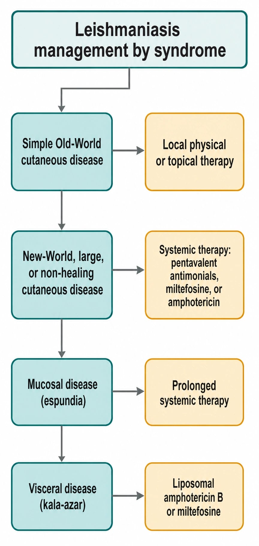
Watchful waiting. Many Old World, uncomplicated lesions caused by L. major heal spontaneously within 6 to 18 months (over 70 percent), and observation alone is a legitimate strategy for small, cosmetically unimportant lesions in a compliant patient. The decision to treat is based on the species (risk of mucosal spread or non-healing), the site (face, near a joint), the size and number of lesions, the cosmetic impact, and the patient's preference.[3][11]
Local (physical and topical) therapy is appropriate for small, uncomplicated Old World lesions and avoids systemic toxicity:[3][11]
- Cryotherapy with liquid nitrogen — repeated freeze-thaw cycles, suitable for small lesions.
- Thermotherapy — localised radiofrequency heat applied to 50 degrees Celsius, killing the heat-sensitive parasite; a single device-delivered session can cure ~75 percent of suitable lesions.
- Topical paromomycin 15% cream (with or without gentamicin or methylbenzethonium chloride) applied twice daily for 20 days — effective for some Old World species but variably effective against New World species.
- Intralesional pentavalent antimonials (sodium stibogluconate / Pentostam, or meglumine antimoniate) injected into the lesion edge every few days — effective for a few Old World and New World lesions but painful and not suitable for multiple or facial lesions. [1]
Systemic therapy is indicated for New World disease (especially L. braziliensis, to prevent mucosal spread), large, multiple, or cosmetically important lesions (especially on the face or near joints), non-healing or relapsing disease, leishmaniasis recidivans, and diffuse cutaneous leishmaniasis:[3][4]
- Pentavalent antimonials — sodium stibogluconate (Pentostam) or meglumine antimoniate (Glucantime) at 20 mg Sb5+ per kg per day, intramuscularly or intravenously, for 20 days for cutaneous disease (28 days for mucosal). They remain first-line in many endemic regions but carry significant toxicity: QT prolongation and arrhythmia (sudden death — requires ECG monitoring), pancreatitis, hepatotoxicity, myalgia, arthralgia, and bone-marrow suppression.[1][11]
- Miltefosine — the only oral antileishmanial, dosed at approximately 2.5 mg per kg per day (adults 50 mg three times daily for those over 30 kg; twice daily for under 30 kg; paediatric from age 2 years) orally for 28 days. Effective against L. braziliensis, L. panamensis, and L. mexicana, and against VL, but teratogenic (contraindicated in pregnancy) and requires contraception. Adverse effects include gastrointestinal upset (nausea, vomiting, diarrhoea), hepatotoxicity, and renal impairment.[12][4]
- Liposomal amphotericin B — 3 mg per kg per day intravenously for up to 7 days for cutaneous disease where antimonials are contraindicated or have failed; expensive but far less toxic than conventional amphotericin.
- Azoles — fluconazole 200 mg daily for 6 weeks or ketoconazole/itraconazole offer a convenient oral option for some Old World species (notably L. major) with lower efficacy than antimonials but good tolerability.[3]
- Pentamidine — an alternative, particularly for some New World species (L. guyanensis), but nephrotoxic and diabetogenic.
Diffuse cutaneous leishmaniasis (DCL, L. aethiopica or L. amazonensis) is refractory and demands combination therapy — typically paromomycin plus sodium stibogluconate for 60 days or longer, or liposomal amphotericin B — and even so relapse is common.[1]
Mucosal (mucocutaneous) leishmaniasis — always systemic
Mucosal disease is always treated systemically and for longer (28 days), because local therapy cannot reach the mucosa and the disease is destructive and recurrent:[3][4]
- Pentavalent antimonials at 20 mg per kg per day for 28 days as first-line, OR liposomal amphotericin B, OR miltefosine.
- Response is often incomplete and relapse is common, requiring retreatment. Surgical reconstruction of the nasal septum, palate, and lips is deferred until the infection is parasitologically cured, because operating on active disease fails. [1]
Visceral leishmaniasis — liposomal amphotericin is the gold standard
Liposomal amphotericin B is the global gold standard for VL, reflecting both efficacy (cure over 95 percent) and a far better safety profile than the older conventional amphotericin or antimonials:[1][5]
- Liposomal amphotericin B — total dose 30 mg per kg given as various regimens: the Indian NVBDCP uses a single 10 mg per kg dose, the WHO recommends 3 mg per kg per day for 7 days (on days 1 to 5, 14, and 21 in some regimens). It is preferred in pregnancy and in HIV co-infection.
- Miltefosine — oral 2.5 mg per kg per day for 28 days, the only oral agent and invaluable in resource-limited settings, but teratogenic and with a long half-life.
- Paromomycin (aminosidine) — intramuscular 15 mg per kg per day for 21 days, inexpensive and effective in some regions.
- Pentavalent antimonials — 20 mg per kg per day for 30 days, still first-line in some endemic regions but being superseded because of toxicity (cardiac, pancreatic) and resistance (notably in Bihar, India, where up to 60 percent of VL was once resistant to antimonials).[5]
Combination therapy — for example, liposomal amphotericin B plus miltefosine, or miltefosine plus paromomycin — shortens the course, reduces the dose and toxicity of each component, and lowers the risk of resistance; it is increasingly recommended by the WHO and the DNDi (Drugs for Neglected Diseases initiative).[1]
Post-kala-azar dermal leishmaniasis (PKDL)
PKDL is treated both for the patient and to clear the human reservoir. Sodium stibogluconate 20 mg per kg per day for 60 days, or liposomal amphotericin B, or miltefosine for 28 days are the principal options. PKDL is central to the Indian and Sudanese elimination programmes because it sustains transmission between epidemics.[7][8]
Antileishmanial drugs and their pitfalls
Specific Subtypes & Scenarios
Chiclero ulcer (L. mexicana, ear pinna)
Forest workers in Mexico and Central America who harvest chicle develop chronic, slowly destructive ulcers of the ear pinna from L. mexicana; the cooler temperature of the auricular cartilage favours this species. Lesions smoulder for years, destroy cartilage, and may warrant systemic therapy.[9]
Visceral leishmaniasis with HIV co-infection
VL is an AIDS-defining illness. HIV-VL co-infection carries a lower treatment response, a much higher relapse rate (over 50 percent within a year), and worse survival. Management pairs antileishmanial therapy (liposomal amphotericin B preferred) with antiretroviral therapy (started early, with immune reconstitution anticipated), and many guidelines recommend secondary prophylaxis with monthly liposomal amphotericin B until immune reconstitution is sustained.[4][6]
Post-kala-azar dermal leishmaniasis — the reservoir
PKDL's importance is epidemiological as much as clinical: in anthroponotic L. donovani regions, the parasites in the skin of PKDL patients are the bridge between epidemics. The Indian and Sudanese elimination programmes depend on finding and treating PKDL as well as active VL. In India PKDL appears 6 months to 3 years after VL; in Sudan it can appear during or shortly after VL treatment and tends to be more inflammatory.[7][8]
Leishmaniasis recidivans (L. tropica)
A chronic, relapsing, lupus-like form in which scaly papules appear at the edge of an old scar, driven by an exuberant but ineffective cell-mediated response. It is paucibacillary (smears often negative), the leishmanin skin test is strongly positive, and treatment is difficult — systemic antimonials or miltefosine, sometimes with adjunctive immunomodulation.[3]
Imported cutaneous leishmaniasis in the returned traveller or soldier
Increasingly common in non-endemic countries among returned soldiers, eco-tourists, and migrants. The key is species identification by PCR to guide therapy: L. major from the Middle East may be observed or treated locally, whereas L. braziliensis from South America warrants systemic therapy to prevent mucosal spread. A high index of suspicion is essential — the chronic painless ulcer of CL is often misdiagnosed as a staphylococcal infection or a spider bite for weeks.[3][11]
Complications & Pitfalls
The complications of leishmaniasis range from cosmetic to fatal.[1][5]
- Disfiguring scars — the face, arms, and legs bear the brunt; chronic CL lesions heal with depressed scars that carry significant cosmetic and psychological impact, especially in children and on the face.
- Mucosal destruction (espundia) — the mutilating 'tapir nose' deformity, palatal perforation, and airway compromise from untreated or recurrent L. braziliensis infection.
- Secondary bacterial infection of cutaneous ulcers (cellulitis, erysipelas).
- Diffuse cutaneous leishmaniasis — chronic, refractory, stigmatising nodular disease.
- Visceral leishmaniasis mortality — bleeding from thrombocytopenia, secondary bacterial infection (pneumonia, tuberculosis), and overwhelming parasitaemia; over 95 percent fatal untreated.
- Post-kala-azar dermal leishmaniasis — the dermal eruption and its reservoir role.
- Antimonial cardiotoxicity — the most feared drug complication: QT prolongation and torsades leading to sudden death, demanding baseline and serial ECG monitoring during therapy; also pancreatitis and hepatotoxicity.[5]
Exam application bank (NEET-PG / INICET)
One-line answer
Leishmaniasis is a protozoal parasitic disease caused by Leishmania species (kinetoplastid flagellates) transmitted by the female phlebotomine sandfly, producing a spectrum from self-healing cutaneous ulcers (the chronic painless ulcer with a raised indurated border, the 'volcano sign') through destructive mucosal disease (espundia, L. braziliensis) to fatal visceral leishmaniasis (kala-azar, L. donovani/infantum/chagasi) with fever, massive splenomegaly, and pancytopenia, and post-kala-azar dermal leishmaniasis after treatment. Old World (L. major, L. tropica, L. aethiopica, L. infantum) and New World (L. mexicana and L. braziliensis complexes) species determine the syndrome and therapy. Diagnosis rests on Giemsa smear/biopsy showing intracellular amastigotes (Leishman-Donovan bodies), culture on NNN medium, PCR for speciation, and the rK39 rapid test for visceral disease. Treatment spa
Worked stems (answer without another resource)
Stem 1 — Classic presentation. Map symptoms to mechanism; name the first investigation and first treatment step with dose/route if drug therapy is standard. [1]
Stem 2 — Unstable / complicated. List red flags that force immediate resuscitation, theatre, ICU, antidote, or reperfusion — and what you do in the first 15 minutes. [1]
Stem 3 — Atypical group. Elderly, pregnancy, child, or immunocompromised: how presentation and thresholds change. [1]
Stem 4 — Differential trap. Name the three closest mimics and one discriminator for each. [1]
Stem 5 — Disposition. Who goes home with safety-netting, who is admitted, who needs HDU/ICU/theatre, and what follow-up is mandatory. [1]
Rapid viva checklist
- Definition + classification
- Pathophysiology chain
- Bedside signs / criteria
- Score with exact components (if any)
- Emergency bundle
- Definitive therapy with doses
- Complications of disease and of treatment
- Special populations
- Guideline/trial name if classic
- Three exam traps
Coverage self-check
If you cannot answer any stem above from this page alone, re-read the matching section — the page is intended to be self-sufficient for final-prof and NEET-PG/INICET questions on Leishmaniasis (cutaneous).
[1]The classic pitfalls are: (1) failing to consider mucosal involvement in any New World cutaneous case; (2) missing visceral leishmaniasis in a febrile splenomegaly patient, attributing it to malaria or typhoid; (3) relying on serology (rK39) for cutaneous disease, where it is unhelpful; (4) failing to speciate by PCR, leading to wrong therapy (treating L. braziliensis locally, or over-treating L. major systemically); (5) not monitoring for antimonial cardiotoxicity; (6) giving miltefosine in pregnancy (teratogenic); and (7) under-treating HIV-VL co-infection without secondary prophylaxis.[3][5]
Prognosis & Disposition
The prognosis of leishmaniasis is set by the syndrome, the species, and the host.[1][5]
Cutaneous leishmaniasis has an excellent prognosis in most cases: Old World L. major and L. mexicana lesions self-heal within 6 to 18 months, leaving a scar. The poorer-prognosis cutaneous forms are L. braziliensis (risk of later mucosal spread), L. tropica (recidivans), and diffuse cutaneous leishmaniasis (chronic, refractory). Mucosal leishmaniasis is mutilating, hard to cure, and tends to recur even after adequate therapy — the prognosis is functional and cosmetic rather than mortal. [1]
Visceral leishmaniasis is over 95 percent fatal untreated, but with liposomal amphotericin B the cure rate exceeds 95 percent. The determinants of poor outcome are delayed diagnosis, malnutrition, HIV co-infection, drug resistance, and secondary infection. Patients must be followed after treatment for relapse and for the emergence of PKDL (skin surveillance for one to three years).[5][7]
Disposition is shared across specialties. Cutaneous leishmaniasis is usually managed as an outpatient by dermatology or infectious diseases. Visceral leishmaniasis requires inpatient admission for intravenous therapy (liposomal amphotericin) and supportive care — transfusion, nutritional support, treatment of secondary infection. Mucosal leishmaniasis demands a multidisciplinary team — infectious diseases for antileishmanial therapy, and ENT and maxillofacial surgery for airway protection and reconstructive surgery once the infection is cured. [1]
Special Populations
Pregnancy
Untreated visceral leishmaniasis is fatal to both mother and fetus, so VL must be treated in pregnancy. Liposomal amphotericin B is the agent of choice (safe in pregnancy). Miltefosine is teratogenic and contraindicated, and pentavalent antimonials are best avoided where possible. Cutaneous leishmaniasis in pregnancy can usually be observed or treated locally.[5]
Children
Children bear a disproportionate burden of VL in endemic regions and may present with fever, failure to thrive, and splenomegaly rather than the full adult syndrome. Antileishmanial therapy is weight-based (mg per kg); miltefosine can be used from age 2 years (paediatric liquid formulations exist). Cutaneous leishmaniasis in children often self-heals but facial lesions are treated to minimise scarring.[1]
HIV co-infection
VL is an AIDS-defining illness and the leading opportunistic parasitic infection in endemic areas. Co-infection carries lower treatment response, higher relapse (over 50 percent within a year), and worse survival. Management pairs liposomal amphotericin B (preferred) with early antiretroviral therapy, and most guidelines recommend secondary prophylaxis until sustained immune reconstitution. The two diseases reinforce one another — VL accelerates HIV progression, and HIV worsens VL outcomes.[4][6]
Malnutrition and the immunosuppressed
Malnutrition worsens VL severity and mortality (protein-energy malnutrition impairs the cellular response on which containment depends). Transplant recipients and patients on anti-TNF therapy may suffer reactivation of latent infection or atypical, disseminated leishmaniasis, demanding a high index of suspicion and prolonged therapy.[6]
Evidence, Guidelines & Regional Differences
The global framework for leishmaniasis control is set by the WHO road map for neglected tropical diseases 2021 to 2030, which targets VL elimination as a public-health problem (under 1 case per 10,000 per year in endemic districts) and the regional elimination of anthroponotic VL in South Asia and East Africa. The WHO's 2010 treatment recommendations established liposomal amphotericin B as first-line for VL and endorsed combination therapy to shorten courses and limit resistance.[1][5]
[1]Controversies and the evidence base. Several questions remain unsettled. When to treat versus observe Old World CL is debated — watchful waiting spares toxicity but leaves the patient with a slowly healing ulcer and scar. The optimal regimen for mucosal leishmaniasis is debated (antimonials versus liposomal amphotericin versus miltefosine), with no single agent clearly superior. The role of combination therapy in VL (notably liposomal amphotericin plus miltefosine or paromomycin) is supported by landmark trials — Sundar and colleagues' 2011 trial of single-dose liposomal amphotericin established it as effective for Indian VL, and combination studies (Sundar 2011; the DNDi programme) have shown non-inferiority with shorter courses and less resistance pressure.[1][5] Vaccine development continues — candidate vaccines (ChAd63-KH, the polyprotein Leish-F3 with the GLA-SE adjuvant, and live-attenuated constructs) have shown promise in phase I and II trials, but no licensed human vaccine exists as of 2026, and leishmaniasis prevention still rests on vector control.[2]
Prevention
Prevention rests on three pillars — vector control, reservoir control, and personal protection — because no licensed human vaccine exists.[1][6]
Vector control is the population-level backbone: indoor residual spraying (IRS) with insecticide in anthroponotic VL regions, insecticide-treated bed nets (sandflies bite from dusk to dawn and are weak fliers, resting near the ground), and environmental management (clearing vegetation around dwellings, improving housing). Reservoir control targets the domestic dog in zoonotic L. infantum regions (delousing collars, vaccines, and culling in Brazil) and rodent control in zoonotic L. major foci. Personal protection for travellers and residents in endemic areas means insect repellent (DEET), long sleeves and trousers, and bed nets — recognising that sandflies are small enough to pass through standard mosquito netting, so fine-mesh nets are needed. Vaccine development is the unfinished chapter: several candidates have reached clinical trials but none is yet licensed for humans; a killed L. major vaccine (leishmanisation, historically practised in the former USSR and Israel) has been abandoned because of safety concerns.[2]
Exam Pearls
Classify leishmaniasis by species and geography, and link each to its typical clinical syndrome.
Old World (Phlebotomus sandfly): L. major (rural, zoonotic, wet cutaneous ulcer, Middle East/North Africa — often self-heals); L. tropica (urban, anthroponotic, dry cutaneous lesion, recidivans variant); L. aethiopica (Ethiopia, diffuse cutaneous and occasional mucosal leishmaniasis); L. infantum (Mediterranean, visceral, zoonotic from dogs); L. donovani (East Africa/South Asia, visceral, anthroponotic). New World (Lutzomyia sandfly): L. mexicana complex (L. mexicana, L. amazonensis — cutaneous, chiclero ulcer, DCL); L. braziliensis complex (L. braziliensis, L. panamensis, L. guyanensis — cutaneous with mucosal/espundia risk); L. infantum chagasi (Latin America, visceral, zoonotic from dogs).[1][9]
A soldier returning from Afghanistan has a chronic painless ulcer with a raised indurated border on the forearm for 6 weeks. Giemsa smear of the ulcer edge shows intracellular amastigotes. What is the diagnosis, the likely species, and the management?
The diagnosis is cutaneous leishmaniasis, transmitted by the female phlebotomine sandfly, confirmed by amastigotes (Leishman-Donovan bodies) within macrophages on Giemsa stain. The likely species is L. major (Old World, rural, Middle East — a soldier's deployment to Afghanistan is classic). Management depends on the lesion: a small, cosmetically unimportant L. major lesion may be managed by watchful waiting (it self-heals in over 70 percent within 6 to 18 months) or local therapy (cryotherapy, thermotherapy, topical paromomycin 15% twice daily for 20 days, or intralesional sodium stibogluconate). Systemic therapy (pentavalent antimonials 20 mg/kg/day for 20 days, miltefosine, or liposomal amphotericin B) is reserved for large, multiple, facial, or non-healing lesions. PCR speciation confirms L. major and excludes a L. braziliensis-complex infection that would carry mucosal risk and mandate systemic therapy.[3][11]
Describe the two morphological forms of Leishmania and where each lives.
Leishmania is dimorphic. The promastigote — an elongated, motile, flagellated form (10 to 20 micrometres) — lives in the gut and proboscis of the female phlebotomine sandfly, and it is this metacyclic promastigote that is inoculated into the skin during a blood meal. The amastigote — a small (2 to 5 micrometres), round, non-motile, intracellular form identified by its nucleus and rod-shaped kinetoplast — lives inside macrophages of the mammalian host (the Leishman-Donovan bodies), dividing in the phagolysosome until the cell ruptures and releases amastigotes to infect new macrophages or be ingested by another sandfly, in which they transform back into promastigotes.[9][10]
A febrile farmer from Bihar, India, has massive splenomegaly, pancytopenia, and greyish skin. What is the diagnosis, the bedside test, the gold-standard confirmatory test, and the treatment of choice?
The diagnosis is visceral leishmaniasis (kala-azar) from L. donovani (anthroponotic, endemic in Bihar), suggested by the tetrad of fever, massive splenomegaly, pancytopenia, and cachexia with greyish hyperpigmentation (kala-azar = 'black disease'). The bedside test is the rK39 rapid immunochromatographic test (positive in South Asian VL). The gold-standard confirmatory test is demonstration of amastigotes (Leishman-Donovan bodies) in a bone marrow or splenic aspirate (splenic aspirate sensitivity over 95 percent, but requires a normal platelet count and coagulation). The treatment of choice is liposomal amphotericin B (single 10 mg/kg dose under the Indian NVBDCP regimen, or 3 mg/kg/day for 7 days per WHO) — preferred over pentavalent antimonials because of antimonial resistance in Bihar. After treatment, the patient must be followed for post-kala-azar dermal leishmaniasis (PKDL) for one to three years.[1][5]
Leishmaniasis rewards the candidate who reads the species and the immune response from the morphology: a volcano-sign ulcer on a soldier's forearm, a mutilated nose years after an Amazonian sore, a farmer with a giant spleen and a grey face, a child with hypopigmented macules after cured kala-azar. Hold the Old World/New World species, the dimorphic life cycle, the amastigote-on-Giemsa signature, and the stepwise therapy from watchful waiting to liposomal amphotericin — and the topic is yours.[1][4]
References
- [1]Burza S, Croft SL, Boelaert M. Leishmaniasis Lancet, 2018.PMID 30126638
- [2]Pareyn M, Alves F, Burza S, et al. Leishmaniasis Nat Rev Dis Primers, 2025.PMID 41266459
- [3]de Vries HJC, Schallig HD. Cutaneous Leishmaniasis: A 2022 Updated Narrative Review into Diagnosis and Management Developments Am J Clin Dermatol, 2022.PMID 36103050
- [4]Aronson NE, Musa AM, Satoskar AR. Leishmaniasis N Engl J Med, 2026.PMID 42202321
- [5]van Griensven J, Diro E. Visceral Leishmaniasis: Recent Advances in Diagnostics and Treatment Regimens Infect Dis Clin North Am, 2019.PMID 30712769
- [6]Mann S, Frasca K, Scherrer S, et al. A Review of Leishmaniasis: Current Knowledge and Future Directions Curr Trop Med Rep, 2021.PMID 33747716
- [7]Zijlstra EE, Musa AM, Khalil EA, et al. Post-kala-azar dermal leishmaniasis Lancet Infect Dis, 2003.PMID 12560194
- [8]Zijlstra EE. Biomarkers in Post-kala-azar Dermal Leishmaniasis Front Cell Infect Microbiol, 2019.PMID 31417876
- [9]Akhoundi M, Kuhls K, Cannet A, et al. A Historical Overview of the Classification, Evolution, and Dispersion of Leishmania Parasites and Sandflies PLoS Negl Trop Dis, 2016.PMID 26937644
- [10]Mathison BA, Bradley BT. Review of the Clinical Presentation, Pathology, Diagnosis, and Treatment of Leishmaniasis Lab Med, 2023.PMID 36468667
- [11]Minodier P, Parola P. Cutaneous leishmaniasis treatment Travel Med Infect Dis, 2007.PMID 17448941
- [12][No authors listed]. Miltefosine 2012.PMID 31643403
- [13]Grevelink SA, Lerner EA. Leishmaniasis J Am Acad Dermatol, 1996.PMID 8642091