Dermatology · Medicine
Lichen planus
Also known as LP · Lichen ruber planus · Oral lichen planus · Lichen planopilaris
Lichen planus is a chronic, immune-mediated, mucocutaneous interface dermatitis that affects skin, hair, nails and mucous membranes. Fellowship-level assessment requires mastery of the classic 6 P's morphology, named variants, Wickham striae and Koebner phenomenon, diagnostic histopathology and dermoscopy, the association with hepatitis C, malignant potential of erosive oral disease, and a tiered treatment ladder from potent topical corticosteroids and calcineurin inhibitors through phototherapy to systemic immunosuppressants and emerging small-molecule therapy.
On this page & tools
Your progress
Saved locally on this device.
Exam tags
Red flags
Definition & Classification
Lichen planus (LP) is a chronic, immune-mediated inflammatory disease of the skin and mucous membranes. The fundamental lesion is a violaceous, flat-topped, polygonal papule that may coalesce into plaques. It is classified by site and morphology rather than by a single severity score. [1]
| Subtype | Features | Notes |
|---|---|---|
| Classical cutaneous LP | Pruritic purple polygonal papules/plaques, often with Wickham striae; flexor wrists, ankles, lumbar region, genitalia | Usually self-limiting over 1–2 years[2] |
| Oral LP | Reticular, papular, plaque-like, erosive, atrophic or bullous forms; buccal mucosa most common | Erosive form is a potentially malignant disorder; requires long-term surveillance[9] |
| Genital LP | Erosive (vulvovaginal, penile), papular or reticular; may scar and cause significant morbidity | Biopsy if uncertain; co-manage with gynaecology/urology[1] |
| Nail LP | Longitudinal ridging, thinning, fissuring, trachyonychia, pterygium, anonychia | Early intervention may prevent permanent scarring[5] |
| Lichen planopilaris | Perifollicular erythema and scale around hair follicles; scarring alopecia | Includes classic LPP and frontal fibrosing alopecia[11] |
| Actinic LP | Annular or plaques in sun-exposed sites (face, dorsal hands) in darker skin | Common in tropical/Middle-Eastern populations[1] |
| Lichen planus pigmentosus | Ashy-brown to grey macules/patches, often in flexures or sun-exposed sites | More common in darker skin types; chronic[1] |
| Hypertrophic LP | Thick, pruritic, verrucous plaques, usually on shins | Chronic, often refractory[1] |
| Bullous LP | Vesicles/bullae arising in established LP or lichen planus pemphigoides | Rare; distinguish from bullous pemphigoid[2] |
| Drug-induced lichenoid eruption | Lichenoid morphology with eosinophils on biopsy; often photodistributed | Improves on drug withdrawal[2] |
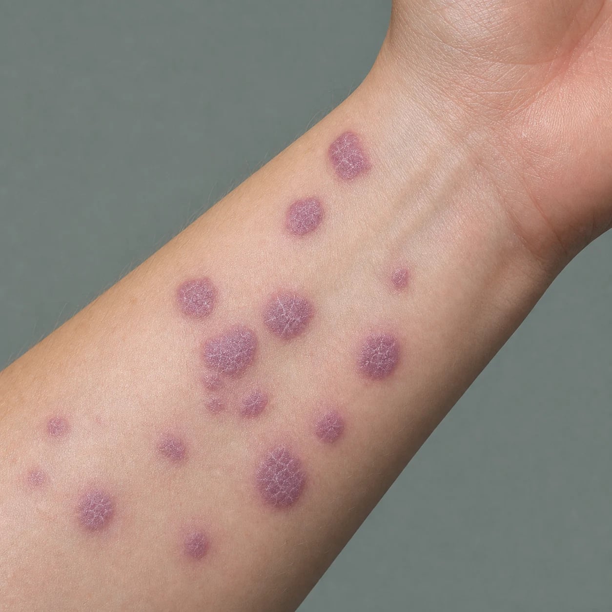
The 6 P's of cutaneous LP [1]
- Pruritic — often intensely so.
- Purple — violaceous hue.
- Polygonal — angular shape.
- Planar — flat-topped.
- Papules / Plaques — may coalesce.
- Penile, Perifollicular, Palmoplantar, Mucosal — sites examiners test.[1][2]
Epidemiology & Risk Factors
Lichen planus affects approximately 0.5–2% of the general population worldwide, with a slight female predominance for oral disease and no clear sex predilection for cutaneous disease. The peak age of onset is 30–60 years, although children and the elderly can be affected.[2]
Triggers and associations [1]
- Hepatitis C virus (HCV) — a consistent, geographically variable association. A 2023 meta-analysis reported HCV seropositivity was approximately four times more frequent in LP patients, and LP was more frequent in HCV-positive patients.[7] For oral LP specifically, meta-analyses show an increased odds of HCV infection.[8][14]
- Drugs — antimalarials, beta-blockers, NSAIDs, ACE inhibitors, methyldopa, penicillamine, gold, lithium, TNF-α inhibitors, immune checkpoint inhibitors, and others can induce lichenoid eruptions.[2]
- Contact allergens — dental amalgam and other oral metal restorations may cause oral lichenoid contact reactions.[1]
- Stress, trauma, and Koebner phenomenon — lesions may appear at sites of injury.
- Autoimmune disease — associations with coeliac disease, primary biliary cholangitis, myasthenia gravis and thymoma are described but not universal.[2]
Pathophysiology
Lichen planus is a cell-mediated autoimmune interface dermatitis directed against basal keratinocytes. [1]
- Initiation — an unknown antigen (possibly modified self-peptide, viral antigen, or drug hapten) is presented by keratinocytes or antigen-presenting cells.
- T-cell trafficking — activated CD8+ cytotoxic T-cells home to the basal epidermis and mucosal epithelium.
- Keratinocyte apoptosis — cytotoxic T-cells induce apoptosis of basal keratinocytes via granzyme B/perforin and Fas-Fas ligand pathways, producing the characteristic vacuolar interface change.
- Cytokine amplification — TNF-α, IFN-γ, IL-6, IL-17 and IL-23 contribute to the inflammatory loop and recruitment of additional lymphocytes.
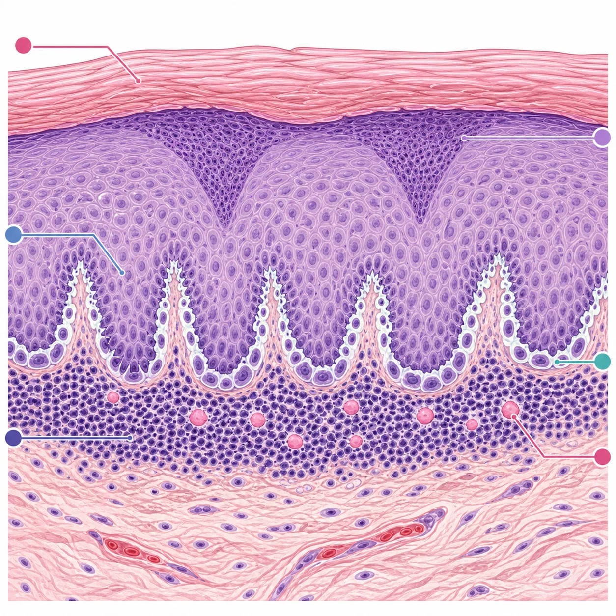
- Histologic consequence — a dense, band-like dermal lymphocytic infiltrate obscures the dermo-epidermal junction, accompanied by Civatte (colloid) bodies, sawtooth rete ridges and wedge-shaped hypergranulosis.[1][2]
Clinical Presentation
Cutaneous lichen planus
- Morphology: 2–10 mm violaceous, polygonal, flat-topped papules that may coalesce into plaques.
- Surface: Wickham striae — fine, white, reticular lines corresponding to hypergranulosis.
- Distribution: Symmetrical; flexor wrists, forearms, ankles, lower back, genitalia; palms/soles and nails in some.
- Symptoms: Moderate to severe pruritus; pain if erosive or ulcerated.
- Koebner phenomenon: new lesions at sites of trauma or scratching.
- Course: Many lesions resolve within 1–2 years; post-inflammatory hyperpigmentation is common, especially in darker skin.[1][2]
Oral lichen planus
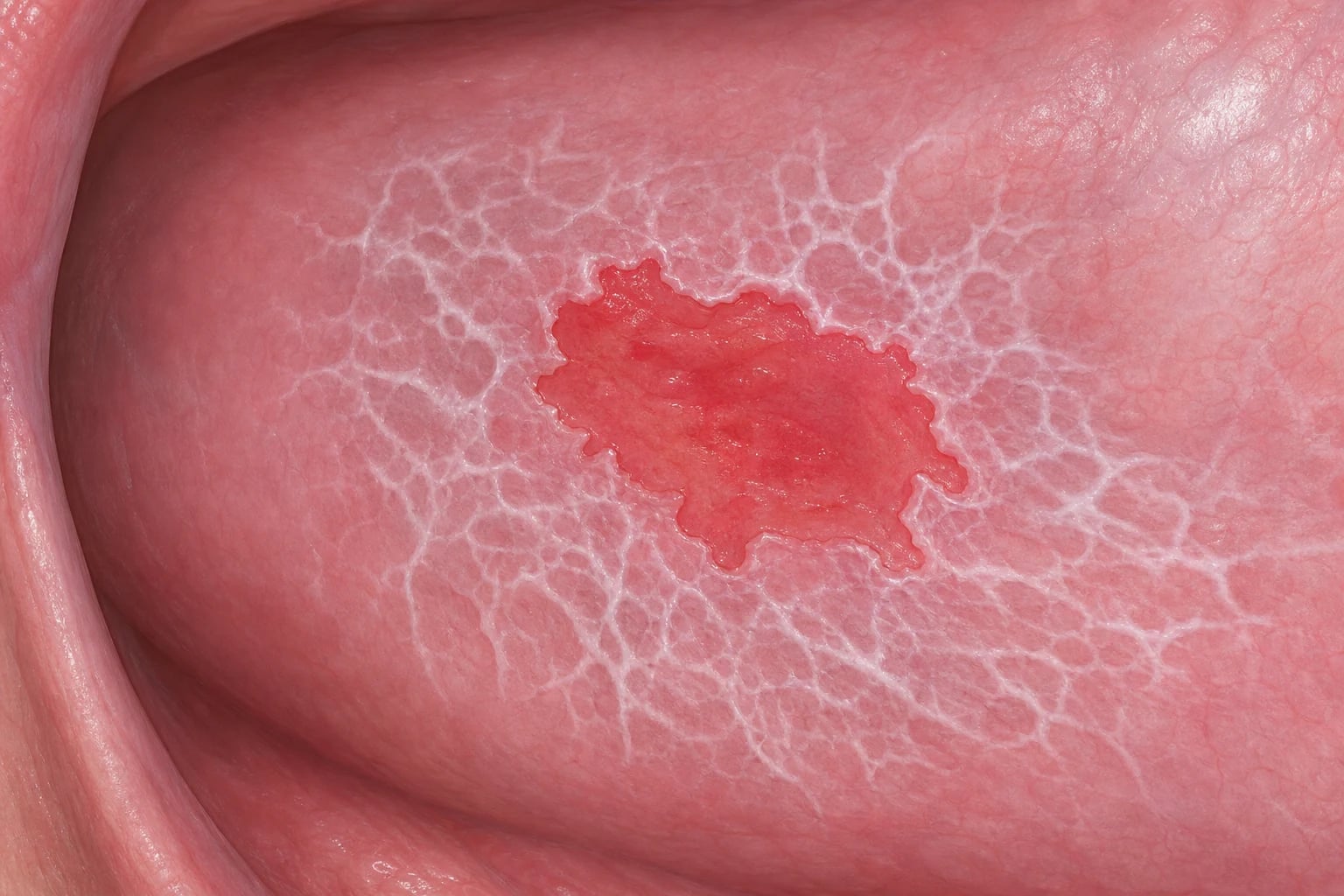
- Reticular — white, lacy Wickham striae; often asymptomatic.
- Erosive — erythematous, painful erosions with peripheral striae; highest morbidity and malignant potential.
- Plaque-like, atrophic, bullous — less common.
- Sites: Buccal mucosa most common; also gingiva, tongue, palate, lips.[1][9]
Nail lichen planus
- Longitudinal ridging, splitting, thinning, trachyonychia, onycholysis.
- Pterygium (dorsal pterygium) indicates matrix scarring.
- Anonychia may result from severe inflammation.
- May be isolated or associated with cutaneous/mucosal disease.[5]
Lichen planopilaris and frontal fibrosing alopecia
- LPP: perifollicular erythema and scale at the scalp margin; progressive scarring alopecia with tufting.
- FFA: band-like frontal hairline recession with loss of vellus hairs, often with eyebrow involvement.
- Early treatment is important because follicular destruction is irreversible.[11]
Atypical presentations examiners test
- Elderly: drug-induced lichenoid eruptions are more common; distribution may be photodistributed or extensive.
- Darker skin types: lichen planus pigmentosus and actinic LP are more common; post-inflammatory dyspigmentation is prominent.
- Children: oral disease and nail involvement occur; consider drug reactions and contact allergens.
- Immunocompromised: atypical morphology, more widespread disease, and overlapping lichenoid drug eruptions.[1][2]
Lichen Planus — Variants & Site-Specific Phenotypes
Fellowship examiners expect precise morphology-based vocabulary. Classical LP accounts for roughly half of all cases; the remainder is made up of named variants that differ in distribution, malignant potential and treatment response. Each variant shares the histopathologic interface pattern of LP but expresses a distinctive clinical face.[1][2]
Lichen planus at a glance — high-yield numbers
Hypertrophic (verrucous) LP
Thick, intensely pruritic, hyperkeratotic, violaceous plaques on the shins and anterior lower legs; psoriasiform surface, often with adherent scale. Ackerman's "hypertrophic LP" usually means more refractory disease, longer duration and highest risk of relapse. First-line therapy is topical clobetasol propionate 0.05% under occlusion or intralesional triamcinolone acetonide 5-10 mg/mL; the systemic agents of choice for recalcitrant disease are acitretin 0.5-1 mg/kg/day or methotrexate 7.5-25 mg weekly. Squamous cell carcinoma can arise within chronic hypertrophic plaques — biopsy any ulcerated or verrucous nodule that fails to respond.[1]
Annular LP
Ring-shaped plaques with a clear or hyperpigmented centre and a raised, often violaceous edge studded with Wickham striae. Common on the penis, axillae, groin and trunk; may mimic granuloma annulare, porokeratosis, tinea corporis, or erythema annulare centrifugum. Dermoscopy shows pearly white Wickham striae at the rim that are absent in the other dermatoses.[1][6]
Atrophic LP
A rare variant in which violaceous plaques evolve into thinned, depressed, hypo- or hyperpigmented patches with loss of the normal skin surface markings. The histology still shows sawtooth rete ridges and a lichenoid infiltrate, but with overlying epidermal thinning. Atrophic LP must be distinguished from lichen sclerosus (which has homogenised papillary dermal sclerosis), atrophic discoid lupus (with follicular plugging and a positive DIF), and congenital / atrophic dermatofibroma.[2]
Actinic LP (subtropical/photosensitive LP)
Annular or pigmented plaques restricted to sun-exposed sites — face, dorsal hands, forearms and V of the chest — in darker-skinned adults from tropical, sub-tropical or Middle-Eastern populations. Lesions are typically well-demarcated, hyperpigmented annular plaques with a hypopigmented centre and an active advancing edge, accentuated in spring and summer. Sun protection (broad-spectrum SPF 50+, UPF clothing, hat) is the cornerstone; pharmacotherapy includes potent topical corticosteroids (clobetasol 0.05%), hydroxychloroquine 200-400 mg/day or acitretin 0.5-1 mg/kg/day for refractory disease.[1]
Pigmented LP (lichen planus pigmentosus, LPPigm)
Diffuse or macular hyperpigmentation in slate-grey to ashy-brown macules and patches, often in the flexures (axillae, groin, neck), upper chest and face. Predominantly affects skin of colour (Fitzpatrick III-V) and tends to be chronic, progressive and cosmetically distressing. The "pigmentosus" form rarely has Wickham striae or violaceous hue — diagnosis rests on the lichenoid infiltrate on biopsy. Treatment options include photoprotection, topical tacrolimus 0.1%, topical retinoids, oral isotretinoin or acitretin, and hydroxychloroquine 200-400 mg/day; response is slow and incomplete. Pigmented LP does not share the malignant potential of erosive oral LP.[1]
Bullous LP and lichen planus pemphigoides (LPPem)
Tense vesicles and bullae that arise on established LP lesions ("bullous LP") or de novo on normal skin within an LP-distribution patient ("lichen planus pemphigoides"). Bullous LP arises from pronounced lichenoid interface damage at the dermo-epidermal junction; LPPem features circulating IgG autoantibodies against BP180 (collagen XVII) and BP230, with linear IgG/C3 along the basement membrane on direct immunofluorescence. LPPem is a true overlap between LP and bullous pemphigoid. Both respond to very potent topical corticosteroids (clobetasol 0.05%) ± short-course oral prednisone 0.5-1 mg/kg/day; refractory LPPem may need doxycycline ± nicotinamide, methotrexate 7.5-25 mg weekly, or omalizumab / dupilumab off-label in expert centres.[1][2]
Linear LP
Linear, Blaschkoid or zosteriform arrays of classical LP papules, often following a Koebner pattern along a trauma line. It typically appears in children or adolescents and may be the only manifestation. The main differential is lichen striatus (which has more eczematous and papular features, hypopigmentation and a shorter course), linear psoriasis and linear Darier disease. No treatment is usually required as it resolves spontaneously; topical corticosteroid or tacrolimus can be used for symptomatic lesions.[2]
Mucosal variants — oral LP
Oral LP is classified by the WHO into six clinical patterns: reticular (the commonest — lacy, asymptomatic Wickham striae on buccal mucosa), erosive / ulcerative (painful, erythematous erosions with the highest malignant potential), atrophic, plaque-like, papular and bullous. Symptoms vary from none to severe pain, burning and dysgeusia. Topical clobetasol propionate 0.05% gel or fluocinonide 0.05% gel/ointment is the cornerstone, with topical tacrolimus 0.1% as steroid-sparing alternative; refractory erosive disease may require methotrexate 7.5-25 mg weekly, hydroxychloroquine 200-400 mg/day, or mycophenolate mofetil 0.5-3 g/day. Surveillance every 3-6 months is essential because of the malignant potential.[4][9][13]
Genital LP — male and female
Penile LP presents as violaceous papules or annular plaques on the glans or shaft, sometimes with phimosis. Vulvovaginal LP in women is dominated by erosive disease with intense pain, dyspareunia, architectural change (introital stenosis, labial resorption, synechiae) and a risk of squamous cell carcinoma that mirrors erosive oral LP. Potent topical corticosteroid (clobetasol 0.05% ointment) or topical tacrolimus 0.1% is first-line; refractory disease may need methotrexate 7.5-25 mg weekly, mycophenolate 1-3 g/day or hydroxychloroquine 200-400 mg/day. Co-management with gynaecology / urology, biopsy of any persistent ulcer or new indurated plaque, and examination of the oral cavity for synchronous disease are all part of the work-up.[1]
Nail LP
Longitudinal ridging, fissuring, splitting, thinning, onycholysis and trachyonychia; dorsal (pterygium) and ventral pterygium indicate matrix scarring and are largely irreversible. Isolated nail LP occurs in up to 10% of patients; conversely, half of patients with nail LP have skin, mucosal or scalp disease. The cornerstone of treatment is intralesional triamcinolone acetonide 2.5-5 mg/mL into the proximal nail fold and matrix every 4-6 weeks, with oral acitretin 0.5-1 mg/kg/day or short-course oral prednisone 0.5-1 mg/kg/day for progressive disease. Early treatment (within 6-12 weeks of symptom onset) is the single most effective way to prevent permanent scarring and anonychia.[1][5]
Scalp LP — lichen planopilaris (LPP) and frontal fibrosing alopecia (FFA)
LPP presents as perifollicular violaceous erythema and hyperkeratosis at the scalp margin with progressive scarring alopecia and tufted folliculitis; FFA is a postmenopausal variant with band-like frontotemporal recession and loss of eyebrows, frequently linked to facial sunscreen and moisturiser use. Follicular destruction is permanent, so the goal is to halt disease activity, not regrow hair. First-line systemic therapy is hydroxychloroquine 200-400 mg/day; alternatives include doxycycline 100 mg twice daily, mycophenolate mofetil 0.5-3 g/day, pioglitazone 15-30 mg/day, and finasteride / dutasteride for FFA. Intralesional triamcinolone 10 mg/mL into active marginal disease and topical clobetasol 0.05% lotion for ongoing scalp inflammation are key adjuncts.[1][11]
[1]LP variant morphologies — six letters, six memories
H-A-A-A-P-B
Hyperkeratotic pruritic plaques on the shins; most refractory to therapy; acitretin 0.5-1 mg/kg/day or methotrexate 7.5-25 mg weekly
Ring-shaped plaques with raised Wickham-edged rim and clear centre; often on penis, axillae or trunk
Thinned violaceous patches; biopsy to exclude lichen sclerosus or atrophic lupus erythematosus
Sun-exposed sites in darker-skinned adults from tropical/Middle-Eastern regions; sun protection is the cornerstone
Slate-grey ashy macules in flexures of skin-of-colour patients; chronic, slow response to treatment
Tense bullae on LP or de novo; lichen planus pemphigoides has anti-BP180/BP230 IgG at the basement membrane
Differential Diagnosis
Clinical & Bedside Assessment
Focused examination
- Skin: inspect flexor wrists/forearms, ankles, lower back, genitalia, palms/soles; look for Wickham striae, Koebnerisation, and nail changes.
- Oral cavity: examine buccal mucosa, gingivae, tongue, palate and lips for reticular striae, erosions, plaques and induration.
- Genitalia: inspect vulva, vagina, glans penis and perianal skin for erosive or papular disease.
- Scalp and hair: look for perifollicular erythema, scale, scarring alopecia, and loss of vellus hairs at the hairline.
- Nails: assess ridging, thinning, pterygium, onycholysis and anonychia. [1]
Dermoscopy
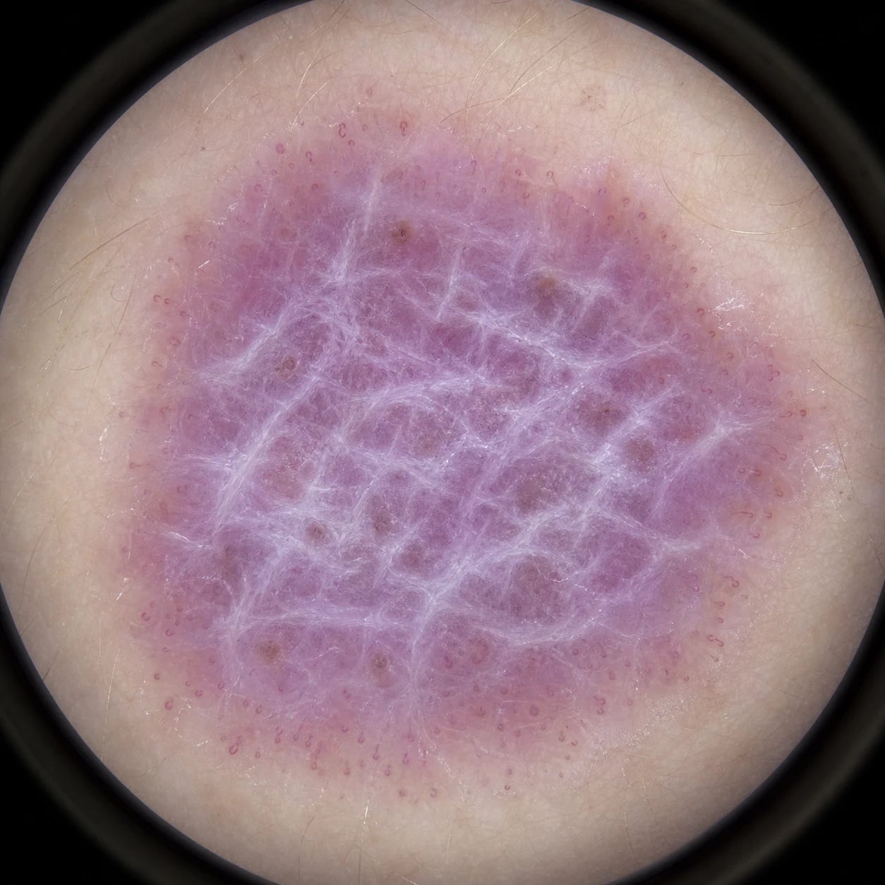
Dermoscopy is a useful bedside adjunct: [1]
- Wickham striae — the hallmark: pearly white or grey reticular lines, corresponding to hypergranulosis.
- Vascular pattern — dotted or linear vessels, often at the periphery of active lesions.
- Pigment pattern — diffuse brownish or grey-black dots/globules in regressing lesions, representing melanophages.
- Background — erythematous or violaceous hue in active lesions. [1]
Wickham striae help distinguish LP from psoriasis (regular dotted vessels, uniform scale) and eczema (yellow scales, irregular vessels).[6]
Investigations
Baseline work-up
- FBC, LFT, U&E, creatinine — before systemic therapy.
- Hepatitis C serology — especially in oral/genital LP or patients from high-prevalence regions; consider hepatitis B and HIV per local protocol.[1][7][8]
Histopathology
A 4-mm punch biopsy from an active lesion is indicated when:
- Diagnosis is uncertain.
- Atypical morphology or rapid change (to exclude malignancy).
- Erosive oral or genital disease.
- Drug-induced lichenoid eruption is suspected. [1]
Characteristic findings
- Epidermis: compact orthokeratosis, wedge-shaped hypergranulosis, sawtooth rete ridges.
- Interface change: vacuolar degeneration of the basal layer with Civatte (colloid) bodies.
- Dermis: dense, band-like lymphocytic infiltrate immediately beneath the epidermis, often obscuring the dermo-epidermal junction; melanophages in regressing lesions.
- Drug-induced lichenoid reactions may show eosinophils, parakeratosis and deeper perivascular infiltrate.[1][2]
Direct immunofluorescence
Not usually required for classic LP. It may be helpful to distinguish erosive oral LP from pemphigus, pemphigoid and lupus:
- LP/lichenoid reaction: shaggy fibrinogen and cytoid bodies; no linear immunoglobulin deposition.
- Pemphigus: intercellular IgG/C3.
- Pemphigoid: linear IgG/C3 at the basement membrane zone.
- Lupus: granular IgG/C3 at the basement membrane zone.[1]
Management — Topical Therapy
Topical therapy is first-line for limited cutaneous and most oral LP. [1]
| Agent | Indication | Notes |
|---|---|---|
| Potent/very potent topical corticosteroids (e.g., clobetasol propionate 0.05%, betamethasone dipropionate 0.05%) | Cutaneous plaques, scalp margin, nail folds | Apply once or twice daily for 2–4 weeks; taper to avoid skin atrophy[1][3] |
| Topical calcineurin inhibitors (tacrolimus 0.03–0.1% ointment, pimecrolimus 1% cream) | Face, intertriginous sites, oral and genital mucosa | First-line or steroid-sparing for mucosal disease; transient burning is common[1][4] |
| Intralesional corticosteroids (triamcinolone acetonide 5–10 mg/mL) | Thick plaques, oral erosions, nail matrix disease | Inject into the lesion; use caution on thin skin and mucosa[1][5] |
| Topical retinoids (tazarotene, adapalene) | Hyperkeratotic or hypertrophic LP | Irritation limits use[1] |
Oral lichen planus specifics
- Topical corticosteroids (clobetasol propionate 0.05% or fluocinonide 0.05% gel/ointment) remain the cornerstone for symptomatic disease.[4]
- Topical tacrolimus 0.1% is an effective steroid-sparing alternative, particularly for erosive and refractory oral LP.[1][13]
- Mouthwashes and oral hygiene optimisation are supportive measures.
- Monitor for candidal superinfection with prolonged topical steroid use.[1]
Management — Phototherapy
For widespread or refractory cutaneous LP: [1]
- Narrowband UVB (NB-UVB, 311 nm) is the most commonly used phototherapy. Typical regimens are 2–3 times weekly for 12–24 sessions, with dose titration based on skin type or minimal erythema dose.[1][3]
- PUVA (psoralen plus UVA) is effective but carries higher long-term photocarcinogenic risk; reserve for refractory disease or when NB-UVB has failed. Contraindications include pregnancy and significant photosensitivity.[1]
- Excimer laser (308 nm) can be used for localised, recalcitrant plaques.[1]
Management — Systemic Therapy
Systemic therapy is reserved for widespread, erosive, hypertrophic, nail or follicular disease that fails topical/phototherapy. [1]
Oral corticosteroids
- Prednis(ol)one 0.5–1 mg/kg/day for 2–6 weeks with a taper can control severe acute flares, including erosive oral/genital LP.[1][2]
- Use the shortest effective course and monitor for glucose intolerance, hypertension, infection and bone protection.
Acitretin
- 10–50 mg daily with food (fatty meal enhances absorption) is useful for hypertrophic LP, palmoplantar LP and nail LP. Teratogenic; avoid in pregnancy and for 3 years after stopping in women of childbearing potential.[1][5]
Methotrexate
- 7.5–25 mg once weekly (oral, subcutaneous or intramuscular) with folic acid supplementation. Monitor FBC, LFT and creatinine. Effective for recalcitrant cutaneous and oral LP.[1][2]
Other systemic agents
| Agent | Dose / notes |
|---|---|
| Mycophenolate mofetil | 0.5–3 g/day in divided doses; used for refractory erosive oral/genital LP[1] |
| Azathioprine | 1–2 mg/kg/day; check TPMT activity before starting[1] |
| Ciclosporin | 2.5–5 mg/kg/day in divided doses; short-term bridge for severe disease[1] |
| Hydroxychloroquine | 200–400 mg/day; used for oral LP and lichen planopilaris; ophthalmologic screening required[1] |
| Apremilast | 30 mg twice daily (after 5-day titration); emerging option supported by case series and small studies for refractory LP[12] |
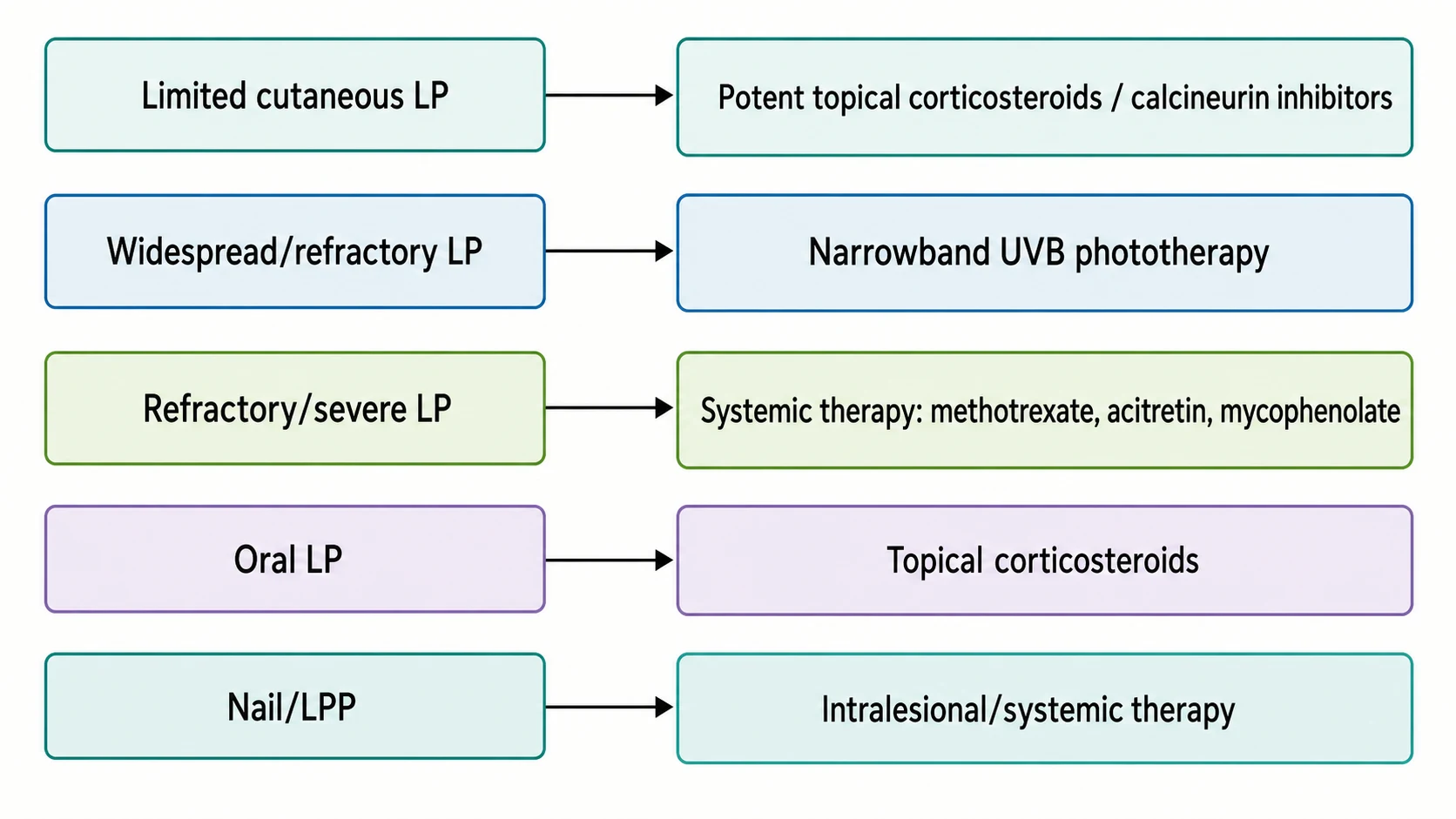
Choosing systemic therapy
- Widespread cutaneous LP: methotrexate, acitretin, or short-course ciclosporin.
- Erosive oral LP refractory to topical therapy: mycophenolate mofetil, azathioprine, ciclosporin, or methotrexate; consider apremilast or JAK inhibitor on specialist advice.
- Nail LP: intralesional triamcinolone into the nail matrix, oral corticosteroid taper, or acitretin; early treatment to prevent scarring.
- Lichen planopilaris: hydroxychloroquine, doxycycline, mycophenolate mofetil, or pioglitazone; intralesional corticosteroids for active marginal disease.[1][5][11]
Management by Special Site or Scenario
Scalp — lichen planopilaris / frontal fibrosing alopecia
- Confirm diagnosis with scalp biopsy if uncertain.
- Treat early; scarring is irreversible.
- Options: hydroxychloroquine, doxycycline, mycophenolate mofetil, intralesional corticosteroids; finasteride/dutasteride may help FFA.[11]
Nails
- Intralesional triamcinolone into the proximal nail fold/matrix.
- Systemic acitretin or short-course oral corticosteroids for progressive disease.
- Protect nails from trauma; treat onychomycosis if present.[5]
Genital/vulvovaginal LP
- Potent topical corticosteroid ointment (e.g., clobetasol) or topical tacrolimus.
- Screen for squamous cell carcinoma; treat scarring and adhesions.
- Co-manage with gynaecology/urology.[1]
Hypertrophic LP
- Potent topical steroids under occlusion, intralesional steroids, acitretin, or methotrexate.[1]
Actinic LP
- Photoprotection; potent topical corticosteroids; acitretin or hydroxychloroquine may be considered.[1]
Complications, Emergencies & Pitfalls
Malignant transformation of oral LP
- Erosive oral LP is an oral potentially malignant disorder.
- The risk of oral squamous cell carcinoma is low but real; estimated cumulative risk varies by cohort and geographic factors.
- Red flags for malignancy: persistent ulceration, induration, new plaque, bleeding, reduced tongue mobility, cervical lymphadenopathy.
- Management: long-term surveillance every 3–6 months; biopsy any suspicious area; smoking and alcohol cessation.[9]
Drug-induced lichenoid eruption
- Always take a careful drug history; common culprits include antimalarials, beta-blockers, ACE inhibitors, NSAIDs, methyldopa, lithium, TNF inhibitors and immune checkpoint inhibitors.
- Biopsy may show eosinophils and parakeratosis.
- Withdraw the suspected agent where possible; lesions usually improve over weeks to months.[2]
Permanent nail and hair loss
- Delayed treatment of nail LP can lead to pterygium and anonychia.
- Lichen planopilaris can cause irreversible scarring alopecia; early therapy preserves follicles.[5][11]
Classic pitfalls
- Missing Koebnerisation — lesions along scratch marks support LP but can be mistaken for linear psoriasis or lichen striatus.
- Misdiagnosing oral LP as candidiasis — Wickham striae and erosions point to LP; KOH and biopsy clarify.
- Attributing all purple papules to LP — biopsy atypical or treatment-resistant lesions to exclude lichenoid drug eruption, lupus or lymphoma.
- Forgetting HCV screening — offer testing in relevant populations and clinical phenotypes. [1]
Prognosis & Follow-Up
Lichen planus is usually chronic and relapsing. Cutaneous disease often resolves within 1–2 years but may leave post-inflammatory hyperpigmentation. Oral, genital, nail and follicular forms tend to persist longer and require ongoing management. [1]
Follow-up intervals [1]
- Limited cutaneous LP: review at 4–8 weeks after starting topical therapy.
- Erosive oral LP: every 3–6 months for malignancy surveillance.
- Nail LP: every 4–6 weeks initially to assess response and scarring.
- Lichen planopilaris: every 3 months to monitor disease activity and treatment toxicity. [1]
Document lesions photographically where possible to detect change.[1][9]
Special Populations
Pregnancy and breastfeeding
- Topical corticosteroids and calcineurin inhibitors are generally preferred; use the lowest effective potency for the shortest time.
- Avoid systemic retinoids (acitretin is highly teratogenic for 3 years after stopping), methotrexate and mycophenolate.
- Oral corticosteroids may be used short-term for severe disease if benefits outweigh risks.[1]
Children
- Oral LP and nail involvement occur; consider drug reactions and contact allergens.
- Use lower-potency topical steroids where possible and monitor growth with systemic therapy.[1]
Elderly
- Higher prevalence of drug-induced lichenoid eruptions and polypharmacy.
- Greater risk of skin atrophy with potent topical steroids; prefer calcineurin inhibitors on sensitive sites.
- Monitor bone health, glucose and blood pressure if systemic steroids are required.[1][2]
Immunocompromised
- Higher risk of widespread and atypical lichenoid drug eruptions, including checkpoint-inhibitor-induced disease.
- Vigilance for infection and malignancy; biopsy uncertain or changing lesions.[2]
Evidence, Guidelines & Regional Differences
Major guidelines
- European S1 guidelines (2020) — the most comprehensive international guidance, covering diagnosis, classification, HCV screening, topical and systemic treatment, phototherapy, and special sites.[1]
- BAD patient information and consensus — UK practice commonly follows BAD consensus; management mirrors European guidance with potent topical corticosteroids first-line and calcineurin inhibitors for mucosal disease.
- AAD — has not published a formal LP guideline; patient education materials endorse topical corticosteroids and calcineurin inhibitors.
Landmark reviews and trials
- Le Cleach & Chosidow, NEJM 2012 — concise clinical practice review defining morphology, variants, associations and treatment ladder.[2]
- Atzmony et al., systematic review 2016 — meta-analysis of cutaneous LP treatments, supporting topical corticosteroids, phototherapy and systemic agents.[3]
- Lodi et al., Cochrane 2020 — corticosteroids remain the mainstay for oral LP; topical calcineurin inhibitors are useful alternatives.[4]
- García-Pola et al., 2023 — updated bidirectional meta-analysis confirming the LP–HCV association.[7]
- González-Moles & Ramos-García, 2025 — contemporary review of malignant transformation risk in oral LP.[9]
Regional nuances
- HCV screening is emphasised in Mediterranean, Middle-Eastern and Asian populations where HCV prevalence is higher; in low-prevalence Northern European populations the yield is lower but testing remains reasonable for oral/genital disease.[7]
- Systemic therapy availability varies: ciclosporin, methotrexate and acitretin are widely used; apremilast and JAK inhibitors are increasingly reported but often off-label and access depends on local formularies and funding.[12]
Exam Pearls
[1]Red Flags
Exam application bank (NEET-PG / INICET)
One-line answer
Lichen planus is a chronic, immune-mediated, mucocutaneous interface dermatitis that affects skin, hair, nails and mucous membranes. Fellowship-level assessment requires mastery of the classic 6 P's morphology, named variants, Wickham striae and Koebner phenomenon, diagnostic histopathology and dermoscopy, the association with hepatitis C, malignant potential of erosive oral disease, and a tiered treatment ladder from potent topical corticosteroids and calcineurin inhibitors through phototherapy to systemic immunosuppressants and emerging small-molecule therapy.
Worked stems (answer without another resource)
Stem 1 — Classic presentation. Map symptoms to mechanism; name the first investigation and first treatment step with dose/route if drug therapy is standard. [1]
Stem 2 — Unstable / complicated. List red flags that force immediate resuscitation, theatre, ICU, antidote, or reperfusion — and what you do in the first 15 minutes. [1]
Stem 3 — Atypical group. Elderly, pregnancy, child, or immunocompromised: how presentation and thresholds change. [1]
Stem 4 — Differential trap. Name the three closest mimics and one discriminator for each. [1]
Stem 5 — Disposition. Who goes home with safety-netting, who is admitted, who needs HDU/ICU/theatre, and what follow-up is mandatory. [1]
Rapid viva checklist
- Definition + classification
- Pathophysiology chain
- Bedside signs / criteria
- Score with exact components (if any)
- Emergency bundle
- Definitive therapy with doses
- Complications of disease and of treatment
- Special populations
- Guideline/trial name if classic
- Three exam traps
Coverage self-check
If you cannot answer any stem above from this page alone, re-read the matching section — the page is intended to be self-sufficient for final-prof and NEET-PG/INICET questions on Lichen planus.
[1]References
- [1]Ioannides D, Vakirlis E, Kemeny L, Marinovic B, Massone C, Murphy R, Nast A, Ronnevig J, Ruzicka T, Cooper SM, Trüeb RM, Pujol Vallverdú RM, Wolf R, Neumann M. European S1 guidelines on the management of lichen planus: a cooperation of the European Dermatology Forum with the European Academy of Dermatology and Venereology J Eur Acad Dermatol Venereol, 2020.PMID 32678513
- [2]Le Cleach L, Chosidow O. Clinical practice. Lichen planus N Engl J Med, 2012.PMID 22356325
- [3]Atzmony L, Reiter O, Hodak E, Gdalevich M, Mimouni D. Treatments for Cutaneous Lichen Planus: A Systematic Review and Meta-Analysis Am J Clin Dermatol, 2016.PMID 26507510
- [4]Lodi G, Manfredi M, Mercadante V, Murphy R, Carrozzo M. Interventions for treating oral lichen planus: corticosteroid therapies Cochrane Database Syst Rev, 2020.PMID 32108333
- [5]Gupta MK, Lipner SR. Review of Nail Lichen Planus: Epidemiology, Pathogenesis, Diagnosis, and Treatment Dermatol Clin, 2021.PMID 33745635
- [6]Güngör Ş, Topal IO, Göncü EK. Dermoscopic patterns in active and regressive lichen planus and lichen planus variants: a morphological study Dermatol Pract Concept, 2015.PMID 26114051
- [7]García-Pola M, Rodríguez-Fonseca L, Suárez-Fernández C, Sanjuán-Pardavila R, Seoane-Romero J, Rodríguez-López S. Bidirectional Association between Lichen Planus and Hepatitis C-An Update Systematic Review and Meta-Analysis J Clin Med, 2023.PMID 37762719
- [8]Alaizari NA, Al-Maweri SA, Al-Shamiri HM, Tarakji B, Shugaa-Addin B. Hepatitis C virus infections in oral lichen planus: a systematic review and meta-analysis Aust Dent J, 2016.PMID 26475515
- [9]González-Moles MÁ, Ramos-García P. Malignant transformation of oral lichen planus: where are we now? Med Oral Patol Oral Cir Bucal, 2025.PMID 39396138
- [10]Fazel N. Cutaneous lichen planus: A systematic review of treatments J Dermatolog Treat, 2015.PMID 24916211
- [11]Husein-ElAhmed H, Husein-ElAhmed S. A Systematic Review and Bayesian Network Meta-Analysis of Medical Therapies for Lichen Planopilaris Dermatology, 2024.PMID 37852211
- [12]Hemrajani P, Godara A, Ghosh A, D'souza P. A Systematic Review of Apremilast as a Therapeutic Option for Lichen Planus Adv Skin Wound Care, 2026.PMID 42296289
- [13]Sandhu S, Klein BA, Al-Hadlaq M, Chirravur P, Bajonaid A, Xu Y, Intini R, Hussein M, Vacharotayangul P, Sroussi H, Treister N, Sonis S. Oral lichen planus: comparative efficacy and treatment costs-a systematic review BMC Oral Health, 2022.PMID 35524296
- [14]Lodi G, Pellicano R, Carrozzo M. Hepatitis C virus infection and lichen planus: a systematic review with meta-analysis Oral Dis, 2010.PMID 20412447