Dermatology · Medicine
Lichen sclerosus
Also known as Lichen sclerosus (LS) · Lichen sclerosus et atrophicus (LSA) · Balanitis xerotica obliterans (BXO) — male genital LS · Kraurosis vulvae · White-spot disease · Csillag disease
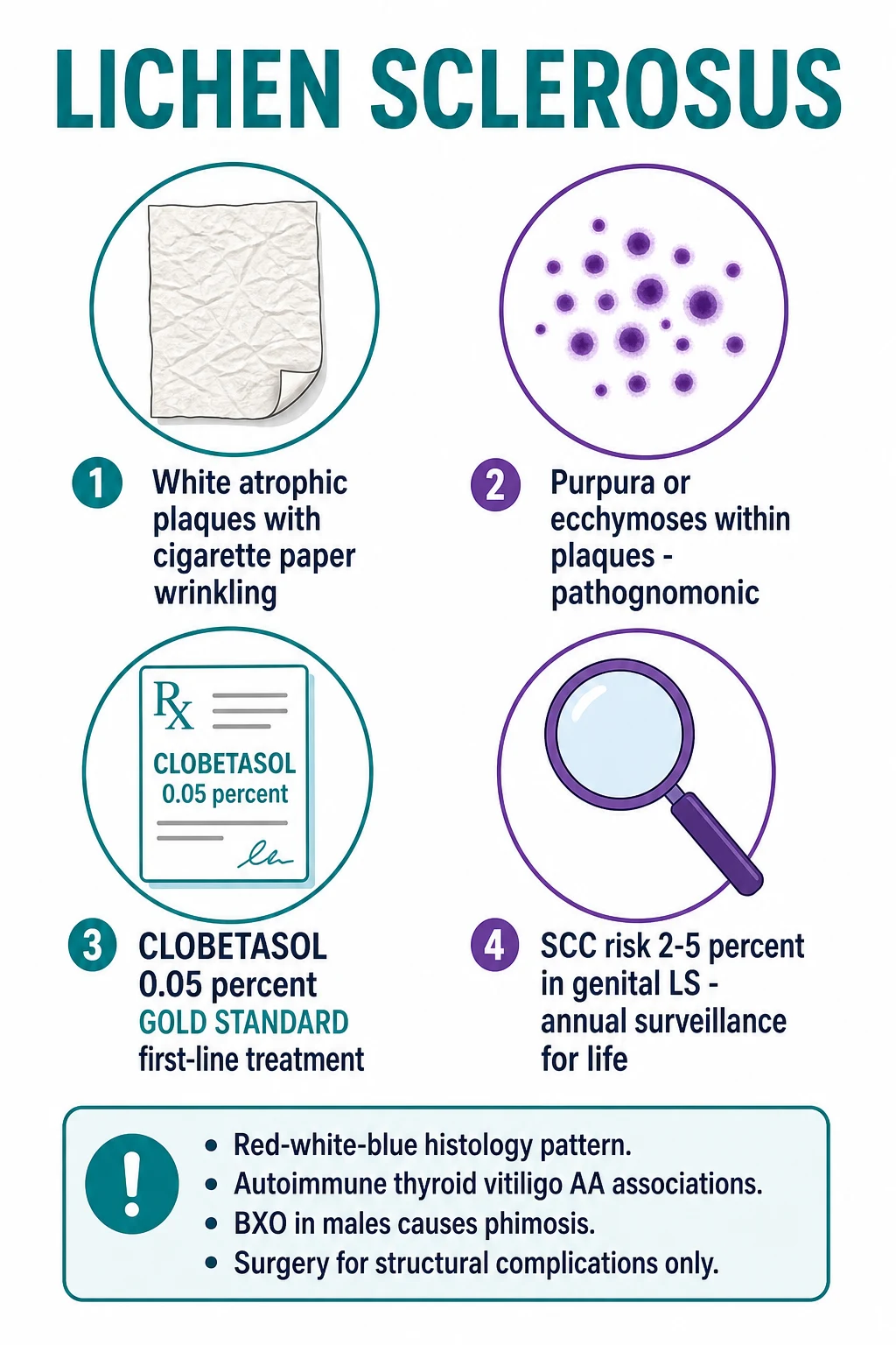
Lichen sclerosus (LS) is a chronic inflammatory dermatosis producing atrophy and sclerosis of the skin — predominantly anogenital (vulva, glans penis, perianal). Classically postmenopausal women; less common in men (BXO, usually post-circumcision or uncircumcised) and rare in prepubertal girls. Pathophysiology: autoimmune (Th1/IFN-gamma, anti-BP180/BP230 autoantibodies), oxidative stress, fibroblast dysfunction, microvascular damage, reduced elastin. Clinical: ivory-white atrophic parchment-like plaques with cigarette-paper wrinkling, purpura/ecchymoses (PATHOGNOMONIC), fissures, labial resorption, introital stenosis in females; phimosis and meatal stenosis in males (BXO). Histology: the RED-WHITE-BLUE pattern. Differential: vitiligo, lichen planus, mucosal pemphigoid, morphoea, atrophy of oestrogen deficiency, child sexual abuse. Management: ultra-potent topical corticosteroid (CLOBETASOL 0.05% ointment) in a 12-week CLOSED tapering course is GOLD STANDARD; emollients; topical calcineurin inhibitors second-line; circumcision for BXO phimosis; surgery for structural complications. Squamous cell carcinoma risk 2-5% in genital LS — annual/biennial LIFELONG surveillance.
On this page & tools
Your progress
Saved locally on this device.
Exam tags
Red flags
What it is
Where it lives
Who gets it
Hallmark sign
Gold-standard treatment
Lifetime malignancy risk
Definition
Lichen sclerosus (LS; formerly lichen sclerosus et atrophicus, LSA) is a chronic, immune-mediated, inflammatory dermatosis of skin and mucocutaneous junctions producing progressive epidermal atrophy and dermal sclerosis, with a striking predilection for anogenital epithelium. The disease is uncommon, frequently missed at first presentation, and clinically important because of three features: (1) disabling symptoms (intractable pruritus, dyspareunia, dysuria, pain), (2) progressive scarring that can destroy vulvar and penile architecture, and (3) a 2-5% lifetime risk of squamous cell carcinoma (SCC) in genital disease, mandating lifelong surveillance.[1][3][4]
The condition has several historical and clinical names: kraurosis vulvae (the old gynaecological term for vulvar LS), white-spot disease and Csillag disease (older terms for extragenital LS), and balanitis xerotica obliterans (BXO) — the male-genital name for LS affecting the prepuce, glans, and meatus. The modern unified term is lichen sclerosus, applied across all sites and both sexes.[1]
LS is a systemic autoimmune disease with a cutaneous signature — the skin is the most visible target, but the disease is associated with a constellation of other autoimmune disorders (autoimmune thyroid disease, vitiligo, alopecia areata, pernicious anaemia, type 1 diabetes) and with circulating autoantibodies to BP180 (collagen XVII) and BP230, the same antigens targeted in bullous pemphigoid.[6]
Classification
LS can be classified along three axes: site, population, and morphology.[1][11]
Genital (vulvar) LS
Genital (penile) LS / BXO
Extragenital LS
Mixed (genital + extragenital)
Prepubertal LS
Bullous LS
The ISSVD 2026 terminology (replacing the 2011 version) standardises the nomenclature for vulvar LS, dVIN (differentiated vulvar intraepithelial neoplasia), and the LS–dVIN–SCC sequence, and is the current reference for biopsy reporting.[11]
Epidemiology & Risk Factors
Risk factors for LS can be grouped into host factors, autoimmune factors, and local factors:[1][3][5]
- Host factors — female sex (10:1), perimenopausal/postmenopausal oestrogen decline, prepubertal hypo-oestrogenism, Caucasian ancestry (rare in deeply pigmented skin), HLA-DR11, DQ7, DQ9 haplotypes in familial cases.
- Autoimmune factors — personal or family history of autoimmune disease; in one series, 20-30% of LS patients had at least one other autoimmune disease (autoimmune thyroid disease the commonest, followed by vitiligo, alopecia areata, pernicious anaemia, type 1 diabetes, morphea, psoriasis).[3]
- Local factors (Koebner / isomorphic phenomenon) — friction, chronic irritation, surgical scars (perineal, episiotomy, circumcision), radiotherapy, lichenification from scratching; LS may develop in old scars or radiotherapy ports. HPV infection is more frequent in vulvar LS plaques than in normal skin, and is a cofactor in malignant transformation.
- Iatrogenic triggers — case reports implicate immune checkpoint inhibitors and antiretroviral initiation; the evidence is anecdotal.
Pathophysiology
The immunopathogenesis of LS is multifactorial — autoimmune, genetic, hormonal, oxidative, and microvascular — and converges on basal-keratinocyte injury, dermal fibroblast dysfunction, and loss of the papillary-dermal elastic-fibre network.[1][3][6]
1. Autoimmune basis. LS is a T-cell-mediated disease. The infiltrate is dominated by CD4+ and CD8+ T lymphocytes with a Th1 cytokine profile (IFN-γ, IL-1, TNF-α), with relatively few B cells. Patsatsi and colleagues (2014) demonstrated circulating IgG autoantibodies to BP180 (collagen XVII) NC16A and BP230 in 23-32% of patients with genital LS — the same antigens targeted in bullous pemphigoid, providing a direct mechanistic link between LS and the BP-spectrum.[6] These antibodies may explain the bullous variant of LS and the histological overlap with bullous pemphigoid.
2. Genetic susceptibility. Familial LS is well documented, with first-degree relatives of patients having a higher prevalence than the background population. HLA associations (DR11, DQ7, DQ9) have been reported, particularly in familial and childhood cases. Loss-of-function variants in barrier and immune genes are under investigation.[1][3]
3. Hormonal influence. LS skin shows reduced expression of oestrogen receptor-β and androgen receptor. The disease is most active in two low-oestrogen states (postmenopause, prepuberty), and there is anecdotal response to topical oestrogen or testosterone (now superseded by topical clobetasol). The mechanistic basis for the female predominance is not fully understood.[3][5]
4. Oxidative stress and fibroblast dysfunction. LS lesional skin has reduced antioxidant defences (decreased glutathione, superoxide dismutase, catalase) with increased reactive oxygen species. Dermal fibroblasts are hyperproliferative and produce excessive type I and III collagen; elastin synthesis is reduced. The result is dermal sclerosis with loss of the papillary-dermal elastic-fibre network — the structural basis of the atrophic, fragile, easily-bruised skin.[3]
5. Microvascular injury and matrix-remodelling dysregulation. Chronic inflammation damages the subepidermal capillary plexus. Matrix metalloproteinases (MMP-2, MMP-9) and tissue inhibitors of metalloproteinases (TIMPs) are dysregulated. The basement-membrane zone becomes fragile, and subepidermal haemorrhage (the pathognomonic purpura) and bullae develop.[1][3]
6. The Koebner (isomorphic) phenomenon. LS can be induced at sites of trauma — old surgical scars, radiotherapy ports, friction, scratching. This is important because it explains why LS may recur at the edge of a vulvectomy scar, why circumcision for BXO does not cure the disease, and why friction should be minimised (loose cotton underwear, lubricants, emollients).[1][3]
Clinical Presentation
Female genital LS — the classic adult presentation
The classic adult is a postmenopausal woman (50-70 years) with a 2- to 10-year history of:[1][5]
- Intractable vulvar pruritus — the most common presenting symptom; worse at night, exacerbated by warmth, sweat, and friction.[1][5]
- Burning and rawness — often described as "the skin feels torn" or "acid is on it".
- Dyspareunia — superficial and deep; from introital stenosis, fissuring, and clitoral involvement.
- Dysuria and post-micturition burning — from involvement of the urethral meatus and anterior vestibule.
- Constipation and perianal pain — from perianal involvement with fissuring, particularly in prepubertal disease but also in adult women with long-standing disease.
- "Cigarette paper" wrinkling of the surface — a hallmark; the surface has a fine, crinkled, hyperepigmented-at-the-margin appearance.
- Purpura and ecchymoses within the white plaques — PATHOGNOMONIC. The purpura is non-palpable, non-blanching, and non-tender, and may be the presenting complaint (the patient thinks it is trauma; it is not).
- Hyperkeratosis — particularly over the clitoral hood and labial minora in long-standing disease.
- Fissures — typically at the posterior fourchette, perineum, and perianal midline; painful; bleed on intercourse or defecation.
- Scarring — the late finding: labia minora resorption (the labia minora disappear), clitoral burying (the clitoris is engulfed by a phimotic hood), introital stenosis, perianal stenosis, and reconfiguration of the vulvar architecture into a flattened, scarred, and narrowed introitus.
Early LS
Established LS
Late / scarred LS
Bullous LS
Male genital LS — BXO
In males, LS presents as BXO — the same disease in the prepuce, glans, and anterior urethra:[3][8][12]
- Phimosis — progressive inability to retract the foreskin; the prepuce becomes tight, sclerotic, and white; the underlying glans shows a white sclerotic ring at the coronal sulcus.[3][8]
- Meatal stenosis — fissuring and narrowing of the urethral meatus; urinary spraying, deflection, straining, and (in severe cases) retention.
- Anterior urethral stricture — extension of the sclerosis into the navicular fossa and beyond; rare but important to recognise.
- Sexual dysfunction — from pain, phimosis, and loss of glans sensation.
- Balanitis — secondary candidal or bacterial infection; fissuring and bleeding.
BXO classically presents in two populations: (1) boys aged 8-15 with apparently primary phimosis (in retrospect, often LS), and (2) adult men 30-40 with phimosis or meatal stenosis, particularly after circumcision for "phimosis" (in retrospect, LS).[8]
Extragenital LS
Extragenital LS occurs in 15-20% of patients and may be the only site, or accompany genital disease.[2] It presents as ivory-white, atrophic, porcelain-like macules and patches with a wrinkled surface, often with follicular plugging (a histological correlate) and a sharply defined edge. Common sites are the trunk, upper back, neck, axillae, breasts, and wrists. The patches are usually asymptomatic and come to attention because of their appearance. Importantly, extragenital LS has NO malignant potential and does not require SCC surveillance.[2]
Prepubertal LS
Prepubertal LS in girls (and rarely in boys) is a well-recognised entity with important clinical and safeguarding implications:[9]
- Peak age 5-8 years; sometimes younger.[9]
- Symptoms — perianal bleeding (from fissures), constipation (from perianal pain and anal fissure), vulvovaginal soreness, dysuria, nocturnal pruritus.
- Signs — ivory-white atrophic plaques and erosions in the vulvoperianal area; haemorrhagic blisters; perianal fissuring and "figure-of-eight" involvement.
- Misdiagnosis — most commonly confused with sexual abuse (the perianal bruising, fissuring, and bleeding can be indistinguishable on inspection alone) or with chronic constipation, candidiasis, or threadworm.
- Treatment — the same ultra-potent topical corticosteroid regimen (clobetasol 0.05% ointment OD for 4 weeks, then alternate days, then twice weekly), with careful counselling of the parents and involvement of safeguarding where appropriate.
- Prognosis — up to 50-75% of girls enter remission at puberty; the remainder have disease into adult life with a persistent SCC risk that mandates continued surveillance.
Atypical presentations examiners test
- Prepubertal LS misdiagnosed as sexual abuse (the most consequential mimicker in paediatric dermatology).[9]
- Male LS presenting as acute phimosis in a previously retractable foreskin; meatal stenosis with obstructive urinary symptoms in a young man.
- Bullous LS — tense haemorrhagic bullae on sclerotic plaques; may mimic bullous pemphigoid or bullous morphoea.
- Haemorrhagic LS with extensive purpura mimicking trauma, vasculitis, or Kaposi sarcoma.
- LS in pregnancy — improves in some patients, worsens in others; clobetasol is safe in small amounts.
- LS in immunosuppressed patients (HIV, transplant) — higher prevalence, more severe, and higher malignant risk.
Differential Diagnosis
The differential diagnosis of the white atrophic anogenital plaque is the most-tested clinical scenario in the topic.[1][5][7]
Vitiligo
Lichen planus (LP)
Mucosal (cicatricial) pemphigoid
Morphoea (localised scleroderma)
Genitourinary syndrome of menopause (atrophic vaginitis)
Lichen simplex chronicus
Chronic candidiasis
Child sexual abuse (prepubertal)
Vulvar intraepithelial neoplasia (VIN, usual type / HPV-related)
Squamous cell carcinoma (SCC) arising in LS
Clinical & Bedside Assessment
Female anogenital exam (in detail). Inspect in good light with the patient in the lithotomy position. Examine in a structured way: [1][5]
- Labia majora — pallor, atrophy, hyperkeratosis, fissuring.[1][5]
- Labia minora — resorption (a late sign; the labia minora "disappear" as the disease progresses), burying of the clitoris, adhesions to the majora.
- Clitoral hood and clitoris — buried clitoris (the clitoris is engulfed by a phimotic, sclerotic hood; this is a sensitive marker of late disease).
- Vestibule and urethral meatus — pallor, erythema, fissuring, stenosis, hyperkeratosis.
- Posterior fourchette and perineum — fissuring, erosions, hyperkeratosis, scarring.
- Perianal skin — figure-of-eight continuity is the hallmark; check for fissures, erosions, ecchymoses, and the radial "purse-string" scarring of long-standing perianal LS.
- Speculum examination — only if introital patency allows; document the degree of introital stenosis and any vaginal involvement (rare in pure LS — think of LP if the vagina is involved).
- Inguinal and femoral lymph nodes — palpate; persistent lymphadenopathy with a new ulcer is suspicious of SCC.
Male genital exam. With the patient supine, retract the foreskin gently (or attempt to — phimosis is often complete). Document:[8][12]
- Prepuce — tightness, sclerotic white ring, fissuring, bleeding; the prepuce may be non-retractable.[8][12]
- Glans — white sclerotic plaques, especially at the coronal sulcus; involvement of the navicular fossa (use a meatoscope or otoscope to inspect).
- Meatus — patency, fissuring, narrowing; a gentle attempt to pass a small Foley or cotton-tipped applicator.
- Urethra — palpate the penile urethra along the ventral shaft; tenderness or induration suggests anterior urethral involvement.
- Inguinal nodes — palpate for lymphadenopathy.
Bedside manoeuvres. [1]
- Cotton-tipped applicator test — light pressure on the vestibule reproduces the patient's pain in vulvodynia / provoked vestibulodynia; useful when there is a co-existent pain syndrome.[1][5]
- Foreskin retraction test — gentle attempted retraction in BXO; incomplete or impossible retraction with a white sclerotic ring is diagnostic.
- Q-tip test for meatal stenosis — passage of a small cotton-tipped applicator through the meatus; difficulty or pain suggests stenosis.
- Photography — serial clinical photographs are useful to document disease activity and response to treatment; also useful for the surveillance of suspicious areas.
- The "scratch sign" — light rubbing of the surface of an LS plaque produces a sharp, well-demarcated purpuric mark (the Darier-like sign of LS); this is the bedside correlate of the structural fragility of the papillary dermis.
Investigations
LS is primarily a clinical diagnosis. The British Association of Dermatologists (BAD) and EuroGuiderm guidelines reserve skin biopsy for: (1) diagnostic uncertainty, (2) refractory disease not responding to ultra-potent topical steroids, (3) any suspected SCC, dVIN, or other premalignant change, and (4) consideration of alternative diagnoses.[4]
Histopathology — the RED-WHITE-BLUE pattern
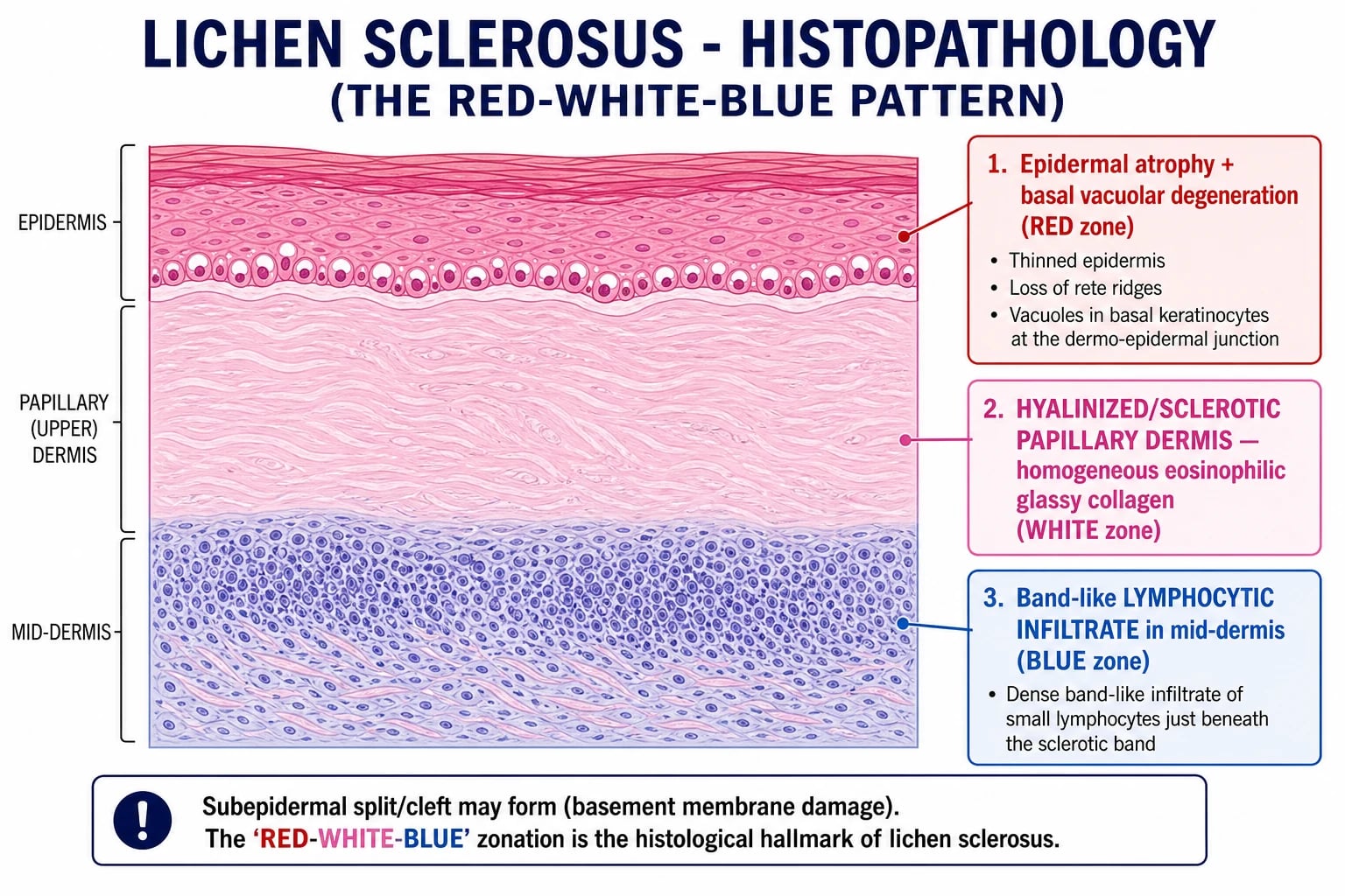
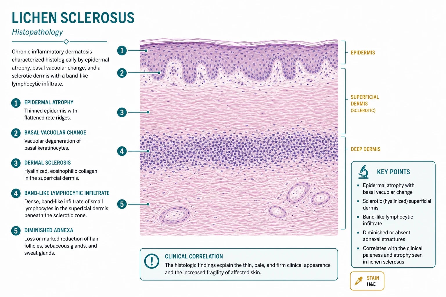
The histology of LS is a layered zonation that is reproduced in dermatology and pathology teaching as the "red-white-blue" pattern:[1][3]
- RED zone (epidermis) — epidermal atrophy (thinned epidermis, loss of rete ridges) + hyperkeratosis (compact orthokeratosis, follicular plugging) + basal vacuolar degeneration (vacuoles in the basal keratinocytes at the dermo-epidermal junction — the interface change).[1][3]
- WHITE zone (upper / papillary dermis) — hyalinisation (sclerosis) of the papillary dermis: a homogenised, eosinophilic, glassy, paucicellular band; loss of elastic fibres; reduced cellularity; oedema in early lesions; sclerosis in late lesions. The basement membrane is thickened and reduplicated.
- BLUE zone (mid-dermis) — a band-like lymphocytic infiltrate with admixed histiocytes, situated BELOW the hyalinised zone (i.e. not at the interface but in the mid-dermis — a key feature distinguishing LS from LP, where the infiltrate is at the interface).
- Subepidermal split / cleft — a frequent finding, due to basement-membrane damage from basal vacuolar degeneration; in the bullous variant, a frank bulla forms.
Lichen sclerosus (RED-WHITE-BLUE)
Lichen planus
Morphea / scleroderma
Bullous pemphigoid
Direct immunofluorescence (DIF)
DIF is NEGATIVE in LS — a useful discriminator. In the bullous variant, linear IgG and C3 at the basement-membrane zone may be seen (overlap with bullous pemphigoid), and the serum may contain anti-BP180 NC16A and anti-BP230 IgG autoantibodies (positive in 23-32% of genital LS).[6] The presence of DIF immunoglobulin deposition at the BMZ should prompt consideration of bullous pemphigoid, mucous membrane pemphigoid, or epidermolysis bullosa acquisita.
Baseline investigations at first visit
- FBC, LFTs, U&E, fasting glucose — to assess general health and to provide baselines for any future systemic therapy.[1][4]
- Thyroid function (TSH) and anti-TPO antibodies — autoimmune thyroid disease is the commonest autoimmune comorbidity (15-20% of LS patients).
- Vitamin B12, intrinsic-factor antibodies — if pernicious anaemia is suspected.
- HbA1c — if type 1 diabetes is suspected.
- Hepatitis B and C serology — if systemic immunosuppression is being considered.
- HIV testing — if risk factors.
- Swab for culture — if secondary candidal or bacterial infection suspected.
- HPV testing — if hyperkeratotic or atypical plaques; may be co-factor in malignant transformation.
- Pregnancy test — in women of childbearing age before any systemic therapy.
Dermoscopy
Dermoscopy is a useful bedside adjunct to distinguish LS from vitiligo and other hypopigmented genital disorders. The dermoscopic features of LS include:[7]
- White structureless areas with rosettes (four-cloverleaf white structures corresponding to follicular plugging) — a relatively specific feature.[7]
- Linear vessels or dotted vessels in the surrounding non-atrophic skin.
- Comedo-like openings (follicular plugging).
- Absence of the pigment network seen in vitiligo.
Management — Resuscitation
LS is a chronic disease, not an emergency, but several clinical scenarios require urgent intervention:[1][4]
1. Acute urinary retention in BXO with meatal stenosis. Try gentle urethral catheterisation; if the meatus will not admit a small Foley, do NOT force it (false passage). Use a suprapubic catheter for decompression, then arrange urgent urology for meatotomy / circumcision. Treat any superimposed UTI with appropriate antibiotics. Clobetasol 0.05% ointment is started as soon as the acute episode is controlled to suppress the underlying inflammation.[1][3][8]
2. Severe erosive / ulcerated LS with secondary infection. Take a swab for bacterial and candidal culture. Apply potassium permanganate 1:10 000 soaks or normal saline compresses. Start empirical flucloxacillin (or per local protocol) pending culture; add fluconazole 150 mg stat if candidiasis is suspected. Barrier protection (zinc oxide paste) until the acute inflammation settles, then clobetasol induction.[1][4]
3. Suspected SCC arising in LS. URGENT 4-mm punch biopsy of any new lump, ulcer, induration, or hyperkeratotic area within an LS plaque. Once SCC is confirmed, stage with ultrasound of the groin nodes and MRI pelvis, and refer to the gynaecological / penile oncology multidisciplinary team. Wide local excision (vulvectomy / glansectomy) with margin assessment is the treatment of choice; inguinal node dissection for high-risk disease.[1][4][5]
4. Severe vulvodynia or sexual dysfunction secondary to LS. Treat the underlying inflammation with clobetasol; add topical lidocaine 2-5% ointment for pain; consider oral amitriptyline 10-25 mg nocte (escalated to 75 mg) or gabapentin 300 mg TDS for neuropathic pain; psychosexual counselling for couples; vaginal dilators once inflammation is controlled. [1]
5. Severe constipation in prepubertal LS. Treat the underlying LS (clobetasol induction) to allow the anal fissure to heal. Add osmotic laxatives (lactulose or polyethylene glycol 3350) to soften stools and reduce straining; encourage adequate fluid and dietary fibre; consider stool softeners (docusate sodium) in the acute phase. Address the parents' anxiety about the bleeding; reassure that the bleeding is from the fissure, not from serious bowel disease. [1]
Management — Definitive & Stepwise
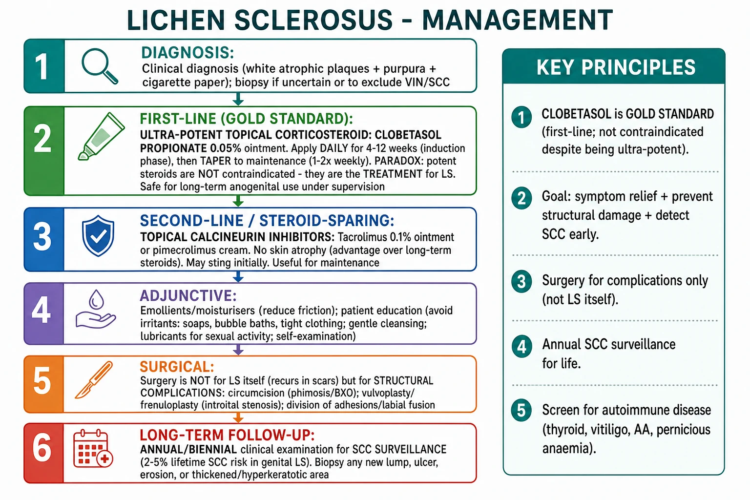
The BAD, EuroGuiderm, and AAD / ISSVD guidance converge on a single algorithm: clobetasol induction → maintenance; topical calcineurin inhibitors as steroid-sparing; surgery for structural complications only; lifelong SCC surveillance.[1][4][11]
First-line: ultra-potent topical corticosteroid (CLOBETASOL)
Clobetasol propionate 0.05% ointment
Dose
Once daily (OD) at night
Mometasone furoate 0.05% ointment
Dose
Once daily
Betamethasone dipropionate 0.05% ointment
Dose
Once daily
Practical application — the BAD and EuroGuiderm guidelines recommend a fingertip unit (FTU) — the amount of ointment expressed from a 5-mm nozzle along the distal phalanx of the index finger — per application, applied thinly at night to the affected areas. For an adult vulva, ~1 FTU is sufficient; for an adult penis, ~0.5 FTU; for a prepubertal vulva, ~0.25-0.5 FTU. [1]
[1]Second-line: topical calcineurin inhibitors (steroid-sparing)
Tacrolimus 0.1% ointment
Dose
Twice daily
Pimecrolimus 1% cream
Dose
Twice daily
Adjunctive therapy
- Emollients / moisturisers — bland emollients (white soft paraffin / liquid paraffin 50:50; petrolatum; or proprietary emollient creams) applied liberally between steroid applications. Reduces friction, irritation, and the Koebner trigger.
- Soap substitutes — aqueous cream, emulsifying ointment, or dedicated soap substitutes; avoid perfumed soaps, washes, wipes, and feminine hygiene sprays.
- Barrier preparations — zinc oxide paste or zinc and castor oil cream for fissuring and perianal involvement; protects the skin from urine, faeces, and friction.
- Avoidance of irritants — tight synthetic underwear, friction, spermicides, perfumed products, prolonged sitting.
- Psychological and psychosexual support — LS has a major impact on quality of life, sexual function, and mental health; psychosexual counselling for couples, and referral to a clinical psychologist for disease-related distress.
- Vaginal dilators — for introital stenosis; used once the acute inflammation is under control; gentle graduated dilation to maintain or restore patency.
- Physiotherapy — pelvic-floor physiotherapy for secondary vaginismus and dyspareunia. [1]
Refractory disease
Oxytetracycline 500 mg BD
Dose
Oral, twice daily
Methotrexate 7.5-15 mg weekly
Dose
Oral or subcutaneous, ONCE WEEKLY with folic acid 5 mg the day after
Acitretin 25-50 mg daily
Dose
Oral, daily with food
Narrowband UVB (NB-UVB, 311 nm) or PUVA
Dose
2-3 sessions per week
- Ablative CO₂ laser — for hyperkeratotic, refractory plaques; vapourises the surface and may relieve symptoms; small series; not a first-line.
- JAK inhibitors (case reports) — emerging; not yet standard of care. [1]
Surgical management — STRUCTURAL COMPLICATIONS ONLY
Circumcision
Meatotomy / meatoplasty
Vulvoplasty / perineoplasty
Vulvectomy
Long-term surveillance — the MANDATORY annual / biennial review
Annual or biennial clinical examination of the genital area for LIFE. This is the single most important management step after induction therapy. The examination includes:[1][4][5]
- Visual inspection of the vulva / penis / perianal area in good light.
- Palpation for induration, nodules, or asymmetry.
- Inspection of the inguinal / femoral nodes.
- Low threshold for 4-mm punch biopsy of any new lump, ulcer, induration, persistent erosion, or hyperkeratotic area.
- Patient education on monthly self-examination and the need to report any new lesion, bleeding, or non-healing area urgently.
- Smoking cessation — smoking is a cofactor in malignant transformation.
- HPV vaccination in adolescents and young adults (where indicated and available) — HPV is a cofactor in dVIN/SCC. [1]
Specific Subtypes & Scenarios
Female genital LS
The commonest subtype. Postmenopausal onset, figure-of-eight anogenital distribution, symptomatic with pruritus and dyspareunia, scarring with labia minora resorption and introital stenosis. Treatment is the standard clobetasol induction → twice-weekly maintenance → annual or biennial review for SCC.[1][4][5]
Male genital LS (BXO)
Phimosis, meatal stenosis, urinary symptoms. Treatment is clobetasol for early or mild disease; circumcision for phimosis (definitive, but does not cure the underlying LS — recurrence in the scar is possible); meatotomy for meatal stenosis. Annual surveillance for penile SCC in men with persistent glanular or preputial disease. The 2026 BASHH national guideline on balanoposthitis and related penile dermatological conditions provides the up-to-date management framework.[8][12]
Prepubertal LS
Up to 50-75% of girls enter remission at puberty, but the remainder have disease into adult life. The Savage 2026 series in Pediatric Dermatology provides long-term outcome data: most children who do not remit continue to have symptoms into adulthood and require ongoing surveillance. The most important clinical task is to distinguish LS from sexual abuse — both are valid differentials in perianal bleeding, fissuring, and constipation, and the management of misdiagnosis is lifelong. Biopsy, photography, and multidisciplinary (paediatric dermatology, gynaecology, safeguarding) assessment are required.[9]
Extragenital LS
Ivory-white atrophic macules and patches on the trunk, neck, upper arms, breasts. Usually asymptomatic. Treatment only if symptomatic or cosmetically distressing (clobetasol, tacrolimus, phototherapy). NO malignant potential — do not include in SCC surveillance.[2]
Bullous LS
Tense haemorrhagic bullae on sclerotic plaques, due to basement-membrane damage. Serum anti-BP180 and anti-BP230 autoantibodies may be positive (overlap with bullous pemphigoid). DIF may show linear IgG/C3 at the BMZ. Treatment is ultra-potent topical steroids; severe cases may require a short course of oral corticosteroids or immunosuppression.[6]
LS in pregnancy
The symptoms of LS may improve in some patients during pregnancy (high oestrogen) and worsen in others. Clobetasol is safe in small amounts; maintenance therapy is usually continued. Vaginal delivery is usually possible if the introitus is patent; caesarean section may be considered for severe vulvovaginal involvement. Post-partum flare is common; reinstitute or escalate clobetasol promptly. [1]
Haemorrhagic LS
Extensive purpura and ecchymoses on the LS plaques, often mistaken for trauma or non-accidental injury. The diagnosis is clinical (white atrophic plaques + intra-plaque purpura), but a biopsy may be required to confirm LS and to exclude vasculitis or Kaposi sarcoma in selected cases. [1]
Complications & Pitfalls
Disease-specific complications
- Scarring — labia minora resorption, clitoris burying, introital stenosis, perianal stenosis; in men, phimosis, meatal stenosis, anterior urethral stricture.
- Sexual dysfunction — dyspareunia, apareunia, secondary vaginismus, loss of glans sensation in BXO, erectile dysfunction from penile pain.
- Urinary complications — spraying, deflection, straining, retention, recurrent UTI (from meatal stenosis in BXO).
- Malignancy — vulvar SCC in 2-5% of female genital LS; penile SCC in men with BXO (rare but reported); extragenital LS has NO malignant potential.
- Psychological impact — depression, anxiety, relationship breakdown, body-image disturbance, loss of sexual confidence; under-recognised and under-treated. [1]
Classic pitfalls
[1]Pitfalls of long-term ultra-potent topical steroid use
Theoretically, chronic use of clobetasol can cause skin atrophy, striae, telangiectasia, contact dermatitis, perioral dermatitis, glaucoma (if used near the eye), and (very rarely) hypothalamic-pituitary-adrenal (HPA) axis suppression. In LS, the practical risk of clinically significant steroid-induced atrophy is LOW (because the skin is already atrophic), but patients should be advised to use the minimum effective amount, to apply it only to affected areas, and to report any new skin change. HPA axis suppression has been reported with extensive use in children and is a reason to use the lowest effective maintenance dose. [1]
Pitfalls of topical calcineurin inhibitors
Transient burning, stinging, or irritation at the application site is common (up to 25% of patients) and usually settles within 1-2 weeks. The FDA black-box warning about theoretical malignancy risk has not been substantiated in human LS studies.[10]
Legal and safeguarding pitfalls
Missed SCC in long-standing LS is a recognised cause of litigation. A clear, documented surveillance plan (annual or biennial review, low biopsy threshold) and patient education on self-examination are the standard of care. In prepubertal disease, the failure to consider LS in a child with perianal symptoms — and the failure to consider sexual abuse in a child with LS-like findings — are both potentially serious errors. [1]
Prognosis & Disposition
Prognostic factors. Good prognostic markers: early presentation, treatment adherence, smoking cessation, no architectural change at first visit, absence of dVIN. Poor prognostic markers: late presentation, scarring at first visit, treatment non-adherence, smoking, immunosuppression, dVIN, and chronic HPV co-infection. [1]
Discharge planning. Patients with LS are usually managed in the community with annual or biennial hospital review. Discharge from specialist care is NOT recommended — the lifetime SCC risk mandates lifelong follow-up. The patient and primary care physician should be given a clear written plan: clobetasol maintenance dose, emollient use, monthly self-examination, and the indications for urgent re-referral (new lump, ulcer, induration, bleeding, non-healing area). [1]
Special Populations
Prepubertal girls
Clobetasol induction regimen is safe and effective. Up to 50-75% remit at puberty (the Savage 2026 series provides the best long-term data).[9] The remainder have disease into adult life. The most important clinical task is to distinguish LS from sexual abuse — both are valid differentials, both may co-exist, and the management of misdiagnosis is lifelong. Biopsy (in selected cases), photography, and multidisciplinary (paediatric dermatology, gynaecology, safeguarding) assessment are required. Long-term follow-up into adult life is recommended.
Pregnancy
Clobetasol is safe in small amounts (category C but extensive clinical experience supports its use). Maintenance therapy is usually continued. Vaginal delivery is usually possible if the introitus is patent; caesarean section may be considered for severe vulvovaginal involvement to avoid perineal trauma. Post-partum flare is common; reinstitute or escalate clobetasol promptly. [1]
Elderly
High prevalence of comorbidities (cardiovascular, diabetes, osteoporosis), polypharmacy, cognitive impairment, and physical disability (hand arthritis, visual impairment) that may limit the ability to apply topical therapy. Consent issues. Higher index of suspicion for SCC given the background risk. Consider a carer or district nurse for medication application. Twice-weekly maintenance is usually well tolerated. [1]
Immunocompromised (HIV, transplant, chemotherapy)
Higher prevalence and severity of LS. Higher risk of malignant transformation. Consider more frequent surveillance (annual rather than biennial). Drug interactions (tacrolimus with systemic immunosuppressants). Biopsy any atypical lesion early. HPV co-infection is more frequent; HPV vaccination should be considered. [1]
Anticoagulated patients
Bruising and purpura in LS may be accentuated by anticoagulation (warfarin, DOACs, heparin). Bleeding from fissures and erosions is more common. Consider short courses of tranexamic acid for acute bleeding episodes; clobetasol is safe in this group. [1]
Evidence, Guidelines & Regional Differences
Major guidelines
- British Association of Dermatologists (BAD) 2018 guideline on LS — the reference UK guideline. Clobetasol propionate 0.05% ointment as first-line; 12-week induction taper; lifelong twice-weekly maintenance; annual or biennial SCC surveillance; tacrolimus as second-line steroid-sparing.
- EuroGuiderm 2024 guideline on LS treatment — the pan-European guideline, an update of the 2017 EDF guideline. Confirms the clobetasol taper; endorses topical calcineurin inhibitors as second-line; emphasises the role of emollients and avoidance of irritants; provides an evidence-graded treatment algorithm; consensus on life-long SCC surveillance for genital LS.[4]
- ISSVD 2026 terminology for vulvar and vaginal disease — standardises the nomenclature for LS, dVIN, and the LS–dVIN–SCC sequence, and is the current reference for biopsy reporting.[11]
- AAD (American Academy of Dermatology) guidelines — broadly consistent with BAD/EuroGuiderm; clobetasol as first-line; emphasises the role of patient education and self-examination.
- IADVL (Indian Association of Dermatologists, Venereologists and Leprologists) consensus — broadly consistent; emphasises cost-effective management with generic clobetasol and emollients; tacrolimus is recommended as second-line where cost is not a constraint.
- BASHH 2026 national guideline on balanoposthitis and related penile dermatological conditions — covers BXO, with a treatment algorithm for phimosis, meatal stenosis, and conservative management.[12]
Landmark reviews and trials
- De Luca 2023 — comprehensive review covering epidemiology, pathophysiology, clinical features, and treatment.[1]
- Kirtschig 2024 (EuroGuiderm) — the most current evidence-based guideline.[4]
- Krapf 2020 — clinical perspectives on vulvar LS, with a focus on the gynaecological assessment.[5]
- Fergus 2020 — systematic review on pathophysiology and treatment, with a particular focus on BXO.[3]
- Arif 2022 — comprehensive review of extragenital LS, with a treatment algorithm.[2]
- Patsatsi 2014 — circulating anti-BP180/BP230 autoantibodies in genital LS, providing the mechanistic link to the bullous pemphigoid spectrum.[6]
- Savage 2026 — long-term outcomes of prepubertal-onset vulvar LS, providing the best data on remission rates at puberty.[9]
- Patel 2026 — systematic review of topical tacrolimus for LS, confirming efficacy and safety.[10]
- Soliman 2024 — dermoscopic features of hypopigmented genital disorders, including LS.[7]
- Fox 2024 — treatment algorithm for severe BXO in boys.[8]
Regional nuances
- United Kingdom — BAD recommends twice-weekly clobetasol maintenance indefinitely; tacrolimus is recommended as second-line where steroid atrophy or steroid-refractory disease occurs.
- Europe — EuroGuiderm 2024 allows step-down to emollients after disease control; tacrolimus is endorsed as steroid-sparing.
- United States — AAD/ISSVD 2026 terminology is the standard; clobetasol as first-line; SCC surveillance is mandated.
- India — IADVL consensus emphasises cost-effective management; generic clobetasol and emollients are the universal regimen; tacrolimus is recommended as second-line where cost is not a constraint.
- Resource-poor settings — generic clobetasol and emollients are the universal regimen; annual surveillance may be the practical limit. [1]
Exam Pearls
Red Flags
Exam application bank (NEET-PG / INICET)
One-line answer
Lichen sclerosus (LS) is a chronic inflammatory dermatosis producing atrophy and sclerosis of the skin — predominantly anogenital (vulva, glans penis, perianal). Classically postmenopausal women; less common in men (BXO, usually post-circumcision or uncircumcised) and rare in prepubertal girls. Pathophysiology: autoimmune (Th1/IFN-gamma, anti-BP180/BP230 autoantibodies), oxidative stress, fibroblast dysfunction, microvascular damage, reduced elastin. Clinical: ivory-white atrophic parchment-like plaques with cigarette-paper wrinkling, purpura/ecchymoses (PATHOGNOMONIC), fissures, labial resorption, introital stenosis in females; phimosis and meatal stenosis in males (BXO). Histology: the RED-WHITE-BLUE pattern. Differential: vitiligo, lichen planus, mucosal pemphigoid, morphoea, atrophy of oestrogen deficiency, child sexual abuse. Management: ultra-potent topical corticosteroid (CLOBETAS
Worked stems (answer without another resource)
Stem 1 — Classic presentation. Map symptoms to mechanism; name the first investigation and first treatment step with dose/route if drug therapy is standard. [1]
Stem 2 — Unstable / complicated. List red flags that force immediate resuscitation, theatre, ICU, antidote, or reperfusion — and what you do in the first 15 minutes. [1]
Stem 3 — Atypical group. Elderly, pregnancy, child, or immunocompromised: how presentation and thresholds change. [1]
Stem 4 — Differential trap. Name the three closest mimics and one discriminator for each. [1]
Stem 5 — Disposition. Who goes home with safety-netting, who is admitted, who needs HDU/ICU/theatre, and what follow-up is mandatory. [1]
Rapid viva checklist
- Definition + classification
- Pathophysiology chain
- Bedside signs / criteria
- Score with exact components (if any)
- Emergency bundle
- Definitive therapy with doses
- Complications of disease and of treatment
- Special populations
- Guideline/trial name if classic
- Three exam traps
Coverage self-check
If you cannot answer any stem above from this page alone, re-read the matching section — the page is intended to be self-sufficient for final-prof and NEET-PG/INICET questions on Lichen sclerosus.
[1]References
- [1]De Luca DA, Papara C, Vorobyev A, et al. Lichen sclerosus: The 2023 update Front Med (Lausanne), 2023.PMID 36873861
- [2]Arif T, Fatima R, Sami M. Extragenital lichen sclerosus: A comprehensive review Australas J Dermatol, 2022.PMID 35950883
- [3]Fergus KB, Lee AW, Baradaran N, et al. Pathophysiology, Clinical Manifestations, and Treatment of Lichen Sclerosus: A Systematic Review Urology, 2020.PMID 31605681
- [4]Kirtschig G, Kinberger M, Kreuter A, et al. EuroGuiderm guideline on lichen sclerosus-Treatment of lichen sclerosus J Eur Acad Dermatol Venereol, 2024.PMID 38822598
- [5]Krapf JM, Mitchell L, Holton MA, et al. Vulvar Lichen Sclerosus: Current Perspectives Int J Womens Health, 2020.PMID 32021489
- [6]Patsatsi A, Kyriakou A, Pavlitou-Tsiontsi A, et al. Circulating anti-BP180 NC16a and anti-BP230 autoantibodies in patients with genital lichen sclerosus do not correlate with disease activity and pruritus Acta Derm Venereol, 2014.PMID 24676719
- [7]Soliman SH, Mangoud AM, Mohamed NN, et al. Evaluation of diagnostic accuracy of dermoscopy in some common hypopigmented skin diseases Arch Dermatol Res, 2024.PMID 39177715
- [8]Fox W, Hidas G, Sampson K, et al. Treatment algorithm for the comprehensive management of severe lichen sclerosus in boys based on the pathophysiology of the disease J Pediatr Urol, 2024.PMID 38918118
- [9]Savage A, Tran JM, Morley KW, et al. Long-Term Outcomes of Prepubertal-Onset Vulvar Lichen Sclerosus Pediatr Dermatol, 2026.PMID 41084133
- [10]Patel B, Lee EB, Gao YH, et al. Topical Tacrolimus for Lichen Sclerosus: Systematic Review of Efficacy and Safety Dermatol Pract Concept, 2026.PMID 41912210
- [11]Day T, Dyall-Smith D, McNally O, et al. 2026 International Society for the Study of Vulvovaginal Disease terminology for vulvar intraepithelial neoplasia and squamous intraepithelial lesions Am J Obstet Gynecol, 2026.PMID 42214713
- [12]Edwards SK, Bunker CB, van der Meijden WI, et al. BASHH national guideline on the management of balanoposthitis (and related penile skin conditions) 2026 Int J STD AIDS, 2026.PMID 42216889