Dermatology · Medicine
Linear IgA bullous dermatosis
Also known as Linear IgA bullous dermatosis (LABD) · Chronic bullous disease of childhood (CBDC) · Linear IgA disease
Linear IgA bullous dermatosis (LABD) is an autoimmune subepidermal blistering disorder defined by LINEAR deposition of IgA along the basement membrane zone on direct immunofluorescence. Two forms exist: the ADULT form (often drug-induced, especially VANCOMYCIN, or idiopathic) and the CHILDHOOD form (chronic bullous disease of childhood, CBDC, with the 'ring of jewels' / 'string of pearls' around the genitals and mouth). Distinguishing LABD from dermatitis herpetiformis hinges on the DIF pattern: LABD is LINEAR, DH is GRANULAR. First-line treatment is DAPSONE 50 to 150 mg/day (check G6PD first); drug-induced disease resolves within weeks of stopping the culprit.
On this page & tools
Your progress
Saved locally on this device.
Exam tags
Red flags
Overview & Definition
Linear IgA bullous dermatosis (LABD) — also called linear IgA disease and, in children, chronic bullous disease of childhood (CBDC) — is an organ-specific autoimmune blistering disease of the skin and mucous membranes. Its defining feature is the continuous, homogeneous (linear) deposition of IgA along the cutaneous basement membrane zone (BMZ) on direct immunofluorescence (DIF), produced by pathogenic IgA autoantibodies directed against antigens of the hemidesmosome–adhesion complex.[1][4] The histological correlate is a subepidermal blister with a predominantly neutrophilic infiltrate, and the clinical correlate is a polymorphic eruption of tense bullae and urticarial plaques that can mimic several other blistering disorders.[2]
LABD occupies a distinctive place in the family of autoimmune blistering diseases. It is the only major blistering disorder mediated by IgA rather than IgG, and it shares target antigens (notably the BP180 ectodomain) with bullous pemphigoid while retaining its own immunoglobulin class and cytokine signature. This explains its two cardinal examinable features: a neutrophil-rich histology (because IgA is a powerful neutrophil chemoattractant) and a rapid, dramatic response to dapsone (because dapsone suppresses neutrophil function) — a response that immediately distinguishes it from the corticosteroid-dependent IgG-mediated pemphigoids.[1][6]
LABD matters to the clinician for four reasons. First, it is a chameleon: its clinical morphology overlaps with bullous pemphigoid, dermatitis herpetiformis, erythema multiforme, and toxic epidermal necrolysis, so the diagnosis rests on immunofluorescence rather than inspection. Second, it is frequently drug-induced — most often by vancomycin — and recognising this transforms management from lifelong immunosuppression to drug withdrawal and observation. Third, it is the commonest autoimmune blistering disorder of childhood, and the paediatric form has a distinctive distribution (the 'ring of jewels' around the genitals and mouth) and a benign self-limiting course. Fourth, its first-line treatment — dapsone — has specific, dangerous adverse effects (haemolysis, methaemoglobinaemia, agranulocytosis) that demand G6PD screening and close monitoring, an examinable safety protocol.[1]
Classification
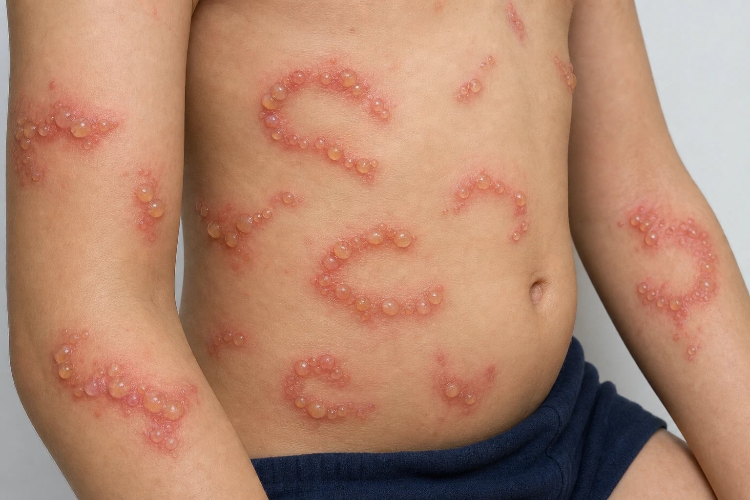
LABD is best classified along two clinically useful axes: the age of onset (which separates the adult and paediatric phenotypes) and the aetiology (which separates drug-induced from idiopathic disease and dictates whether treatment is drug withdrawal or immunosuppression). [1]
By age of onset: adult versus childhood (CBDC)
Adult LABD
Childhood LABD (CBDC)
By aetiology: idiopathic versus drug-induced
Drug-induced LABD
Idiopathic LABD
A small but examinable subset is mucosal-dominant LABD, in which disease is concentrated on the oral, conjunctival, or genital mucosa and mimics mucous membrane pemphigoid, with a real risk of ocular scarring.[3]
Epidemiology & Risk Factors
LABD is the rarest of the major autoimmune blistering disorders, considerably less common than bullous pemphigoid and pemphigus vulgaris. Population data are limited by the disease's rarity, but the available incidence figures cluster between 0.2 and 1.0 per million per year in European and North American cohorts, with a roughly equal sex distribution.[7]
The risk factors divide cleanly into pharmacological and systemic. By far the most important pharmacological risk factor is drug exposure, with vancomycin consistently the most commonly implicated agent across series and the answer to the canonical exam question.[7] The list of implicated drugs is long and reflects the drug's tendency to act as an immune hapten at the basement membrane zone:
VANCOMYCIN
The single most common drug cause — the classic exam answer
Penicillins, cephalosporins, rifampicin
Especially diclofenac
And other cardiovascular drugs (amiodarone)
Interferon-alpha, G-CSF, somatostatin
Lithium
A drug history is mandatory in every suspected case
Anti-PD-1/PD-L1 increasingly reported
Rare reports
If no drug, consider lymphoproliferative disease or IBD
The systemic risk factors are the second strand. Lymphoproliferative malignancy (B-cell lymphoma, chronic lymphocytic leukaemia, multiple myeloma) is the most consistently reported neoplastic association and warrants targeted screening when LABD is idiopathic or atypical.[3] Inflammatory bowel disease, particularly ulcerative colitis, is over-represented, supporting an immune-deviation hypothesis. Infections (upper respiratory and urinary tract infections, particularly in children) often precede CBDC. Solid malignancies (bladder, thyroid, renal, endometrial) have been reported as paraneoplastic associations. Increasingly, immune checkpoint inhibitors (nivolumab, pembrolizumab) are recognised as triggers, reflecting their broad amplification of autoimmunity.[6]
Epidemiological patterns are broadly similar worldwide, but access to confirmatory immunofluorescence, dapsone, and intravenous immunoglobulin differs sharply between high- and low-resource settings. In India, the IADVL consensus highlights delayed diagnosis because perilesional DIF and salt-split IIF are available only at tertiary centres, and dapsone — cheap and effective — is often the pragmatic first choice even before specialised testing. In Europe (EADV S2k) and the UK, centralised immunodermatology laboratories support early, precise diagnosis.[1]
Pathophysiology
IgA autoantibodies at the basement membrane zone
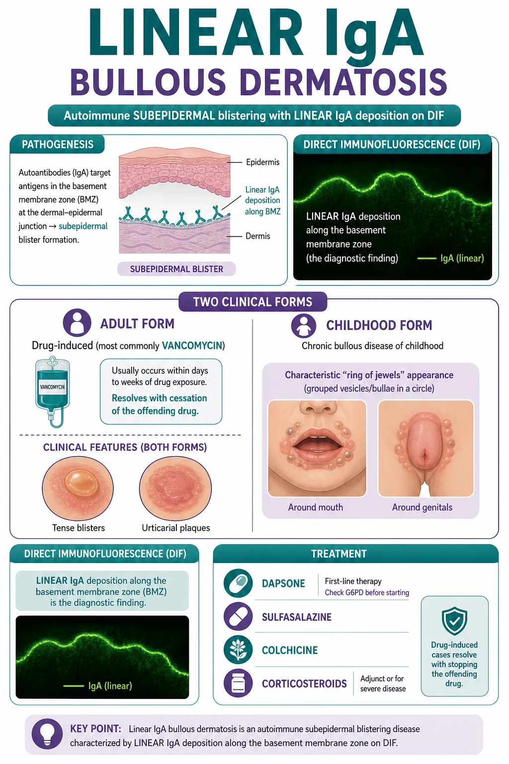
LABD is an organ-specific autoimmune disease caused by pathogenic IgA autoantibodies directed against antigens of the hemidesmosome–epidermal basement membrane adhesion complex. The hallmark — and the diagnostic sine qua non — is the linear, continuous band of IgA deposited along the basement membrane zone on DIF of perilesional skin. This linear deposition reflects antibody binding to distributed target antigens across the lamina lucida, lamina densa, and sublamina densa, in contrast to the granular, papillary-tip IgA deposits of dermatitis herpetiformis (immune complexes) and the linear IgG/C3 deposition of bullous pemphigoid.[2]
The dominant target antigens overlap with those of bullous pemphigoid. The most important are: [1]
BP180 (collagen XVII)
BP97 / LAD-285 / 120 kDa antigen
BP230 (BPAG1)
Type VII collagen (in EBA-overlap)
The mechanism of blister formation
The pathogenic cascade begins with IgA-class autoantibodies binding to the basement membrane adhesion complex. IgA is unique among immunoglobulins as a potent activator of neutrophils through Fc-alpha receptors, and this dictates the histological and clinical phenotype.[2] Antibody deposition recruits neutrophils to the basement membrane zone, where they release proteolytic enzymes (elastase, gelatinase) and reactive oxygen species that cleave the anchoring filaments and the lamina lucida. The result is subepidermal clefting and a neutrophil-rich blister — the histological signature. The neutrophil predominance is the biological reason LABD responds so dramatically to dapsone, which impairs neutrophil chemotaxis, myeloperoxidase activity, and the respiratory burst.[6]
Drug-induced LABD — why vancomycin?
The mechanism of drug-induced LABD is not fully resolved. Three hypotheses coexist and are not mutually exclusive. The hapten / neoantigen hypothesis proposes that the drug (or its reactive metabolites) binds to basement membrane components, creating a novel immunogenic complex that elicits an IgA response — analogous to drug-induced pemphigus. The immune-deviation hypothesis proposes that the drug skews T-cell help toward a Th2/IgA-promoting cytokine milieu. The direct inflammatory / cytokine-release hypothesis proposes that vancomycin, which is well known to trigger histamine release and immune activation, non-specifically amplifies a pre-existing low-grade autoimmune response. Whatever the mechanism, vancomycin's disproportionate implication (it is by far the single most cited culprit) makes drug history the single most important aetiological question in any adult presenting with new blistering, particularly in a hospitalised patient.[3][7]
The timing of drug-induced LABD is characteristic: blisters typically appear within 1 to 2 weeks of drug exposure (range 1 to 15 days), and resolve within 2 to 6 weeks of withdrawal. This predictable latency supports a hapten-driven immune response rather than a simple idiosyncratic reaction.[7]
Clinical Presentation
LABD is clinically polymorphic and the diagnosis is never secure without immunofluorescence. However, the two age-defined forms have sufficiently distinctive morphologies and distributions that an alert clinician can raise the diagnosis at the bedside. [1]
Adult LABD
The adult form presents with tense, fluid-filled bullae arising on normal or urticarial (erythematous, annular, or polycyclic) plaques on the trunk (especially the lower trunk, groin, and flexures), proximal limbs, and, less often, the face and scalp.[3][7] A characteristic and examinable morphology is the 'string of pearls' sign: new, small, tense blisters arranged in an arcuate or annular pattern around the advancing edge of an urticarial plaque, reflecting centrifugal spread. The blisters are tense (subepidermal) and may contain clear, haemorrhagic, or pustular fluid; they rupture to leave erosions that heal without scarring unless secondarily infected.
Pruritus is common and ranges from mild to intense, occasionally mimicking the intense itch of dermatitis herpetiformis. Burning and tenderness are also described, particularly over erosions. Mucosal involvement occurs in roughly 50 per cent of adult cases — most often the oral mucosa (erosions, stomatitis) and conjunctiva (keratoconjunctivitis, rarely scarring); genital involvement is less common. Severe mucosal disease can mimic mucous membrane pemphigoid and carries the same risk of ocular scarring (symblepharon, synechiae) and oesophageal or laryngeal strictures.[3]
The tempo is variable. Drug-induced disease appears abruptly in a patient on a culprit drug (typically within 1 to 2 weeks of vancomycin), often in the context of serious illness or recent surgery. Idiopathic disease is more insidious, with relapsing waves of blistering over weeks to months. [1]
Childhood LABD (chronic bullous disease of childhood, CBDC)
CBDC is the commonest autoimmune blistering disease of children and has a presentation so distinctive that experienced paediatric dermatologists often diagnose it clinically before immunofluorescence. The onset is under 5 years (median 1 to 5 years), with a slight female predominance in some series.[5][8]
The morphology mirrors the adult disease — tense bullae on urticarial, annular, or polycyclic plaques — but the distribution is characteristic: the genital and perineal region, the perioral face, and the buttocks and lower abdomen are the classical sites, with clusters of new blisters arranged around the periphery of older lesions producing the celebrated 'ring of jewels' (also called 'cluster of jewels' or 'string of beads') sign.[5] The face, scalp, palms, soles, and oral mucosa may also be involved. Children are often systemically well apart from itch, discomfort, and (occasionally) feeding difficulty from oral erosions.
Adult LABD
Childhood LABD (CBDC)
Atypical presentations
Several atypical presentations are examinable because they mislead the clinician. Mucosal-dominant LABD presents primarily with oral, conjunctival, or genital erosions and minimal skin disease, mimicking mucous membrane pemphigoid and risking ocular scarring.[3] Drug-induced LABD in the ICU or perioperative setting can erupt as a widespread blistering eruption resembling bullous pemphigoid or even toxic epidermal necrolysis in a patient on vancomycin. Adult-type LABD in an adolescent blurs the age boundary. Pregnancy-associated LABD overlaps clinically with pemphigoid gestationis and requires DIF to distinguish (LABD = linear IgA; PG = linear C3/IgG along BMZ).[6]
Evolution of a drug-induced LABD lesion
Differential Diagnosis
LABD sits within the subepidermal blistering family, and the differential turns on histology and immunofluorescence as much as on morphology. Every patient with suspected LABD should be considered against the full subepidermal blistering differential, because the clinical appearances overlap substantially.[2]
Bullous pemphigoid (BP)
Dermatitis herpetiformis (DH)
Epidermolysis bullosa acquisita (EBA)
Mucous membrane pemphigoid (MMP)
Pemphigoid gestationis (PG)
Erythema multiforme / SJS-TEN
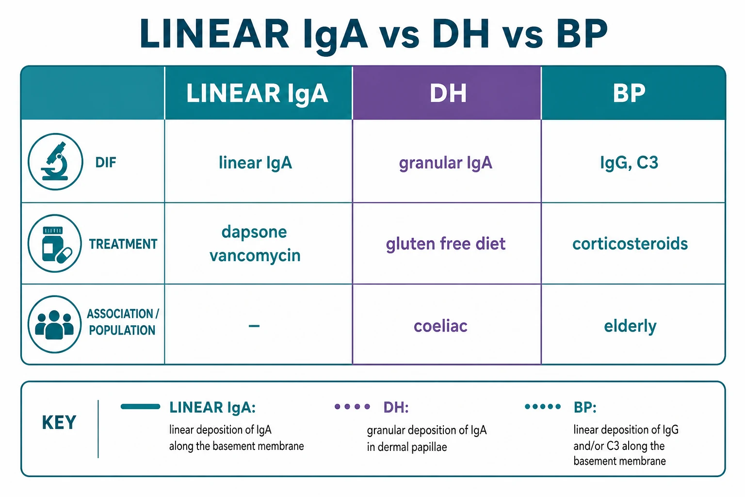
The two discriminating principles are, first, the DIF pattern: linear IgA at the BMZ is LABD; granular IgA in dermal papillae is dermatitis herpetiformis; linear IgG/C3 (without IgA) is bullous pemphigoid or EBA.[2] Second, the salt-split skin localisation separates the IgG-mediated dermopathies: epidermal (roof) labelling favours BP, dermal (floor) labelling favours EBA; LABD usually labels the roof (the 97 kDa antigen lies in the lamina lucida), and a subset labels the floor (EBA-overlap).[2] The clinical context — coeliac disease for DH, advanced age for BP, pregnancy for PG, trauma-prone scarring for EBA — refines the picture.
Clinical & Bedside Assessment
The diagnosis of LABD is immunopathological, but the bedside examination is essential to raise the diagnosis, characterise the eruption, screen for drug triggers and systemic associations, and assess disease severity. [1]
The focused skin examination begins with inspection of the trunk (lower abdomen, groin, flexures), proximal limbs, face, and scalp for tense bullae and urticarial, annular, or polycyclic plaques. Look specifically for the 'string of pearls' sign — small, new tense vesicles arranged in an arc around the advancing edge of a plaque — which is the single most suggestive cutaneous sign. Note the distribution: lower-trunk and flexural predominance in adults; genital, perineal, and perioral predominance in children (the 'ring of jewels'). Characterise the blisters (tense versus flaccid, clear versus haemorrhagic, size, and grouping) and the secondary changes (erosion, crusting, post-inflammatory pigmentation). Examine the mucous membranes — oral cavity, conjunctiva, and genitalia — because mucosal involvement occurs in about half of adult cases and carries a risk of ocular scarring.[3]
The drug history is the single most important bedside step in any adult with new blistering. Elicit every drug received in the preceding 1 to 2 weeks, paying particular attention to vancomycin, NSAIDs, lithium, ACE inhibitors, antibiotics, interferon, G-CSF, and immune checkpoint inhibitors. In the hospitalised patient, the medication chart and pharmacy records should be reviewed directly — vancomycin given for a line infection or surgical prophylaxis is easily missed. The systems review screens for lymphoproliferative disease (B symptoms, lymphadenopathy), inflammatory bowel disease (change in bowel habit, rectal bleeding), and malignancy, because these are over-represented in idiopathic LABD.[3]
[1]Investigations
The diagnosis of LABD rests on a coordinated set of investigations: histology of a lesional biopsy to characterise the blister, direct immunofluorescence (DIF) of perilesional skin to demonstrate the defining linear IgA band, indirect immunofluorescence (IIF) on salt-split skin to localise the antigen, and serological ELISA to characterise the antibody specificity. None of these can be omitted in a definitive work-up.[1][2]
Histology
A punch or shave biopsy of an early, intact blister shows a subepidermal blister with a predominantly neutrophilic infiltrate in the papillary dermis and blister cavity. The neutrophils tend to cluster along the papillary tips, a pattern that overlaps with dermatitis herpetiformis — but the DIF pattern distinguishes the two (linear in LABD, granular in DH). Eosinophils may be present in varying numbers; a predominantly eosinophilic infiltrate points more toward bullous pemphigoid. Fibrin deposition and occasional neutrophilic microabscesses at the papillary tips can be seen.[2]
Direct immunofluorescence (DIF) — the diagnostic test
Perilesional skin (within 1 cm of an active lesion) is biopsied and submitted in Michel's medium or saline (not formalin) for DIF. The diagnostic finding is a continuous, homogeneous LINEAR band of IgA along the basement membrane zone. [1][2] This single finding:
- Confirms LABD when clinical and histological features are consistent.
- Distinguishes LABD from dermatitis herpetiformis, where IgA is deposited in a granular pattern within the dermal papillae (immune complexes).
- Distinguishes LABD from bullous pemphigoid, where the linear band is IgG and C3 (with or without minor IgA).
- Occasionally shows concomitant IgG or C3 in a subset (mixed immunodeposition), which can complicate interpretation. [1]
Site of biopsy
Transport medium
What DIF shows
Indirect immunofluorescence (IIF) on salt-split skin
Serum is tested by IIF on 1 M NaCl salt-split human skin to determine the precise level of antibody binding. In most LABD cases, IgA labels the epidermal roof of the split, because the 97 kDa / BP180 ectodomain antigen lies in the lamina lucida.[2] This localisation:
- Supports LABD (and BP, which also labels the roof via BP180).
- Distinguishes LABD from EBA, where antibodies label the dermal floor (type VII collagen / anchoring fibrils).
- A minority of LABD sera label the dermal floor, indicating an EBA-overlap phenotype with anti-type VII collagen IgA, which is typically more dapsone-resistant. [1]
Serological ELISA
IgA anti-BP180 NC16A ELISA is positive in a substantial proportion of LABD sera, reflecting the shared BP180 ectodomain target. The IgA (rather than IgG) reactivity distinguishes LABD from bullous pemphigoid, where IgG anti-BP180 NC16A predominates. ELISA is useful for diagnosis where immunofluorescence is equivocal and for monitoring disease activity (titres tend to fall with treatment).[2]
Pre-treatment blood tests
Before commencing dapsone, several blood tests are mandatory: [1]
The diagnostic algorithm is therefore: (1) suspect LABD clinically; (2) biopsy an early blister for histology and perilesional skin for DIF; (3) send serum for IIF on salt-split skin and IgA anti-BP180 ELISA; (4) check G6PD, FBC, U&E before starting dapsone; (5) exclude mimics (DH, BP, EBA) on the immunofluorescence pattern.[1]
Management — Resuscitation
LABD is rarely an immediate threat to life, but extensive blistering (skin failure), severe mucosal disease, or a drug-induced eruption in a sick patient require prompt supportive care. The resuscitation priorities are: stop any culprit drug, manage skin failure as a burn, protect the airway and eyes, and address pain, fluid, and infection. [1]
**Stop the culprit drug** — review every drug received in the preceding 1 to 2 weeks; vancomycin is the prime suspect. In drug-induced LABD, withdrawal alone often resolves the disease within 2 to 6 weeks.
**Manage skin failure like a burn** — extensive denudation of skin requires fluid balance, temperature control, sterile handling, and a low-friction environment; involve the burns/plastics team if more than 10 per cent body surface area is affected.
**Protect eyes and mucosa** — any conjunctival involvement needs same-day ophthalmology, topical lubricants, and consideration of topical corticosteroids to prevent symblepharon and scarring; oral erosions need mouthwashes and analgesia.
**Pain and itch control** — paracetamol and oral antihistamines (non-sedating by day, sedating at night) for pruritus; opioid analgesia for extensive erosions.
**Topical therapy** — potent topical corticosteroids to active lesions; emollients and non-adherent dressings to erosions; topical antibiotics for localised secondary infection.
**Diagnose before immunosuppression** — secure biopsies for histology and DIF BEFORE starting systemic corticosteroids, which can confound immunofluorescence.
The key principle is that drug-induced LABD is treated by drug withdrawal, and idiopathic LABD is treated by dapsone. Conflating the two — putting a vancomycin-induced patient on long-term dapsone without attempting withdrawal — exposes the patient to avoidable toxicity.[7]
Management — Definitive & Stepwise
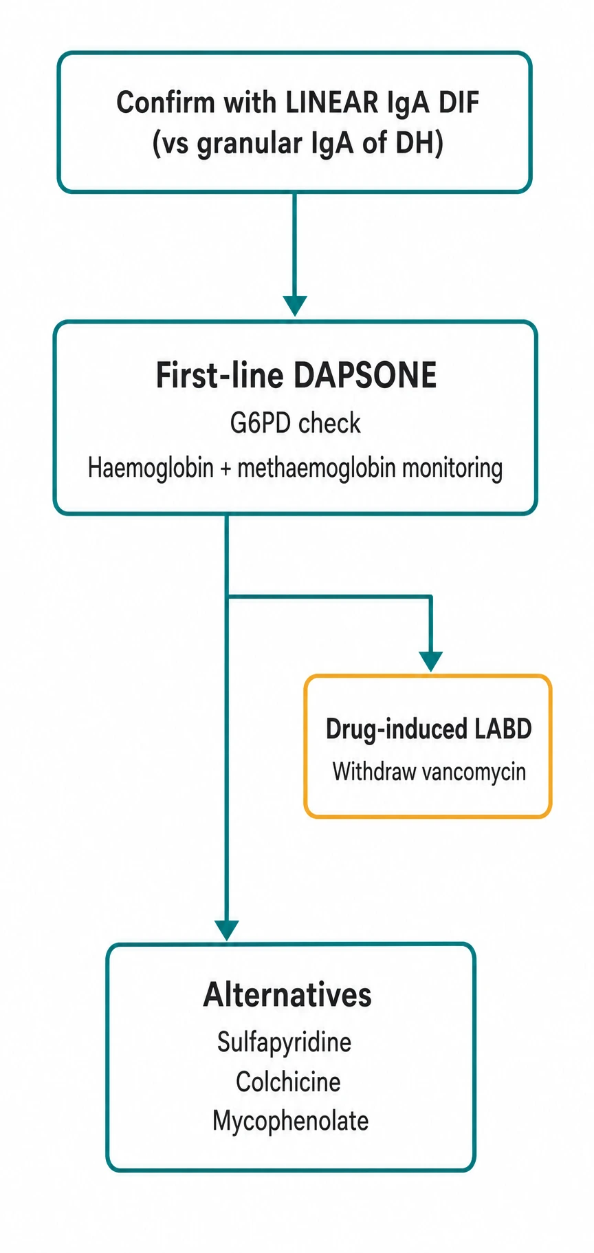
The definitive management of LABD follows a stepwise ladder calibrated to the aetiology and severity. The first-line agent for idiopathic disease is dapsone, with colchicine, sulfonamides, corticosteroids, and immunosuppressants as second- and third-line options.[1][6]
First-line: dapsone
Dapsone
Dose
50 to 150 mg orally once daily (start 50 mg and titrate); children 0.5 to 2 mg/kg/day
Dapsone is the cornerstone of LABD therapy and produces a dramatic response within 24 to 72 hours in most patients — a response so rapid that it is itself a diagnostic clue when DIF is pending. The starting dose is 50 mg once daily in adults, titrated up to a typical maintenance of 100 to 150 mg daily according to response and tolerance. Children receive 0.5 to 2 mg/kg/day.[1]
The pre-dapsone checklist is examinable and mandatory. G6PD activity must be checked before the first dose because dapsone causes haemolysis and methaemoglobinaemia in G6PD-deficient individuals; G6PD deficiency is common in Mediterranean, African, Middle Eastern, and South-East Asian populations. A baseline full blood count, reticulocyte count, and methaemoglobin level are obtained, with monitoring weekly for the first month and then monthly. Patients should be counselled about the symptoms of haemolysis (pallor, fatigue, jaundice) and methaemoglobinaemia (cyanosis, breathlessness, headache) and warned to report them promptly.[6]
Second-line and adjunctive agents
When dapsone is contraindicated, insufficient, or poorly tolerated, several alternatives exist: [1]
Sulfasalazine / sulfapyridine
Colchicine
Corticosteroids
Immunosuppressants
Biologics and IVIG
Stepwise algorithm
**Step 1 — Identify and stop any culprit drug** (vancomycin first). For drug-induced LABD, this alone resolves most cases within 2 to 6 weeks.
**Step 2 — First-line idiopathic LABD: dapsone** 50 mg daily titrated to 100 to 150 mg daily. Check G6PD first. Response within 24 to 72 hours.
**Step 3 — Add or switch**: if dapsone insufficient or poorly tolerated, add colchicine or switch to sulfasalazine/sulfapyridine.
**Step 4 — Short-course prednisolone** 0.5 mg/kg/day for severe or refractory disease; combine with dapsone or an immunosuppressant.
**Step 5 — Steroid-sparing immunosuppression** (mycophenolate, azathioprine, ciclosporin) for chronic idiopathic disease requiring prolonged therapy.
**Step 6 — Biologics/IVIG** (rituximab, IVIG) for refractory, mucosal-dominant, or immunosuppression-intolerant disease.
**Adjuncts throughout**: potent topical corticosteroids, emollients, antihistamines, wound care, and ophthalmology for any ocular involvement.
Children
In CBDC, dapsone 0.5 to 2 mg/kg/day is first-line, with the same G6PD and FBC monitoring as adults. Colchicine and short courses of prednisolone are alternatives. The prognosis is excellent, with most children remitting before puberty, so the aim is symptomatic control with the least toxic regimen.[5][8]
Specific Subtypes & Scenarios
Vancomycin-induced LABD
Vancomycin is the single most common drug cause of LABD and the classic exam answer. The mechanism is presumed to be hapten-driven, with vancomycin or its metabolites creating a neoantigen at the basement membrane zone that elicits an IgA response. The latency is 1 to 2 weeks (range 1 to 15 days), and the eruption typically begins on the trunk and proximal limbs as urticarial plaques that develop tense bullae in the characteristic 'string of pearls' pattern. Management is drug withdrawal — the single most effective intervention — with bridging dapsone or potent topical corticosteroids for symptomatic control if lesions are extensive. Resolution occurs within 2 to 6 weeks of withdrawal, and relapse is expected on re-challenge, which should therefore be avoided.[3][7]
Other antibiotics (penicillins, cephalosporins, rifampicin) and an ever-growing list of drugs (lithium, diclofenac and other NSAIDs, captopril and ACE inhibitors, amiodarone, interferon-alpha, G-CSF, somatostatin, iodinated contrast) and increasingly immune checkpoint inhibitors (nivolumab, pembrolizumab) are also reported, so a complete drug history is mandatory.[6]
Chronic bullous disease of childhood (CBDC)
CBDC is the commonest autoimmune blistering disease of children. It presents under 5 years with tense bullae in annular clusters around the genitalia, perineum, perioral face, buttocks, and lower abdomen (the 'ring of jewels'). The DIF is identical to adult LABD (linear IgA along the BMZ). Treatment is dapsone 0.5 to 2 mg/kg/day (check G6PD first), with colchicine or short-course prednisolone as alternatives. The prognosis is excellent: most children remit before puberty, usually within 2 to 5 years, and treatment can be tapered and withdrawn as the disease burns out.[5][8]
Mucosal-dominant and ocular LABD
A subset of LABD has predominantly mucosal involvement — oral erosions, conjunctivitis, and genital erosions — that mimics mucous membrane pemphigoid and carries the same risk of ocular scarring (symblepharon, synechiae, corneal opacity, visual loss). Any patient with conjunctival involvement requires same-day ophthalmology, topical lubricants and corticosteroids, and aggressive systemic therapy (dapsone plus an immunosuppressant; rituximab or IVIG for refractory disease). Dapsone may be insufficient for mucosal disease, and combination therapy is often needed.[3]
Pregnancy-associated LABD
LABD in pregnancy is rare and overlaps clinically with pemphigoid gestationis (intensely pruritic urticarial plaques and vesicles, often periumbilical). The two are distinguished by DIF: linear IgA along the BMZ in LABD versus linear C3 (with or without IgG) along the BMZ in pemphigoid gestationis. Dapsone (after G6PD check) is the preferred first-line agent in pregnancy; systemic corticosteroids are reserved for severe disease. There is a risk of transient neonatal blistering from transplacental passage of maternal IgA, which resolves as the antibody clears.[6]
Malignancy-associated and immune checkpoint inhibitor LABD
LABD may be paraneoplastic, most often associated with lymphoproliferative disease (B-cell non-Hodgkin lymphoma, chronic lymphocytic leukaemia, multiple myeloma) and less often with solid tumours (bladder, thyroid, renal, endometrial). An idiopathic or atypical adult case warrants targeted malignancy screening (FBC, blood film, LDH, serum electrophoresis, and imaging as guided by history). Immune checkpoint inhibitor-associated LABD is increasingly recognised and sits within the broader spectrum of immune-related adverse events; management combines drug review (with the oncology team), dapsone, and topical or systemic corticosteroids.[3]
Complications & Pitfalls
The complications of LABD arise from the disease itself (mucosal scarring, secondary infection, fluid loss), from misdiagnosis, and from the adverse effects of its principal treatment, dapsone. [1]
Disease complications
Dapsone adverse effects
Diagnostic pitfalls
Exam application bank (NEET-PG / INICET)
One-line answer
Linear IgA bullous dermatosis (LABD) is an autoimmune subepidermal blistering disorder defined by LINEAR deposition of IgA along the basement membrane zone on direct immunofluorescence. Two forms exist: the ADULT form (often drug-induced, especially VANCOMYCIN, or idiopathic) and the CHILDHOOD form (chronic bullous disease of childhood, CBDC, with the 'ring of jewels' / 'string of pearls' around the genitals and mouth). Distinguishing LABD from dermatitis herpetiformis hinges on the DIF pattern: LABD is LINEAR, DH is GRANULAR. First-line treatment is DAPSONE 50 to 150 mg/day (check G6PD first); drug-induced disease resolves within weeks of stopping the culprit. [1]
Worked stems (answer without another resource)
Stem 1 — Classic presentation. Map symptoms to mechanism; name the first investigation and first treatment step with dose/route if drug therapy is standard. [1]
Stem 2 — Unstable / complicated. List red flags that force immediate resuscitation, theatre, ICU, antidote, or reperfusion — and what you do in the first 15 minutes. [1]
Stem 3 — Atypical group. Elderly, pregnancy, child, or immunocompromised: how presentation and thresholds change. [1]
Stem 4 — Differential trap. Name the three closest mimics and one discriminator for each. [1]
Stem 5 — Disposition. Who goes home with safety-netting, who is admitted, who needs HDU/ICU/theatre, and what follow-up is mandatory. [1]
Rapid viva checklist
- Definition + classification
- Pathophysiology chain
- Bedside signs / criteria
- Score with exact components (if any)
- Emergency bundle
- Definitive therapy with doses
- Complications of disease and of treatment
- Special populations
- Guideline/trial name if classic
- Three exam traps
Coverage self-check
If you cannot answer any stem above from this page alone, re-read the matching section — the page is intended to be self-sufficient for final-prof and NEET-PG/INICET questions on Linear IgA bullous dermatosis.
Prognosis & Disposition
The prognosis of LABD is generally favourable and depends almost entirely on the aetiology. The three subtypes have predictably different outcomes. [1]
Drug-induced LABD
Idiopathic adult LABD
Childhood LABD (CBDC)
The disposition depends on severity. Most patients are managed as outpatients with dapsone and topical therapy. Admission is warranted for extensive skin failure, severe mucosal disease (especially ocular), systemic upset, or diagnostic uncertainty in a sick patient. Ophthalmology follow-up is essential for any ocular involvement. Dermatology follow-up monitors treatment response, dapsone adverse effects (FBC, methaemoglobin), and tapers therapy as the disease remits.[5][7]
Disease course and remission
Special Populations
Children
Pregnancy
G6PD-deficient patients
The hospitalised / ICU patient
Evidence, Guidelines & Regional Differences
The evidence base for LABD management is limited by the disease's rarity: there are no large randomised controlled trials, and recommendations rest on retrospective cohort studies, case series, and expert consensus.[1][6]
Landmark guidelines
Diagnostic evidence
Therapeutic evidence
Regional differences are modest in principle but sharp in access to specialised diagnostics and biologics. In Europe (EADV S2k) and the UK, centralised immunodermatology laboratories support early, precise diagnosis (perilesional DIF, salt-split IIF, IgA anti-BP180 ELISA), and rituximab/IVIG are available for refractory disease through regional centres. In the United States, the approach is similar, with the American Academy of Dermatology endorsing the same diagnostic and therapeutic ladder. In India and other resource-limited settings, the IADVL consensus acknowledges that perilesional DIF is available only at tertiary centres, so dapsone is often the pragmatic first choice even before specialised testing, given its low cost, safety (after G6PD screening), and rapid, highly characteristic response. In Australasia, the RANZCD-aligned approach mirrors the European guidance.[1]
Exam Pearls
LINEAR
The defining DIF finding — a continuous IgA band along the BMZ
Idiopathic or drug-induced (vancomycin) in adults; idiopathic in children
Subepidermal blister with neutrophils — the IgA-driven infiltrate
Target = BP180 (collagen XVII) ectodomain / 97 kDa antigen; ELISA for IgA anti-BP180 NC16A
Adults over 60 (vancomycin) and children under 5 (CBDC, ring of jewels)
Dapsone 50 to 150 mg/day first-line (check G6PD) — response within 24 to 72 hours
Why does LABD respond to dapsone while bullous pemphigoid does not?
LABD is driven by IgA autoantibodies that recruit neutrophils via Fc-alpha receptors; the neutrophil proteases cleave the lamina lucida. Dapsone selectively impairs neutrophil chemotaxis, myeloperoxidase activity, and the respiratory burst, so it shuts down the neutrophil-driven blistering within 24 to 72 hours. Bullous pemphigoid is driven by IgG with eosinophil predominance and complement activation, which dapsone does not effectively suppress — hence its dependence on corticosteroids.[6]
How do you distinguish LABD from dermatitis herpetiformis (DH) on immunofluorescence?
Both deposit IgA, but the pattern differs. LABD shows a continuous LINEAR band of IgA along the basement membrane zone (antibody bound to a distributed BMZ antigen). Dermatitis herpetiformis shows GRANULAR IgA in the dermal papillae (immune complexes). The distinction is critical because DH is coeliac-associated and gluten-responsive, while LABD is not.[2]
What is the 'ring of jewels' sign?
The 'ring of jewels' (also called 'cluster of jewels' or 'string of beads') is the characteristic sign of chronic bullous disease of childhood (CBDC): new, small, tense bullae arranged in an annular or polycyclic cluster around the periphery of an older lesion, typically on the genitalia, perineum, perioral face, and lower abdomen. It is the paediatric equivalent of the adult 'string of pearls' sign.[5]
References
- [1]Caux F, Patsatsi A, Karakioulaki M, et al. S2k guidelines on diagnosis and treatment of linear IgA dermatosis initiated by the European Academy of Dermatology and Venereology J Eur Acad Dermatol Venereol, 2024.PMID 38421060
- [2]van Beek N, Holtsche MM, Atefi I, et al. State-of-the-art diagnosis of autoimmune blistering diseases Front Immunol, 2024.PMID 38903493
- [3]Juratli HA, Sárdy M. [Linear IgA bullous dermatosis] Hautarzt, 2019.PMID 30874843
- [4]Egan CA, Zone JJ. Linear IgA bullous dermatosis Int J Dermatol, 1999.PMID 10583613
- [5]Wang KL, Lehman JS, Davis DMR. Linear IgA bullous dermatosis of childhood: Retrospective single-center cohort Pediatr Dermatol, 2024.PMID 38378007
- [6]Khan M, Park L, Skopit S. Management Options for Linear Immunoglobulin A (IgA) Bullous Dermatosis: A Literature Review Cureus, 2023.PMID 37090290
- [7]Mar K, Landuffs F, Khalid B, et al. Clinical characteristics and treatment outcomes of linear IgA bullous dermatosis J Dtsch Dermatol Ges, 2025.PMID 40018881
- [8]Lara-Corrales I, Pope E. Autoimmune blistering diseases in children Semin Cutan Med Surg, 2010.PMID 20579597
- [9]Bernett CN, Yadlapati S, Rosario-Collazo JA. Linear IGA Dermatosis 2026.PMID 30252369