Dermatology · Medicine
Lymphoid and histiocytic infiltrates
Also known as Cutaneous lymphoid infiltrates · Cutaneous pseudolymphoma · Cutaneous lymphoma dermatopathology · Cutaneous histiocytosis · Lymphohistiocytic infiltrates of skin
Pattern-based dermatopathology of lymphoid and histiocytic infiltrates of skin: reactive cutaneous lymphoid hyperplasia (pseudolymphoma) versus WHO-EORTC primary cutaneous T- and B-cell lymphomas, CD30+ lymphoproliferative disorders, and histiocytoses (LCH, juvenile xanthogranuloma family, Rosai–Dorfman disease, multicentric reticulohistiocytosis). Emphasises clinical–pathologic correlation, immunohistochemistry lineage panels, clonality pitfalls, staging triggers, and exam-yield management ladders.
On this page & tools
Your progress
Saved locally on this device.
Exam tags
Red flags
Definition & Classification
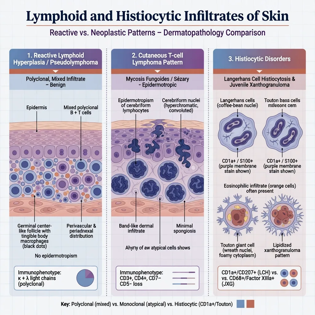
A cutaneous lymphoid infiltrate is a dermal and/or epidermal accumulation of lymphocytes (± other inflammatory cells). The central diagnostic task is to decide whether it is reactive (cutaneous lymphoid hyperplasia / pseudolymphoma) or neoplastic (primary or secondary lymphoma), and—when histiocytes dominate—to place the process in the modern histiocytosis classification.[1][3][9]
WHO-EORTC primary cutaneous lymphomas (conceptual map)
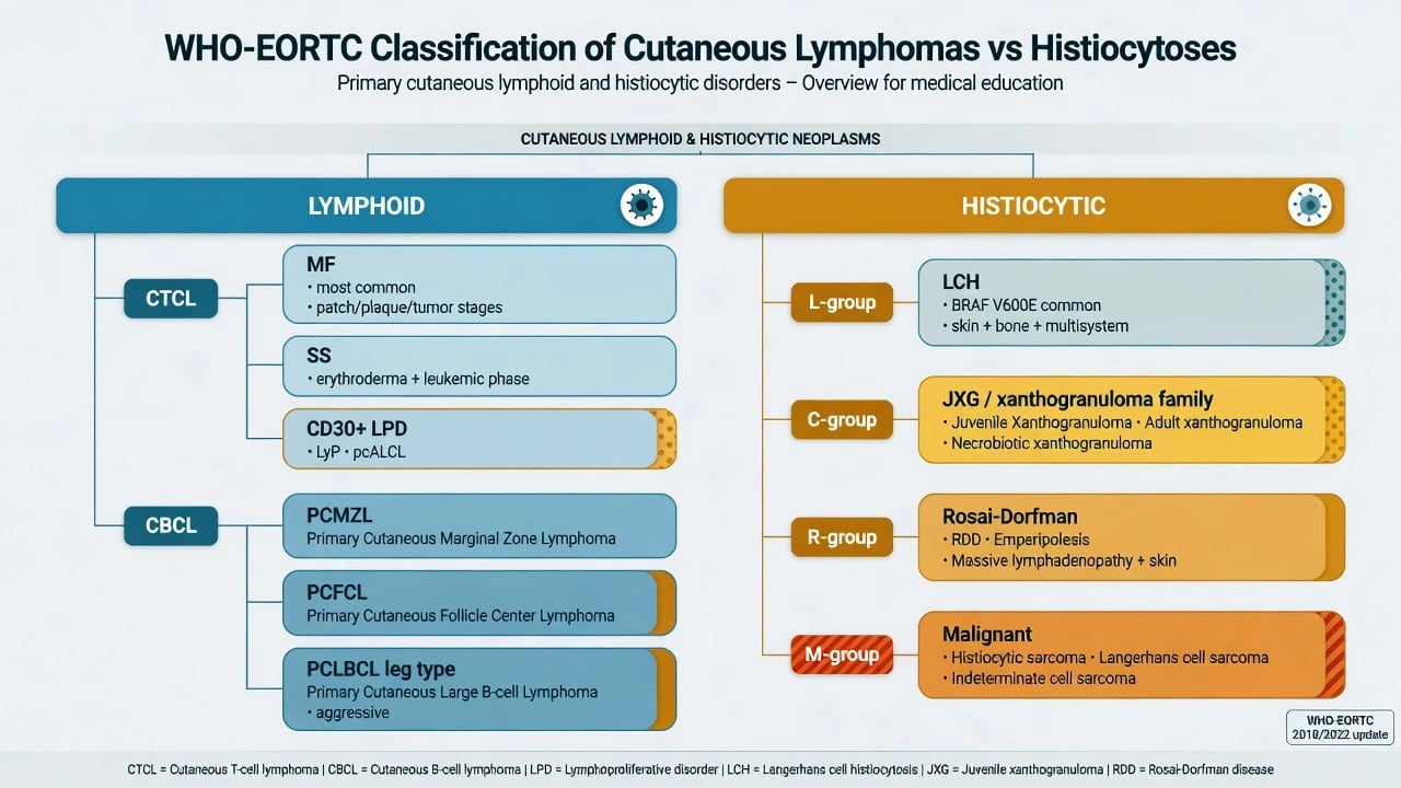
The WHO-EORTC system separates entities by cell lineage, clinical behaviour, and site, not by histology alone.[1][2]
| Group | High-yield entities | Behaviour (typical) |
|---|---|---|
| CTCL | Mycosis fungoides (MF), Sézary syndrome (SS), CD30+ LPD (LyP, pcALCL) | MF often indolent early; SS aggressive |
| CBCL | Primary cutaneous marginal zone lymphoma (PCMZL); primary cutaneous follicle-centre lymphoma (PCFCL); primary cutaneous large B-cell lymphoma, leg type | First two usually indolent; leg type aggressive |
| Other | Rare NK/T, blastic plasmacytoid dendritic cell neoplasm (systemic often) | Variable / aggressive |
Histiocytoses (Emile revised framework)
| Group | Examples relevant to skin | Lineage cue |
|---|---|---|
| L (Langerhans) | LCH | CD1a+, langerin (CD207)+, S100+; often BRAF V600E |
| C (cutaneous / JXG family) | Juvenile xanthogranuloma, adult xanthogranuloma, related non-LCH | CD68+/CD163+; usually CD1a−/langerin− |
| R (Rosai–Dorfman) | Cutaneous or systemic RDD | S100+ large histiocytes; emperipolesis |
| M (malignant) | Histiocytic sarcoma and related | Rare; aggressive |
Classification (pattern-first dermatopathology)
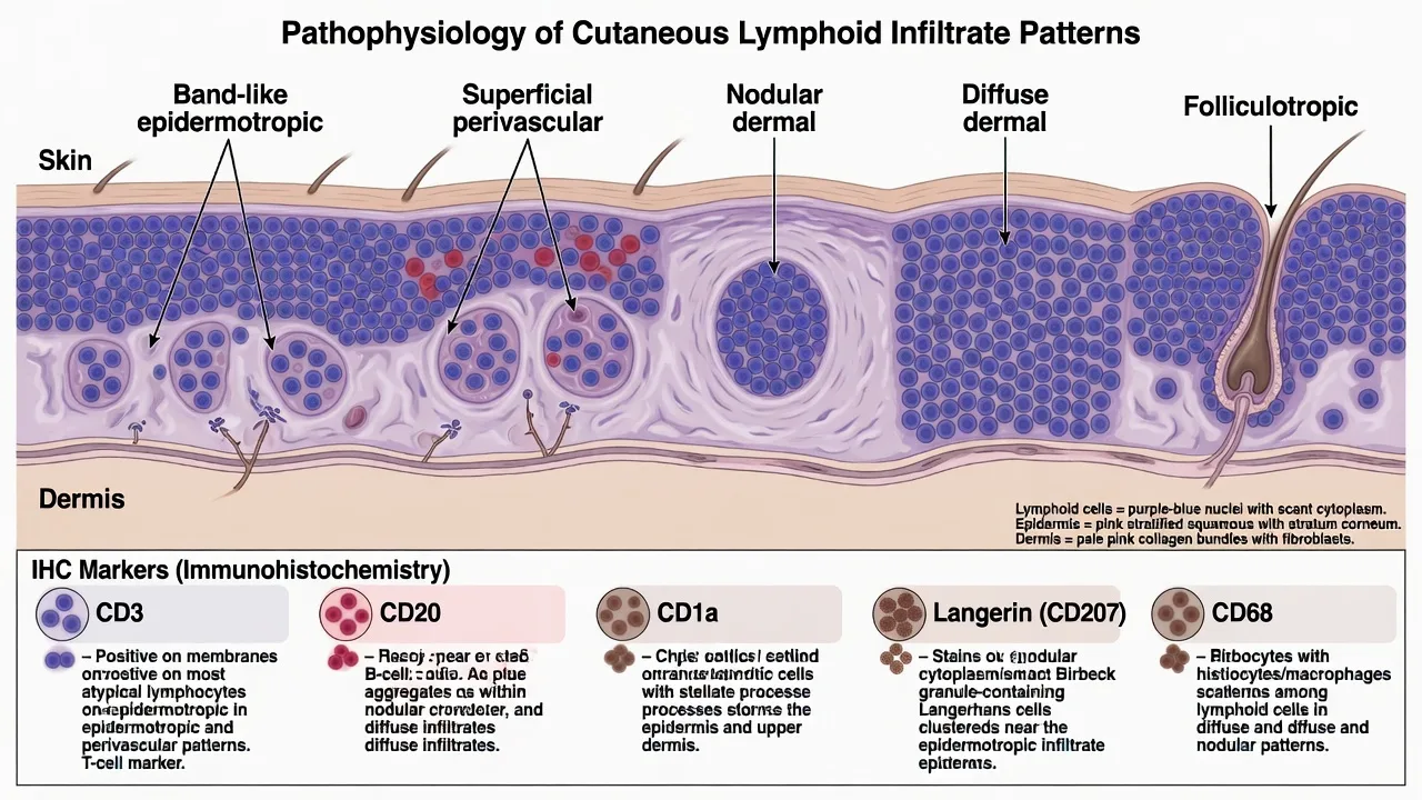
Before naming a disease, describe the architectural pattern: [1]
- Band-like / epidermotropic — MF spectrum until proven otherwise (with caveats for spongiotic mimics).[5]
- Superficial perivascular — often reactive or early inflammatory; still can hide early MF.
- Nodular dermal — common in CBCL and B-cell pseudolymphoma.[6][3]
- Diffuse dermal — broad differential (aggressive lymphoma, leukaemia cutis, dense reactive).
- Folliculotropic / syringotropic — folliculotropic MF and follicular mucinosis spectrum.[5]
Pseudolymphoma is a clinicopathologic diagnosis of a reactive lymphoid hyperplasia that can closely simulate lymphoma; triggers include arthropod bites, tattoos, infections, drugs, and idiopathic cases.[3][4]
Epidemiology & Risk Factors
- MF is the most common primary CTCL in adults; primary CBCL are less common but important exam entities.[1][5]
- Pseudolymphoma occurs across ages; drug-induced and arthropod-related forms are classic teaching associations.[3]
- JXG predominates in infants/young children; multisystem LCH is a paediatric emergency spectrum when disseminated.[8][7]
- Immunosuppression and EBV-associated lymphoproliferative disease widen the differential for atypical infiltrates (special population).
Pathophysiology
- CTCL: neoplastic skin-homing memory T cells (often CLA+/CCR4+ concepts in MF/SS) with epidermotropism and progressive immune dysregulation.[5]
- Indolent CBCL: antigen-driven B-cell proliferations in skin microenvironments; PCMZL often mixed with reactive T cells and plasma cells.[6]
- LCH: clonal neoplastic proliferation of Langerhans-type dendritic cells frequently harbouring MAPK pathway mutations (commonly BRAF V600E).[8][7]
- RDD: S100-positive large histiocytes engulf intact lymphocytes (emperipolesis).[10]
- Pseudolymphoma: sustained antigenic stimulation produces polyclonal or oligoclonal lymphoid expansion that can still look “lymphoma-like” on H&E.[3]
Clinical Presentation
Lymphoid
- MF: patches → plaques → tumours; poikiloderma; special sites (bathing-suit, breast, buttocks); great imitator of eczema/psoriasis.[5]
- SS: erythroderma + lymphadenopathy + circulating neoplastic cells (see dedicated topic).
- CD30+ LPD: LyP — recurring self-healing papulonodules; pcALCL — larger solitary/grouped tumours, often better prognosis than systemic ALCL when truly primary cutaneous.[1]
- PCMZL / PCFCL: red-to-violaceous papules, plaques, or nodules, often head/trunk or trunk/head respectively.[6]
- PCLBCL, leg type: rapidly growing red-blue tumours on lower legs of elderly (often women).[6][1]
Histiocytic
- LCH: seborrhoeic-like scalp/flexural eruption, purpuric papules, erosions; bone, DI, marrow, lung may coexist in multisystem disease.[8]
- JXG: yellow-orange papulonodules; may be solitary or multiple; ocular risk in multiple facial lesions in infants (exam pearl).[7]
- Cutaneous RDD: nodules/plaques; may be skin-limited or part of systemic RDD.[10]
- Multicentric reticulohistiocytosis: coral-bead periungual papules with destructive arthritis (systemic association).
Differential Diagnosis
| Dense dermal lymphoid infiltrate | Distinguishing approach |
|---|---|
| Pseudolymphoma | Clinical trigger, mixed cells, germinal centres, polyclonal light chains, benign course |
| MF / CTCL | Epidermotropism, atypical cerebriform cells, loss of pan-T antigens, clone + clinical stage |
| Primary CBCL | Nodular B-cell sheets, architecture of follicles vs marginal zone, BCL2/MUM1 profile for leg type |
| Secondary systemic lymphoma / leukaemia cutis | Systemic history, blood film, broad IHC |
| Lymphomatoid drug eruption | Drug timeline; often resolves on withdrawal |
| LyP vs pcALCL | Self-healing crops vs persistent larger tumours; shared CD30+ large cells |
| Histiocyte-rich dermis | Distinguishing approach |
|---|---|
| LCH | CD1a+/langerin+ coffee-bean nuclei |
| JXG family | Touton giant cells; CD68+ CD1a− |
| RDD | Emperipolesis; S100+ |
| Granulomatous infection / sarcoid / GA | Special stains, polarisation, clinical pattern |
| Xanthomas | Lipidised macrophages; metabolic context |
Clinical & Bedside Assessment
- Full skin map, photos of morphology, and lymph node basins when lymphoma is plausible.
- Ask about drugs, arthropod exposure, tattoos, radiation, immunosuppression, B symptoms, polyuria/polydipsia (DI in LCH).
- Document spontaneous regression (favours LyP) versus relentless progression (favours aggressive lymphoma).
- In infants with JXG-like lesions plus systemic signs, do not assume “benign histiocytosis” without staging consideration.[8][7]
Investigations
Biopsy strategy
- Choose developed lesions; for MF, multiple biopsies over time may be needed.[5]
- Request H&E + IHC; add molecular only when it will change management interpretation.
- For bullous/interface mimics of lymphoma, ensure the clinical question is clear to the lab (separate DIF pathway if immunobullous disease is also possible — see special-stains/IF topic).
Lineage IHC cheat-sheet (exam gold)
| Question | Panel (teaching core) |
|---|---|
| T vs B | CD3 vs CD20 (plus CD79a/PAX5 as needed) |
| MF phenotype clues | CD4-predominant epidermotropism; CD7 loss; CD8 variants exist |
| CD30 LPD | CD30 large cells ± cytotoxic markers |
| Indolent vs leg-type CBCL cues | BCL6/CD10 (follicle centre) vs strong MUM1/BCL2 in leg type concepts |
| LCH | CD1a, langerin (CD207), S100 |
| Non-LCH histiocytes | CD68, CD163; S100 variable (RDD often S100+) |
Clonality
- TCR/Ig gene rearrangement supports neoplasia but is not diagnostic alone — reactive infiltrates can be clonal/oligoclonal and early lymphomas may be negative.[3][5]
Staging when indicated
- CTCL: skin score concepts, blood flow (if erythrodermic), nodes/viscera imaging per pathway.[5]
- Aggressive CBCL / systemic suspicion: staging CT/PET and haemato-oncology labs.[6]
- Multisystem LCH: skeletal survey/imaging, endocrine (DI), marrow as indicated.[8]
Management — Resuscitation / Urgent Priorities
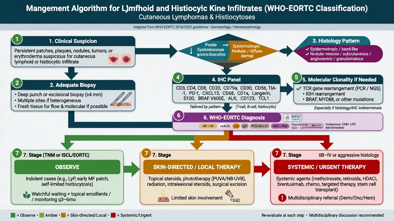
- Erythroderma / tumour-stage CTCL / suspected SS: barrier care, infection control, urgent derm–haemato-oncology pathway (not “more topical steroids alone”).
- Infant multisystem LCH suspicion: same-day paediatric oncology referral; airway, cytopenias, and DI assessment.[8]
- Ulcerated aggressive leg-type CBCL: stage and plan systemic therapy promptly.[6]
Management — Definitive & Stepwise
Reactive lymphoid hyperplasia (pseudolymphoma)
- Remove trigger (drug, antigen) when identified.
- Local therapy: potent topical/intralesional corticosteroid; excision of solitary lesions when diagnostic/therapeutic.
- Evidence for treatment is heterogeneous; many lesions are managed conservatively after confident diagnosis.[4][3]
Early MF / indolent CTCL concepts
- Skin-directed therapy first (topical corticosteroids, phototherapy, topical mechlorethamine/others per centre, local radiotherapy for limited tumours).
- Escalate systemically for advanced stage or refractory disease (see CTCL/SS topics).[5][1]
Indolent primary CBCL (PCMZL, PCFCL)
- Often local radiotherapy, excision, or watchful waiting for selected low-burden disease; systemic rituximab-based approaches in multifocal or refractory disease depending on pathway.[6]
Aggressive CBCL (leg type)
- Treat analogous to systemic large B-cell lymphoma concepts with haemato-oncology (not simple excision alone).[6][1]
Histiocytoses
- Solitary JXG: often observation or simple removal; eye exam when indicated by lesion pattern/age.[7]
- LCH: risk-stratified (single-system skin vs multisystem); dermatology alone is insufficient for multisystem disease — oncology protocols apply; MAPK-targeted therapy appears in selected refractory pathways in contemporary practice.[8][7]
- Cutaneous RDD: observation, excision, or systemic options for extensive disease; exclude systemic RDD when indicated.[10]
Pseudolymphoma
- Trigger-related / mixed cells
- Often polyclonal
- Local therapy / observe
- Follow clinically
Indolent primary lymphoma
- WHO-EORTC entity
- Clone supports but not sole criterion
- Local RT/excision common
- Long survival typical
Aggressive lymphoma / multisystem LCH
- Rapid growth or organ risk
- Full staging
- Systemic protocols
- Haemato-oncology lead
Specific Subtypes & Scenarios
- Folliculotropic MF: head/neck plaques, alopecia, acneiform lesions; may behave more stubbornly than classic patch MF.[5]
- LyP: chronic, relapsing, self-healing; association risk with other lymphomas warrants long-term skin surveillance.[1]
- PCFCL vs leg-type DLBCL: architecture and immunophenotype (MUM1/BCL2 emphasis in leg type teaching) separate indolent from aggressive CBCL.[6]
- Skin-limited vs systemic RDD: cutaneous disease can be isolated; still screen clinically for nodes/systemic signs.[10]
Complications & Pitfalls
- Under-calling MF as chronic eczema for years.[5]
- Over-calling lymphoma on a reactive nodular B-cell infiltrate without clinical correlation.[3]
- Clonality false reassurance or false alarm.[3]
- Missing multisystem LCH complications (DI, cytopenias, lung/liver).[8]
- Treating leg-type CBCL as a benign cyst or “infected boil.”
Prognosis & Disposition
- Early MF and indolent CBCL: often excellent long-term survival with skin-directed care.[1][5]
- SS and leg-type CBCL: substantially worse — specialist systemic care.[1][6]
- Single-system skin LCH may do well; multisystem risk-organ LCH drives prognosis.[8]
- Disposition: dermatology follow-up for indolent entities; shared haemato-oncology care whenever systemic risk exists.
Special Populations
- Children: JXG vs LCH is the key histiocyte fork; avoid adult CTCL heuristics.[7][8]
- Elderly: higher yield for aggressive CBCL and SS work-up.
- Immunosuppressed: broader viral/lymphoproliferative differential.
- Resource-limited settings: prioritise adequate biopsy + basic IHC (CD3/CD20/CD30/CD1a) and clinical staging over unavailable molecular tests.
Evidence, Guidelines & Regional Differences
- WHO-EORTC 2018 update refined entity boundaries and remains the classification language for exams and pathology reports.[1]
- Emile 2016 reorganised histiocytoses into L/C/R/M groups used internationally.[9]
- Pseudolymphoma treatment evidence is largely observational/systematic-review level rather than large RCTs.[4]
- Regional access differs for phototherapy, radiotherapy, rituximab, and MAPK inhibitors — algorithms must be adapted without changing diagnostic standards.
Exam Pearls
PATTERN
PATTERN
Band, nodular, diffuse, folliculotropic
Reactive triggers vs neoplastic sheets
CD3 / CD20 / CD1a–langerin–CD68
Name the entity, not just 'lymphoma'
S100+ large histiocytes
Supportive, not standalone
SS, leg-type, multisystem LCH
- WHO-EORTC language is non-negotiable in answers.[1]
- Pseudolymphoma is real — clinical trigger + mixed histology.[3]
- Leg-type CBCL is the aggressive CBCL exam trap.[6]
- Emperipolesis → Rosai–Dorfman.[10]
- MF is a clinicopathologic diagnosis; one biopsy rarely ends the story.[5]
Exam application bank (NEET-PG / INICET)
One-line answer
Pattern-based dermatopathology of lymphoid and histiocytic infiltrates of skin: reactive cutaneous lymphoid hyperplasia (pseudolymphoma) versus WHO-EORTC primary cutaneous T- and B-cell lymphomas, CD30+ lymphoproliferative disorders, and histiocytoses (LCH, juvenile xanthogranuloma family, Rosai–Dorfman disease, multicentric reticulohistiocytosis). Emphasises clinical–pathologic correlation, immunohistochemistry lineage panels, clonality pitfalls, staging triggers, and exam-yield management ladders.
Worked stems (answer without another resource)
Stem 1 — Classic presentation. Map symptoms to mechanism; name the first investigation and first treatment step with dose/route if drug therapy is standard. [1]
Stem 2 — Unstable / complicated. List red flags that force immediate resuscitation, theatre, ICU, antidote, or reperfusion — and what you do in the first 15 minutes. [1]
Stem 3 — Atypical group. Elderly, pregnancy, child, or immunocompromised: how presentation and thresholds change. [1]
Stem 4 — Differential trap. Name the three closest mimics and one discriminator for each. [1]
Stem 5 — Disposition. Who goes home with safety-netting, who is admitted, who needs HDU/ICU/theatre, and what follow-up is mandatory. [1]
Rapid viva checklist
- Definition + classification
- Pathophysiology chain
- Bedside signs / criteria
- Score with exact components (if any)
- Emergency bundle
- Definitive therapy with doses
- Complications of disease and of treatment
- Special populations
- Guideline/trial name if classic
- Three exam traps
Coverage self-check
If you cannot answer any stem above from this page alone, re-read the matching section — the page is intended to be self-sufficient for final-prof and NEET-PG/INICET questions on Lymphoid and histiocytic infiltrates.
Expanded exam teaching (depth pass)
Clinical reasoning
For Lymphoid and histiocytic infiltrates, examiners test whether you can prioritise life threats, choose the right first test, and give specific therapy (agent, dose, route, timing). Generic phrases without numbers score poorly.
Mechanism → feature map
Build a short chain: cause → pathophysiologic intermediate → clinical feature → complication. Every major symptom in the classic vignette should sit on that chain.
Investigation strategy
- Bedside/first-line tests that change immediate management
- Confirmatory or staging tests
- What a normal result does not exclude
- When not to delay treatment for imaging (unstable patient)
Management ladder
- Resuscitation / ABC / sepsis or haemorrhage bundle as relevant
- Specific antidote / procedure / antimicrobial / reperfusion / surgery
- Supportive care and monitoring targets
- Definitive long-term therapy and secondary prevention
- Disposition and safety-net advice
Special populations
Always prepare one line each for children, pregnancy, elderly, renal/hepatic impairment, and immunocompromised patients when the topic allows.
Pitfalls that fail candidates
- Treating the number not the patient
- Missing pregnancy status when relevant
- Imaging before stabilisation
- Wrong empiric cover or wrong antidote timing
- Incomplete counselling on recurrence, adherence, or red-flag return
Pattern-based dermatopathology of lymphoid and histiocytic infiltrates of skin: reactive cutaneous lymphoid hyperplasia (pseudolymphoma) versus WHO-EORTC primary cutaneous T- and B-cell lymphomas, CD30+ lymphoproliferative disorders, and histiocytoses (LCH, juvenile xanthogranuloma family, Rosai–Dorfman disease, multicentric reticulohistiocytosis). Emphasises clinical–pathologic correlation, immunohistochemistry lineage panels, clonality pitfalls, staging triggers, and exam-yield management ladd [1]
Structured revision sheet
Must-know numbers and names
List every score, size threshold, dose, and time window from this topic on a blank page from memory, then check against the sections above.
Three classic MCQ angles
- Most likely diagnosis given a vignette
- Next best step in management
- Most appropriate investigation
Three classic SAQ angles
- Pathophysiology in five steps
- Management algorithm with doses
- Complications and prevention
Clinical station flow
Greet → focused history → targeted exam → investigations → explain diagnosis → emergency care → definitive plan → safety-net / follow-up → answer examiner questions on mechanism and pitfalls.
[1] [1]References
- [1]Willemze R, Cerroni L, Kempf W, et al. The 2018 update of the WHO-EORTC classification for primary cutaneous lymphomas Blood, 2019.PMID 30635287
- [2]Willemze R, Jaffe ES, Burg G, et al. WHO-EORTC classification for cutaneous lymphomas Blood, 2005.PMID 15692063
- [3]Mitteldorf C, Kempf W. Cutaneous pseudolymphoma-A review on the spectrum and a proposal for a new classification J Cutan Pathol, 2020.PMID 31237707
- [4]Miguel D, Peckruhn M, Elsner P. Treatment of Cutaneous Pseudolymphoma: A Systematic Review Acta Derm Venereol, 2018.PMID 29136262
- [5]Jawed SI, Myskowski PL, Horwitz S, et al. Primary cutaneous T-cell lymphoma (mycosis fungoides and Sézary syndrome): part I. Diagnosis: clinical and histopathologic features and new molecular and biologic markers J Am Acad Dermatol, 2014.PMID 24438969
- [6]Goyal A, LeBlanc RE, Carter JB. Cutaneous B-Cell Lymphoma Hematol Oncol Clin North Am, 2019.PMID 30497672
- [7]McClain KL, Bigenwald C, Collin M, et al. Histiocytic disorders Nat Rev Dis Primers, 2021.PMID 34620874
- [8]Krooks J, Minkov M, Weatherall AG. Langerhans cell histiocytosis in children: History, classification, pathobiology, clinical manifestations, and prognosis J Am Acad Dermatol, 2018.PMID 29754885
- [9]Emile JF, Abla O, Fraitag S, et al. Revised classification of histiocytoses and neoplasms of the macrophage-dendritic cell lineages Blood, 2016.PMID 26966089
- [10]Bruce-Brand C, Schneider JW, Schubert P. Rosai-Dorfman disease: an overview J Clin Pathol, 2020.PMID 32591351