Dermatology · Medicine
Mastocytosis (cutaneous and systemic) — urticaria pigmentosa
Also known as Cutaneous mastocytosis · Urticaria pigmentosa · Maculopapular cutaneous mastocytosis (MPCM) · Darier sign · Solitary mastocytoma · Telangiectasia macularis eruptiva perstans (TMEP)
Mastocytosis is a clonal neoplastic proliferation of mast cells affecting the skin (cutaneous) and/or internal organs (systemic). The hallmark of cutaneous disease is the DARIER SIGN — reddish-brown maculopapular lesions that urticate (wheal and flare) when rubbed, from mechanical degranulation of lesional mast cells. Cutaneous variants include maculopapular cutaneous mastocytosis (urticaria pigmentosa, most common in children), solitary mastocytoma, diffuse cutaneous mastocytosis, and telangiectasia macularis eruptiva perstans (TMEP, adults). Systemic mastocytosis (adults; KIT D816V gain-of-function mutation in over 90%) involves the bone marrow and causes episodic flushing, diarrhoea, palpitations, hepatosplenomegaly, osteoporosis and potentially life-threatening anaphylaxis — especially after Hymenoptera stings. Diagnosis: skin biopsy (mast cell infiltrate; CD117/tryptase/CD25+), serum tryptase (elevated above 20 ng/mL in systemic disease), KIT D816V mutation analysis, bone marrow biopsy. Management: trigger avoidance, H1 + H2 antihistamines, sodium cromoglycate, leukotriene antagonists, phototherapy, adrenaline auto-injector for anaphylaxis; midostaurin/avapritinib for advanced systemic disease.
On this page & tools
Your progress
Saved locally on this device.
Exam tags
Red flags
Overview and Definition
Mastocytosis is best understood as a spectrum of disorders united by one abnormality — an abnormal, clonal accumulation of mast cells in one or more tissues.[2] Mast cells are long-lived tissue-resident cells of haematopoietic origin, loaded with cytoplasmic granules of preformed mediators (histamine, tryptase, heparin, chymase) and capable of rapidly synthesising prostaglandin D2, leukotrienes and platelet-activating factor on activation. In health they defend against pathogens and orchestrate wound healing; in mastocytosis their sheer number and their hair-trigger sensitivity to degranulation turn a useful cell into a source of chronic symptoms and episodic danger.
The disease is conventionally divided by where the clonal mast cells accumulate. Cutaneous mastocytosis (CM) is confined to the skin; systemic mastocytosis (SM) involves extracutaneous organs, most often the bone marrow, but also the gastrointestinal tract, liver, spleen, lymph nodes and skeleton.[2][6] The two overlap — most children have cutaneous-only disease, while most adults with skin lesions have occult systemic involvement — and the same KIT mutation drives both, so the distinction rests on the distribution of the mast cell burden rather than on a different biology.
Two clinical facts anchor every assessment. First, the skin lesion of cutaneous mastocytosis is unmistakable: reddish-brown to yellow-tan macules, papules or nodules that, when firmly stroked, develop an urtical wheal, erythema and pruritus within minutes — the Darier sign, pathognomonic because the lesional mast cells are mechanically fragile and degranulate on trivial trauma.[1] Second, the symptoms patients complain of — flushing, itching, abdominal cramping, diarrhoea, palpitations, and occasionally syncope or anaphylaxis — are the downstream effects of mast cell mediator release, not of tissue infiltration per se, and they are triggerable by heat, friction, exercise, alcohol, opioids, NSAIDs, iodinated contrast and Hymenoptera venom.[9] Holding these two facts together — a mechanically fragile pigmented lesion and a multimediator symptom complex provoked by identifiable triggers — is the entire diagnostic concept of the disease.
Classification
Mastocytosis is classified by tissue distribution (cutaneous vs systemic), and systemic disease is then sub-stratified by mast cell burden and the presence or absence of organ damage (C-findings). The current framework reflects harmonisation of the 2016 World Health Organization criteria, the 2022 International Consensus Classification, and the AIM/ECNM proposals.[6][10]
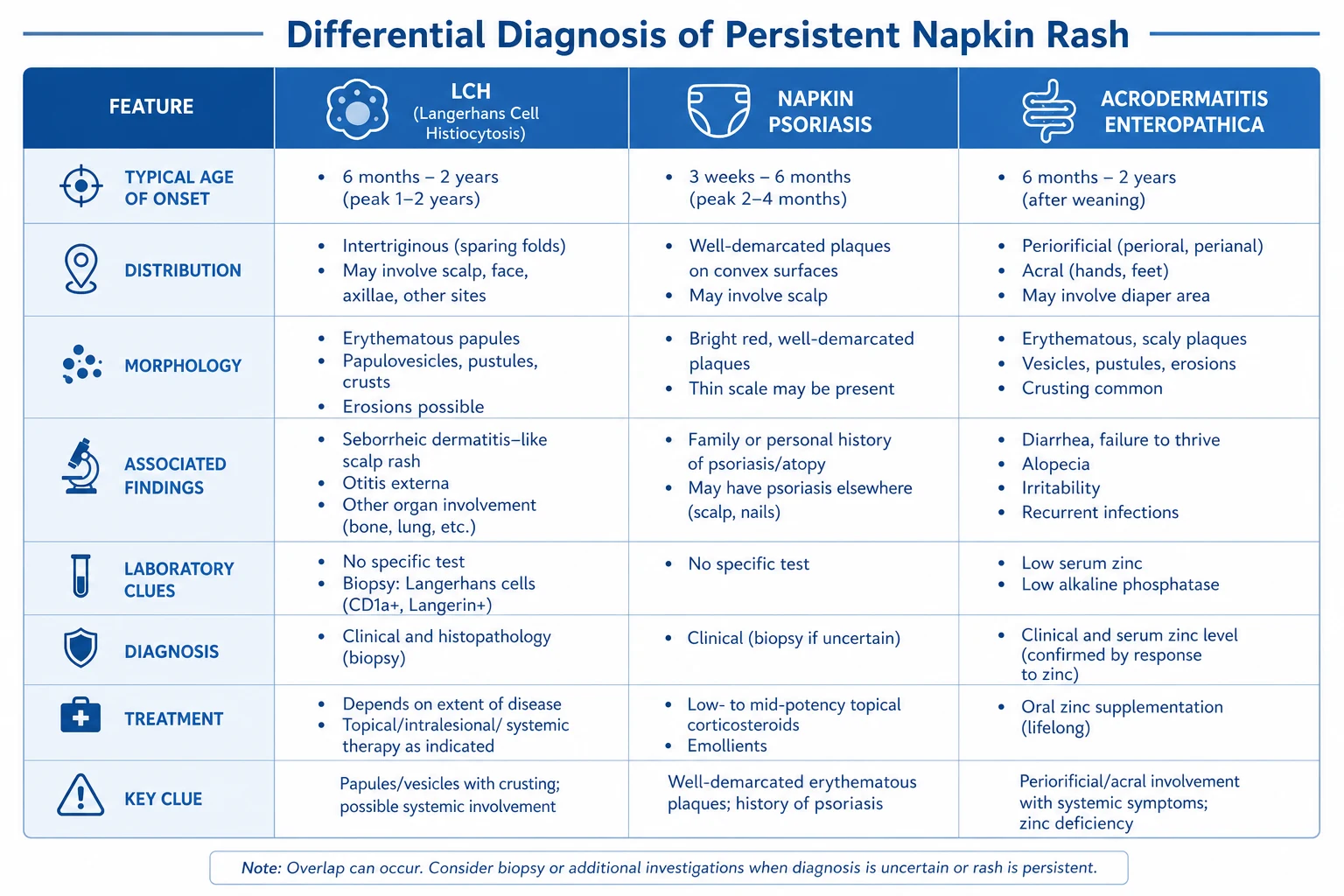
Cutaneous mastocytosis (CM)
Cutaneous disease has four recognised morphological variants:[1][4]
Systemic mastocytosis (SM)
Systemic disease is subcategorised by mast cell burden and organ damage into B-findings (markers of high burden but no organ impairment) and C-findings (evidence of organ damage that signals the need for cytoreductive therapy).[2][6]
The B-findings and C-findings that govern this stratification are worth memorising because they determine whether a patient is simply observed or treated with cytoreductive drugs:[2][6]
Epidemiology and Risk Factors
Mastocytosis is uncommon but not rare, and its epidemiology is sharply age-dependent. Children account for the majority of cutaneous cases, and roughly 65% of paediatric cutaneous mastocytosis begins before the age of two years, with most of the remainder presenting before adolescence.[4] The sex distribution is approximately equal (male-to-female roughly 1:1), and there is no clear racial predilection.[1]
The decisive epidemiological divide is paediatric versus adult-onset disease. In children, mastocytosis is overwhelmingly cutaneous and confined to the skin; systemic involvement is uncommon and progression to aggressive disease is rare.[4] In adults, the reverse holds: new-onset skin lesions almost always reflect underlying systemic mastocytosis, and adult disease is usually chronic and persistent rather than self-resolving.[2][5] The major risk factors for severe outcomes are an elevated serum tryptase (a marker of mast cell burden), advanced age at onset, the presence of C-findings or an associated haematological neoplasm, and Hymenoptera venom allergy — the single most important provokable trigger of life-threatening anaphylaxis in these patients.[9]
Familial clustering is recognised but exceptional; most cases are sporadic.[1]
Pathophysiology
The unifying molecular event in systemic mastocytosis is an activating somatic mutation in KIT, the gene encoding the transmembrane receptor tyrosine kinase for stem cell factor (SCF). The dominant mutation is KIT D816V — a substitution of aspartate by valine at codon 816, lying in the activation loop of the intracellular kinase domain — which renders the receptor constitutively active and ligand-independent.[2][6] This single change converts a tightly regulated growth-factor receptor into a continuously signalling molecule, driving survival, proliferation and accumulation of clonal mast cells in the bone marrow and extracutaneous tissues.
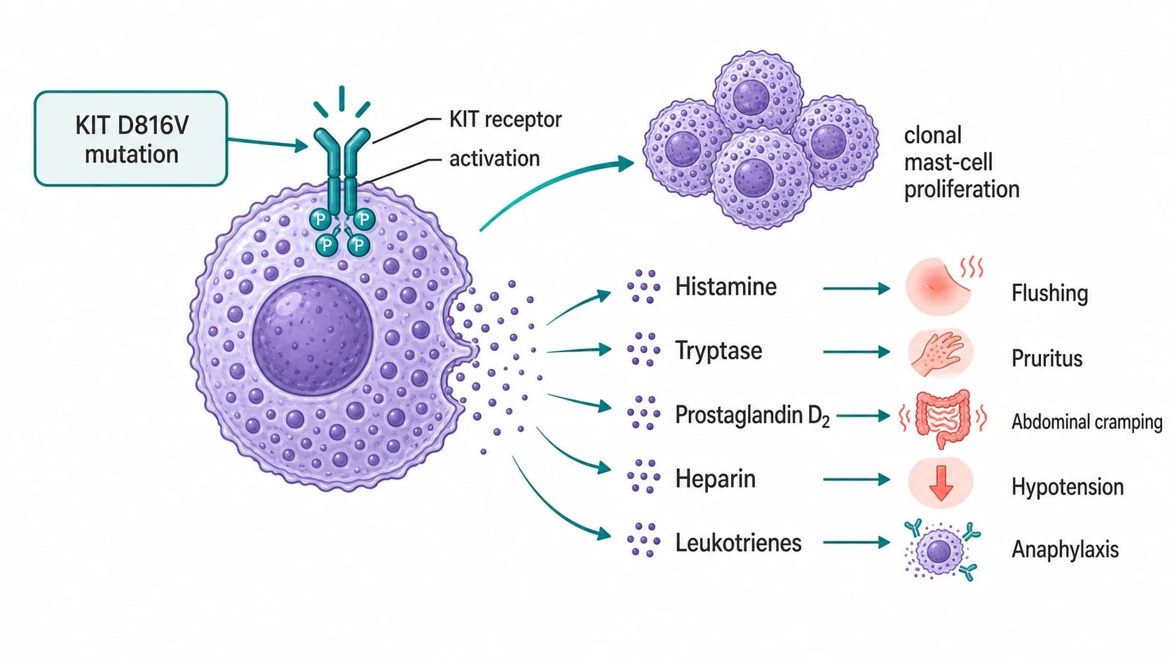
The mast cell lineage
Mast cells arise from CD34+ haematopoietic progenitors in the bone marrow, circulate as committed precursors, and complete their differentiation in peripheral tissues under the influence of SCF acting on KIT. Normal mast cell development is therefore SCF/KIT-dependent, which is precisely why an activating KIT mutation is so effective at producing mast cell excess — the proliferative brake is removed at the receptor level.[2] The mature cells take up residence in tissues rich in blood vessels and nerves (skin, gut mucosa, airway, bone marrow), where their granules await degranulation.
Why D816V matters for therapy
The location of the mutation within the kinase domain has a direct therapeutic consequence: imatinib does not inhibit the D816V-mutant kinase because the mutation locks the activation loop in a conformation that the drug cannot bind. Imatinib remains effective only for the rare mastocytosis driven by wild-type KIT or non-D816V mutations (and for a few KIT rearrangements).[2][7] This is why the modern cytoreductive agents for advanced systemic mastocytosis are midostaurin (a multi-kinase inhibitor active against D816V) and avapritinib (a selective D816V inhibitor), both of which circumvent imatinib resistance.[7]
Mast cell mediators and the symptom complex
The accumulated mast cells in mastocytosis are not merely numerous — they are hypersensitive, degranulating in response to trivial physical and chemical triggers. The resulting symptoms reflect the collective action of multiple mediators, not a one-mediator-per-symptom mapping:[8][9]
- Histamine (preformed) — H1-receptor effects produce flushing, pruritus, urticaria and bronchospasm; H2-receptor effects produce gastric acid hypersecretion, peptic ulceration, abdominal cramping and diarrhoea, and contribute to tachycardia and hypotension.
- Prostaglandin D2 (newly synthesised) — a potent vasodilator causing flushing, headache and hypotension, and a bronchoconstrictor contributing to wheeze.
- Leukotrienes (LTC4, LTD4, LTE4) — bronchoconstriction, increased vascular permeability and gut motility, contributing to wheeze, cramping and anaphylaxis.
- Heparin (preformed) — anticoagulant effect; may contribute to bruising and bleeding tendency, and co-released with histamine.
- Platelet-activating factor — augments vascular permeability and bronchoconstriction in anaphylaxis.
- Tryptase (preformed) — released in parallel with histamine, but its clinical role is as a diagnostic marker of mast cell burden and activation, not as a direct mediator of symptoms. Serum tryptase reflects total body mast cell load, which is why it rises in systemic disease.[8]
This multimediator basis explains two features examiners probe: first, why combined H1 and H2 antihistamine blockade is needed (histamine acts on both receptor families, and other mediators are not histamine at all); and second, why leukotriene antagonists and mast cell stabilisers add incremental benefit in refractory mediator symptoms.[1][9]
Why rubbing a lesion causes a wheal
The Darier sign is simply the clinical expression of this biology. The lesional skin is densely packed with fragile, granule-laden mast cells. Firm stroking mechanically disrupts their membranes, triggering an explosive local release of histamine and other vasoactive mediators into the surrounding dermis. Within two to five minutes the overlying vessels dilate and leak, producing the erythematous wheal and surrounding flare, and the accompanying histamine stimulates local C-fibres to produce pruritus. The same mechanism explains why hot baths, friction from clothing, exercise, alcohol, opioids and iodinated contrast provoke systemic flushing and itching — each is a degranulation stimulus acting on a mast cell population that is both enlarged and hyperreactive.[1][9]
Clinical Presentation
The clinical presentation of mastocytosis is a composite of cutaneous signs, mediator symptoms, and — in systemic disease — signs of organ infiltration. The tempo differs by age: children typically present with skin lesions alone, while adults present with skin lesions plus mediator symptoms and are found to have systemic involvement on investigation.[1][2]
Cutaneous manifestations (the Darier sign and its lesions)
The skin lesions of maculopapular cutaneous mastocytosis (urticaria pigmentosa) are the most diagnostically powerful findings in the disease. They are:[1][4]
- Reddish-brown to yellow-tan macules and papules, occasionally small nodules, distributed chiefly on the trunk, thighs and extremities, tending to spare the palms, soles and scalp.
- The lesions may be discrete and scattered, or numerous and confluent; in adults they are often monomorphic and small, while in children they are more polymorphic.
- A useful bedside clue is that the lesions blanch only partially on diascopy, and bullae may form in infants and in diffuse cutaneous disease, reflecting the massive subepidermal mast cell load. [1]
The Darier sign is elicited by firmly stroking a pigmented lesion with the blunt end of a pen or a tongue depressor and observing over two to five minutes for the development of an urtical wheal and erythematous flare with pruritus. A positive Darier sign is pathognomonic for cutaneous mastocytosis.[1] Two cautions: stroking should be gentle rather than traumatic (vigorous rubbing can provoke a systemic mediator response), and the sign may be absent in TMEP, where the mast cell burden per lesion is low — its absence does not exclude the diagnosis.
Mediator symptoms (triggerable, episodic)
The symptom complex of mastocytosis reflects episodic mast cell degranulation and is characteristically provoked by identifiable triggers:[9]
The mastocytosis mediator complex and its triggers
TELEPATH
Heat, hot bath, exercise, emotion, alcohol — episodic redness
Itching, often with the flush; worst over lesions
Pathognomonic — wheal on rubbing a lesion
Cramping, diarrhoea, nausea; histamine-driven acid hypersecretion
Histamine on H2 receptors and vasodilation
Hypotension, bronchospasm — esp. Hymenoptera stings
Alcohol, NSAIDs, opiates, iodinated contrast, friction, heat
Vasodilation from mediator release; can be the presenting event
The classic triggers — hot baths, sudden temperature change, vigorous exercise, emotional stress, alcohol, spicy foods, NSAIDs, opioids (morphine, codeine), iodinated radiocontrast and Hymenoptera venom — should be asked about directly, because identifying and avoiding them is the cornerstone of management.[1][9]
Systemic manifestations and organ involvement
In systemic mastocytosis, symptoms of organ infiltration accompany the mediator complex:[2][6]
- Gastrointestinal tract — abdominal cramping, diarrhoea, nausea, vomiting, peptic ulcer disease (histamine-driven acid hypersecretion), and occasionally malabsorption with weight loss in aggressive disease.
- Liver and spleen — hepatomegaly and splenomegaly; in aggressive disease, portal hypertension, ascites and transaminitis.
- Lymph nodes — lymphadenopathy, often found on imaging.
- Skeleton — bone pain, osteoporosis, osteosclerosis and osteolyses; pathological fractures may be the presenting feature, and unexplained osteoporosis in an adult warrants a serum tryptase.
- Haematological — cytopenias (anaemia, leucopenia, thrombocytopenia) in aggressive disease or SM-AHN; circulating mast cells indicate mast cell leukaemia.
- Neuropsychiatric — headache, fatigue, impaired concentration, depression — often under-recognised and disabling. [1]
Specific cutaneous variants
Solitary mastocytoma presents as a single tan-yellow or reddish-brown nodule or plaque, most often on the trunk or wrist of an infant; it too has a positive Darier sign and may blister.[4] Diffuse cutaneous mastocytosis is the rarest and most severe cutaneous form: the skin is diffusely thickened, doughy and yellowish with prominent folds, and blistering, erosions and fluid-electrolyte loss complicate infancy; mediator load is high and anaphylaxis risk is substantial.[3] TMEP (telangiectasia macularis eruptiva perstans) is an adult-onset variant of small, lightly pigmented, telangiectatic macules on the trunk, with minimal or absent Darier sign; because the mast cell burden is low in the skin but high systemically, TMEP should always prompt a search for systemic disease.[1]
Atypical presentations and pitfalls
Examiners deliberately probe atypical presentations. Recurrent idiopathic anaphylaxis with a normal-looking skin examination may be the only clue to bone marrow mastocytosis — a serum tryptase and KIT D816V test should be sent before labelling the episodes idiopathic.[9] Unexplained osteoporosis or a pathological fracture in a middle-aged adult may be the presenting feature of indolent systemic mastocytosis. Adult-onset cutaneous lesions almost always indicate systemic disease and must be worked up, not dismissed as a benign skin condition. In pregnancy, mediator symptoms may intensify and anesthetic planning for labour is essential because of the risk of perioperative anaphylaxis. Finally, in children with diffuse cutaneous mastocytosis, the combination of blistering, fluid loss and mediator release can mimic a severe bullous disorder or even sepsis, and early specialist involvement is mandatory.[3]
Differential Diagnosis
The pigmented, urticating papule has a finite differential, and each mimic has a distinguishing feature.[1][4]
The discriminating logic is mechanical. Ask first whether the lesions urticate when rubbed — only cutaneous mastocytosis does this, so a positive Darier sign essentially settles the question. Among Darier-negative pigmented lesions, ask whether they are elevated and granuloma-like (juvenile xanthogranuloma, histiocytosis), flat and uniform from birth (cafe-au-lait, melanocytic naevi), or transient and migratory (ordinary urticaria, papular urticaria). When doubt persists, a punch biopsy with Giemsa or tryptase stain resolves the diagnosis definitively by demonstrating the mast cell infiltrate.[1]
Clinical and Bedside Assessment
There is no substitute for eliciting the Darier sign at the bedside. A focused assessment answers three questions in sequence: (1) Is this cutaneous mastocytosis? (2) Could this be systemic? (3) What is the anaphylaxis risk?[1][5]
General inspection. Examine the entire skin under good light, noting the distribution, colour and morphology of the lesions. Document the body habitus and any flushing, and look for the stigmata of systemic disease. [1]
Eliciting the Darier sign. Select a representative pigmented lesion on the trunk or thigh. Firmly but not traumatically stroke it with the blunt end of a pen or a tongue depressor for several seconds, then observe for two to five minutes. A positive Darier sign is the appearance of an urtical wheal with surrounding erythema and pruritus over the stroked lesion. Document the result; photograph it for the record. Avoid testing multiple lesions at once in a heavily burdened patient, as the cumulative mediator release can provoke systemic symptoms.[1]
Systemic examination. In any adult with cutaneous mastocytosis, examine for hepatosplenomegaly, lymphadenopathy and bone tenderness, and assess for signs of cytopenias (pallor, petechiae). These findings push the diagnosis toward systemic disease and lower the threshold for bone marrow biopsy.[2]
Bedside risk stratification. A single serum tryptase drawn at a baseline (non-acute) visit is the most useful bedside blood test: a value persistently above 20 ng/mL is a WHO minor criterion for systemic mastocytosis and triggers formal systemic workup.[5][6] Note that tryptase also rises transiently during and after an acute anaphylactic episode, so a baseline level between attacks is more informative.
Investigations
The investigation strategy is two-tiered. Cutaneous mastocytosis is confirmed by skin biopsy, and systemic mastocytosis is confirmed and sub-categorised by bone marrow biopsy with mutational and laboratory assessment.[5][6]
Skin biopsy (confirms cutaneous disease)
A punch biopsy of a representative lesion demonstrates the diagnostic infiltrate:[1]
- Increased mast cells in the papillary and upper dermis — either as perivascular and periadnexal aggregates or, in more densely infiltrated lesions, as sheets; cells may be cuboidal or spindle-shaped (atypical).
- The threshold for cutaneous mastocytosis is a dense infiltrate of mast cells (typically more than 15 mast cells per high-power field in an aggregate, or perivascular clusters); isolated scattered mast cells are non-specific.
- Stains: the metachromatic granules are seen on Giemsa or toluidine blue; immunohistochemistry for tryptase, CD117 (KIT) and CD25 confirms the mast cell lineage and the aberrant phenotype (CD25 expression is a clonal marker). [1]
Systemic workup (mandatory in adults; guided by burden in children)
When cutaneous mastocytosis is confirmed in an adult, or when a child has high lesion burden or mediator symptoms, the systemic screen is undertaken:[2][5]
- Serum tryptase — the best non-invasive screening test. A baseline value persistently above 20 ng/mL is a WHO minor criterion for systemic mastocytosis. It correlates with total body mast cell burden and is used to monitor disease activity.[6]
- Bone marrow aspirate and trephine biopsy — the gold standard for systemic mastocytosis, demonstrating the major criterion (multifocal dense infiltrates of mast cells, typically 15 or more in aggregate) and allowing sub-categorisation. Mast cells are identified by tryptase/CD117/CD25 immunostain and often show atypical (spindle or bilobed) morphology.[10]
- KIT D816V mutation analysis — performed on bone marrow aspirate or peripheral blood (high-sensitivity allele-specific PCR). A positive result is a WHO minor criterion and confirms the clonal nature of the disease.[6]
- Full blood count, blood film and comprehensive metabolic panel — to detect cytopenias (C-finding), transaminitis and electrolyte disturbance.
- Skeletal survey and DEXA densitometry — in patients with bone pain, elevated tryptase or advanced disease, to detect osteolyses, osteosclerosis or osteoporosis; DEXA is now recommended at baseline in systemic disease.[2]
- Abdominal imaging (ultrasound or CT) — to define hepatosplenomegaly, lymphadenopathy, ascites or portal hypertension when organ involvement is suspected.
- Allergy and venom testing — specific IgE and skin testing to Hymenoptera venom (bee, wasp) in patients with a history of sting anaphylaxis, because venom allergy is the single largest provokable cause of life-threatening anaphylaxis in mastocytosis and indicates venom immunotherapy.[9]
The WHO diagnostic criteria for systemic mastocytosis are reproduced here because they are examinable. The diagnosis requires the major criterion plus one minor criterion, or three minor criteria in the absence of the major criterion:[5][6]
Management — Resuscitation (Acute Mediator Crisis and Anaphylaxis)
Acute severe mast cell mediator release presents as anaphylaxis: rapid-onset flushing, urticaria, angioedema, bronchospasm, abdominal cramping, hypotension and syncope, most often triggered by a Hymenoptera sting, an opioid or iodinated contrast, or an NSAID in a sensitised patient.[9] The management is standard anaphylaxis therapy applied without delay.[9]
[1]The single most important anticipatory step is ensuring that every patient with systemic mastocytosis (and any patient with a history of mediator-induced anaphylaxis) carries two adrenaline auto-injectors at all times and knows how to use them.[9] Patients on beta-blockers should be switched to alternative antihypertensives wherever possible, since beta-blockade both increases mast cell mediator release and renders adrenaline ineffective in anaphylaxis.
Management — Definitive and Stepwise
Definitive management is built in three layers: trigger avoidance, anti-mediator pharmacotherapy, and — only for advanced systemic disease — cytoreductive therapy. Cutaneous disease in children is managed almost entirely with the first two layers and often needs nothing more than reassurance.[1][5]
Layer 1 — Trigger avoidance (universal)
Every patient receives structured counselling to avoid degranulation triggers:[1][9]
- Heat and friction — avoid very hot baths and showers, sauna and hot tubs; pat (do not rub) the skin dry.
- Medications — avoid NSAIDs (if previously reactive; test challenge if essential), opioids (especially morphine and codeine; prefer fentanyl/pethidine if an opioid is required), iodinated radiocontrast (premedicate if essential), and decreased-strength alcohol tinctures.
- Alcohol and spicy foods — common flushing triggers.
- Hymenoptera stings — insect avoidance measures; carry adrenaline; venom immunotherapy if venom-allergic.
- Perioperative planning — alert anaesthesia; premedicate with antihistamines; avoid histamine-releasing agents; monitor for intraoperative mediator crisis. [1]
Layer 2 — Anti-mediator pharmacotherapy (the standard ladder)
The pharmacological ladder addresses mediator symptoms across multiple receptor and mediator pathways. Therapy is titrated to symptoms, not to tryptase.[1][5]
Cutaneous disease — topical and physical measures
For troublesome cutaneous lesions resistant to systemic anti-mediator therapy, two modalities are useful:[1]
- Topical corticosteroids — a very potent topical steroid (e.g. clobetasol propionate 0.05%) under occlusion can reduce lesional mast cell load and improve pruritus and pigmentation, particularly in localised or paediatric disease; use time-limited courses to avoid skin atrophy.
- Phototherapy — PUVA (psoralen plus UVA) or narrowband UVB (NB-UVB) over several weeks reduces lesional mast cell burden, pruritus and cosmetic pigmentation. Improvement is often gratifying but temporary; relapse within months is typical, so reserve phototherapy for symptomatic disease rather than for cosmesis alone. NB-UVB is preferred in children for its better safety profile. [1]
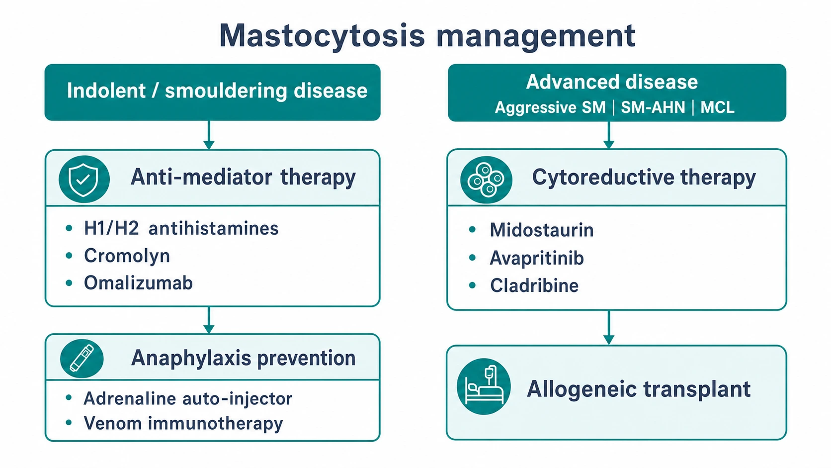
Anaphylaxis prevention
- Adrenaline auto-injector — two devices carried at all times; teach the patient and family. Essential for any patient with systemic mastocytosis, a history of anaphylaxis, or extensive cutaneous disease in children.[9]
- Venom immunotherapy — for patients with Hymenoptera venom allergy and systemic mastocytosis, lifelong venom immunotherapy dramatically reduces the risk of sting anaphylaxis and is strongly recommended.[9]
- Omalizumab (anti-IgE) — reduces the frequency of recurrent anaphylaxis and refractory mediator symptoms in selected patients; an off-label but increasingly used option.[8]
- MedicAlert identification and a written anaphylaxis action plan.
Layer 3 — Cytoreductive therapy (advanced systemic disease)
Cytoreduction is reserved for advanced systemic mastocytosis — aggressive SM, SM-AHN and mast cell leukaemia — defined by the presence of C-findings (organ damage). It is NOT used for indolent disease, where it adds toxicity without benefit.[2][7]
For SM-AHN, therapy is directed at the associated haematological neoplasm (e.g. azacitidine/decitabine for MDS, AML-type regimens), often combined with midostaurin for the mast cell component.[2] Bone disease (osteoporosis, fractures) is managed with bisphosphonates or denosumab alongside calcium and vitamin D optimisation.
Specific Subtypes and Scenarios
Solitary mastocytoma
A single tan-yellow or reddish nodule in an infant, typically on the trunk or wrist, with a positive Darier sign and occasional blistering. The natural history is almost uniformly self-resolving over years; management is reassurance and trigger avoidance, with symptomatic H1 antihistamines for pruritus. Excision is rarely needed and reserved for severely symptomatic lesions.[4]
Diffuse cutaneous mastocytosis
The rarest and most severe cutaneous form, presenting in infancy with diffusely thickened, doughy skin, prominent skin folds, and a high mast cell burden. Blistering, erosions, fluid and electrolyte loss, and a high risk of anaphylaxis dominate the clinical course. Management demands specialist input: aggressive anti-mediator therapy (combined H1/H2, cromoglycate, consider ketotifen), two adrenaline auto-injectors, and supportive skin care; bullae are managed as burns. Prognosis is guarded in infancy but often improves with age as the burden declines.[3]
TMEP (telangiectasia macularis eruptiva perstans)
An adult-onset variant of small, telangiectatic, lightly pigmented macules on the trunk, often with minimal Darier sign. The clinical trap is that the cutaneous burden appears trivial while systemic involvement is common — a serum tryptase and systemic assessment are mandatory in every case of TMEP.[1]
Mast cell leukaemia
The most aggressive category, defined by at least 20% mast cells on bone marrow aspirate smears (or circulating mast cells). Acute MCL carries C-findings and has a very poor prognosis (median survival measured in months); chronic MCL lacks C-findings and runs a more indolent but still serious course. Management is intensive cytoreduction (midostaurin, avapritinib, cladribine) and consideration of allogeneic transplant.[2]
Recurrent idiopathic anaphylaxis and bone marrow mastocytosis
A patient with recurrent anaphylaxis and a normal skin examination may have bone marrow mastocytosis — marrow involvement without skin lesions and with low burden. The diagnostic key is to check a serum tryptase and KIT D816V before labelling the episodes idiopathic; a marrow biopsy confirms the clonal mast cell population. These patients benefit from anti-mediator therapy, two adrenaline auto-injectors, and venom immunotherapy if a trigger is identified.[9][10]
Complications and Pitfalls
The complications of mastocytosis are the complications of mediator release and, in advanced disease, of organ infiltration.[2][9]
- Anaphylaxis — the most feared complication, most often triggered by Hymenoptera venom in adults; carries a real mortality risk. Every systemic mastocytosis patient needs an adrenaline auto-injector and, if venom-allergic, venom immunotherapy.[9]
- Peptic ulcer disease and GI bleeding — histamine-driven acid hypersecretion; prevent with H2 blockers and PPIs.
- Osteoporosis and pathological fractures — a common and under-recognised complication of systemic disease; screen with DEXA and treat with bisphosphonates/denosumab.[2]
- Malabsorption and weight loss — in aggressive disease with gut infiltration; a C-finding prompting cytoreduction.
- Portal hypertension and ascites — in aggressive disease with liver/spleen infiltration.
- Cytopenias — in aggressive SM, SM-AHN and MCL.
The classic pitfalls deserve specific attention:[5][9]
- Mislabelling recurrent idiopathic anaphylaxis without checking tryptase for occult systemic mastocytosis.
- Missing adult-onset cutaneous mastocytosis as a systemic disease by failing to perform a systemic workup.
- Overlooking osteoporosis as the presenting feature of indolent systemic mastocytosis in a middle-aged adult.
- Perioperative mediator crisis from histamine-releasing anaesthetic agents (opioids, muscle relaxants) — avoid them and premedicate.
- Aggressive rubbing to elicit the Darier sign provoking systemic symptoms — stroke gently, test one lesion.
- Using ranitidine — withdrawn in many markets for NDMA contamination; prescribe famotidine or nizatidine instead. [1]
Prognosis and Disposition
Prognosis is sharply age-dependent and, in adults, burden-dependent.[1][2]
- Childhood cutaneous mastocytosis — benign. Up to 50% resolve by puberty, and most of the remainder improve substantially; progression to systemic or aggressive disease is rare.[4]
- Adult-onset disease — chronic and persistent; cutaneous lesions rarely resolve. Indolent systemic mastocytosis has a near-normal life expectancy, but patients carry a lifelong anaphylaxis risk and require monitoring.[2]
- Smouldering SM — intermediate; a minority progress to aggressive disease or SM-AHN.
- Aggressive SM, SM-AHN and mast cell leukaemia — poor prognosis, with median survival ranging from roughly two to four years for aggressive SM down to months for acute MCL, though outcomes have improved with midostaurin and avapritinib.[7]
Disposition. A child with cutaneous mastocytosis is managed jointly by primary care and dermatology/paediatrics with reassurance, trigger advice and symptomatic antihistamines; a baseline tryptase is reasonable but bone marrow biopsy is reserved for high-burden or atypical cases. An adult with cutaneous mastocytosis is referred to haematology/allergy for systemic assessment (tryptase, KIT D816V, and bone marrow biopsy if tryptase is elevated or symptoms suggest organ involvement). Any patient with a history of anaphylaxis, high tryptase, or Hymenoptera venom allergy is enrolled in an anaphylaxis-prevention programme (adrenaline auto-injector, venom immunotherapy, MedicAlert).[5][9]
Special Populations
Children (paediatric weight-based management)
Childhood cutaneous mastocytosis is the commonest scenario and is usually benign. Weight-based dosing applies to antihistamines and adrenaline: cetirizine 0.25 mg/kg (or age-banded doses), and an adrenaline auto-injector at 0.15 mg (Junior) for children under 25-30 kg and 0.3 mg (adult device) above that. Diffuse cutaneous mastocytosis and extensive blistering disease in infants warrant specialist paediatric dermatology input because of the fluid, electrolyte and anaphylaxis risks.[3][4]
Parents should be counselled that the disease usually improves and often resolves, that the child should avoid known triggers and carry adrenaline if there has been any mediator event, and that school and childcare staff must be trained in auto-injector use. A MedicAlert is advisable for children with a history of anaphylaxis.[4]
Pregnancy
Mastocytosis symptoms may intensify or, less often, remit during pregnancy. The principal risk is perioperative anaphylaxis during labour and delivery: a written anaesthetic plan, avoidance of histamine-releasing agents (prefer fentanyl or remifentanil over morphine; use propofol-based induction), and availability of adrenaline are essential. Mastocytosis is not itself a contraindication to pregnancy or vaginal delivery, and the disease does not cross the placenta. Adrenaline auto-injectors are safe in pregnancy and should be continued.[9]
Adults with adult-onset cutaneous disease
Adult-onset cutaneous mastocytosis should be approached as systemic until proven otherwise: a serum tryptase, KIT D816V and — if tryptase is elevated — a bone marrow biopsy are mandatory. The patient is enrolled in long-term monitoring (annual tryptase, blood count, symptom review; DEXA at baseline).[2][5]
The immunocompromised and those with associated neoplasms
Patients with SM-AHN are managed jointly with haematology, with therapy directed at the associated neoplasm (MDS, MPN, AML) combined with midostaurin for the mast cell component. Immunosuppression for comorbid disease does not by itself worsen mastocytosis but complicates cytoreductive choices.[2]
Evidence, Guidelines, and Regional Differences
The diagnostic and therapeutic framework for mastocytosis has been progressively refined by the WHO (2001, 2008, 2016), the International Consensus Classification (2022), and the harmonisation proposals of the European Competence Network on Mastocytosis (ECNM) and the American Initiative in Mast Cell Diseases (AIM), which have converged the WHO and ICC criteria for clinical use.[6][10]
The landmark therapy trial is the Gotlib et al NEJM 2016 study of midostaurin in advanced systemic mastocytosis, which demonstrated clinically meaningful responses (overall response around 60%, including organ-damage responses) and established midostaurin as the standard cytoreductive agent for advanced disease.[7] Avapritinib, building on this, has extended effective targeted inhibition to D816V-driven disease in both advanced and selected indolent/smouldering settings. The ECNM diagnostic algorithm guides the workup of suspected mastocytosis and stratifies the decision to perform bone marrow biopsy.[5]
[1] [1]Australian and New Zealand practice follows ECNM-derived protocols. The PBS funds midostaurin and avapritinib for advanced systemic mastocytosis under specialist authority. Adrenaline auto-injectors (EpiPen, Anapen, Emerade) are PBS-subsidised; patients with venom allergy and mastocytosis receive lifelong venom immunotherapy. Ranitidine withdrawal applies (TGA, 2020).
The disease biology is identical worldwide; what varies is drug availability (cromoglycate availability differs between US "cromolyn" and UK/EU "cromoglicate"; ketotifen availability is country-specific), adrenaline auto-injector brand (EpiPen, Emerade, Anapen, Jext), and the degree of specialist access for bone marrow mutational testing and cytoreductive therapy.
Controversies. The boundary between indolent systemic mastocytosis and mast cell activation syndrome (MCAS) is debated — MCAS describes patients with recurrent mediator symptoms, a clonal mast cell abnormality that does not meet full SM criteria, and a response to anti-mediator therapy.[8] The role of omalizumab and phototherapy rests on observational data rather than randomised trials; ketotifen evidence is likewise largely historical. The threshold tryptase at which to recommend bone marrow biopsy is conventionally 20 ng/mL, but this is calibrated to the systemic-mastocytosis population and may over-investigate patients with isolated anaphylaxis.[5]
Exam Pearls
Triggers to AVOID in mastocytosis
HOT-FAIM
Temperature change, sauna, friction
Morphine, codeine — histamine-releasing; prefer fentanyl
Alcohol and spicy foods trigger flushing
Rubbing lesions, tight clothing
If previously reactive; also iodinated contrast
Hymenoptera venom — carry adrenaline, consider VIT
Premedicate if essential; alert anaesthesia
Self-test: A 42-year-old woman has new-onset flushing, diarrhoea and reddish-brown trunk macules that wheal when rubbed. Tryptase is 38 ng/mL. What is the diagnosis, and what is the next step?
The clinical picture is systemic mastocytosis: adult-onset cutaneous lesions with a positive Darier sign, mediator symptoms (flushing, diarrhoea) and a serum tryptase persistently above 20 ng/mL (a WHO minor criterion). The next step is bone marrow aspirate and trephine biopsy with KIT D816V mutation analysis, plus a full blood count, biochemistry and DEXA. She should start trigger avoidance and H1 (cetirizine 10 mg) plus H2 (famotidine 20 mg BD) antihistamines, carry two adrenaline auto-injectors, and have Hymenoptera venom testing. Cytoreductive therapy is NOT indicated unless C-findings (organ damage) are present.
Red Flags
Exam application bank (NEET-PG / INICET)
One-line answer
Mastocytosis is a clonal neoplastic proliferation of mast cells affecting the skin (cutaneous) and/or internal organs (systemic). The hallmark of cutaneous disease is the DARIER SIGN — reddish-brown maculopapular lesions that urticate (wheal and flare) when rubbed, from mechanical degranulation of lesional mast cells. Cutaneous variants include maculopapular cutaneous mastocytosis (urticaria pigmentosa, most common in children), solitary mastocytoma, diffuse cutaneous mastocytosis, and telangiectasia macularis eruptiva perstans (TMEP, adults). Systemic mastocytosis (adults; KIT D816V gain-of-function mutation in over 90%) involves the bone marrow and causes episodic flushing, diarrhoea, palpitations, hepatosplenomegaly, osteoporosis and potentially life-threatening anaphylaxis — especially after Hymenoptera stings. Diagnosis: skin biopsy (mast cell infiltrate; CD117/tryptase/CD25+), seru
Worked stems (answer without another resource)
Stem 1 — Classic presentation. Map symptoms to mechanism; name the first investigation and first treatment step with dose/route if drug therapy is standard. [1]
Stem 2 — Unstable / complicated. List red flags that force immediate resuscitation, theatre, ICU, antidote, or reperfusion — and what you do in the first 15 minutes. [1]
Stem 3 — Atypical group. Elderly, pregnancy, child, or immunocompromised: how presentation and thresholds change. [1]
Stem 4 — Differential trap. Name the three closest mimics and one discriminator for each. [1]
Stem 5 — Disposition. Who goes home with safety-netting, who is admitted, who needs HDU/ICU/theatre, and what follow-up is mandatory. [1]
Rapid viva checklist
- Definition + classification
- Pathophysiology chain
- Bedside signs / criteria
- Score with exact components (if any)
- Emergency bundle
- Definitive therapy with doses
- Complications of disease and of treatment
- Special populations
- Guideline/trial name if classic
- Three exam traps
Coverage self-check
If you cannot answer any stem above from this page alone, re-read the matching section — the page is intended to be self-sufficient for final-prof and NEET-PG/INICET questions on Mastocytosis (cutaneous and systemic) — urticaria pigmentosa.
References
- [1]Asna N, Suri A, Sarkar R, Garg VK. Cutaneous mastocytosis: A dermatological perspective Australas J Dermatol, 2021.PMID 33040350
- [2]Lim KH, Pardanani A, Tefferi A. Review and Updates on Systemic Mastocytosis and Related Entities Cancers (Basel), 2023.PMID 38067330
- [3]Brockow K, Akin C, Huber M, Metcalfe DD. Diffuse Cutaneous Mastocytosis: A Current Understanding of a Rare Disease Int J Mol Sci, 2024.PMID 38338679
- [4]Akoglu G, Erkin G, Cakir B, et al. Pediatric cutaneous mastocytosis: a review of 180 patients Isr Med Assoc J, 2005.PMID 15909466
- [5]Valent P, Escribano L, Broesby-Olsen S, et al. Proposed diagnostic algorithm for patients with suspected mastocytosis: a proposal of the European Competence Network on Mastocytosis Allergy, 2014.PMID 24836395
- [6]Valent P, Hartmann K, Hoermann G, et al. Harmonization of Diagnostic Criteria in Mastocytosis for Use in Clinical Practice: WHO vs ICC vs AIM/ECNM J Allergy Clin Immunol Pract, 2024.PMID 39216803
- [7]Gotlib J, Kluin-Nelemans HC, George TI, et al. Efficacy and Safety of Midostaurin in Advanced Systemic Mastocytosis N Engl J Med, 2016.PMID 27355533
- [8]Castells M, Giannetti MP, Hamilton MJ, et al. Mast cell activation syndrome: Current understanding and research needs J Allergy Clin Immunol, 2024.PMID 38851398
- [9]Gulen T, Akin C. Anaphylaxis and Mast Cell Disorders Immunol Allergy Clin North Am, 2022.PMID 34823750
- [10]Zanotti R, Bonifacio M, Lucchini G, et al. Refined diagnostic criteria for bone marrow mastocytosis: a proposal of the European competence network on mastocytosis Leukemia, 2022.PMID 34545185