Dermatology · Medicine
Melanocytic naevi
Also known as Melanocytic naevi · Moles · Naevocellular naevi · Melanocytic nevi
Melanocytic naevi (moles) are benign proliferations of melanocytes (naevus cells), classified as acquired (junctional, compound, intradermal), congenital (small, medium, large/giant), or special types (Spitz, blue, halo/Sutton, dysplastic/atypical). Dermoscopy reveals age-dependent benign patterns (globular in children, reticular in young adults, homogeneous in older adults). The key clinical task is distinguishing benign naevi from melanoma (ABCDE, ugly duckling sign, dermoscopy criteria) and identifying high-risk lesions requiring biopsy — especially Spitz naevi (excisional biopsy to exclude Spitzoid melanoma) and giant congenital naevi (melanoma and neurocutaneous melanosis risk). Fellowship-level assessment demands mastery of the naevus classification and maturation sequence, dermoscopy patterns, the special subtypes (Spitz, halo, blue, congenital) and their management, and the indications for biopsy and surveillance.
On this page & tools
Your progress
Saved locally on this device.
Practise this topic
Exam tags
Red flags
Definition & Classification
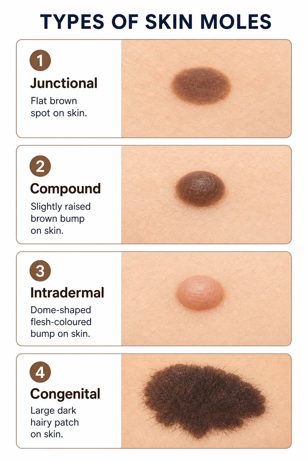
Melanocytic naevi (moles) are benign proliferations of naevus cells (modified melanocytes of neural-crest origin). They are classified by onset, histological location of the naevus cells, and morphology:[4]
Acquired naevi (the commonest type)
| Type | Location of naevus cells | Clinical appearance |
|---|---|---|
| Junctional | Epidermo-dermal junction | Flat, dark brown-black macule; reticular dermoscopy |
| Compound | Junction + dermis | Slightly raised, brown papule; combined reticular + globular |
| Intradermal | Dermis only | Dome-shaped, flesh-coloured or light brown papule; commonest in adults; homogeneous dermoscopy |
Maturation sequence: junctional → compound → intradermal as naevus cells migrate from the junction into the dermis with age. This explains why flat dark naevi (junctional) are commoner in youth, while dome-shaped flesh-coloured naevi (intradermal) predominate in older adults.[4]
Congenital melanocytic naevi (CMN)
Present at birth or within the first year of life. Classified by projected adult size:[5]
| Size category | Projected adult diameter | Melanoma risk |
|---|---|---|
| Small | less than 1.5 cm | ~1% (same as general population) |
| Medium | 1.5-19.9 cm | ~1-2% |
| Large / Giant | ≥20 cm or ≥5% BSA | ~5-10% (highest) |
Special types
| Type | Clinical | Key features |
|---|---|---|
| Spitz naevus | Pink-red firm hairless papule in children | Spindled/epithelioid cells; "Spitzoid" spectrum; excise to exclude melanoma |
| Blue naevus | Blue-black papule | Dermal melanocytes; common on hands, feet, scalp |
| Halo naevus (Sutton) | Naevus with depigmented halo | CD8+ T-cell-mediated; vitiligo association; self-resolving |
| Dysplastic / atypical | Irregular, >5mm, variable colour | See dedicated topic; FAMMM syndrome |
| Meyerson naevus | Naevus with eczematous halo | Type IV hypersensitivity; topical steroid |
| Recurrent (pseudomelanoma) | Pigmentation at excision scar | Not a true recurrence; review histology |
Epidemiology
- Average adult has 15-40 acquired naevi; number peaks in the 3rd-4th decade then declines with age (immune clearance).[4]
- Fair-skinned (Fitzpatrick I-II) individuals have more naevi than dark-skinned.
- High naevus count (>50 common or >5 atypical) is a major melanoma risk factor.[2]
- Genetic: CDKN2A (p16) germline mutations → familial atypical multiple mole melanoma (FAMMM) syndrome; NRAS mutations in CMN.[2]
Dermoscopy — Benign Patterns
Quick numbers for the examiner
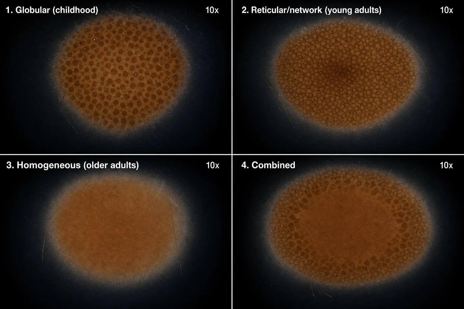
Dermoscopy reveals age-dependent benign patterns:[4]
- Globular pattern: uniform brown globules; children/adolescents (compound naevi).
- Reticular (network) pattern: regular pigment network with uniform mesh; young adults (junctional naevi).
- Homogeneous pattern: uniform structureless brown; older adults (intradermal naevi).
- Combined pattern: two patterns coexisting (e.g. reticular periphery + central globules). [1]
The key task is confirming symmetry, uniformity and absence of melanoma-specific criteria (atypical network, blue-white veil, regression, polymorphous vessels, multiple colours).[4]
ABCDE of melanoma — the screening bedside rules
One half doesn't match the other; benign naevi are usually symmetric.
Irregular, ragged, notched, blurred edges; benign naevi have smooth, well-defined borders.
Variation in pigmentation (brown, black, red, white, blue); benign naevi are uniform in colour.
>6 mm (size of a pencil eraser) is suspicious; benign naevi usually smaller.
Change in size, shape, colour, elevation, or new symptoms (itch, bleed, crust); the most important feature.
Clinical Presentation — Special Types [1]

- Spitz naevus: a pink-red, firm, hairless, dome-shaped papule (usually 5-10 mm) in children; may be pigmented. Histologically shows spindled and epithelioid melanocytes. The diagnostic challenge is distinguishing benign Spitz from Spitzoid melanoma — hence excisional biopsy is recommended for all Spitzoid lesions.[1][6]
- Blue naevus: a blue-black papule caused by dermal melanocytes (deep Tyndall effect makes it appear blue); common on the dorsum of hands/feet, scalp, buttocks. Stable; excise only if changing or symptomatic.
- Halo naevus (Sutton naevus): a central pigmented naevus surrounded by a symmetrical depigmented halo; caused by CD8+ T-cell-mediated immune response against naevus cells (which also destroys surrounding normal melanocytes → halo). Multiple, common in adolescents. Associated with vitiligo. Self-resolving (the central naevus eventually disappears). No treatment needed; check for vitiligo elsewhere.
- Giant CMN: a large dark brown-black plaque (>20 cm projected adult diameter) with hypertrichosis and small satellite lesions. May be associated with neurocutaneous melanosis (leptomeningeal melanocytosis → neurological symptoms, hydrocephalus, seizures).[5]
Differential Diagnosis
| Mimic | Distinguishing features |
|---|---|
| Melanoma | ABCDE, dermoscopy chaos-and-clues, ugly duckling |
| Seborrhoeic keratosis | "Stuck-on", milia-like cysts, comedo-like openings |
| Dermatofibroma | Dimple sign, firm, central white patch on dermoscopy |
| Pigmented BCC | Pearly border, arborising vessels |
| Lentigo | Flat, uniform, no naevus cells on histology |
| Ephelis (freckle) | Sun-induced, flat, no naevus cells, darkens with UV |
Investigations
- Clinical diagnosis + dermoscopy is sufficient for the vast majority of benign naevi.[4]
- Biopsy (excisional) is indicated for: any naevus with atypical features (ABCDE, change, ugly duckling), all Spitzoid lesions, symptomatic or rapidly growing naevi, and naevi with atypical dermoscopy.
- Total body photography + sequential digital dermoscopy for patients with many naevi or atypical naevus syndrome.[2]
- MRI brain and spine for giant CMN (screen for neurocutaneous melanosis).[5]
- Histology confirms diagnosis and excludes melanoma (especially for Spitzoid lesions requiring expert dermatopathology).[6]
Management
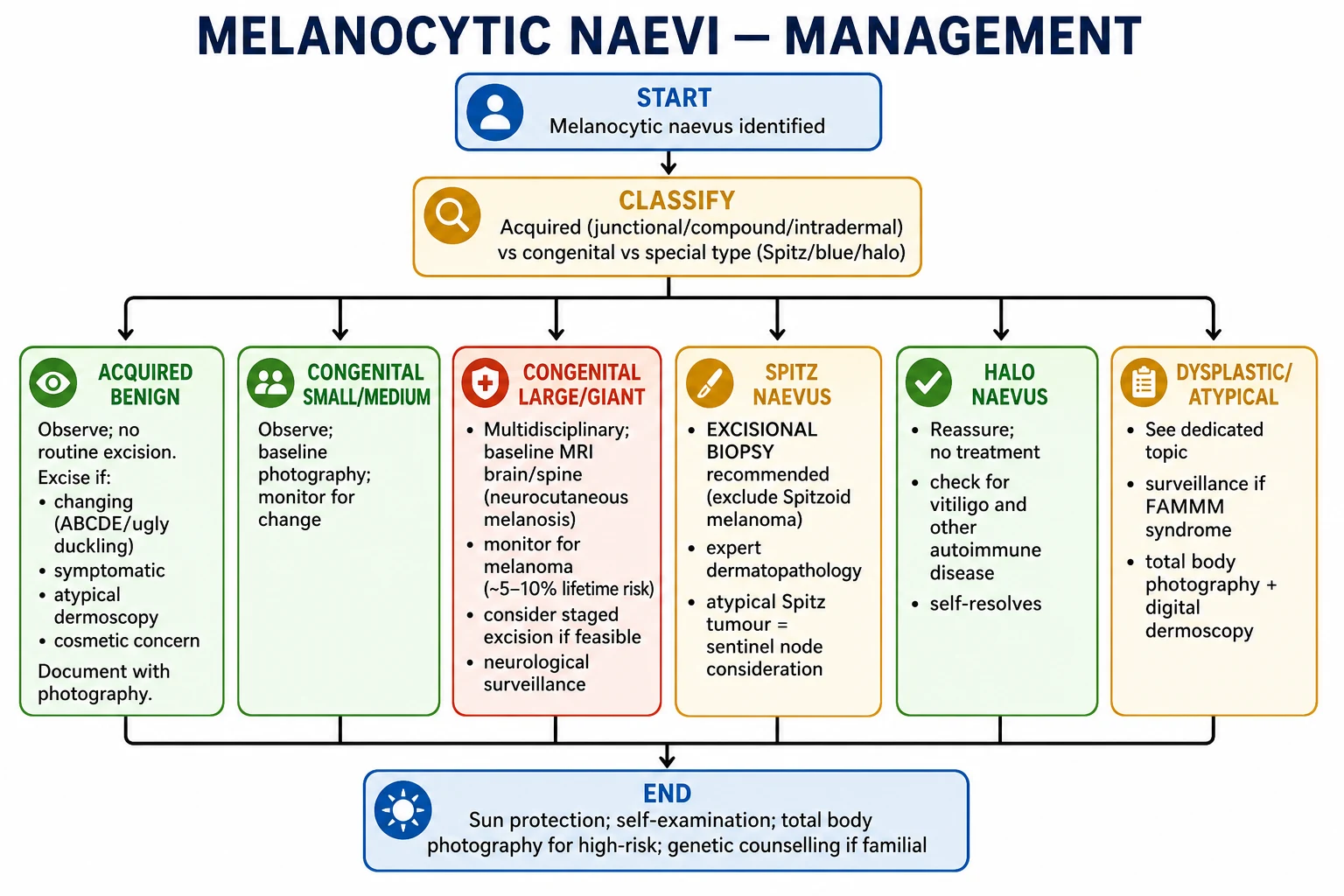
- Acquired benign naevi: observe; no routine excision. Excise if changing, symptomatic, atypical dermoscopy or cosmetic concern. Document with photography.[4]
- CMN (small/medium): observe; baseline photography; monitor for change; reassure parents.[5]
- CMN (large/giant): multidisciplinary management; baseline MRI brain/spine for neurocutaneous melanosis; monitor for melanoma (clinical exam every 3-6 months); consider staged surgical excision or laser if feasible; neurological surveillance.[3][5]
- Spitz naevus: excisional biopsy is recommended for ALL Spitzoid lesions (to exclude Spitzoid melanoma); expert dermatopathology; atypical Spitz tumours may warrant sentinel node consideration.[1][6]
Pathophysiology & Molecular Biology
Naevi arise from clonal proliferation of melanocytes driven by activating mutations in the MAPK/ERK pathway — primarily BRAF V600E in approximately 80% of acquired benign naevi and NRAS Q61 in congenital melanocytic naevi (especially giant CMN, where NRAS mosaicism is the dominant molecular driver).[2][4]
The classic "oncogene-induced senescence" explains why a single mutant melanocyte clone expands into a clinically visible naevus but then arrests: BRAF/NRAS activation drives initial proliferation, followed by p16/INK4a and p53-mediated senescence. This is why benign naevi are stably growth-arrested — they do not progress to melanoma unless additional "second hits" (CDKN2A loss, TERT promoter mutations, additional MAPK effectors) break the senescence barrier. [1]
Key molecular correlates: [1]
- BRAF V600E (acquired naevi, dermoscopy globular/reticular): benign stable clone
- NRAS Q61K/R (CMN, deep-penetrating naevus): larger clones, deeper dermal involvement
- BAP1 loss (BAP1-inactivated naevus, atypical Spitzoid): seen in BAP1 tumour predisposition syndrome; larger and more numerous epithelioid Spitzoid naevi
- HRAS Q61 (Spitz naevus, some congenital): Spitzoid morphology
- CDKN2A germline loss: familial atypical multiple mole melanoma (FAMMM) syndrome; high melanoma risk [1]
Specific Subtypes & High-Yield Scenarios
- Recurrent (persistent) naevus / pseudomelanoma: pigmentation at the edge of an excision scar; not a true recurrence. Review original histology carefully; re-excise if atypical.
- Meyerson naevus: a halo of acute eczema around a benign naevus; type IV hypersensitivity; settles with topical steroid; not malignant.
- Combined naevus: two histological patterns in one lesion (e.g. blue + compound); typically benign.
- Deep penetrating naevus: dark dermal naevus with wedge-shaped deep extension; benign but can simulate melanoma; expert pathology review.
- BAP1-inactivated naevus: pink-tan epithelioid Spitzoid naevi seen in BAP1 tumour predisposition syndrome (germline BAP1 mutation); risk of uveal melanoma, mesothelioma, RCC; multiple naevi; cutaneous melanoma.
- Pigmented spindle cell naevus (of Reed): dark brown-black flat well-defined papule on thigh of young women; dermoscopy shows regular pigmented striae radiating from centre; benign.
- Desmoplastic naevus: thick collagenous stroma; firm, sclerotic; can mimic dermatofibroma.
- Naevus spilus (speckled lentiginous naevus): tan background macule with superimposed darker macules/papules; risk of melanoma within lesion (especially giant variants).
- Halo naevus in children: most common cause of a depigmented halo around a mole; immune-mediated; usually resolves spontaneously over 2-5 years. [1]
Spitz Naevus & Spitzoid Melanoma — The Diagnostic Spectrum [1]
The Spitzoid spectrum is one of the most challenging areas in dermatopathology because it spans from a clearly benign childhood papule to an aggressive adult melanoma that may be histologically indistinguishable at one end of the spectrum. The current WHO classification (2018) collapses the older Spitz tumour / atypical Spitz tumour / Spitzoid melanoma terminology into a continuum of "Spitz melanocytic tumours" with annotation of risk category.[1][6]
Spitz naevus (classic / benign)
- Demographics: 70% occur in patients under 20 years of age; peak incidence 3-10 years.
- Morphology: solitary, rapidly growing, pink-red, dome-shaped, hairless, firm papule, usually 5-10 mm; face and lower extremities are the commonest sites.
- Dermoscopy (classic "starburst" pattern): a peripheral radiating pattern of brown or pink streaks/pseudopods at the edge (a true "starburst"), often combined with a central homogeneous pink-tan area with dotted vessels. This pattern is highly suggestive of a benign Spitz naevus in a child.
- Histology: large spindled and/or epithelioid melanocytes arranged in vertically-oriented nests with clefts around nests (artefactual retraction), eosinophilic cytoplasm, occasional Kamino bodies (PAS-positive eosinophilic globules at the dermo-epidermal junction). Maturation with depth is preserved and mitoses are rare or confined to the superficial dermis.
- Molecular profile: HRAS Q61 mutations or rearrangement of ALK, ROS1, NTRK1, NTRK3, BRAF, or RET — none of which are typically seen in conventional melanoma.
- Behaviour: benign; spontaneous regression documented; no malignant transformation in classic childhood Spitz naevi. [1]
Atypical Spitz tumour (AST) / Spitz melanocytoma
- Lesions with one or more worrisome clinical or histological features that fall between classic Spitz naevus and Spitzoid melanoma.
- Features of concern: size >10 mm, ulceration, deep mitoses, asymmetry, poor maturation, dense inflammation, or patient age >10 years.
- Molecular work-up is mandatory — FISH (RREB1, MYB, CCND1, CEP6) and next-generation sequencing for kinase fusions (ALK/ROS1/NTRK) help stratify risk; BAP1 loss suggests BAP1-inactivated melanocytic tumour (often germline BAP1 tumour predisposition syndrome — consider uveal melanoma and renal cell carcinoma screening in the patient and family).[6]
- Management: complete excisional biopsy with 1-3 mm margins; consider sentinel lymph node biopsy (SLNB) in adults or atypical lesions, although its prognostic value is debated.
Spitzoid melanoma
- Most often an adult presentation (>20 years), or a childhood lesion with frankly malignant architectural and cytological features.
- Dermoscopy: an atypical starburst (irregular, asymmetric distribution of streaks), homogeneous blue pigmentation, ulceration, or atypical vascular pattern — the classic reassuring starburst pattern is lost.
- Histology: pagetoid spread, deep dermal mitoses, marked cytological atypia, loss of maturation with depth, necrosis, and an expansile nodular growth pattern.
- Molecular profile: lacks the kinase fusions typical of benign Spitz; instead, shows conventional melanoma drivers (BRAF V600E, NRAS, TERT promoter mutations, CDKN2A loss, PTEN loss) — useful adjunct when histology is borderline.
- Management: wide local excision with 1-2 cm margins (per melanoma guidelines), SLNB, staging imaging, and adjuvant therapy as per conventional melanoma protocols. [1]
Practical approach to a Spitzoid lesion
SPITZ — when to excise
Larger than a pencil eraser; atypical.
Brown/black Spitzoid lesions are more suspicious than the classic pink papule.
Loss of the symmetric radiating pattern on dermoscopy; architectural disorder.
Spitz naevi in adults carry significantly higher melanoma risk; biopsy.
Ulceration, bleeding, rapid growth, or evolution — all red flags.
Congenital Melanocytic Naevi (CMN) — Variants & Risk Stratification
CMN are present at birth or appear within the first year of life (tardive CMN) and show distinct clinical and molecular features from acquired naevi. The most important clinical correlate is the size-dependent melanoma risk and the association with neurocutaneous melanosis (NCM).[5]
Size categories and projected adult diameter
| Category | Projected adult diameter | Approximate melanoma risk | Neurocutaneous melanosis risk |
|---|---|---|---|
| Small | less than 1.5 cm | ~1% (population baseline) | Negligible |
| Medium | 1.5-19.9 cm | ~1-2% | Low |
| Large / Giant | ≥20 cm OR ≥5% BSA in newborns | 5-10% lifetime | 5-15% (highest in axial/head/neck) |
| Multiple medium CMN (≥3) | Multiple | Slightly elevated | Higher if >20 satellites |
The projected adult diameter (PAD) is calculated by multiplying the measured diameter in infancy by 1.7 (head) or by 3 (other body sites) to estimate final adult size — the basis for early risk stratification.[5]
Clinical variants
- Giant CMN ("bathing trunk" / "cape" / "stocking" distribution): extensive dark brown-black plaque with hypertrichosis, rugosity, and multiple satellite lesions (smaller CMN scattered over the body). Subtypes based on distribution: bolero (upper back/arms), cape (head/neck/upper back), bathing trunk (lower trunk/perineum/legs).
- "Neurocutaneous melanosis" (NCM): proliferation of melanocytes in the leptomeninges. Risk factors: giant CMN in axial/head/neck distribution AND >20 satellite lesions. May be asymptomatic (found incidentally on MRI) or present with neurological symptoms (seizures, cranial nerve palsies, hydrocephalus, developmental delay) — symptomatic NCM carries a 50% mortality in early childhood. Screening MRI brain ± spine with contrast is recommended in the first year of life.[3][5]
- "Satellite" lesions: smaller CMN scattered beyond the main lesion; >20 satellites significantly increases NCM risk.
- "Halo congenital naevus": a CMN developing a depigmented halo (CD8+ T-cell-mediated) — usually benign and may herald regression.
- "Proliferative nodule" within a giant CMN: a rapidly growing nodule within a CMN in infancy. Most are benign but difficult to distinguish histologically from melanoma; expert dermatopathology review is mandatory, and complete excision is preferred.
Giant CMN + CNS — the NEURO mnemonic for neurocutaneous melanosis screening
More than 20 satellite naevi dramatically increase NCM risk; full skin examination at birth.
Bathing trunk, cape, or bolero distribution; leptomeningeal melanocytes track the dorsal mid-line neural crest.
Seizures, cranial nerve palsy, hydrocephalus, vomiting, developmental delay — symptomatic NCM carries up to 50% mortality.
Screening MRI with contrast in infancy; look for T1-hyperintense leptomeningeal deposits, especially in the temporal lobes and cerebellum.
Repeat imaging if symptomatic; multidisciplinary care with paediatric dermatology, neurosurgery, and oncology.
Melanoma risk — the numbers that matter
- Giant CMN ("bathing trunk" / "cape" / "stocking" distribution): extensive dark brown-black plaque with hypertrichosis, rugosity, and multiple satellite lesions (smaller CMN scattered over the body). Subtypes based on distribution: bolero (upper back/arms), cape (head/neck/upper back), bathing trunk (lower trunk/perineum/legs).
- "Neurocutaneous melanosis" (NCM): proliferation of melanocytes in the leptomeninges. Risk factors: giant CMN in axial/head/neck distribution AND >20 satellite lesions. May be asymptomatic (found incidentally on MRI) or present with neurological symptoms (seizures, cranial nerve palsies, hydrocephalus, developmental delay) — symptomatic NCM carries a 50% mortality in early childhood. Screening MRI brain ± spine with contrast is recommended in the first year of life.[3][5]
- "Satellite" lesions: smaller CMN scattered beyond the main lesion; >20 satellites significantly increases NCM risk.
- "Halo congenital naevus": a CMN developing a depigmented halo (CD8+ T-cell-mediated) — usually benign and may herald regression.
- "Proliferative nodule" within a giant CMN: a rapidly growing nodule within a CMN in infancy. Most are benign but difficult to distinguish histologically from melanoma; expert dermatopathology review is mandatory, and complete excision is preferred.
Surgical and laser management of giant CMN
- Complete excision (serial excision or tissue expansion) reduces melanoma risk and improves cosmetic/functional outcome, but may not be feasible for very extensive lesions; risk/benefit must be individualised.
- Curettage or dermabrasion in the first 2-4 weeks of life removes the superficial pigmented component (when naevus cells are still confined to the upper dermis) — reduces pigmentation but does NOT eliminate melanoma risk (deeper residual cells remain).
- Laser (Q-switched ruby/Nd:YAG, ablative CO2/Er:YAG) can lighten pigmentation; does not reduce melanoma risk; may obscure future dermoscopic assessment.
- Multidisciplinary care (paediatric dermatology, plastic surgery, neurosurgery for NCM, oncology, psychology) is the standard for giant CMN. [1]
Familial Melanoma Syndromes (FAMMM & Related)
Familial atypical mole-multiple melanoma (FAMMM) syndrome (also called dysplastic naevus syndrome, atypical mole syndrome) describes kindreds with multiple atypical/dysplastic naevi and a strong family history of melanoma. The major susceptibility gene is CDKN2A, encoding the tumour suppressors p16/INK4a and p14/ARF — both cell-cycle regulators on chromosome 9p21.[2]
Diagnostic criteria for FAMMM
The Amsterdam-style criteria for FAMMM kindreds require: [1]
- One individual with melanoma, AND
- One first- or second-degree relative with melanoma, AND
- Multiple (>50) naevi with multiple (>5) clinically atypical naevi in the proband or family members. [1]
Genetic basis and cancer spectrum
- CDKN2A (p16/INK4a loss → cell-cycle deregulation; p14/ARF loss → MDM2-mediated p53 degradation) accounts for ~20-40% of familial melanoma kindreds.
- CDK4 mutations (rare) — same phenotype as CDKN2A.
- MC1R variants (RHC variant alleles) — red hair, fair skin, freckling phenotype; modest melanoma risk.
- BAP1 tumour predisposition syndrome — germline BAP1 loss; cutaneous BAP1-inactivated melanocytic tumours (BIMT, "atypical Spitz"), uveal melanoma, renal cell carcinoma, mesothelioma, and other cancers.
- MITF p.E318K — moderate melanoma and renal cell carcinoma risk.
- Hereditary breast/ovarian (BRCA1/2) and Lynch syndrome also confer modest melanoma risk in some kindreds. [1]
Clinical and surveillance implications
- Lifelong melanoma risk is markedly elevated: CDKN2A carriers have a 30-90% lifetime melanoma risk (varies by geography and penetrance), often with younger onset (median ~40 years) and multiple primary melanomas.
- Intensive surveillance is mandatory: total body photography + sequential digital dermoscopy; full skin examination every 6-12 months from age 10-12 onwards; monthly self-examination.
- Genetic counselling and testing should be offered to first-degree relatives after appropriate informed consent; cascade testing follows confirmation of a pathogenic variant.
- Pancreatic cancer screening (annual MRI/EUS from age 40-50) is now recommended in CDKN2A carriers because of a 10-20-fold increased pancreatic adenocarcinoma risk in many kindreds — referral to a high-risk pancreatic screening programme is appropriate.
- Renal cell carcinoma screening (annual renal MRI/ultrasound) for BAP1 and MITF p.E318K carriers.
- Strict photoprotection, sun avoidance in peak hours, no tanning beds; behavioural counselling for adolescents.
- Ocular examination for uveal melanoma in BAP1 carriers (annual dilated fundoscopy). [1]
Complications & Pitfalls
- Missed melanoma: the commonest medicolegal pitfall. Any changing naevus, ugly duckling, or dermoscopy chaos demands biopsy.
- Inadequate biopsy: a partial shave biopsy of a melanoma can underestimate Breslow thickness; full-thickness excisional biopsy with 1-3 mm margins for suspicious lesions.
- Confusing Spitz naevus with Spitzoid melanoma: the histological spectrum is one of dermatopathology's hardest problems; expert review, molecular testing (FISH, CGH, gene fusions), and possibly sentinel lymph node biopsy for atypical Spitz tumours.
- Cosmetic damage from unnecessary excision: a benign naevus in a cosmetically sensitive area (face) should generally be observed, not routinely excised.
- Psychological harm of unnecessary anxiety; counsel patients about the difference between high naevus count (risk marker) and individual naevus (mostly benign).
- Pregnancy misinterpretation: darkening naevi in pregnancy are usually physiological; biopsy if ABCDE or ugly-duckling features, not just because of pregnancy.
- Cosmetics confusion: cosmetic procedures (laser hair removal, tattooing) on undiagnosed pigmented lesions can mask melanoma. Always examine before cosmetic intervention. [1]
Evidence, Guidelines & Regional Differences (Extended)
- NCCN Melanoma 2024: total body photography + sequential digital dermoscopy for high-risk patients; baseline full skin examination at age 20-25 for FAMMM/CDKN2A carriers; excisional biopsy preferred for suspicious lesions.
- Australian guidelines: more aggressive surveillance given highest melanoma incidence globally; melanoma risk approximately 1 in 17 by age 85.
- European Euromelanoma: annual skin check campaigns; public education on ABCDE and sun protection.
- NRAS-targeted therapies for NRAS-mutant CMN and melanoma are in clinical trials (MEK inhibitors such as binimetinib, selumetinib).
- BAP1 testing indicated for atypical Spitzoid naevi clusters, especially with personal/family history of uveal melanoma or mesothelioma.
- Sun-protection public-health campaigns: Australian "Slip Slop Slap" (1980s onwards) reduced melanoma incidence in younger cohorts; WHO recommends broad-spectrum SPF 30+ sunscreen, hats, clothing, sunglasses, shade 10 am-4 pm.
- Artificial UV (tanning beds): IARC group 1 carcinogen for melanoma; banned in Australia for under-18s and commercial use; multiple US states have restrictions. [1]
Exam Pearls (Extended)
[1]- Halo naevus: reassure; no treatment; self-resolving; check for vitiligo.[4]
- Blue naevus: observe; excise only if changing or symptomatic.[4]
Special Populations
- Children: naevus count increases through childhood; monitor for CMN, Spitz naevi; genetic counselling if familial melanoma.[5]
- Pregnancy: naevi may darken and enlarge (hormonal); this is usually benign; biopsy only if suspicious features.[4]
- Immunosuppressed: higher naevus count; higher melanoma risk; surveillance essential.[2]
Prognosis
- Benign naevi carry an excellent prognosis; the vast majority never transform to melanoma.
- High naevus count is a melanoma risk marker, not a precursor (most melanomas arise de novo, not from pre-existing naevi).[2]
- Giant CMN carry a ~5-10% lifetime melanoma risk (mostly in the first 10 years of life) and neurocutaneous melanosis risk.[3][5]
Evidence, Guidelines & Regional Differences
- FAMMM syndrome: CDKN2A carriers → genetic counselling; intensive surveillance (total body photography + digital dermoscopy from early adulthood).[2]
- CMN management: evolving — surgical excision vs conservative observation; laser (limited role); NRAS inhibitor trials for CMN.[5]
- Spitzoid tumour classification: the spectrum from Spitz naevus to atypical Spitz tumour to Spitzoid melanoma is one of dermatopathology's most challenging areas; molecular testing (BAP1 loss, TERT promoter, HRAS, kinase fusions) is increasingly used.[6]
Prevention
- Sun protection (reduces naevus count in children and melanoma risk).[2]
- Self-examination monthly; report any changing lesion.
- Total body photography + digital dermoscopy for high-risk patients (many naevi, atypical naevus syndrome, FAMMM, CMN).[2]
- Genetic counselling for familial melanoma kindreds.[2]
Exam Pearls
[1]Red Flags
Exam application bank (NEET-PG / INICET)
One-line answer
Melanocytic naevi (moles) are benign proliferations of melanocytes (naevus cells), classified as acquired (junctional, compound, intradermal), congenital (small, medium, large/giant), or special types (Spitz, blue, halo/Sutton, dysplastic/atypical). Dermoscopy reveals age-dependent benign patterns (globular in children, reticular in young adults, homogeneous in older adults). The key clinical task is distinguishing benign naevi from melanoma (ABCDE, ugly duckling sign, dermoscopy criteria) and identifying high-risk lesions requiring biopsy — especially Spitz naevi (excisional biopsy to exclude Spitzoid melanoma) and giant congenital naevi (melanoma and neurocutaneous melanosis risk). Fellowship-level assessment demands mastery of the naevus classification and maturation sequence, dermoscopy patterns, the special subtypes (Spitz, halo, blue, congenital) and their management, and the indic
Worked stems (answer without another resource)
Stem 1 — Classic presentation. Map symptoms to mechanism; name the first investigation and first treatment step with dose/route if drug therapy is standard. [1]
Stem 2 — Unstable / complicated. List red flags that force immediate resuscitation, theatre, ICU, antidote, or reperfusion — and what you do in the first 15 minutes. [1]
Stem 3 — Atypical group. Elderly, pregnancy, child, or immunocompromised: how presentation and thresholds change. [1]
Stem 4 — Differential trap. Name the three closest mimics and one discriminator for each. [1]
Stem 5 — Disposition. Who goes home with safety-netting, who is admitted, who needs HDU/ICU/theatre, and what follow-up is mandatory. [1]
Rapid viva checklist
- Definition + classification
- Pathophysiology chain
- Bedside signs / criteria
- Score with exact components (if any)
- Emergency bundle
- Definitive therapy with doses
- Complications of disease and of treatment
- Special populations
- Guideline/trial name if classic
- Three exam traps
Coverage self-check
If you cannot answer any stem above from this page alone, re-read the matching section — the page is intended to be self-sufficient for final-prof and NEET-PG/INICET questions on Melanocytic naevi.
[1]References
- [1]Brown A, Sawyer JD, Neumeister MW. Spitz Nevus: Review and Update Clin Plast Surg, 2021.PMID 34503728
- [2]Toussi A, Mans N, Welborn J, et al. Germline mutations predisposing to melanoma J Cutan Pathol, 2020.PMID 32249949
- [3]Alos L, Carrasco A, Teixidó C, et al. Melanoma on congenital melanocytic nevi Pathol Res Pract, 2024.PMID 38518732
- [4]Yeh I. Melanocytic naevi, melanocytomas and emerging concepts Pathology, 2023.PMID 36642570
- [5]Mologousis MA, Tsai SY, Tissera KA, et al. Updates in the Management of Congenital Melanocytic Nevi Children (Basel), 2024.PMID 38255375
- [6]Yeh I, Busam KJ. Spitz melanocytic tumours - a review Histopathology, 2022.PMID 34958498