Dermatology · Medicine
Cutaneous melanoma
Also known as Melanoma · Malignant melanoma · Cutaneous melanoma
Cutaneous melanoma is a malignant tumour of melanocytes driven predominantly by UV-induced oncogenic mutations in the MAPK and PI3K pathways (BRAF V600E ~50%, NRAS, NF1), clinically presenting as a new or changing pigmented lesion (ABCDE, ugly-duckling, dermoscopic chaos-and-clues), histologically defined by Breslow thickness, ulceration and mitotic rate, and staged by the AJCC 8th edition (T by thickness/ulceration; N by sentinel-node burden). Prognosis spans 99% 5-year survival for stage IA to less than 25% for stage IV. Management is stage-stratified: excisional biopsy, wide local excision with Breslow-guided margins, sentinel lymph node biopsy for ≥T1b, adjuvant anti-PD-1 or BRAF+MEK for resected IIB/IIC and III, and anti-PD-1 ± ipilimumab, BRAF+MEK or anti-LAG3 for advanced disease — backed by landmark trials (CheckMate 067, KEYNOTE-006, COMBI-AD, COLUMBUS, RELATIVITY-047, MSLT-II). Fellowship-level assessment demands mastery of subtype morphology, dermoscopy, AJCC 8th staging, biopsy technique, WLE margins, SLNB criteria, and the full adjuvant and metastatic algorithm.
On this page & tools
Your progress
Saved locally on this device.
Exam tags
Red flags
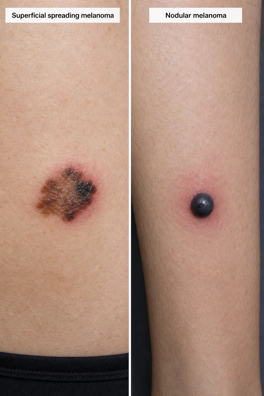
Definition & Classification
Melanoma is a malignant tumour arising from melanocytes (neural-crest–derived pigment cells), most commonly in the skin but also mucosa, nail apparatus, conjunctiva and uveal tract. It is defined histologically by architectural disorder, cytological atypia and pagetoid scatter of melanocytes, with invasion (in situ vs invasive) defined by breach of the basement membrane.[3]
Major cutaneous subtypes (distinguished by morphology, site and behaviour):[3][5]
| Subtype | Frequency | Clinical clue | Site / host |
|---|---|---|---|
| Superficial spreading melanoma (SSM) | ~70% | ABCDE; irregular pigment network; slow horizontal growth phase then vertical | Trunk (men), legs (women); intermittent-UV skin |
| Nodular melanoma | ~15-20% | Raised blue-black/pink nodule; rapid vertical growth; may lack ABCDE | Any site; older men |
| Lentigo maligna melanoma | ~10% | Macular, variegated brown-black on chronically sun-damaged skin (Hutchinson's freckle) | Face, neck, scalp of elderly |
| Acral lentiginous melanoma (ALM) | ~5% (commonest type in dark skin) | Pigmented macule on palm/sole or subungual; Hutchinson's sign | Soles, palms, nail bed |
| Desmoplastic / neurotropic | less than 2% | Flesh-coloured/firm scar-like plaque; perineural spread; often pain | Head/neck; high local recurrence |
| Amelanotic melanoma | ~2-8% | Pink, vascular-appearing lesion; frequently missed | Any |
| Mucosal / uveal melanoma | rare | Pigmented or bleeding lesion in oral/nasal/vulvovaginal mucosa or the eye | Non-UV; worse prognosis |
Epidemiology & Risk Factors
- Incidence continues to rise worldwide, disproportionately in white populations; Australia/New Zealand have the highest global incidence. Lifetime risk in high-incidence regions approaches 1 in 25.[3][6]
- Host risk factors: fair skin (Fitzpatrick I-II), red hair (MC1R variants), ≥50 common naevi or ≥5 atypical naevi, personal/family history of melanoma, large congenital naevi, xeroderma pigmentosum, solid-organ transplant/immunosuppression, prior melanoma.[2]
- Genetic predisposition: CDKN2A (p16) germline mutations (familial atypical multiple mole melanoma syndrome), CDK4, and inherited telomere biology syndromes; account for a minority but warrant genetic counselling and intensive surveillance.[3]
- Exposure: intermittent intense UV exposure (sunburn history, indoor tanning) is the dominant modifiable risk for SSM; chronic cumulative UV drives lentigo maligna. Sunscreen reduces incidence of invasive melanoma (randomised evidence in high-risk adults).[2]
- Behavioural prevention (sun protection, avoiding indoor tanning, self-examination) is the cornerstone of public-health melanoma control.[2]
Pathophysiology
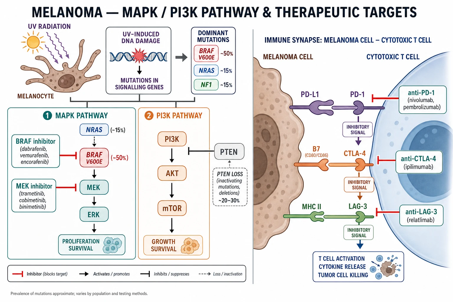
- UV-driven mutagenesis: ultraviolet radiation produces C→T pyrimidine dimer transitions; signature UV damage is concentrated in key oncogenes.[3]
- MAPK pathway is the dominant oncogenic driver. BRAF V600E mutations occur in ~50% of cutaneous melanomas (V600K in ~5-10%, commoner in older patients with chronic sun damage); NRAS in ~15%; NF1 loss in ~15%. Combined BRAF+MEK inhibition exploits this dependency.[4][8]
- PI3K-AKT-mTOR pathway is co-activated via PTEN loss (in ~30%), promoting invasion and resistance.[4]
- Telomerase (TERT promoter) mutations and CDKN2A loss confer replicative immortality; these accumulate as melanocytes progress from naevus → dysplasia → radial → vertical growth phase.[3]
- Immune evasion: melanoma is one of the most immunogenic solid tumours; engagement of inhibitory checkpoints PD-1/PD-L1 and CTLA-4 disables cytotoxic T cells. Blockade of these checkpoints (anti-PD-1, anti-CTLA-4, anti-LAG-3) restores anti-tumour immunity and underpins the modern management of advanced disease.[5]
- Progression model: benign naevus → atypical/dysplastic naevus → melanoma in situ (radial growth) → invasive melanoma (vertical growth) → regional then distant metastasis (lymphatic then haematogenous to lung, liver, brain, bone, gut).[3]
Clinical Presentation
The most reliable clinical sign is a new or changing pigmented lesion. Apply the ABCDE rule plus the "ugly duckling" (a naevus that looks different from the patient's other naevi):[1][6]
- Asymmetry, Border irregularity, Colour variegation (≥3 colours including blue/black/red/white), Diameter >6 mm (low specificity on its own), Evolving (change in size/shape/symptom).
- Subtype-specific morphology as in the table above. Nodular melanoma is the classic trap — a vertically growing, often symmetrical nodule that may be pink (amelanotic) and skip the ABCDE.
- Atypical presentations examiners test: acral melanoma on the sole or under a nail (longitudinal melanonychia with widening, triangular shape, and Hutchinson's sign — pigment extending onto the proximal/lateral nailfold); amelanotic pink papule mimicking BCC, pyogenic granuloma or a keratoacanthoma; desmoplastic scar-like plaque, often with neuropathic pain; mucosal ulceration or bleeding; ocular (uveal) melanoma with visual change; metastatic disease of unknown primary (especially in-transit, nodal or visceral disease with regressed cutaneous primary).[3][5]
Differential Diagnosis
| Mimic | Distinguishing features |
|---|---|
| Atypical (dysplastic) naevus | Stable, symmetrical, lacks chaos-and-clues on dermoscopy; diagnosed on history/dermoscopy, excised if suspicious |
| Spitz naevus | Pink, symmetrical, fast-growing in children; "Spitzoid" histology requires expert dermatopathology |
| Pigmented BCC | Pearly border, arborising vessels, slow growth; dermoscopy differentiates |
| Seborrhoeic keratosis | "Stuck-on", milia-like cysts, comedo-like openings; non-polarised dermoscopy |
| Dermatofibroma | Dimple sign, central white patch, stable |
| Subungual haematoma | History of trauma; distal progression with nail growth; no Hutchinson's sign |
| Merkel cell carcinoma | Rapid, painless, red-violaceous nodule on sun-damaged head/neck of elderly (CK20, MCPyV) |
| Pigmented SCC / pigmented actinic keratosis | Rough, keratotic, sun-damaged site |
| Pyogenic granuloma | Friable bleeding red papule; short history; biopsy if atypical |
Dermoscopy
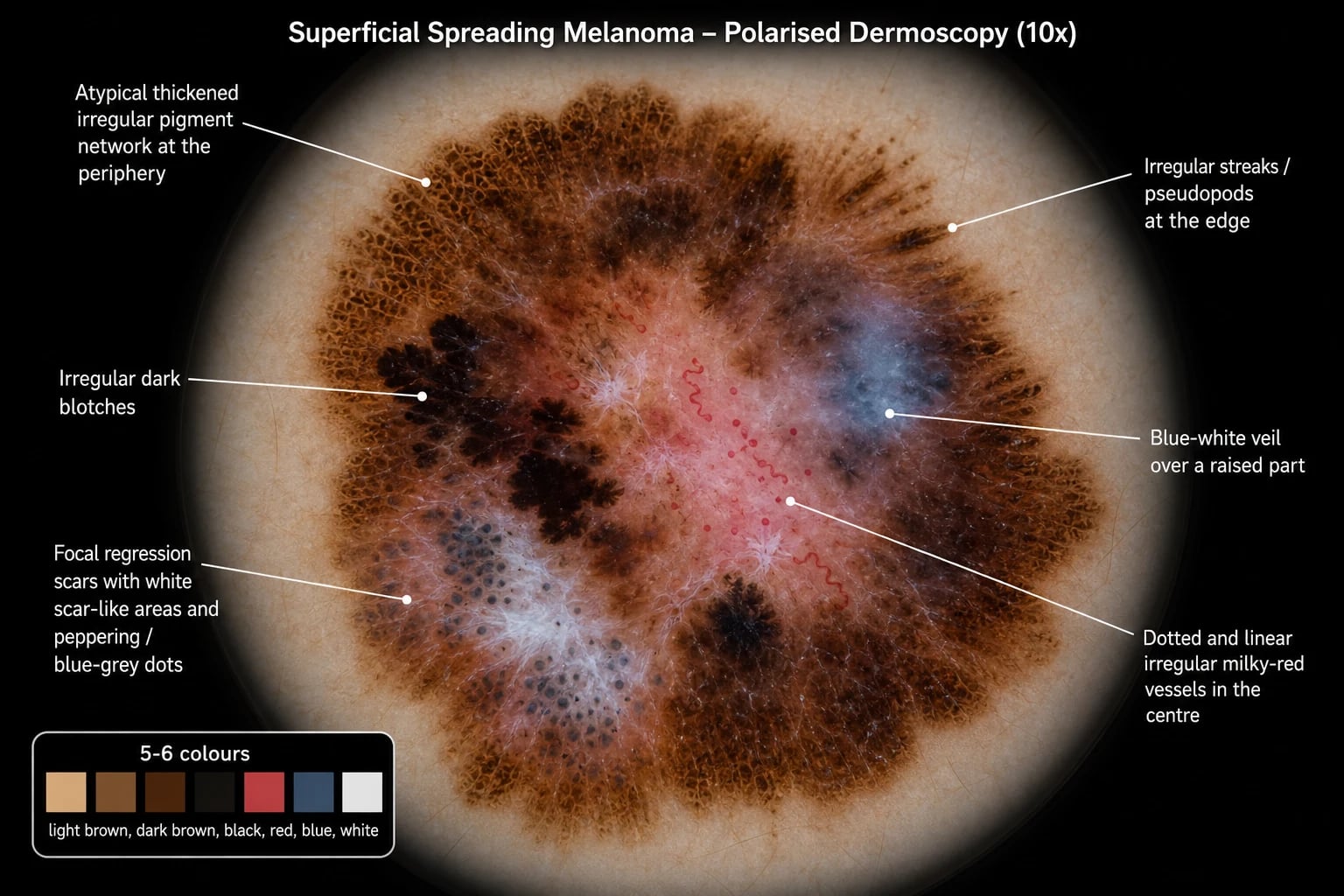
Dermoscopy improves melanoma sensitivity and specificity. Apply the two-step algorithm (benign vs melanocytic, then benign vs malignant melanocytic). The melanoma-specific criteria are:[7]
- Atypical pigment network (thickened, irregular lines).
- Irregular streaks (pseudopods/radial streaming) at the periphery.
- Irregular blotches and multiple colours (≥5-6 colours is highly specific).
- Regression — scar-like white areas with peppering (blue-grey dots).
- Blue-white veil — over a raised invasive component; highly specific.
- Atypical (polymorphous) vessels — dotted, linear-irregular, milky-red areas, chrysalis.
- Chaos and clues approach: an asymmetric lesion with ≥1 clue (the practical short algorithm for non-experts).[7]
Histopathology
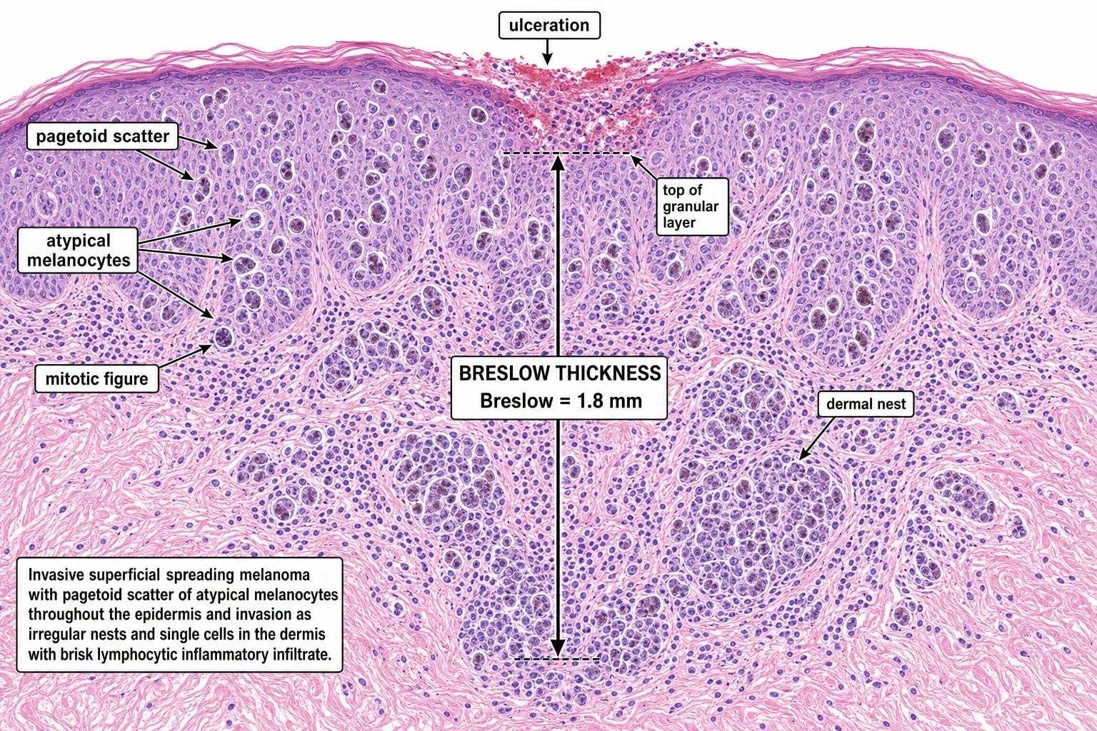
The histology report determines staging and management. Essential elements:[3][5]
- Breslow thickness (mm) — the single most important prognostic factor; measured from the top of the granular layer to the deepest invasive cell.
- Ulceration — adverse prognostic factor; defines the "b" subcategory.
- Mitotic rate (per mm²) — adverse; required on every report.
- Lymphovascular invasion, microsatellites (≥0.05 mm, within 1-2 cm of primary; = N1c), perineural (neurotropic) invasion.
- Regression — pathological regression present.
- Clark level — superseded for staging except it remains relevant in thin (T1) melanomas where thickness and ulceration are borderline.
- Margins — involved/close/positive.
- Histological subtype (SSM, nodular, LM, ALM, desmoplastic, Spitzoid). [1]
Clinical & Bedside Assessment
- Examine the whole skin including scalp (part the hair), retro-auricular, oral mucosa, conjunctiva, neck, axillae, trunk (use a chaperone), genitals/perianal, palms, soles, interdigital web spaces, and all nails.[6]
- Palpate regional lymph nodes (cervical, axillary, inguinal).
- Use total-body photography and sequential digital dermoscopy for high-risk patients (dysplastic naevus syndrome, personal/family history). Hand-held dermatoscopy at every visit.[3]
Investigations
- Biopsy technique (critical): full-thickness excisional biopsy with 1-3 mm clinical margins, orientated vertically (to avoid lymphatic disruption and allow accurate Breslow measurement). Avoid shave/scoop biopsies for suspected melanoma. A punch may suffice for small lesions. For large lesions (e.g. lentigo maligna), an incisional biopsy of the most atypical area is acceptable for diagnosis, followed by definitive excision.[6][5]
- Staging investigations are driven by stage and symptoms. Localised (clinical stage I-II): no routine imaging/bloods; LDH not required. Stage III/IV: whole-body CT or PET-CT and brain MRI (more sensitive than CT for brain metastases); LDH (an independent M-category prognostic factor).[3][5]
- Sentinel lymph node biopsy (SLNB): offered for pathological stage ≥T1b (≥0.8 mm, or less than 0.8 mm with ulceration); discussed selectively for thin (T1a) melanomas with adverse features (mitoses, regression, lymphovascular invasion, young age). Performed with lymphoscintigraphy and blue dye and/or radiotracer.[3]
- Gene-expression profiling (e.g. DecisionDx-Melanoma, CP-GEP) is an emerging adjunct to refine SLNB decisions in borderline-thickness tumours; not yet universally adopted.[3]
AJCC 8th Edition Staging

T (primary tumour):[3]
| T | Thickness | Notes |
|---|---|---|
| Tis | in situ | — |
| T1 | ≤1.0 mm | T1a less than 0.8 mm and non-ulcerated; T1b 0.8-1.0 mm OR less than 0.8 mm ulcerated |
| T2 | 1.01-2.0 mm | a = non-ulcerated, b = ulcerated |
| T3 | 2.01-4.0 mm | a/b as above |
| T4 | >4.0 mm | a/b as above |
N (regional nodes): N1 (1 node), N2 (2-3 nodes), N3 (≥4 nodes or matted); suffix (a) clinically occult / sentinel-detected, (b) clinically detected, (c) in-transit/satellite/microsatellite metastases without nodal involvement (N1c).[3]
M (distant): M1a (skin/soft tissue/non-regional node), M1b (lung), M1c (other visceral), M1d (brain); each further subdivided by LDH normal (0) vs elevated (1) — elevated LDH is an adverse, independent prognostic marker.[3]
M-category anatomy and LDH stratification (AJCC 8th edition): [1]
M1a — skin/soft tissue/nodal
- Distant cutaneous, subcutaneous or nodal metastases beyond the regional nodal basin
- Includes in-transit metastases outside the treated limb
- Best prognosis of the M1 subcategories; 5-year OS approximately 40-55 percent
- Often a candidate for surgery, regional therapy (isolated limb infusion, T-VEC) or systemic therapy
- LDH stratification: (0) normal, (1) elevated — elevated LDH roughly halves the 5-year OS
M1b — pulmonary
- Lung metastases with or without M1a disease
- Commonest single visceral site of first distant relapse
- Median OS 24-36 months in modern immunotherapy series
- Often asymptomatic — picked up on surveillance CT chest
- LDH (0) versus (1) — adverse, independent prognostic factor in AJCC 8th
M1c — other visceral
- Non-CNS visceral metastases (liver, bone, adrenal, GI tract, spleen)
- Median OS 18-24 months with modern anti-PD-1 +/- ipilimumab
- Liver and bone carry the worst single-site prognosis within M1c
- LDH elevation is the single strongest independent adverse factor
- Re-biopsy often considered to confirm histology and obtain fresh tissue for molecular profiling
M1d — CNS (brain)
- Brain metastasis with or without any other metastatic site — its own AJCC 8th subcategory
- Worst median OS (15-22 months with ipi+nivo; 12-15 months with single-agent anti-PD-1)
- Up to 40 percent of BRAF V600 mutant patients develop brain metastases
- Asymptomatic lesions common — MRI brain is mandatory in stage III/IV staging
- Local therapy (stereotactic radiosurgery, surgery) plus systemic therapy; ipi+nivo has intracranial activity
Management — Biopsy & Surgery
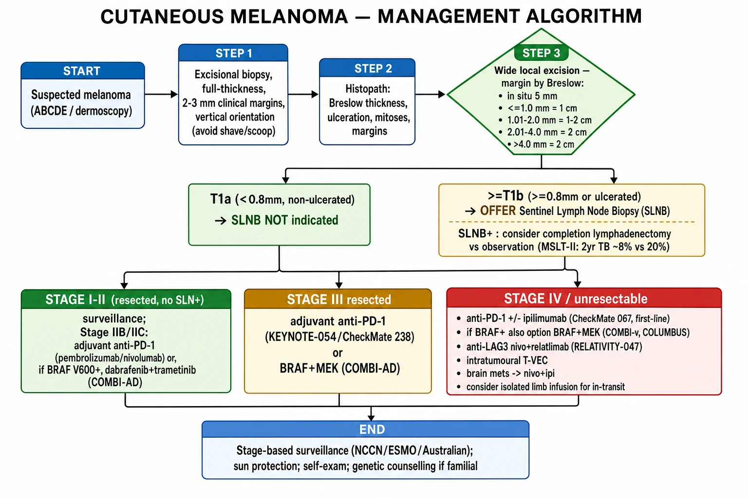
Wide local excision (WLE) margins (clinical, to muscular fascia), by Breslow thickness:[5][6]
| Breslow | Clinical margin |
|---|---|
| In situ | 0.5-1.0 cm (5 mm) |
| ≤1.0 mm | 1 cm |
| 1.01-2.0 mm | 1-2 cm |
| 2.01-4.0 mm | 1-2 cm |
| >4.0 mm | 2 cm |
- Sentinel lymph node biopsy for ≥T1b disease; provides staging and prognostic information.
- Completion lymph node dissection (CLND) vs observation after a positive SLNB: MSLT-II and DeCOG-SLT showed no melanoma-specific survival benefit from immediate CLND, with a small absolute reduction in regional nodal recurrence; CLND is now reserved for bulky/clinically apparent nodal disease, with ultrasound surveillance an alternative.[3][5]
- Lentigo maligna may be excised with Mohs micrographic surgery (or staged mapped excision) for tissue conservation on the face.
Management — Adjuvant Systemic Therapy (Stage IIB/IIC and resected Stage III)
Adjuvant therapy improves recurrence-free survival in resected high-risk melanoma:[5]
- Anti-PD-1: pembrolizumab (KEYNOTE-054) or nivolumab (CheckMate 238), ~1 year, for resected IIB/IIC/III regardless of BRAF status.
- BRAF + MEK combination (dabrafenib + trametinib; COMBI-AD) ~1 year, for BRAF V600-mutated resected stage III.
- Choice between anti-PD-1 and BRAF+MEK in BRAF+ patients is individualised (efficacy comparable; differing toxicity profiles). [1]
Management — Advanced / Metastatic Disease (unresectable III or IV)
Modern first-line therapy has transformed stage IV melanoma into a manageable disease with durable long-term survival in a subset:[5][3]
- Anti-PD-1 +/- ipilimumab (anti-CTLA-4): nivolumab + ipilimumab (CheckMate 067 — 10-year follow-up showing the longest reported survival in advanced melanoma) or anti-PD-1 monotherapy (pembrolizumab KEYNOTE-006; nivolumab). First-line for most patients regardless of BRAF status.
- Anti-PD-1 dosing (FDA/EMA approved schedules):
- Pembrolizumab 200 mg IV every 3 weeks OR 400 mg IV every 6 weeks (weight-independent flat dosing).
- Nivolumab 240 mg IV every 2 weeks OR 480 mg IV every 4 weeks (flat dosing).
- Nivolumab + ipilimumab induction: nivolumab 1 mg/kg PLUS ipilimumab 3 mg/kg IV every 3 weeks for FOUR doses, followed by nivolumab 240 mg every 2 weeks or 480 mg every 4 weeks monotherapy.
- Nivolumab + relatlimab (RELATIVITY-047): nivolumab 480 mg + relatlimab 160 mg IV every 4 weeks (flat dosing).
- BRAF + MEK inhibition for BRAF V600-mutant disease (only after confirmation of V600E/K/D by validated assay — IHC is a screen, sequencing/NGS confirms):
- Dabrafenib 150 mg orally twice daily + trametinib 2 mg orally once daily (COMBI-d/COMBI-v).
- Encorafenib 450 mg orally once daily + binimetinib 45 mg orally twice daily (COLUMBUS).
- Vemurafenib 960 mg orally twice daily + cobimetinib 60 mg orally once daily for 21 days of each 28-day cycle (coBRIM; vemurafenib monotherapy is the historical BRIM-3 regimen and is now superseded by the combination).
- Rapid disease control (responses within 1-2 weeks); useful for symptomatic/bulky or brain disease; resistance typically emerges within 9-12 months with combination therapy.
- Anti-LAG-3: nivolumab + relatlimab (RELATIVITY-047) — improved progression-free survival over nivolumab alone; a newer first-line option.
- Intratumoural therapy: T-VEC (talimogene laherparepvec) — a modified oncolytic herpes simplex virus type 1 expressing human GM-CSF. Injected directly into cutaneous, subcutaneous or nodal lesions at a concentration of 10⁶ PFU/mL for the first dose (to seroconvert) then 10⁸ PFU/mL every 2 weeks until response, intolerance or lesion disappearance. Indicated for injectable, locally advanced or low-burden cutaneous disease (OPTiM trial).
- Brain metastases: nivolumab + ipilimumab has intracranial activity; stereotactic radiosurgery for limited disease; whole-brain radiotherapy reserved for diffuse symptomatic disease.
- In-transit / regionally advanced disease: isolated limb infusion/perfusion with melphalan (with or without actinomycin D or TNF-alpha), electrochemotherapy, or intralesional T-VEC.
- Uveal melanoma (HLA-A*02:01+): tebentafusp — a TCR-CD3 bispecific (gp100 × CD3) — 20 mcg IV on day 1, 30 mcg on day 8, then 30 mcg weekly thereafter, continued while benefit persists (Tebentafusp was the first systemic agent to improve overall survival in metastatic uveal melanoma).
- PET/CT and multidisciplinary tumour board review is standard for stage III/IV.
- Treatment selection principles: anti-PD-1 monotherapy for most patients with favourable disease tempo; ipi+nivo combination for symptomatic, bulky, rapidly progressing or brain-met disease in patients who can tolerate the toxicity; BRAF+MEK targeted therapy first-line in BRAF V600 mutant disease needing a fast response (rapid symptomatic control) or when immune checkpoint inhibitors are contraindicated. [1]
A practical timeline for advanced BRAF-mutant melanoma (illustrative): [1]
Radiotherapy
Radiotherapy has a limited but defined role:[5]
- Adjuvant after therapeutic lymph node dissection for high-risk nodal disease (multiple involved nodes, extracapsular extension) to reduce regional recurrence (especially desmoplastic and head/neck sites).
- Palliative for symptomatic brain, bone and soft-tissue metastases. [1]
Special Populations
- Paediatric melanoma: rare; Spitzoid histology commoner and biologically distinct; congenital melanocytic naevi carry a small lifetime risk. Management principles mirror adults with age-adjusted dosing.
- Pregnancy: it is safe to biopsy and excise melanoma during pregnancy; sentinel node mapping with radiotracer is generally avoided in the first trimester but feasible later. Systemic immunotherapy (anti-PD-1, BRAF+MEK) decisions weigh maternal benefit against fetal risk and are individualised.[3]
- Immunosuppressed (transplant): melanoma behaves more aggressively; immunotherapy is complicated by allograft rejection (anti-PD-1 carries graft-rejection risk).
- Familial (CDKN2A): refer for genetic counselling; intensive whole-skin surveillance from early adulthood; consider pancreatectomy screening (associated pancreatic cancer risk).
Prognosis & Surveillance
5-year survival by AJCC 8th stage (approximate):[3][5]
| Stage | 5-year survival |
|---|---|
| IA | ~99% |
| IB | ~93-97% |
| IIA-IIC | ~82-87% (IIC ~82%) |
| IIIA | ~93% |
| IIIB-IIID | ~30-83% (IIID ~30-50%) |
| IV (M1) | ~25-35% (M1d/raised LDH worse; less than 25%) |
Prognostic factors: stage (Breslow thickness, ulceration, sentinel-node status, LDH), mitotic rate, site (acral/mucosal/uveal worse), BRAF status, age, male sex.[3]
Surveillance schedule is stage-stratified (NCCN v3.2025, ESMO 2022, Cancer Council Australia); typically every 3-12 months for 5 years with skin and node examination, with imaging reserved for stage III/IV or symptoms. Lifelong whole-skin surveillance (new primaries in ~5-10%).[5]
Evidence, Guidelines & Regional Differences
- Landmark trials (named in vivas): CheckMate 067 (nivo+ipi advanced, longest reported survival), KEYNOTE-006 (pembrolizumab), COMBI-AD (adjuvant dab+trameti, BRAF+), KEYNOTE-054/CheckMate 238 (adjuvant anti-PD-1), COLUMBUS (encorafenib+binimetinib), RELATIVITY-047 (nivo+relatlimab), MSLT-II/DeCOG-SLT (no CLND benefit), BRIM-3 (vemurafenib).[3][5]
- Guidelines (regional): ESMO (Europe), NCCN (North America), Cancer Council Australia / New Zealand guidelines (highest-incidence region; aggressive surveillance). Principles agree; surveillance intensity, SLNB thresholds and reimbursement of gene-expression tests differ regionally.[5]
- Controversies: SLNB in thin melanomas; role of completion dissection vs surveillance; optimal sequence of immunotherapy vs targeted therapy in BRAF+ advanced disease; neoadjuvant systemic therapy in resectable stage III (emerging).[3]
Prevention
- Sun protection: shade, protective clothing, broad-spectrum SPF 30+ sunscreen applied adequately and frequently; avoid indoor tanning. Sunscreen reduces invasive melanoma incidence (randomised evidence in high-risk adults).[2]
- Self-examination and professional surveillance for high-risk individuals (dysplastic naevus syndrome, personal/family history, immunosuppression, CDKN2A carriers).
- No proven chemoprevention; nicotinamide and retinoids are investigational.[2]
Exam Pearls
[1]Drug-specific patterns and doses
[1]Melanoma systemic therapy quick numbers
ABCDEF - melanoma adjuvant and metastatic therapy
Landmark systemic-therapy trials and dosing reference
The numbers below are the doses used in the registration trials and now codified in FDA/EMA labels. Fellowship vivas test both the trial name and the regimen, so commit them as a single bundle. [1]
Anti-PD-1 dosing (flat, weight-independent in adults): [1]
- Pembrolizumab 200 mg IV every 3 weeks OR 400 mg IV every 6 weeks — KEYNOTE-006 (metastatic) used 10 mg/kg q2w or 200 mg q3w; KEYNOTE-054 (adjuvant) used 200 mg q3w for up to 12 months. The 400 mg q6w schedule is pharmacokinetically equivalent and approved to ease clinic burden.
- Nivolumab 240 mg IV every 2 weeks OR 480 mg IV every 4 weeks — CheckMate 066 (dacarbazine-controlled first-line), CheckMate 238 (adjuvant vs ipilimumab) and CheckMate 067 (combination arm) used 3 mg/kg q2w; the flat 240/480 mg schedules are now standard.
- Nivolumab + ipilimumab induction (CheckMate 067): nivolumab 1 mg/kg PLUS ipilimumab 3 mg/kg IV every 3 weeks for FOUR induction doses, then nivolumab monotherapy 240 mg q2w (or 480 mg q4w) until progression or intolerable toxicity. The reverse dose schedule (ipi 1 mg/kg + nivo 3 mg/kg, CheckMate 511) halves the grade 3-5 immune-related AE rate (34% to 18%) with comparable efficacy, now preferred in many patients.
- Nivolumab + relatlimab (RELATIVITY-047): nivolumab 480 mg + relatlimab 160 mg IV every 4 weeks (flat). [1]
BRAF + MEK inhibitor dosing (oral, BRAF V600-mutant disease): [1]
- Dabrafenib 150 mg orally twice daily PLUS trametinib 2 mg orally once daily — COMBI-d / COMBI-v (metastatic) and COMBI-AD (adjuvant) regimen; continue until progression or unacceptable toxicity (adjuvant: 12 months).
- Encorafenib 450 mg orally once daily PLUS binimetinib 45 mg orally twice daily — COLUMBUS regimen; encorafenib's longer half-life enables once-daily dosing and the 450 mg dose delivers a higher cumulative exposure than 300 mg. Continue until progression.
- Vemurafenib 960 mg orally twice daily PLUS cobimetinib 60 mg orally once daily days 1-21 of each 28-day cycle (coBRIM); vemurafenib monotherapy 960 mg BD (BRIM-3) is now superseded by the combination. [1]
Tumour-infiltrating lymphocyte (TIL) therapy — lifileucel: [1]
- Lifileucel 7.5 billion viable cells IV once (single infusion; range of 1 to 150 billion cells per the FDA label) after non-myeloablative lymphodepletion (cyclophosphamide 60 mg/kg IV days -7 and -6; fludarabine 25 mg/m² IV days -5 to -1) followed by short-course IL-2 (aldesleukin 600,000 IU/kg IV q8h, up to 6 doses) and prophylactic tocilizumab for cytokine release. The C-144-01 study (single-arm phase 2, advanced melanoma post-anti-PD-1 ± anti-CTLA-4) reported an objective response rate of approximately 31% with durable complete responses lasting beyond 24 months; lifileucel is the first FDA-approved TIL therapy for melanoma (2024) and is indicated for unresectable stage III/IV disease that has progressed on a prior anti-PD-1 and, if BRAF V600 mutant, a BRAF inhibitor ± MEK inhibitor. [1]
CheckMate 067 — long-term follow-up (advanced first-line nivo+ipi vs nivo vs ipi, treatment-naive unresectable III/IV melanoma, n = 945): [1]
- 6.5-year analysis: median OS 72.1 months (nivo+ipi) vs 36.9 months (nivo) vs 19.9 months (ipi); 6.5-year OS 49% vs 42% vs 23%. 10-year follow-up (2024) confirmed a 10-year OS of 43% (nivo+ipi) vs 37% (nivo) vs 19% (ipi) — the longest survival ever reported in a phase 3 trial of advanced melanoma. Median melanoma-specific survival was >120 months (nivo+ipi) vs 49.4 months (nivo) vs 21.9 months (ipi).
- Median PFS: 11.5 months (nivo+ipi) vs 6.9 months (nivo) vs 2.9 months (ipi). Treatment-related grade 3-4 adverse events: 59% (nivo+ipi) vs 24% (nivo) vs 28% (ipi); treatment-related deaths 1.4% in the combination arm (most commonly immune-related colitis, pneumonitis and myocarditis). Treatment-free interval: a substantial fraction of durable responders on nivo+ipi remained off any systemic therapy beyond 5 years, the strongest evidence that cure is achievable in a subset of advanced melanoma patients. [1]
KEYNOTE-054 / EORTC 1325 (adjuvant pembrolizumab vs placebo, resected stage IIIA-IIIC, n = 1019): [1]
- 200 mg IV q3w for up to 12 months vs placebo. 3.5-year analysis: RFS hazard ratio 0.59 (95% CI 0.49-0.70), 5-year RFS 55.4% (pembro) vs 38.3% (placebo); benefit was seen regardless of BRAF status and PD-L1 expression, although PD-L1-positive tumours derived somewhat larger absolute benefit. Distant-metastasis-free survival HR 0.60; the trial supports adjuvant pembrolizumab for any resected stage IIIA-IIIC patient regardless of BRAF status. Treatment-related grade 3-4 AEs: 14% (mostly immune-related hepatitis, colitis, pneumonitis and endocrinopathies); treatment-related deaths 0.2%. [1]
COMBI-AD (adjuvant dabrafenib + trametinib vs placebo, resected BRAF V600-mutant stage III, n = 870): [1]
- Dabrafenib 150 mg BD + trametinib 2 mg daily for 12 months vs placebo. 5-year analysis: relapse-free survival 52% (dab+tram) vs 36% (placebo); distant-metastasis-free survival 65% vs 54%; OS HR 0.81 (95% CI 0.61-1.06) at 5 years (a numerically but not statistically significant OS difference at that read-out, with longer follow-up continuing to favour the combination). Benefit was largest in stage IIIC-IIID; grade 3-4 AEs ~41% (pyrexia, chills, fatigue and hypertension are the signature toxicities of dab+tram; serious AEs ~36%). This is the trial that establishes BRAF+MEK as an alternative to anti-PD-1 for BRAF-mutant resected stage III, with comparable RFS and OS outcomes to KEYNOTE-054.
Red Flags [1]
Exam application bank (NEET-PG / INICET)
One-line answer
Cutaneous melanoma is a malignant tumour of melanocytes driven predominantly by UV-induced oncogenic mutations in the MAPK and PI3K pathways (BRAF V600E ~50%, NRAS, NF1), clinically presenting as a new or changing pigmented lesion (ABCDE, ugly-duckling, dermoscopic chaos-and-clues), histologically defined by Breslow thickness, ulceration and mitotic rate, and staged by the AJCC 8th edition (T by thickness/ulceration; N by sentinel-node burden). Prognosis spans >99% 5-year survival for stage IA to less than 25% for stage IV. Management is stage-stratified: excisional biopsy, wide local excision with Breslow-guided margins, sentinel lymph node biopsy for ≥T1b, adjuvant anti-PD-1 or BRAF+MEK for resected IIB/IIC and III, and anti-PD-1 ± ipilimumab, BRAF+MEK or anti-LAG3 for advanced disease — backed by landmark trials (CheckMate 067, KEYNOTE-006, COMBI-AD, COLUMBUS, RELATIVITY-047, MSLT-II)
Worked stems (answer without another resource)
Stem 1 — Classic presentation. Map symptoms to mechanism; name the first investigation and first treatment step with dose/route if drug therapy is standard. [1]
Stem 2 — Unstable / complicated. List red flags that force immediate resuscitation, theatre, ICU, antidote, or reperfusion — and what you do in the first 15 minutes. [1]
Stem 3 — Atypical group. Elderly, pregnancy, child, or immunocompromised: how presentation and thresholds change. [1]
Stem 4 — Differential trap. Name the three closest mimics and one discriminator for each. [1]
Stem 5 — Disposition. Who goes home with safety-netting, who is admitted, who needs HDU/ICU/theatre, and what follow-up is mandatory. [1]
Rapid viva checklist
- Definition + classification
- Pathophysiology chain
- Bedside signs / criteria
- Score with exact components (if any)
- Emergency bundle
- Definitive therapy with doses
- Complications of disease and of treatment
- Special populations
- Guideline/trial name if classic
- Three exam traps
Coverage self-check
If you cannot answer any stem above from this page alone, re-read the matching section — the page is intended to be self-sufficient for final-prof and NEET-PG/INICET questions on Cutaneous melanoma.
[1]References
- [1]Ahmed B, Qadir MI, Ghafoor S. Malignant Melanoma: Skin Cancer-Diagnosis, Prevention, and Treatment Crit Rev Eukaryot Gene Expr, 2020.PMID 32894659
- [2]Dzwierzynski WW. Melanoma Risk Factors and Prevention Clin Plast Surg, 2021.PMID 34503715
- [3]Tasdogan A, Sullivan RJ, Katalinic A, et al. Cutaneous melanoma Nat Rev Dis Primers, 2025.PMID 40180935
- [4]Guo W, Wang H, Li C. Signal pathways of melanoma and targeted therapy Signal Transduct Target Ther, 2021.PMID 34924562
- [5]Joshi UM, Kashani-Sabet M, Kirkwood JM. Cutaneous Melanoma: A Review JAMA, 2025.PMID 40853557
- [6]Lauters R, Brown AD, Harrington KA. Melanoma: Diagnosis and Treatment Am Fam Physician, 2024.PMID 39418569
- [7]Marghoob NG, Liopyris K, Jaimes N. Dermoscopy: A Review of the Structures That Facilitate Melanoma Detection J Am Osteopath Assoc, 2019.PMID 31135866
- [8]Castellani G, Buccarelli M, Arasi MB, et al. BRAF Mutations in Melanoma: Biological Aspects, Therapeutic Implications, and Circulating Biomarkers Cancers (Basel), 2023.PMID 37627054