Dermatology · General Medicine
Melanoma & Skin Cancer
Also known as Melanoma · Cutaneous melanoma · Basal cell carcinoma · BCC · Squamous cell carcinoma · SCC · Non-melanoma skin cancer · Merkel cell carcinoma · Rodent ulcer
Skin cancer is the commonest malignancy worldwide and splits into melanoma (aggressive melanocytic cancer with high metastatic potential) and the non-melanoma skin cancers (NMSC) — basal cell carcinoma (BCC, commonest and locally invasive, rarely metastasises) and squamous cell carcinoma (SCC, keratinising, can metastasise to nodes) — plus the rarer, aggressive Merkel cell carcinoma (neuroendocrine, polyomavirus-related). Risk factors centre on ultraviolet radiation (sun exposure, sunburn, tanning beds), fair skin, freckles, red hair (MC1R), immunosuppression, older age, family/genetic predisposition (FAMM/CDKN2A, xeroderma pigmentosum, Gorlin), and previous skin cancer. Melanoma is recognised by the ABCDE criteria and the ugly-duckling sign; subtypes are superficial spreading (commonest), nodular (worst), lentigo maligna, acral lentiginous and amelanotic. Staging rests on Breslow thickness, ulceration, and sentinel lymph node biopsy, with AJCC TNM 8th edition defining the T categories. Treatment is surgical (wide local excision with margins dictated by Breslow thickness; Mohs for facial/high-risk BCC/SCC) and, for advanced melanoma, transformed by anti-PD-1 immunotherapy (pembrolizumab, nivolumab) +/- anti-CTLA-4 (ipilimumab) and BRAF/MEK targeted therapy (dabrafenib + trametinib). Sun protection and early detection remain the preventive cornerstones.
On this page & tools
Your progress
Saved locally on this device.
Exam tags
Red flags
Overview & Definition
Skin cancer is the commonest malignancy in humans, and it is also the most preventable. Clinically it divides into melanoma — a cancer of melanocytes that is uncommon relative to the others but responsible for most skin-cancer deaths — and the non-melanoma skin cancers (NMSC), dominated by basal cell carcinoma (BCC) and squamous cell carcinoma (SCC), which are far more common but usually curable. To these the examiner expects you to add Merkel cell carcinoma, a rare neuroendocrine skin tumour that behaves aggressively and is driven by the Merkel cell polyomavirus.[1][12]
The clinical skill that saves lives is early recognition of melanoma: applying the ABCDE criteria and the ugly-duckling sign to a new or changing pigmented lesion, then performing a prompt full-thickness excisional biopsy. Early, thin melanoma is cured by surgery alone; thick, nodular or metastatic melanoma requires systemic therapy, and the prognosis collapses. The same examination identifies the pearly telangiectatic facial papule of a rodent ulcer (BCC) and the scaly keratinising nodule of SCC on chronically sun-exposed skin. Because almost all skin cancer is UV-driven, sun protection and skin self-examination are the two interventions that bend the incidence curve.[1][8]
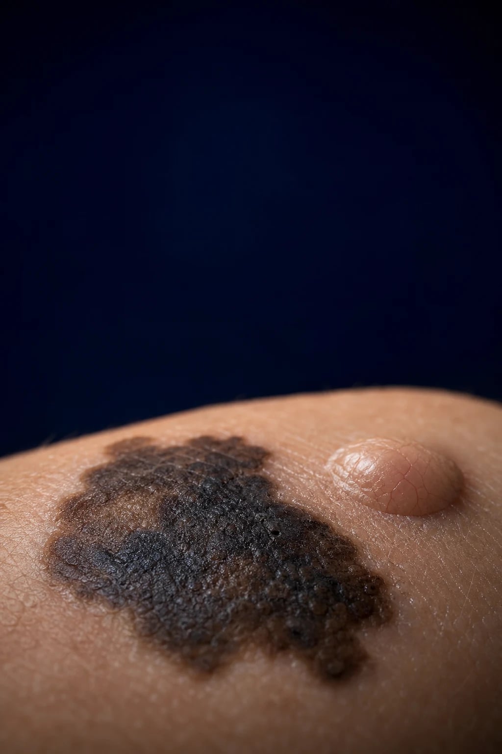
Classification
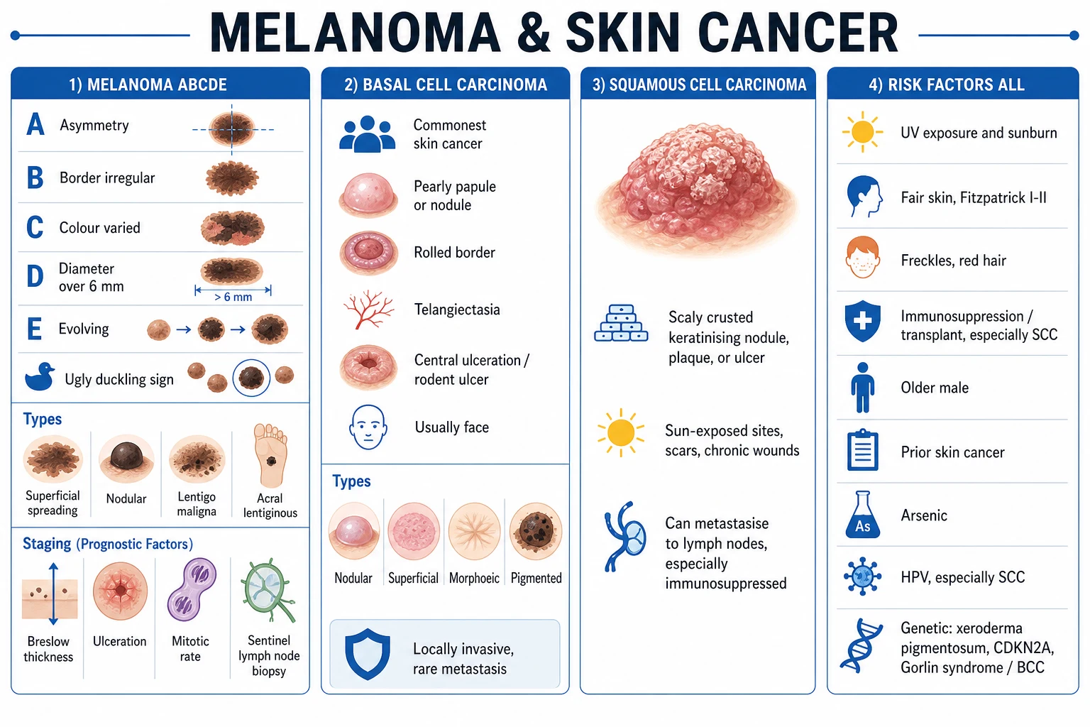
Skin cancer is classified first by cell of origin, then — for melanoma — by clinical subtype and Breslow/AJCC stage. The high-yield distinctions are between melanoma (melanocyte) and the keratinocyte carcinomas (BCC = basal keratinocyte, SCC = squamous keratinocyte), with Merkel cell carcinoma a separate neuroendocrine entity.[1][12]
Melanoma
- Melanocyte origin; aggressive, metastasises early
- ABCDE + ugly duckling; subtypes superficial spreading, nodular, lentigo maligna, acral, amelanotic
- Breslow thickness + ulceration + SLNB drive stage
- WLE by Breslow; anti-PD-1 / BRAF-MEK for advanced
BCC
- Basal keratinocyte; commonest cancer in humans
- Pearly papule, rolled border, telangiectasia, rodent ulcer; face
- Locally invasive; metastasis under 0.1 percent
- Excision 4 to 6 mm, Mohs, imiquimod, vismodegib, RT
SCC
- Squamous keratinocyte; arises from actinic keratosis / Bowen disease
- Scaly crusted keratinising nodule or ulcer on sun-exposed skin
- Regional nodal metastasis 2 to 5 percent (higher if immunosuppressed)
- Excision 4 to 6 mm or Mohs; cemiplimab if advanced
Merkel cell
- Neuroendocrine; Merkel cell polyomavirus in about 80 percent
- Rapidly growing painless red/violaceous nodule, elderly/immunosuppressed
- CK20 perinuclear dot; AEIOU clinical clue; high mortality
- Wide excision + SLNB + adjuvant RT; avelumab if advanced
A benign but important precursor is the actinic (solar) keratosis, a scaly erythematous patch on sun-damaged skin with a small annual risk of progression to SCC; Bowen disease is SCC in situ (a fixed, scaly, well-demarcated red plaque). Lentigo maligna (Hutchinson melanotic freckle) is melanoma in situ of chronically sun-exposed elderly skin, and keratoacanthoma is a low-grade, rapidly growing crateriform variant of SCC that may involute but is usually excised as SCC. The congenital and acquired melanocytic naevi (junctional, compound, intradermal; dysplastic/atypical naevi) are the benign lesions from which melanoma must be distinguished.[1][13]
Epidemiology & Risk Factors
Skin cancer is the commonest malignancy in fair-skinned populations, and melanoma incidence has risen faster over the past half-century than almost any other cancer — driven by recreational UV exposure, tanning beds, ageing populations, and earlier detection. Globally, GLOBOCAN 2020 estimated roughly 325,000 new melanoma cases and 57,000 melanoma deaths annually; Australia and New Zealand have the highest incidence in the world, followed by North America and Europe, reflecting the predominantly fair-skinned, UV-exposed populations at temperate latitudes.[11]
Skin cancer by the numbers
Ultraviolet radiation is the central carcinogen. Both intermittent, intense, blistering sun exposure (melanoma, particularly in childhood) and chronic cumulative exposure (SCC, BCC, actinic keratoses) are implicated; indoor tanning beds are classified by the IARC as Group 1 carcinogens and multiply melanoma risk, especially when use begins before age 35. The host factors that multiply UV risk are the classical fair-skin phenotype: fair skin, freckling, light hair (red hair carries a two- to fourfold melanoma risk via MC1R variants), blue/green eyes, and an inability to tan, alongside a high naevus count (more than 50 to 100 common naevi, or more than 5 atypical naevi).[1][8]
UV / host factors
- Sunburn, tanning beds, chronic sun exposure (SCC/BCC)
- Fair skin, freckles, red hair (MC1R), light eyes, poor tanning
- High naevus count, multiple atypical naevi
Immunosuppression
- Solid-organ transplant: 65 to 100x SCC risk, 10x BCC risk
- SCC becomes commoner than BCC; HPV co-factor; aggressive course
- Chronic lymphocytic leukaemia, HIV, methotrexate also increase risk
Genetic syndromes
- FAMM syndrome / dysplastic naevus syndrome (CDKN2A): high melanoma risk
- Xeroderma pigmentosum: defective nucleotide excision repair, 1000x risk
- Gorlin (basal cell naevus) syndrome (PTCH1): multiple BCCs from childhood
- Oculocutaneous albinism: high SCC and melanoma risk
Other
- Previous skin cancer (highest predictor of a second primary)
- PUVA photochemotherapy, arsenic, radiation (chronic)
- Older age, male sex (melanoma in younger women on legs)
- Large or giant congenital melanocytic naevus (melanoma risk)
Pathophysiology
Skin carcinogenesis is, in almost every case, a story of ultraviolet-induced DNA damage that outstrips repair. UVB (290 to 320 nm) is directly absorbed by DNA and produces cyclobutane pyrimidine dimers and 6-4 photoproducts, generating the signature UV mutations C to T and CC to TT transitions. If the nucleotide excision repair pathway cannot clear these adducts — as in xeroderma pigmentosum — mutations accumulate at oncogenes and tumour suppressors and the cell progresses to malignancy. UVA (320 to 400 nm) contributes by indirect oxidative damage. The result, across all skin cancers, is activation of growth-promoting pathways (chiefly the MAPK and PI3K cascades) coupled with loss of cell-cycle control and apoptosis.[1]
In melanoma, the dominant oncogenic driver is an activating mutation in BRAF (the BRAF V600E substitution in roughly half of all cutaneous melanomas, and V600K in a further minority), which constitutively switches on the MAPK pathway; NRAS mutations occur in roughly 15 to 25 percent, and NF1 and PTEN loss in further subsets. Melanoma evolves through a radial growth phase (spread along the epidermis, low metastatic potential) into a vertical growth phase (invasion into the dermis, acquisition of metastatic competence). In BCC, the molecular signature is loss of the PTCH1 tumour suppressor — usually by UV mutation — which releases the Hedgehog signalling pathway (constitutive SMO activation and GLI1 transcription); this explains why the Hedgehog inhibitors vismodegib and sonidegib work in advanced disease, and why Gorlin syndrome (germline PTCH1) produces hundreds of BCCs.[14]
In SCC, UV drives early TP53 loss in actinic keratoses, with subsequent NOTCH1/2 and CDKN2A inactivation; in immunosuppressed patients the human papillomavirus oncoproteins E6 and E7 (degrading p53 and Rb) are a powerful co-carcinogen. The sequence is actinic keratosis to SCC in situ (Bowen disease) to invasive SCC. Merkel cell carcinoma is distinct: in roughly 80 percent of cases the Merkel cell polyomavirus (MCPyV) is clonally integrated into the tumour genome, expressing truncated large T antigen that inactivates Rb; the remaining UV-driven tumours carry a heavy UV mutational signature and TP53 mutations. Histologically Merkel cell carcinoma is a small round blue cell tumour, and immunohistochemistry shows the pathognomonic perinuclear dot-like CK20 staining (CK7 negative, TdT negative).[12]
Clinical Presentation
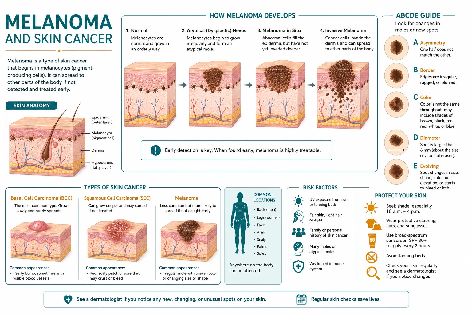
Melanoma classically presents as a new or changing pigmented lesion in a sun-exposed or intermittently exposed site, most often the trunk (back) in men and the lower legs in women, although any site is possible including palms, soles, nail beds and mucosa. The clinical recognition tool is the ABCDE criteria: Asymmetry (one half unlike the other), irregular Border, Colour variegation (mixtures of brown, black, blue, red, white), Diameter greater than 6 mm, and Evolving (change in size, shape, colour, elevation, or new symptoms). The complementary ugly-duckling (outlier) sign — a mole that looks different from the patient's other moles — is particularly powerful in patients with many naevi, because an individual mole may not individually meet ABCDE yet still stand out as the melanoma.[8]
ABCDE
One half does not match the other in shape
Irregular, notched, scalloped or blurred edges
Variegated — mixed brown, black, blue, red, white; uneven pigmentation
Over 6 mm (about the size of a pencil eraser), though smaller melanomas occur
Change in size, shape, colour, elevation, or new symptoms (bleeding, itch) — the most sensitive single sign
The melanoma subtypes present differently. Superficial spreading melanoma (about 70 percent) is the commonest, with a prolonged radial growth phase producing the textbook irregular variegated lesion. Nodular melanoma (15 to 30 percent) is the dangerous exception: a blue-black raised nodule that grows vertically and rapidly, may be symmetric and monochromatic, and is the subtype most often missed because it fails the classical ABCD — it is the E (evolving, rapidly growing) that catches it; roughly 5 percent are amelanotic (fleshy pink). Lentigo maligna melanoma arises in the elderly on chronically sun-damaged face, scalp or neck from its in-situ precursor (lentigo maligna / Hutchinson melanotic freckle). Acral lentiginous melanoma occurs on palms, soles and subungual sites — it is not UV-driven, is the commonest melanoma in darker-skinned and Asian populations, and subungual melanoma presents as longitudinal melanonychia with the Hutchinson sign (pigment extending onto the proximal nail fold); it carries a poorer prognosis because it is diagnosed late.[1]
Basal cell carcinoma presents as a slow-growing, pearly or translucent papule or nodule with a rolled border, arborising (branching) telangiectasia, and frequently central ulceration — the classical rodent ulcer — almost always on a sun-exposed area of the face (nose, nasolabial fold, periorbital, ears, scalp). The nodular subtype accounts for roughly 80 percent; superficial BCC is a scaly pink patch (often on the trunk) that can mimic eczema or psoriasis; the morphoeic (sclerosing/infiltrative) subtype is a poorly defined scar-like plaque whose true extent is much greater than it appears, demanding Mohs surgery; and the pigmented BCC mimics melanoma. BCC essentially never metastasises but is locally destructive — neglected tumours erode deeply into cartilage, bone and orbit.[14]
Squamous cell carcinoma presents as an indurated, scaly, crusted, keratinising nodule or ulcer on chronically sun-exposed skin (scalp, face, ears, lower lip, dorsum of the hands), often arising within a pre-existing actinic keratosis; it may be tender, bleed easily, and develop a hyperkeratotic crust or cutaneous horn. Regional lymphadenopathy at presentation signals metastatic spread. Merkel cell carcinoma presents as a rapidly growing, painless, firm, shiny red or violaceous nodule on sun-exposed skin of the elderly or immunosuppressed (head, neck and extremities); the clinical clue to its aggressive behaviour is growth over weeks (faster than an SCC, which grows over months) and the tumour is classically painless despite its size.[12][13]
[1]Differential Diagnosis
The differential depends on the morphology and colour of the lesion. For a pigmented lesion the crucial task is separating melanoma from its benign mimics, while for a non-pigmented nodule or ulcer the keratinocyte carcinomas and their mimics come into play.[1][14]
Melanoma mimics
- Benign melanocytic naevus (junctional/compound/intradermal): symmetric, uniform colour, stable
- Seborrhoeic keratosis: 'stuck-on', waxy, sharply demarcated (dermoscopy: milia-like cysts, comedo-like openings)
- Pigmented BCC: pearly, telangiectatic, slow on the face
- Dermatofibroma: firm dimple sign, stable on the leg
- Subungual haematoma (trauma history, grows out with the nail), pyogenic granuloma (amelanotic, bleeding)
BCC mimics
- SCC, amelanotic melanoma, keratoacanthoma
- Sebaceous hyperplasia (umbilicated yellow papule)
- Intradermal naevus, fibrous papule, molluscum contagiosum
- Nummular eczema, psoriasis (superficial BCC mimic)
SCC mimics
- Keratoacanthoma: rapid cup-shaped growth then involution; treated as SCC
- Actinic keratosis (precursor), Bowen disease (SCC in situ)
- Viral wart, hypertrophic lichen planus, pyogenic granuloma
- Venous/stasis ulcer, artefactual ulcer, amelanotic melanoma
Merkel mimics
- Amelanotic melanoma, SCC, lymphoma cutis, epidermoid cyst
- Metastatic small round blue cell tumour, cutaneous abscess
- Basal cell carcinoma, atypical fibroxanthoma (elderly sun-exposed)
A general principle the examiner rewards: seborrhoeic keratoses are stuck-on and symmetric; naevi are uniform and stable; the ugly-duckling lesion that does not fit the patient's pattern is the one to biopsy. Never let a reassuring-looking sticker ("looks like a wart") defer biopsy of a lesion that is growing, bleeding, or standing out. [1]
Clinical & Bedside Assessment
The single most important bedside action is a complete skin examination — total-body, including the scalp (part the hair), retroauricular areas, face and ears, oral mucosa, neck, trunk, back, gluteal cleft, genitals, palms, soles, interdigital web spaces and all nails (lift any nail polish). Examine the patient in good lighting, use dermoscopy where available, and always palpate the regional lymph node basins draining any suspicious lesion (cervical, axillary, inguinal). In melanoma, document the ABCDE features, the ugly-duckling status, the anatomical site and diameter, and look for satellite lesions (within 2 cm) and in-transit metastases (more than 2 cm from the primary but not beyond the regional nodes).[1][8]
The seven-point checklist is an alternative recognition aid: three major features (change in size, shape and colour) and four minor features (inflammation, crusting or bleeding, sensory change, and diameter over 7 mm). Any one major, or any combination of minor, warrants referral. Dermoscopy adds specificity: suspicious melanoma features include an atypical pigment network, blue-white veil, irregular dots and globules, streaks/pseudopods, regression structures (scar-like and peppering), and polymorphous vascular pattern with milky-red areas — none of which a benign naevus shows in combination.
Melanoma dermoscopy
- Atypical (irregular, thickened) pigment network
- Blue-white veil (orthokeratosis overlying pigmentation)
- Irregular dots/globules and streaks (pseudopods) at periphery
- Regression: scar-like depigmentation and peppering (blue-grey dots)
- Polymorphous vascular pattern: dotted, linear-irregular, milky-red areas
BCC dermoscopy
- Arborising (tree-like) telangiectatic vessels
- Blue-grey ovoid nests, multiple blue-grey globules
- Leaf-like areas, spoke-wheel structures at periphery
- Ulceration; shiny white-red structureless areas
SCC dermoscopy
- Hairpin (glomerular) vessels, often with a white halo
- White circles/clods, white structureless areas (keratin)
- Central keratin (yellow-white scale-crust), ulceration
- Actinic keratosis: strawberry pattern with pseudo-network
Benign naevus dermoscopy
- Regular, symmetric pigment network fading at edge
- Homogeneous or globular pattern, regular distribution
- Comma vessels (dermal naevus), point vessels (junctional)
- Stable on serial imaging (mole-mapping)
In the patient with many moles, dermoscopy is combined with total-body photography and sequential digital dermoscopy (mole-mapping) to detect the changing lesion rather than the merely atypical one — because the melanoma is the mole that changes, not necessarily the most irregular-looking mole at a single visit. Confocal microscopy and optical coherence tomography are emerging non-invasive tools that reduce unnecessary biopsies in specialist centres but are not yet routine. The clinical principle remains: any lesion the clinician cannot confidently diagnose as benign on examination and dermoscopy is biopsied.[1][8]
Investigations
The diagnosis of any suspected skin cancer is histological, and for melanoma the biopsy technique is non-negotiable: a full-thickness excisional biopsy with a 1 to 2 mm clinical margin of normal skin, oriented along the long axis of the limb (to allow subsequent wide local excision), down to subcutaneous fat. A shave or curettage biopsy is forbidden in suspected melanoma because it cannot measure true Breslow depth (it transects the lesion), may upstage the patient, and confounds margin assessment. For large lesions on the face (suspected lentigo maligna), or where excision would be mutilating, a punch biopsy of the most atypical area(s) is acceptable for diagnosis, with definitive excision planned after. For BCC and SCC a shave or punch biopsy for histology is acceptable because Breslow depth is irrelevant; curettage may even be therapeutic for low-risk BCC.[1][7]
The histopathology report for melanoma must record the determinants of stage and prognosis: [1]
T1
- Breslow under or equal to 1.0 mm
- T1a: under 0.8 mm, no ulceration
- T1b: under 0.8 mm with ulceration, OR 0.8 to 1.0 mm with or without ulceration
- SLNB offered for T1b and discussed for thin melanoma with adverse features
T2
- Breslow 1.01 to 2.0 mm
- T2a: no ulceration; T2b: with ulceration
- WLE margin 1 to 2 cm; SLNB recommended
- WPE margin determined by final Breslow
T3
- Breslow 2.01 to 4.0 mm
- T3a: no ulceration; T3b: with ulceration
- WLE margin 2 cm; SLNB recommended
- Adjuvant therapy considered if node-positive or ulcerated
T4
- Breslow over 4.0 mm
- T4a: no ulceration; T4b: with ulceration
- WLE margin 2 cm; SLNB recommended
- High recurrence risk; adjuvant anti-PD-1 or BRAF-MEK if BRAF-mutant
The sentinel lymph node biopsy (SLNB) is the most important staging investigation for intermediate and thick melanomas. It is recommended for melanomas over 0.8 mm Breslow thickness and for T1b lesions (under 0.8 mm with ulceration, or 0.8 to 1.0 mm with or without ulceration); it is discussed case-by-case for thin melanomas with adverse features (mitotic rate, lymphovascular invasion, positive deep margin, Clark IV/V). SLNB uses radiotracer and blue dye to identify the first draining (sentinel) node, which is then examined by immunohistochemistry; a positive sentinel node upstages the patient to stage III and was, historically, followed by completion lymph node dissection — until the MSLT-II trial showed that observation with ultrasound surveillance was non-inferior for survival while sparing patients the lymphoedema of completion dissection.[5][7]
For stage III or IV disease, the work-up adds BRAF V600 mutation testing (which guides targeted therapy), PET-CT for whole-body staging, and brain MRI (melanoma has a marked tropism for the brain, and small metastases change management). Ultrasound-guided fine-needle aspiration of clinically suspicious nodes is preferred over open biopsy. For BCC and SCC, imaging (CT/MRI) is reserved for suspected deep invasion, perineural involvement, parotid or nodal disease — routine staging is unnecessary for the typical lesion.[1][13]
Management — Resuscitation & Referral
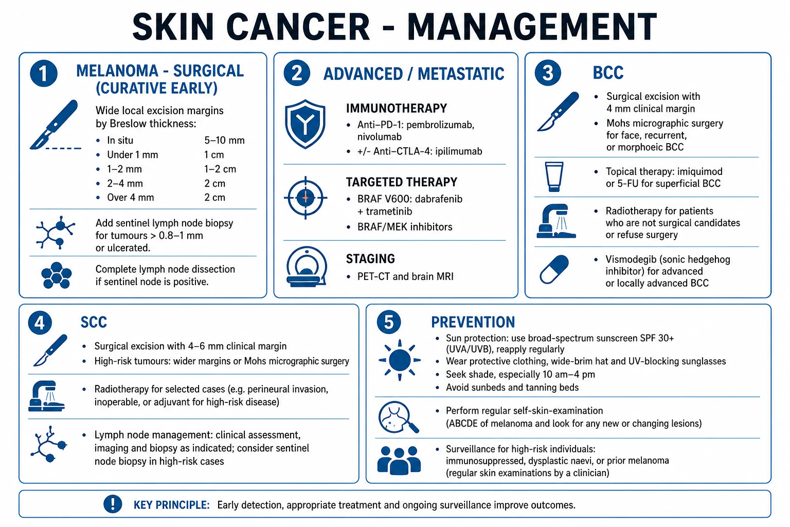
Skin cancer is rarely a resuscitation emergency, but two scenarios demand urgent action: an exsanguinating or deeply eroding neglected tumour (rodent ulcer invading the orbit or a fungating SCC) and sepsis from an infected neglected lesion. The routine "emergency" is the suspicious lesion: any pigmented or non-pigmented lesion meeting ABCDE, the ugly-duckling sign, or the seven-point checklist must be referred urgently under a suspected-cancer pathway (the UK two-week wait or the 28-day faster diagnosis standard) and biopsied without delay. Never shave, curette, or partially excise a suspected melanoma — a full-thickness excisional biopsy is mandatory. At every clinical contact, counsel the patient on sun protection (SPF 30 or higher broad-spectrum sunscreen, protective clothing, shade, avoidance of tanning beds), because prevention is the only way to reduce incidence.[1][8]
Management — Definitive & Stepwise
Melanoma — surgery
Wide local excision (WLE) is the curative treatment for primary melanoma, and the excision margin is dictated by the Breslow thickness. The margins below reflect the NCCN, ESMO and Australian guidelines, which have converged on narrower margins than historical practice after randomised evidence showed that wide margins do not improve survival. [1]
Melanoma WLE margins by Breslow thickness
For patients with a positive sentinel node, MSLT-II established that observation with ultrasound surveillance of the regional basin is the standard — completion lymph node dissection does not improve survival and adds lymphoedema.[5] Therapeutic node dissection is reserved for clinically or radiologically detected nodal disease.
Melanoma — adjuvant and advanced systemic therapy
The systemic treatment of melanoma has been transformed since 2011 by immune checkpoint inhibition and BRAF-targeted therapy. For resected stage IIB to stage IV disease, adjuvant anti-PD-1 (pembrolizumab in KEYNOTE-054; nivolumab in CheckMate 238) significantly improves recurrence-free survival versus placebo or ipilimumab, and is now standard.[9][10] For BRAF V600-mutant resected stage III melanoma, adjuvant dabrafenib plus trametinib (COMBI-AD) improved relapse-free and overall survival versus placebo.[6]
For unresectable or metastatic (stage IV) melanoma, first-line therapy is combination immune checkpoint blockade — nivolumab plus ipilimumab (CheckMate 067) produced the longest survival ever reported in advanced melanoma, with roughly a quarter of patients alive and disease-free at 10 years.[3] Where combination immunotherapy is not tolerated, anti-PD-1 monotherapy (pembrolizumab in KEYNOTE-006; nivolumab) is highly active and less toxic than ipilimumab, and is preferred in patients with comorbidity or asymptomatic low-burden disease.[4] For BRAF V600-mutant disease, BRAF plus MEK inhibition (dabrafenib plus trametinib, or encorafenib plus binimetinib, or vemurafenib plus cobimetinib) gives rapid responses and is preferred for symptomatic, bulky, rapidly progressive disease or brain metastases, with immunotherapy reserved for the BRAF-wild-type or slower-growing tumour. The sequencing of immunotherapy and targeted therapy in BRAF-mutant disease remains an area of debate, but immunotherapy first is the usual default.[1]
Key systemic regimens in melanoma (agent, dose, schedule)
Basal cell carcinoma
BCC is treated according to risk. Standard surgical excision with a 4 to 6 mm margin cures the majority of low-risk nodular BCCs. Mohs micrographic surgery — intraoperative frozen-section margin control — is indicated for facial lesions (especially around the eyes, nose, lips, ears), recurrent BCC, the morphoeic/infiltrative subtype, tumours over 2 cm, and any lesion with indistinct clinical margins, because it maximises cure while sparing tissue. Imiquimod 5 percent cream (applied five times weekly for six weeks) is an option for superficial BCC in patients unsuitable for surgery. Curettage and electrodessication is acceptable for small low-risk nodular BCCs off the face. Radiotherapy (often hypofractionated) is an alternative when surgery is contraindicated, particularly in the elderly.[2][14] For locally advanced or metastatic BCC unsuitable for surgery or radiotherapy, the Hedgehog pathway inhibitors vismodegib (150 mg PO daily) and sonidegib (200 mg PO daily) shrink tumours, though resistance and side-effects (muscle cramps, alopecia, dysgeusia) limit use.[14]
Squamous cell carcinoma
Cutaneous SCC is treated by surgical excision with a 4 to 6 mm margin for low-risk lesions (over 6 mm for higher-risk tumours), or Mohs surgery for facial, recurrent, perineural, large or poorly differentiated tumours. Elective regional node dissection or SLNB is considered for high-risk SCC (over 2 cm, over 6 mm depth, immunosuppressed, perineural invasion, recurrent). Radiotherapy is an alternative primary treatment or adjuvant in high-risk disease. For advanced, metastatic SCC (including in immunosuppressed transplant recipients), cemiplimab (anti-PD-1, 350 mg IV every three weeks) is approved, and options include EGFR inhibitors and chemotherapy.[13]
Merkel cell carcinoma
Merkel cell carcinoma is managed aggressively because of its high mortality. Wide local excision with a 1 to 2 cm margin (typically 1 cm) is combined with routine sentinel lymph node biopsy (performed in almost all patients, as nodal involvement is common even when clinically negative) and adjuvant radiotherapy to the primary site and draining basin in most patients. For advanced or metastatic disease, the anti-PD-L1 antibody avelumab (10 mg/kg IV every two weeks), and pembrolizumab or nivolumab, produce durable responses — a striking finding for a virus-associated tumour.[12]
Prevention and field therapy
For actinic keratoses and field cancerisation, options include cryotherapy for discrete lesions and field-directed therapy for extensive disease: 5-fluorouracil 5 percent cream (twice daily for three to four weeks), imiquimod 5 percent, photodynamic therapy, diclofenac 3 percent gel, and ingenol mebutate. Acitretin or capecitabine may reduce SCC incidence in heavily immunosuppressed transplant recipients. Sun protection (broad-spectrum SPF 30 or higher sunscreen applied 20 minutes before exposure and every two hours, protective clothing, wide-brimmed hats, shade between 10 am and 4 pm, and total avoidance of tanning beds) and regular skin self-examination are the foundation of prevention and of surveillance in high-risk individuals.[1]
Specific Subtypes & Scenarios
Each melanoma subtype has clinical and prognostic features the examiner probes. Superficial spreading melanoma carries the best prognosis if caught thin, because its long radial phase delays invasion. Nodular melanoma has the worst prognosis for a given thickness because of its pure vertical growth and rapid progression; it is over-represented among thick melanomas and is the classic "interval" cancer that appears between screening visits. Lentigo maligna melanoma arises in elderly sun-damaged skin; its in-situ precursor (lentigo maligna) may be present for years, and the diagnostic challenge is sampling error — multiple punches or staged mapped biopsy are needed. Acral lentiginous melanoma is not UV-driven, is the commonest melanoma in Asian and Black populations, and presents late on the palm, sole or under a nail — subungual melanoma shows longitudinal melanonychia and the Hutchinson sign (pigment on the nail fold). Amelanotic melanoma is the great mimic: a pink, fleshy, bleeding or growing lesion that fails the ABCD criteria and is diagnosed only by a high index of suspicion and biopsy.[1]
Desmoplastic and neurotropic melanoma is a scar-like variant with a high rate of perineural invasion and local recurrence but a lower rate of nodal metastasis; it is managed with wider margins and consideration of radiotherapy. Keratoacanthoma is a low-grade SCC variant that grows rapidly over weeks into a cup-shaped nodule with a central keratin plug, then classically involutes — but because it cannot be reliably distinguished from SCC clinically or histologically, it is excised as SCC. Bowen disease is SCC in situ (a fixed, scaly, well-demarcated red plaque) treated by excision, curettage, 5-fluorouracil, imiquimod or photodynamic therapy. Gorlin (basal cell naevus) syndrome — germline PTCH1 mutation — produces multiple BCCs from childhood, palmar pits, odontogenic keratocysts, bifid ribs and calcification of the falx, and is managed with lifelong surveillance, strict sun protection and Hedgehog inhibitors for severe disease.[14]
Complications & Pitfalls
The complications of skin cancer are those of local destruction (BCC and SCC eroding cartilage, bone, orbit, nose — the neglected rodent ulcer is a surgical disaster), metastasis (melanoma to lymph nodes, lung, liver, brain, bone and bowel — the last causing small-bowel intussusception or anaemia; SCC to regional nodes, with perineural spread along the trigeminal or facial nerve), and treatment toxicity. After surgery, wound dehiscence, skin-graft failure, infection and lymphoedema (especially after inguinal or axillary node dissection) are the common problems.[1]
The immune-related adverse events (irAEs) of checkpoint inhibitors are now bread-and-butter exam material and can be life-threatening: colitis (with ipilimumab, may perforate), pneumonitis, hepatitis, nephritis, and a range of endocrinopathies (thyroiditis causing hypo- or hyperthyroidism, hypophysitis with hypopituitarism, type 1 diabetes, primary adrenal insufficiency). CTLA-4 (ipilimumab) is more toxic than PD-1 monotherapy, and combination therapy is the most toxic of all. Management is by holding the drug, grading severity, and giving corticosteroids (prednisone 1 to 2 mg/kg/day) with other immunosuppressants (infliximab for severe colitis, mycophenolate for nephritis) as needed, and hormone replacement for endocrinopathies (which are often permanent). BRAF inhibitors paradoxically activate the MAPK pathway in wild-type cells, producing keratoacanthomas and SCCs (within weeks), alongside pyrexia, photosensitivity, rash and, with MEK inhibitors, ocular toxicity (uveitis, retinal vein occlusion) and cardiac effects.[3][4]
Exam application bank (NEET-PG / INICET)
One-line answer
Skin cancer is the commonest malignancy worldwide and splits into melanoma (aggressive melanocytic cancer with high metastatic potential) and the non-melanoma skin cancers (NMSC) — basal cell carcinoma (BCC, commonest and locally invasive, rarely metastasises) and squamous cell carcinoma (SCC, keratinising, can metastasise to nodes) — plus the rarer, aggressive Merkel cell carcinoma (neuroendocrine, polyomavirus-related). Risk factors centre on ultraviolet radiation (sun exposure, sunburn, tanning beds), fair skin, freckles, red hair (MC1R), immunosuppression, older age, family/genetic predisposition (FAMM/CDKN2A, xeroderma pigmentosum, Gorlin), and previous skin cancer. Melanoma is recognised by the ABCDE criteria and the ugly-duckling sign; subtypes are superficial spreading (commonest), nodular (worst), lentigo maligna, acral lentiginous and amelanotic. Staging rests on Breslow thickn
Worked stems (answer without another resource)
Stem 1 — Classic presentation. Map symptoms to mechanism; name the first investigation and first treatment step with dose/route if drug therapy is standard. [1]
Stem 2 — Unstable / complicated. List red flags that force immediate resuscitation, theatre, ICU, antidote, or reperfusion — and what you do in the first 15 minutes. [1]
Stem 3 — Atypical group. Elderly, pregnancy, child, or immunocompromised: how presentation and thresholds change. [1]
Stem 4 — Differential trap. Name the three closest mimics and one discriminator for each. [1]
Stem 5 — Disposition. Who goes home with safety-netting, who is admitted, who needs HDU/ICU/theatre, and what follow-up is mandatory. [1]
Rapid viva checklist
- Definition + classification
- Pathophysiology chain
- Bedside signs / criteria
- Score with exact components (if any)
- Emergency bundle
- Definitive therapy with doses
- Complications of disease and of treatment
- Special populations
- Guideline/trial name if classic
- Three exam traps
Coverage self-check
If you cannot answer any stem above from this page alone, re-read the matching section — the page is intended to be self-sufficient for final-prof and NEET-PG/INICET questions on Melanoma & Skin Cancer.
[1]Prognosis & Disposition
Breslow thickness is the single strongest prognostic factor in localised melanoma, and ulceration and sentinel-node status add the next layers. The AJCC 8th edition five-year survival figures illustrate the precipitous fall with stage: stage I (T1a/T1b/T2a, node-negative) around 98 to 99 percent; stage IIA about 93 percent; stage IIB about 83 percent; stage IIC about 71 percent; stage IIIA about 93 percent (microscopic nodal disease, thin primary, non-ulcerated); stage IIIB about 83 percent; stage IIIC about 69 percent; stage IIID about 32 percent; and stage IV around 25 to 30 percent at five years — though immunotherapy has improved even stage-IV outcomes markedly since these historic baselines were set.[7]
BCC has an excellent prognosis — over 95 percent cure with appropriate excision or Mohs — and almost never metastasises (under 0.1 percent); its morbidity is local recurrence (higher with the morphoeic subtype or incomplete excision). SCC is cured in about 95 percent of cases when excised adequately, but nodal metastasis drops survival to 30 to 50 percent, hence the importance of identifying high-risk tumours. Merkel cell carcinoma has a five-year survival of roughly 50 to 70 percent for local disease, falling to about 25 percent with regional nodes and 10 to 15 percent with distant metastasis — one of the highest skin-cancer mortalities.[12]
Follow-up after melanoma is intensive: six-monthly full skin and node examination for five years (UK practice), with total-body skin examination lifelong because of the high rate of second primaries. Patients with stage III/IV disease, or on adjuvant therapy, have PET-CT and brain MRI surveillance as indicated. The safety-net for any patient with a previous skin cancer is lifetime self-examination and low threshold for re-presentation.[1]
Special Populations
Immunosuppressed solid-organ transplant recipients carry a 65- to 100-fold increased risk of SCC (and a roughly 10-fold increased risk of BCC); SCC becomes more common than BCC, the tumours are more aggressive, multiple, and HPV-related, and metastasis is common. Management combines mTOR inhibitors (sirolimus/everolimus, which may reduce skin-cancer risk), reduction of immunosuppression where feasible, aggressive field therapy (5-fluorouracil, acitretin, photodynamic therapy), and low threshold for excision. Chronic lymphocytic leukaemia and HIV also raise SCC and Merkel cell risk.[13]
Xeroderma pigmentosum (defective nucleotide excision repair) carries a more than 1000-fold increase in skin-cancer risk with childhood onset; management is strict photoprotection and frequent surveillance. Familial atypical multiple mole melanoma (FAMM) syndrome (germline CDKN2A) produces multiple atypical naevi and a high lifetime melanoma risk, warranting intensive screening from adolescence. Gorlin syndrome patients develop hundreds of BCCs. Oculocutaneous albinism produces high SCC and melanoma risk without protection. Pregnancy does not clearly worsen melanoma prognosis, but melanoma can rarely metastasise to the placenta and fetus — management is otherwise as standard. Children rarely develop melanoma; the differential includes Spitz naevus, and giant congenital melanocytic naevi (over 20 cm) carry a meaningful lifetime melanoma risk warranting surveillance. The elderly present with thicker melanoma at diagnosis, have more lentigo maligna and extensive actinic damage, and their comorbidities shape the choice between surgery, radiotherapy and systemic therapy.[1][12]
Evidence, Guidelines & Regional Differences
Modern melanoma management rests on a sequence of practice-defining trials. CheckMate 067 (Larkin et al, NEJM 2015) established nivolumab plus ipilimumab as the most effective first-line therapy for advanced melanoma, with long-term follow-up showing a 10-year survival of roughly 52 percent with combination therapy — unprecedented in this disease.[3] KEYNOTE-006 (Robert et al, NEJM 2015) showed pembrolizumab superior to ipilimumab in advanced melanoma with less toxicity.[4] In the surgical arena, MSLT-II (Faries et al, NEJM 2017) changed practice by showing that observation with ultrasound surveillance is non-inferior to completion lymph node dissection after a positive sentinel node, sparing patients lymphoedema.[5] In the adjuvant setting, COMBI-AD (Long et al, NEJM 2017) established dabrafenib plus trametinib for resected BRAF-mutant stage III melanoma,[6] while KEYNOTE-054 (Eggermont et al, NEJM 2018) established adjuvant pembrolizumab for stage III disease,[9] and CheckMate 238 (Weber et al, NEJM 2017) showed adjuvant nivolumab superior to ipilimumab.[10] The staging framework itself was revised by the AJCC 8th edition (Gershenwald et al, 2017), which moved the T1a/T1b threshold to 0.8 mm and down-weighted Clark level and mitotic rate.[7]
Guidelines are broadly concordant across regions: the US NCCN, the UK NICE/British Association of Dermatologists, European ESMO/EORTC, and the Australian Cancer Council / New Zealand guidelines agree on ABCDE recognition, excisional biopsy, WLE margins by Breslow, SLNB for T1b and thicker, anti-PD-1-based therapy for advanced disease, and sun protection. Differences are largely in Mohs utilisation (higher in the US, where it is well reimbursed, than in the UK/Australia, where standard excision with margin control predominates), checkpoint-inhibitor funding and first-line choices (combination immunotherapy more readily funded in Australia/Europe than in some settings), and follow-up intensity.
Exam Pearls
- ABCDE = Asymmetry, Border irregular, Colour variegated, Diameter over 6 mm, Evolving (the "E" was added later to the original ABCD of 1985; Evolving is the most sensitive single sign and the only one that catches nodular melanoma).[8]
- Ugly-duckling sign: the mole that looks different from the patient's other moles — high yield, especially in patients with many naevi.
- Breslow thickness is measured in millimetres from the top of the granular layer to the deepest tumour cell; Clark level (I = in situ to V = subcutaneous fat) is now secondary and used only for thin melanomas.
- AJCC 8th edition: T1a under 0.8 mm without ulceration; T1b under 0.8 mm with ulceration, or 0.8 to 1.0 mm with or without ulceration. The SLNB threshold is over 0.8 mm, or ulcerated.
- Nodular melanoma is the exception that fails ABCD — small, symmetric, monocolour, fast-growing. Amelanotic melanoma is pink.
- BCC = rodent ulcer: pearly papule, arborising telangiectasia, rolled border, central ulceration. BCC essentially never metastasises (under 0.1 percent).
- SCC: keratinising scaly nodule on sun-exposed skin; keratin pearls on histology; metastasises to regional nodes (2 to 5 percent). Keratoacanthoma grows in weeks then involutes — treat as SCC.
- Merkel cell carcinoma — AEIOU: Asymptomatic, Expanding rapidly, Immunosuppression, Older white, UV-exposed site; CK20 perinuclear dot, Merkel cell polyomavirus in 80 percent.
- Subungual melanoma — Hutchinson sign (pigment on the nail fold); not UV-related (acral lentiginous). Bob Marley died of acral lentiginous melanoma.
- Mohs surgery for BCC/SCC: face (eyes, nose, lips, ears), recurrent, morphoeic/infiltrative, over 2 cm, indistinct margins.
- Never shave-biopsy a suspected melanoma. Full-thickness excisional biopsy, 1 to 2 mm margin.
- Hedgehog inhibitors (vismodegib, sonidegib) for advanced BCC; cemiplimab for advanced SCC; avelumab for advanced Merkel cell carcinoma.
- Checkpoint-inhibitor irAEs: colitis (ipilimumab), pneumonitis, hepatitis, thyroiditis, hypophysitis, type 1 diabetes — manage with hold, steroids, hormone replacement.
- Sunscreen: broad-spectrum SPF 30 or higher, applied 20 minutes before exposure and every two hours, shade between 10 am and 4 pm, no tanning beds.
Rapid self-test — what is the SLNB threshold, and why?
SLNB is offered for melanoma over 0.8 mm Breslow thickness, or under 0.8 mm if ulcerated (T1b), because the risk of occult nodal disease rises sharply above 0.8 mm (from roughly 5 percent below to over 10 percent above). AJCC 8th edition moved the T1a/T1b boundary to 0.8 mm on this basis.[7]
Rapid self-test — a 68-year-old man has a 1 cm rapidly growing painless red nodule on his temple. What is the diagnosis and the first test?
Think Merkel cell carcinoma (AEIOU: rapidly Expanding, Older white, UV-exposed, painless). The diagnosis is histological — biopsy (a full-thickness excisional or incisional biopsy), with CK20 perinuclear-dot staining and Merkel cell polyomavirus testing. Management is wide excision, routine sentinel node biopsy, adjuvant radiotherapy, and avelumab if advanced.[12]
References
- [1]Schadendorf D, van Akkooi ACJ, Berking C, et al. Melanoma Lancet, 2018.PMID 30238891
- [2]Zaorsky NG, Lee CT, Zhang E, et al. Hypofractionated radiation therapy for basal and squamous cell skin cancer: A meta-analysis Radiother Oncol, 2017.PMID 28843727
- [3]Larkin J, Chiarion-Sileni V, Gonzalez R, et al. Combined Nivolumab and Ipilimumab or Monotherapy in Untreated Melanoma N Engl J Med, 2015.PMID 26027431
- [4]Robert C, Schachter J, Long GV, et al. Pembrolizumab versus Ipilimumab in Advanced Melanoma N Engl J Med, 2015.PMID 25891173
- [5]Faries MB, Thompson JF, Cochran AJ, et al. Completion Dissection or Observation for Sentinel-Node Metastasis in Melanoma N Engl J Med, 2017.PMID 28591523
- [6]Long GV, Hauschild A, Santinami M, et al. Adjuvant Dabrafenib plus Trametinib in Stage III BRAF-Mutated Melanoma N Engl J Med, 2017.PMID 28891408
- [7]Gershenwald JE, Scolyer RA, Hess KR, et al. Melanoma staging: Evidence-based changes in the American Joint Committee on Cancer eighth edition cancer staging manual CA Cancer J Clin, 2017.PMID 29028110
- [8]Abbasi NR, Yancovitz M, Gutkowicz-Krusin D, et al. Early diagnosis of cutaneous melanoma: revisiting the ABCD criteria JAMA, 2004.PMID 15585738
- [9]Eggermont AMM, Blank CU, Mandala M, et al. Adjuvant Pembrolizumab versus Placebo in Resected Stage III Melanoma N Engl J Med, 2018.PMID 29658430
- [10]Weber J, Mandala M, Del Vecchio M, et al. Adjuvant Nivolumab versus Ipilimumab in Resected Stage III or IV Melanoma N Engl J Med, 2017.PMID 28891423
- [11]Sung H, Ferlay J, Siegel RL, et al. Global Cancer Statistics 2020: GLOBOCAN Estimates of Incidence and Mortality Worldwide for 36 Cancers in 185 Countries CA Cancer J Clin, 2021.PMID 33538338
- [12]Becker JC, Stang A, DeCaprio JA, et al. Merkel cell carcinoma Nat Rev Dis Primers, 2017.PMID 29072302
- [13]Que SKT, Zwald FO, Schmults CD. Cutaneous squamous cell carcinoma: Incidence, risk factors, diagnosis, and staging J Am Acad Dermatol, 2018.PMID 29332704
- [14]Dika E, Scarpa F, Lambertini M, et al. Basal Cell Carcinoma: A Comprehensive Review Int J Mol Sci, 2020.PMID 32759706