Dermatology · Medicine
Melasma
Also known as Chloasma · Mask of pregnancy
Melasma is an acquired, bilateral, symmetrical hyperpigmentation of sun-exposed facial skin, classically affecting women with Fitzpatrick III-IV skin. It is driven by UV and visible light, hormonal triggers, genetic predisposition and local skin microenvironmental changes. Wood's lamp examination separates epidermal (accentuated, responds to topicals), dermal (not accentuated, resistant) and mixed types. Management is built on strict photoprotection, then topical depigmenting agents such as hydroquinone, tretinoin and azelaic acid, with oral tranexamic acid and carefully selected procedures for refractory disease. It is chronic and relapsing.
On this page & tools
Your progress
Saved locally on this device.
Exam tags
Red flags
Overview & Definition
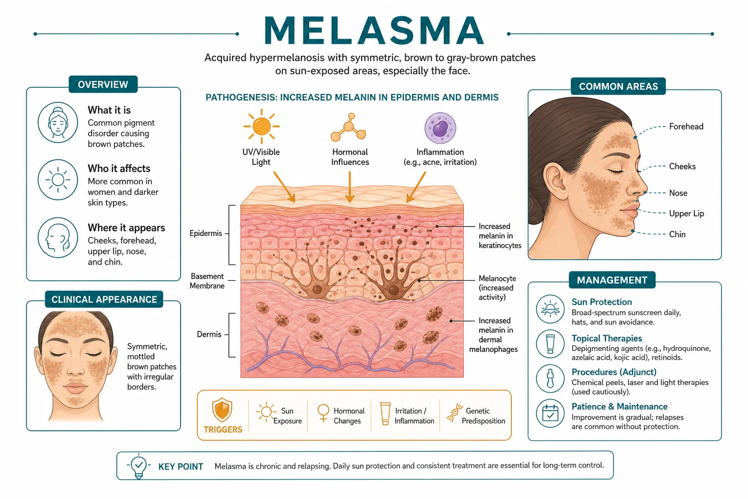
Melasma is an acquired, usually bilateral and symmetrical disorder of facial hyperpigmentation that affects the sun-exposed skin of the face and, less commonly, the neck and forearms.[5][6] It is also known historically as chloasma or the mask of pregnancy, because it frequently appears or worsens during gestation. The pigmentation is caused by increased deposition of melanin within the epidermis and/or dermis, rather than by an increase in melanocyte number, although the melanocytes that are present are metabolically hyperactive.[5]
The diagnosis is clinical, but the condition is best understood as a multifactorial photodermatosis rather than a simple cosmetic problem. Sun exposure, particularly ultraviolet A and B, and more recently recognised visible light, are the dominant environmental triggers. Hormonal influences, including pregnancy, oral contraceptives and hormone replacement therapy, explain the female predominance and the gestational form. Genetic predisposition accounts for strong familial clustering and the tendency for the disease to recur. [5][6]
The term chloasma comes from the Greek word for "greenish", and mask of pregnancy reflects the common presentation in gestation. Modern nomenclature prefers melasma because the condition is not limited to pregnancy and can occur in men and in the absence of hormonal therapy. The older term chloasma is still used in many textbooks and examinations, and candidates should be familiar with both names in clinical practice. [1]
[6] [1] [6]Classification
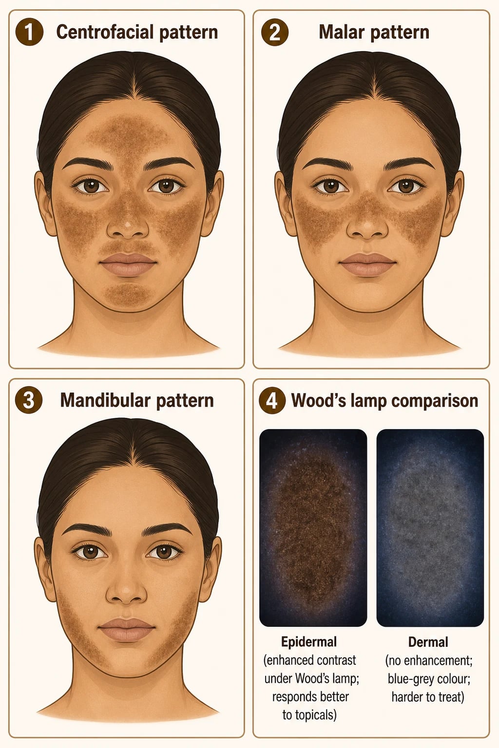
Melasma can be classified in two complementary ways: by anatomical distribution and by Wood's lamp depth. Both classifications are examinable and directly guide management. [1]
[6]Anatomical patterns are usually described as three major distributions.[5]
- Centrofacial is the most common pattern, affecting the forehead, cheeks, nose, upper lip and chin. It is often the pattern that patients notice first and that causes the greatest psychosocial impact.
- Malar melasma is limited to the cheeks and nose, producing a butterfly-like mask. It is frequently mistaken for blushing or rosacea in its early stages.
- Mandibular melasma affects the jawline and is more common in older patients. It is sometimes thought to be more therapy-resistant, possibly because of deeper dermal pigment in this region. [1]
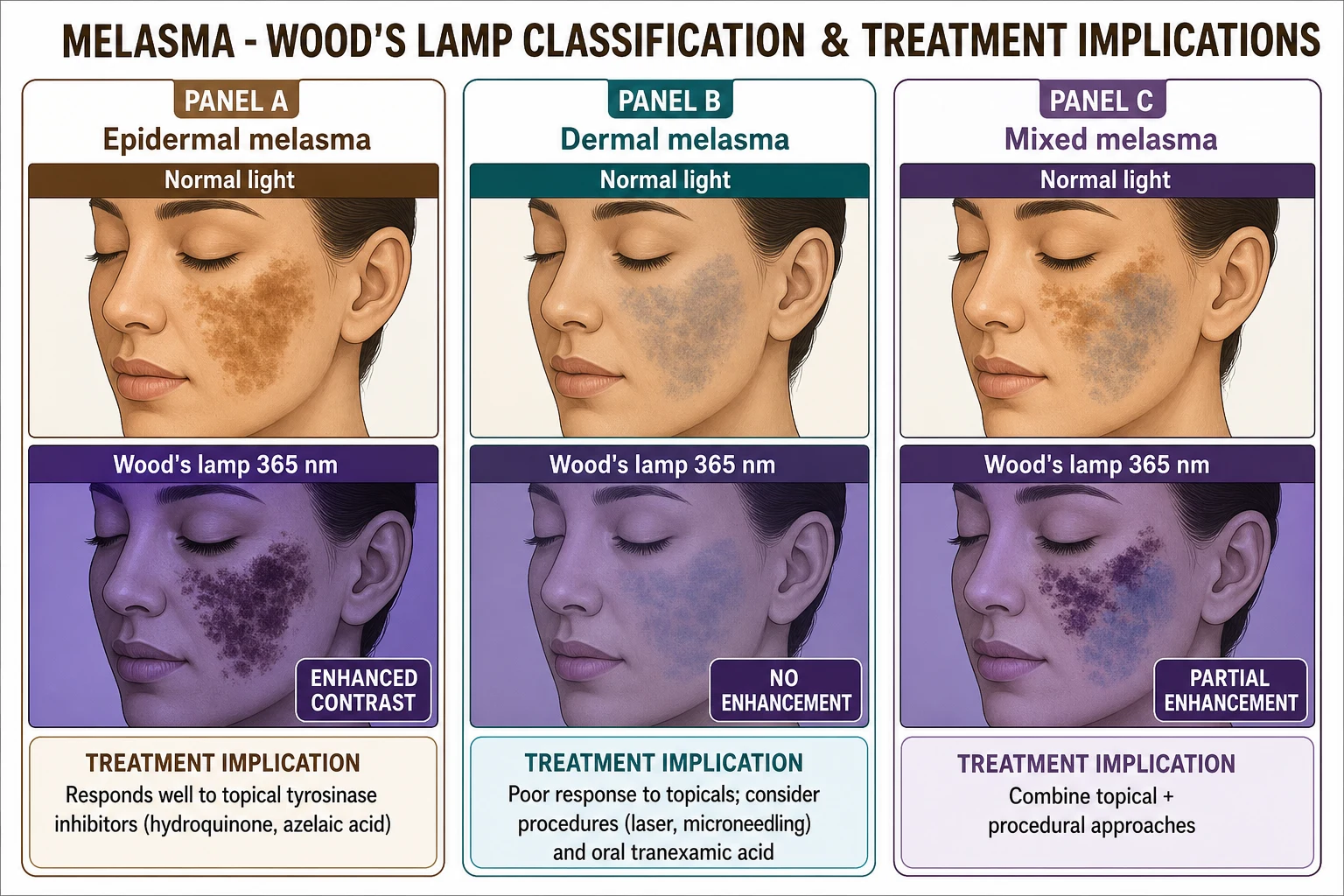
Wood's lamp classification is the clinically important system that predicts treatment response. Wood's lamp emits long-wave ultraviolet A light at approximately 365 nm. The excess melanin in the epidermis absorbs this light and makes the lesion appear darker and more sharply demarcated, whereas dermal melanin, because it is deeper and shielded by the overlying epidermis, does not accentuate.[6]
Epidermal
Best prognosis
- Colour: brown or tan.
- Wood's lamp: accentuated, darker and sharper.
- Mechanism: melanin in basal and suprabasal keratinocytes.
- Response: good to topical tyrosinase inhibitors and retinoids.
Dermal
Most resistant
- Colour: blue-grey or slate-grey.
- Wood's lamp: no accentuation, poorly defined.
- Mechanism: melanin within dermal melanophages.
- Response: poor to topicals; may need oral agents or procedures.
Mixed
Intermediate
- Colour: brown-grey, variable.
- Wood's lamp: partial accentuation.
- Mechanism: both epidermal and dermal melanin.
- Response: intermediate; combined approach.
Epidemiology & Risk Factors
Melasma is a global condition but shows striking variation by geography, sex, skin phototype and hormonal status. Estimates of prevalence vary widely because survey methods differ and because many cases are mild and unreported, but the disease is consistently more common in regions with high ambient ultraviolet exposure and in populations with darker skin.[5]
Epidemiological headline numbers
The strongest risk factors are female sex, Fitzpatrick III-IV skin, pregnancy, oral contraceptive or hormone replacement therapy, ultraviolet and visible light exposure, and family history. Patients with Fitzpatrick III-IV skin have more labile melanocytes that are more easily activated by UV and visible light; they also have larger melanosomes and more efficient transfer of melanosomes to keratinocytes, so once triggered the pigment is more conspicuous.[5][6]
Hormonal triggers are central. During pregnancy, rising oestrogen and progesterone stimulate melanogenesis, particularly in the centrofacial distribution. Oral contraceptive pills and hormone replacement therapy can produce the same effect, and stopping or changing these agents is often part of management. Thyroid disease, particularly autoimmune thyroid disease, has been reported more frequently in patients with melasma, so targeted thyroid testing may be appropriate in selected cases.[5][6]
Photosensitising agents and cosmetics are less commonly recognised but important precipitants. Some fragrances, essential oils and cosmetic ingredients can cause photocontact dermatitis followed by post-inflammatory hyperpigmentation that merges clinically with melasma. Systemic drugs that increase photosensitivity, including some anticonvulsants, non-steroidal anti-inflammatory agents and photosensitising antibiotics, can also worsen the condition.[3]
Risk factors for melasma
MELASMA
Fitzpatrick III-IV skin with easily activated melanocytes.
UV and visible light, the dominant environmental triggers.
Photosensitising medications and fragranced cosmetics.
Family history and genetic predisposition.
Pregnancy, OCPs and HRT.
High-UV climates and outdoor occupations.
Women of reproductive age.
Pathophysiology
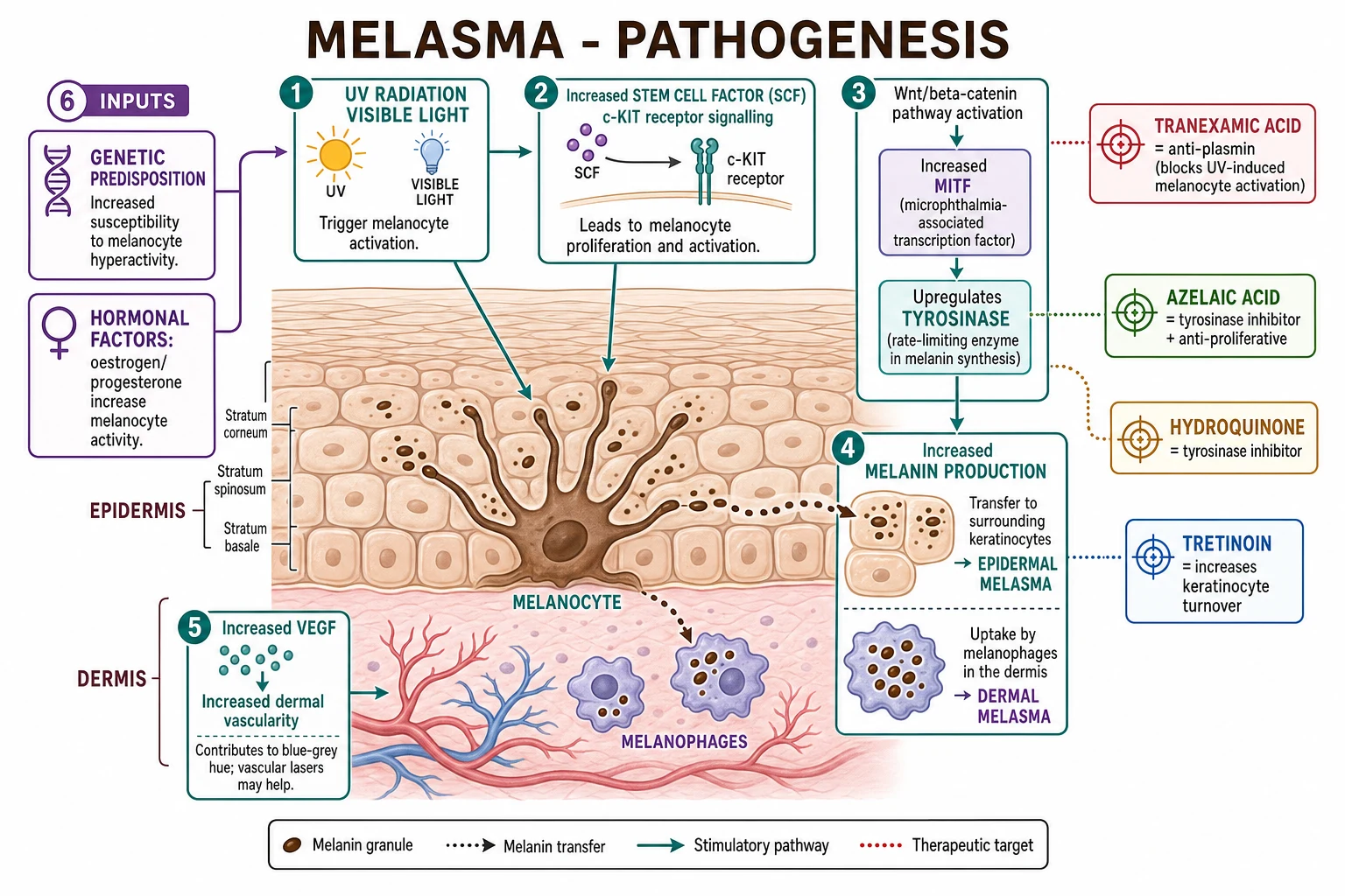
Melasma is not simply an excess of melanin. It is a disorder of melanocyte-keratinocyte interaction driven by ultraviolet and visible light, amplified by hormonal and genetic factors, and sustained by changes in the local skin microenvironment.[5][7]
The central cascade begins when keratinocytes detect UV radiation. They release stem cell factor (SCF), which binds the c-KIT receptor on melanocytes and triggers proliferation, dendrite extension and melanogenesis. Parallel activation of the Wnt/β-catenin pathway increases microphthalmia-associated transcription factor (MITF), the master regulator of melanocyte function. MITF upregulates tyrosinase, the rate-limiting enzyme in melanin synthesis, as well as tyrosinase-related protein 1 and dopachrome tautomerase. The result is increased production of eumelanin and deposition in surrounding keratinocytes.[5]
Recent work has expanded this model. The basement membrane is disrupted in melasma, with thinning, duplication and gap formation. These gaps allow melanin to fall into the dermis, where it is engulfed by dermal melanophages. This mechanism explains why some lesions become dermal or mixed and why dermal melasma is so resistant to topical agents: the pigment is below the reach of creams and gels.[7]
The melanin synthesis cascade begins when the enzyme tyrosinase oxidises the amino acid tyrosine to L-DOPA and then to dopaquinone. Subsequent steps generate eumelanin or pheomelanin, which are packaged into melanosomes and transferred to neighbouring keratinocytes. In melasma, both tyrosinase activity and melanosome transfer are increased, and the transferred melanosomes are larger and more heavily pigmented. This explains why the condition is clinically dark and why it responds to agents that block tyrosinase, accelerate keratinocyte turnover or inhibit melanosome transfer.[5]
Vascularisation is also increased. Lesional skin shows higher expression of vascular endothelial growth factor (VEGF) and more prominent superficial vessels. This vascular component contributes to the blue-grey or erythematous undertone sometimes seen in melasma, and it may explain why some patients improve with vascular-targeting lasers or oral tranexamic acid, which has anti-angiogenic as well as anti-plasmin effects.[5][7]
Mast cells are more numerous in melasma skin and release mediators such as histamine, tryptase and VEGF that can stimulate melanocytes and promote vascularity. This mast-cell-driven inflammation helps explain the chronicity and photosensitivity of the condition. The local microenvironment also shows increased matrix metalloproteinase activity and altered extracellular matrix, suggesting that melasma is partly a disorder of photodamage as well as pigmentation.[7]
The hormonal component is mediated by oestrogen and progesterone receptors on melanocytes, which amplify melanogenesis under the same UV stimulus. This is why pregnancy and exogenous hormones can precipitate melasma even in the absence of increased sun exposure. Genetic studies show familial clustering and polymorphisms in genes related to melanogenesis, supporting the clinical observation that a positive family history is common.[5]
Clinical Presentation
Patients typically present with symmetrical, poorly defined, brown to grey-brown patches on the face. The colour is determined by the depth of pigment: epidermal lesions are brown, dermal lesions are blue-grey or slate, and mixed lesions are brown-grey. The patches are usually asymptomatic, though some patients report mild burning or stinging after sun exposure.[6]
The distribution follows the anatomical patterns described above. Centrofacial melasma is the most common and most psychologically distressing because it involves the central face. Malar melasma produces a mask-like appearance across the cheeks and nose. Mandibular melasma is more common in older patients and is often more resistant to treatment. Rarely, melasma can affect the neck or forearms, termed extra-facial melasma; these sites are usually associated with severe cumulative sun exposure.[5]
Atypical presentations include male melasma, which is less common but often more resistant and more strongly associated with sun exposure, thyroid disease and occasionally androgenic factors. Elderly-onset melasma, particularly in the mandibular pattern, may be misdiagnosed as actinic pigmentation or solar lentigines. Darker skin types can develop very intense pigment that is more likely to be mixed or dermal, and they are at higher risk of post-inflammatory hyperpigmentation from aggressive treatments. Pregnancy-associated melasma often appears in the second or third trimester and may fade after delivery, though it frequently recurs with subsequent pregnancies or with later sun exposure.[5][6]
Clinical clues to depth are important. Lesions that are sharply demarcated and uniformly brown are more likely epidermal; lesions that are slate-grey, poorly defined and diffuse are more likely dermal. The definitive bedside test is Wood's lamp examination. Some lesions also show a subtle erythematous component related to increased vascularity; this can make the patches appear brown-pink rather than pure brown and may predict a less robust response to topical therapy alone. [7]
Differential Diagnosis
Several conditions can mimic melasma. The key differentiators are the history of preceding inflammation, distribution, colour, Wood's lamp findings and response to therapy. A unilateral lesion, violaceous hue, marked scale, or systemic symptoms should always prompt reconsideration.[6]
Post-inflammatory hyperpigmentation
History of rash or acne
- Follows acne, eczema, psoriasis or drug eruption.
- Often asymmetric or follows the pattern of prior inflammation.
- Wood's lamp may show variable accentuation.
- Treat the underlying inflammation and photoprotect.
Solar lentigo
Discrete sun spots
- Sharply demarcated, discrete, uniform brown macules.
- Usually smaller and more numerous than melasma patches.
- Wood's lamp accentuates strongly.
- Histology shows elongated rete ridges with increased melanin.
Ephelides
Freckles
- Small, well-defined tan macules, often in childhood.
- Darken with sun and fade in winter.
- Wood's lamp accentuates; no hormonal trigger.
Other important mimics include exogenous ochronosis, which presents as blue-black or grey-brown patches, often on the cheeks, after prolonged use of topical hydroquinone. It is a paradoxical darkening caused by deposition of homogentisic acid-like pigment in the dermis and does not improve with further hydroquinone.[2][6] Drug-induced pigmentation can be caused by minocycline (slate-grey), amiodarone (blue-grey), antimalarials, chemotherapy and heavy metals. The pigmentation is usually more diffuse and may involve the sclerae, mucosa or nails.[3]
Riehl melanosis is a contact dermatitis-related pigmentary dermatosis that produces reticulated brown-grey facial pigmentation, often after exposure to certain fragrances or cosmetics. It is thought to represent a delayed hypersensitivity reaction followed by pigment incontinence. The history of a new cosmetic or fragrance, and sometimes a positive patch test, points toward the diagnosis. Erythromelanosis follicularis presents with follicular erythema and hyperpigmentation, usually on the neck and face, and often has a rough, follicular texture on palpation. Addison's disease causes generalised hyperpigmentation with accentuation in scars, palmar creases and mucous membranes, and it is accompanied by systemic symptoms such as fatigue, weight loss, postural hypotension and hyponatraemia. Acanthosis nigricans is velvety, flexural and associated with insulin resistance; it is usually easily distinguished by texture and site, but it can coexist with facial melasma in patients with metabolic syndrome.[6]
Clinical & Bedside Assessment
The focused assessment of a patient with suspected melasma has three parts: history, examination under ambient light and examination with Wood's lamp. Photographs are helpful for monitoring response because improvement is often gradual and patients may not notice incremental change.[6]
History should establish onset, duration, relationship to pregnancy or hormonal therapy, sun exposure, family history, cosmetic and drug use, and prior treatments. Ask specifically about hydroquinone use duration and any paradoxical darkening, which suggests exogenous ochronosis. Enquire about thyroid symptoms and systemic symptoms that might point to Addison's disease. [1]
[6]Wood's lamp examination should be performed in a darkened room. The lamp is held 10-15 cm from the skin and moved systematically across the face. Areas that become darker and more sharply defined are epidermal; areas that do not change are dermal; mixed areas show partial accentuation. Document the pattern and depth because this guides treatment and helps set patient expectations. Dermoscopy can support the classification: epidermal melasma often shows a regular pigment network, while dermal melasma shows irregular grey-brown structureless areas or slate-grey blotches.[3]
Bedside clues to alternative diagnoses include unilateral distribution, violaceous or violaceous-brown colour, scale, punctate follicular openings, mucosal involvement, palmar crease darkening or systemic symptoms. Any of these should prompt a broader differential and targeted investigations. [1]
[6]Investigations
Melasma is usually a clinical diagnosis. No blood test or imaging is required in typical cases. The role of investigations is to confirm depth, exclude mimics and identify contributing systemic disease when suggested by history or examination.[6]
Wood's lamp is the single most useful bedside investigation. It is inexpensive, quick and directly separates epidermal from dermal disease. Dermoscopy can be used in centres where it is available, but it is not mandatory. Photography with standardised lighting and background is valuable for monitoring. [1]
[6]Skin biopsy is rarely needed. It is reserved for cases with atypical features, failure to respond to first-line therapy, or diagnostic uncertainty. Histology in melasma shows increased melanin in the epidermis, with or without melanophages in the dermis, and basement membrane changes. Solar lentigo shows elongated rete ridges; exogenous ochronosis shows banana-shaped ochre deposits in the dermis; lichen planus pigmentosus shows interface dermatitis with pigment incontinence. A biopsy should not be performed lightly on the face because of the risk of post-inflammatory hyperpigmentation and scarring, especially in darker skin.[6]
Thyroid function tests, typically thyroid-stimulating hormone, are reasonable in patients with symptoms or signs of thyroid disease, because autoimmune thyroid disease is over-represented. Serum cortisol and ACTH or a short synacthen test should be considered if there is generalised pigmentation, mucosal pigmentation, postural hypotension, fatigue, weight loss or hyponatraemia, because Addison's disease can mimic melasma. Drug levels or medication review are useful when drug-induced pigmentation is suspected. [1]
[6]When would you order a skin biopsy in melasma?
A biopsy is reserved for atypical presentations, diagnostic uncertainty, failure to respond to appropriate therapy, or suspicion of an alternative diagnosis such as exogenous ochronosis, lichen planus pigmentosus or drug-induced pigmentation. In most patients the clinical and Wood's lamp findings are sufficient.
Management — Immediate Measures
Every patient with melasma should begin with strict photoprotection and trigger avoidance, regardless of subtype or severity. This is the non-negotiable foundation on which all other therapies depend. Without photoprotection, even the most effective topical or oral agents will fail.[6]
Photoprotection means broad-spectrum, high-protection sunscreen with SPF 50 or higher and a PA rating of ++++. The product must block both UVB and UVA, because UVA penetrates deeply and is a major driver of melanocyte activation. Sunscreen should be applied generously to the entire face and neck, 15-30 minutes before exposure, and reapplied every two hours during outdoor activity and after swimming or sweating. A tinted sunscreen containing iron oxide is preferred because iron oxide blocks visible light, which is increasingly recognised as a melasma trigger, especially in darker skin types.[2][6]
Physical measures are equally important: wide-brimmed hats, sunglasses, seeking shade, and avoiding the midday sun. Patients should also be aware that visible light from screens and indoor lighting is a minor contributor compared with sunlight, but cumulative exposure can matter in sensitive individuals. UV-protective film on car and home windows can help those with long commutes or significant indoor UV exposure. [1]
[6]Trigger avoidance includes reviewing oral contraceptive pills, hormone replacement therapy and photosensitising medications. Where possible, switching to a non-hormonal contraceptive method may reduce relapse, though it is not always practical. Stopping unnecessary photosensitising drugs, or at least minimising sun exposure while taking them, is advisable. Pregnant patients should avoid hydroquinone and tretinoin entirely and rely on photoprotection and azelaic acid.[6]
[1] [6]Management — Definitive & Stepwise
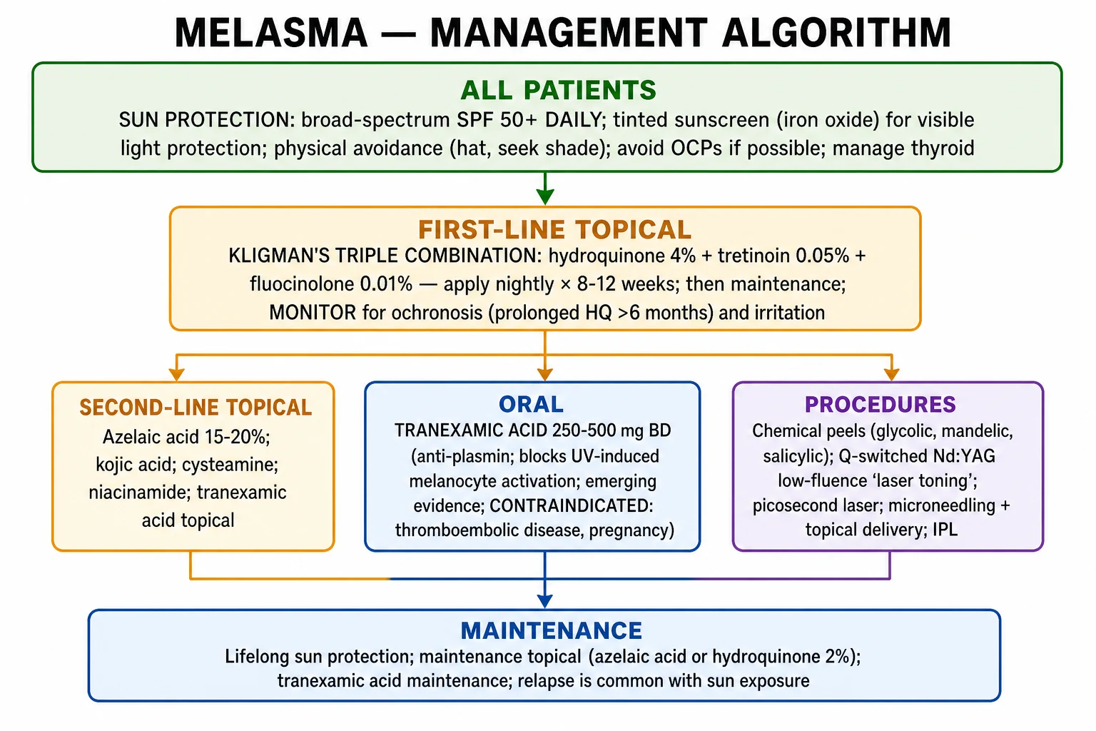
Once photoprotection is established, management follows a stepwise ladder. The choice at each step depends on disease depth, severity, patient preference, skin type, cost and availability.[1][8]
Stepwise management ladder
Photoprotection for all
Broad-spectrum SPF 50+ PA++++ sunscreen, tinted iron oxide if possible, hats, shade, trigger review.
First-line topical therapy
Kligman's triple combination (hydroquinone 2-4% + tretinoin + fluocinolone) nightly for 8-12 weeks, or azelaic acid if pregnancy or intolerance.
Second-line topical agents
Hydroquinone monotherapy, tretinoin, azelaic acid, kojic acid, arbutin, cysteamine or topical tranexamic acid.
Oral therapy
Oral tranexamic acid 250 mg two to three times daily for refractory disease, after excluding thromboembolic risk.
Procedures
Chemical peels, Q-switched or picosecond laser, fractional laser, microneedling — only in experienced hands, with caution in darker skin.
Maintenance
Lifelong photoprotection plus intermittent topical agents such as azelaic acid or low-dose hydroquinone.
The Kligman formula, or triple combination cream, is the most evidence-based first-line topical therapy. The standard preparation contains hydroquinone 2-4%, tretinoin 0.05% and fluocinolone acetonide 0.01%. Hydroquinone inhibits tyrosinase and reduces melanin synthesis. Tretinoin increases keratinocyte turnover, disperses melanin and improves penetration of hydroquinone. The mild corticosteroid reduces irritation and post-inflammatory hyperpigmentation from the other two agents. It is applied once nightly for 8 to 12 weeks and then stopped or tapered to maintenance.[1][6]
For patients who cannot tolerate the triple combination, or who are pregnant, azelaic acid 15-20% is a safe and effective alternative. It inhibits tyrosinase and is anti-proliferative. It has a favourable safety profile and is generally considered safe in pregnancy, unlike hydroquinone and tretinoin.[1][6]
Second-line topical agents
Hydroquinone monotherapy 2-4% is effective but should be used in short courses, typically up to 8-12 weeks, followed by a break. Continuous use beyond 6 months risks exogenous ochronosis, a paradoxical blue-black darkening caused by dermal deposition of homogentisic acid-like pigment. If this occurs, hydroquinone must be stopped immediately.[2][6]
Tretinoin 0.025-0.05% can be used alone or in combination. It is a category C agent in pregnancy and is avoided in pregnant or breastfeeding patients. Kojic acid is a tyrosinase inhibitor but can cause contact dermatitis. Arbutin is a naturally occurring hydroquinone derivative with a similar mechanism but lower potency. Cysteamine is a potent emerging tyrosinase inhibitor with a characteristic sulphurous odour. Niacinamide inhibits melanosome transfer. Topical tranexamic acid is an emerging option, often used in combination with other agents.[2][3]
Topical agents at a glance
| Agent | Typical concentration | Mechanism | Key considerations |
|---|---|---|---|
| Hydroquinone | 2-4% | Tyrosinase inhibitor | Gold-standard depigmenting agent; limit to 8-12 weeks, then break; risk of exogenous ochronosis with prolonged continuous use. |
| Tretinoin | 0.025-0.05% | Increases keratinocyte turnover | Improves penetration of hydroquinone; irritant; avoid in pregnancy. |
| Azelaic acid | 15-20% | Tyrosinase inhibition, anti-proliferative | Safe in pregnancy; useful for maintenance and as monotherapy. |
| Kojic acid | 1-4% | Tyrosinase inhibitor | Can cause contact dermatitis; often combined with hydroquinone. |
| Arbutin | 3-7% | Tyrosinase inhibition, hydroquinone derivative | Gentler than hydroquinone; slower onset. |
| Cysteamine | 5% | Potent tyrosinase inhibitor | Sulphurous odour; emerging first-line alternative in some guidelines. |
| Niacinamide | 2-5% | Inhibits melanosome transfer | Well tolerated; useful for maintenance and sensitive skin. |
| Topical tranexamic acid | 2-5% | Reduces plasmin-mediated melanocyte activation | Emerging; often combined with other agents. |
The choice of agent depends on the patient's skin type, depth of pigment, pregnancy status, tolerability and local availability. Combination therapy is generally more effective than monotherapy, but it also increases the risk of irritation. [1]
[6]Oral therapy
Oral tranexamic acid has become an important treatment for refractory melasma, particularly mixed and dermal types. The usual dose is 250 mg two to three times daily. It works by inhibiting plasmin, which reduces the release of arachidonic acid and thereby lowers prostaglandin-driven melanocyte activation. It also appears to have anti-angiogenic effects that may reduce the vascular component of melasma.[2]
Tranexamic acid is contraindicated in patients with a history of thromboembolic disease, active malignancy, severe renal impairment or pregnancy. A full medical history and review of risk factors are essential before prescribing. Treatment is usually continued for 8-12 weeks under specialist supervision. Side effects are usually gastrointestinal but serious thromboembolic events have been reported rarely.[2]
Procedures
Procedures are third-line and should be used cautiously, especially in darker skin types, because they can cause post-inflammatory hyperpigmentation and paradoxical worsening. Chemical peels with glycolic acid, salicylic acid, mandelic acid or Jessner's solution can accelerate epidermal turnover and remove superficial pigment. Glycolic acid 30-70% is commonly used, but darker skin types require lower concentrations and careful supervision. Mandelic acid is considered safer in darker skin because of its larger molecular size and slower penetration.[1][6]
Laser and light therapy includes Q-switched Nd:YAG low-fluence laser toning, picosecond lasers, fractional lasers and intense pulsed light. These target dermal melanin or remodel the skin. They should be performed by experienced operators, at conservative settings, with test patches, and always in the context of strict photoprotection. Multiple sessions are usually required, and relapse is common.[2]
Management of procedures must be individualised. Chemical peels are usually performed every 2-4 weeks, with a series of 4-6 sessions. Pre-treatment with a topical retinoid and hydroquinone for 2-4 weeks reduces the risk of post-inflammatory hyperpigmentation. Post-peel care involves strict photoprotection, bland emollients and avoidance of picking or scrubbing. Glycolic acid peels at 30-50% are suitable for epidermal and mixed melasma. Salicylic acid peels at 20-30% have additional anti-inflammatory effects. Jessner's peel combines resorcinol, salicylic acid and lactic acid and provides superficial exfoliation. Mandelic acid is a larger alpha-hydroxy acid with slower penetration, making it safer in darker skin. [1]
[6]Laser therapy should never be the first-line treatment. Low-fluence Q-switched Nd:YAG 1064 nm laser is the most commonly used laser toning modality. It targets dermal melanin with minimal epidermal damage when used at low fluences and large spot sizes. Picosecond lasers deliver shorter pulse durations and may produce less thermal damage and less risk of post-inflammatory hyperpigmentation. Fractional non-ablative lasers can improve texture and pigment but require careful parameter selection. Intense pulsed light is less specific and has a higher risk of adverse effects in darker skin; it is generally avoided in Fitzpatrick IV-VI. Vascular lasers such as pulsed dye or 532 nm KTP may help lesions with a prominent erythematous component. A test patch is mandatory, and settings should be conservative. [1]
[6]Microneedling creates trans-epidermal channels that enhance delivery of depigmenting agents. It can be combined with topical tranexamic acid or vitamin C. Radiofrequency microneedling delivers thermal energy to the dermis and may remodel the abnormal microenvironment, but evidence is still emerging and it carries a risk of post-inflammatory hyperpigmentation. All procedures are best reserved for patients who have failed optimised medical therapy and who can adhere to strict photoprotection. [4][8]
Regional availability of hydroquinone varies. In the United States and many Asian countries, hydroquinone 2-4% remains available by prescription, and some low-strength products are sold over the counter. In the European Union, Japan and several other jurisdictions, hydroquinone is banned in cosmetic products and is available only as a prescription drug for specific indications. These regulatory differences affect what can be prescribed in daily practice, but the principle remains the same: short courses, monitoring, and avoidance of continuous long-term use because of ochronosis risk.
Specific Subtypes & Scenarios
Epidermal melasma is the most responsive subtype. It usually improves with topical depigmenting agents and strict photoprotection alone. Patients can expect noticeable lightening within 8-12 weeks of triple-combination therapy. Maintenance with azelaic acid or intermittent low-dose hydroquinone is often needed.[6]
Dermal melasma is the most resistant because the pigment is sequestered in dermal melanophages beyond the reach of topical agents. These patients often require oral tranexamic acid, procedures or a combination. Expectations must be set realistically: complete clearance is uncommon, and the goal is usually substantial improvement rather than cure.[6]
Mixed melasma requires a combination approach. Treat the epidermal component with topicals and photoprotection, and consider oral tranexamic acid or carefully selected procedures for the dermal component. Many patients will have partial improvement and require long-term maintenance.[6]
Refractory or recurrent melasma should prompt reassessment of photoprotection, trigger control and diagnosis. Consider whether exogenous ochronosis, drug-induced pigmentation or an alternative diagnosis has been missed. If the diagnosis is secure, escalate to oral tranexamic acid or specialist-led procedures, and counsel the patient about the chronic nature of the disease.[2]
Pregnancy-associated melasma usually presents in the second or third trimester. It may fade spontaneously within months after delivery, but it often recurs. Management during pregnancy is limited to photoprotection and azelaic acid; hydroquinone and tretinoin are avoided. After delivery and breastfeeding, the full treatment ladder can be used if the pigmentation persists.[6]
Complications & Pitfalls
The most important complication of treatment is exogenous ochronosis. This is a paradoxical darkening caused by prolonged use of hydroquinone, typically beyond six months continuously. It presents as blue-black or grey-brown patches, often in the same sites as the original melasma. Histologically, it shows banana-shaped ochre deposits in the dermis. Management is to stop hydroquinone, use alternatives such as azelaic acid or cysteamine, and consider laser in selected cases. Prevention is key: limit hydroquinone to short courses.[2][6]
Other adverse effects of topical agents include irritant or allergic contact dermatitis from hydroquinone, tretinoin or kojic acid; skin atrophy from prolonged corticosteroid use in the triple combination; periorbital irritation; and telangiectasia with retinoids. These can all worsen post-inflammatory hyperpigmentation, particularly in darker skin. [1]
[6]Procedural pitfalls are common. Aggressive chemical peels, laser toning or IPL in darker skin can cause post-inflammatory hyperpigmentation, paradoxical darkening or even hypopigmentation and scarring. Patients should always be treated conservatively, with test patches, gradual escalation and strict photoprotection. Oral tranexamic acid must be avoided in patients with thromboembolic risk, and it should be prescribed only after a careful medical history.[1][4]
A common diagnostic pitfall is to treat every brown facial patch as melasma. Unilateral lesions, rapid progression, mucosal involvement, systemic symptoms or atypical colour should prompt reconsideration of the differential diagnosis and appropriate investigation. [1]
[6]Prognosis & Disposition
Melasma is a chronic, relapsing condition. Complete and permanent cure is uncommon. The disease behaves as a lifelong tendency to develop facial hyperpigmentation in response to sun, hormones and other triggers. This reality must be communicated clearly to patients at the first visit.[5][6]
Epidermal melasma has the best prognosis and can often be controlled with topicals and photoprotection. Mixed melasma has an intermediate prognosis. Dermal melasma is the most resistant and may require repeated courses of therapy or long-term oral tranexamic acid. Even after successful treatment, relapse is common with sun exposure, pregnancy or hormonal changes. [1]
[6]The psychosocial impact can be significant. Facial disfigurement affects self-esteem, social interaction and quality of life. Patients may become frustrated by the slow response and recurrent nature of the disease. Counselling, realistic goal-setting and a written maintenance plan improve adherence and satisfaction. Disposition is usually outpatient. Patients with atypical features or systemic symptoms should be referred for endocrine or internal medicine evaluation. [1]
[6]Counselling and adherence are as important as the choice of drug. At the first visit, explain that melasma is controllable but not curable, and that the aim is lightening and stability rather than complete elimination. Set a realistic timeline: most topical regimens take 8-12 weeks to show noticeable improvement. Emphasise that visible light, heat and hormonal triggers, not just sunburn, can reactivate pigment. Provide a written photoprotection plan and demonstrate the correct amount of sunscreen. Address cost and cosmetic concerns, because daily sunscreen adherence is often the weakest link. Schedule follow-up at 8-12 weeks to assess response, and if improvement is inadequate, escalate therapy or reconsider the diagnosis. When improvement plateaus, move to a maintenance regimen to reduce relapse. [6]
Special Populations
Pregnancy is the classic scenario for melasma. Management is limited to strict photoprotection and azelaic acid 15-20%. Hydroquinone and tretinoin are avoided because of potential teratogenicity and limited safety data. Most pregnancy-related melasma improves postpartum, but it may persist or recur with future pregnancies. Oral tranexamic acid is contraindicated in pregnancy. Patients should be reassured that many cases fade after delivery, and that more aggressive therapy can be deferred until after pregnancy and breastfeeding. [6]
Lactation has limited data. Topical azelaic acid is generally considered low risk because systemic absorption is minimal. Hydroquinone and tretinoin are usually avoided as a precaution. Oral tranexamic acid is not recommended during breastfeeding because of lack of safety data. Photoprotection remains the safest and most important measure. If a breastfeeding mother requires treatment, start with sunscreen and azelaic acid, and discuss the uncertain risk-benefit balance of any systemic agent. [6]
Men with melasma are less common but deserve the same systematic evaluation. They often have more sun exposure and may be more likely to have thyroid disease or occupational photosensitisation. Treatment response can be more variable, and the same photoprotection and topical ladder applies. Because male melasma is less hormonally driven, identifying and reducing UV exposure is particularly important. [5]
Darker skin types are at higher risk of both melasma and treatment-related post-inflammatory hyperpigmentation. Start with gentle agents, avoid aggressive procedures, and always use high-protection broad-spectrum sunscreen. Tinted sunscreens with iron oxide are particularly important because visible light plays a larger role in darker skin. Peels and lasers should be performed at lower settings with test patches, and hydroquinone courses should be monitored carefully. [2][6]
Elderly patients may have concurrent solar lentigines, actinic damage and skin fragility. Their melasma is often mandibular and may be more dermal. Treatment should be cautious to avoid irritation and further dyspigmentation. In older patients, the differential diagnosis with actinic bronzing, poikiloderma and drug-induced pigmentation should be considered, and a biopsy is sometimes required to confirm the diagnosis before long-term therapy. [1]
[6]Evidence, Guidelines & Regional Differences
The evidence base for melasma is built on numerous randomised controlled trials and systematic reviews. The landmark systematic review by Neagu and colleagues in 2022 demonstrated that the triple combination cream was superior to any single agent for moderate to severe melasma, with faster onset and greater efficacy, although it was associated with more local irritation. [1] The network meta-analysis also ranked non-hydroquinone combinations such as azelaic acid plus tretinoin as effective alternatives for patients who cannot tolerate hydroquinone.
A 2025 network meta-analysis of randomised controlled trials compared multiple topical, oral and procedural interventions and confirmed that triple-combination therapy and oral tranexamic acid are among the most effective options, while highlighting the need for larger trials with standardised outcome measures. [8] Outcomes in melasma trials are most commonly assessed with the Melasma Area and Severity Index (MASI), a clinician-rated tool that grades area, darkness and homogeneity. More recently, patient-reported outcomes, quality-of-life scores and objective colourimetry have been incorporated, but heterogeneity remains a limitation across studies. The 2024 review by Gan and Rodrigues summarised emerging agents including cysteamine, topical tranexamic acid, picosecond lasers and oral tranexamic acid, emphasising that none replace photoprotection. [2]
Key evidence-based messages for clinical practice are: photoprotection is the single most effective preventive and maintenance measure; combination topical therapy outperforms monotherapy; hydroquinone should be used in short courses to avoid ochronosis; oral tranexamic acid is effective for refractory disease but requires thromboembolic risk stratification; and procedural therapies carry a real risk of worsening pigmentation in darker skin and should be used conservatively. [1][6][8]
Regional differences are substantial. In the United States, hydroquinone remains widely available in prescription and, in some states, over-the-counter formulations. In the European Union and Japan, hydroquinone is restricted to prescription use for specific indications and is banned in cosmetic products. This means that European clinicians more commonly use azelaic acid, kojic acid, cysteamine and procedures as first-line therapy. The availability of laser and oral tranexamic acid also varies by country and by specialist setting.[6]
Guidelines generally agree on the following principles: photoprotection is foundational; triple-combination therapy is first-line where available; second-line agents include azelaic acid, tretinoin and other tyrosinase inhibitors; oral tranexamic acid is effective for refractory disease; and procedures should be used cautiously. There is no universal guideline that mandates one specific regimen, so practice is often tailored to local availability, cost and patient preference. [1]
[6]Prevention
Prevention of melasma centres on two strategies: photoprotection and trigger avoidance. Daily broad-spectrum SPF 50+ sunscreen, ideally tinted with iron oxide, should be used habitually, even on cloudy days and during indoor activities near windows. Patients should reapply every two hours when outdoors. Wide-brimmed hats, sunglasses and shade-seeking behaviour are essential adjuncts.[6]
Trigger avoidance includes reviewing hormonal medications. Women who develop melasma while taking oral contraceptives or hormone replacement therapy may consider switching to a non-hormonal alternative if medically appropriate. Photosensitising drugs and cosmetics should be minimised. Management of thyroid disease and general skin health also supports prevention. [1]
[6]Patients who have achieved clearance must continue maintenance photoprotection indefinitely. Intermittent use of a mild topical agent such as azelaic acid or low-strength hydroquinone in short courses may reduce relapse, but it is no substitute for sunscreen. [1]
[6]Public health messaging should emphasise that melasma is not simply a cosmetic concern. It disproportionately affects women of colour and can have significant psychological and occupational consequences. Accessible, culturally appropriate advice about sunscreen use, including tinted formulations that do not leave a white cast, improves adherence. Primary care and dermatology services should provide consistent messaging that photoprotection is the single most effective intervention for prevention and maintenance. [6]
Exam Pearls
[6]Red Flags
Exam application bank (NEET-PG / INICET)
One-line answer
Melasma is an acquired, bilateral, symmetrical hyperpigmentation of sun-exposed facial skin, classically affecting women with Fitzpatrick III-IV skin. It is driven by UV and visible light, hormonal triggers, genetic predisposition and local skin microenvironmental changes. Wood's lamp examination separates epidermal (accentuated, responds to topicals), dermal (not accentuated, resistant) and mixed types. Management is built on strict photoprotection, then topical depigmenting agents such as hydroquinone, tretinoin and azelaic acid, with oral tranexamic acid and carefully selected procedures for refractory disease. It is chronic and relapsing.
Worked stems (answer without another resource)
Stem 1 — Classic presentation. Map symptoms to mechanism; name the first investigation and first treatment step with dose/route if drug therapy is standard. [1]
Stem 2 — Unstable / complicated. List red flags that force immediate resuscitation, theatre, ICU, antidote, or reperfusion — and what you do in the first 15 minutes. [1]
Stem 3 — Atypical group. Elderly, pregnancy, child, or immunocompromised: how presentation and thresholds change. [1]
Stem 4 — Differential trap. Name the three closest mimics and one discriminator for each. [1]
Stem 5 — Disposition. Who goes home with safety-netting, who is admitted, who needs HDU/ICU/theatre, and what follow-up is mandatory. [1]
Rapid viva checklist
- Definition + classification
- Pathophysiology chain
- Bedside signs / criteria
- Score with exact components (if any)
- Emergency bundle
- Definitive therapy with doses
- Complications of disease and of treatment
- Special populations
- Guideline/trial name if classic
- Three exam traps
Coverage self-check
If you cannot answer any stem above from this page alone, re-read the matching section — the page is intended to be self-sufficient for final-prof and NEET-PG/INICET questions on Melasma.
[1] [6]References
- [1]Neagu N, Conforti C, Agozzino M, et al. Melasma treatment: a systematic review J Dermatolog Treat, 2022.PMID 33849384
- [2]Gan C, Rodrigues M. An Update on New and Existing Treatments for the Management of Melasma Am J Clin Dermatol, 2024.PMID 38896402
- [3]Thawabteh AM, Jibreen A, Karaman D, et al. Skin Pigmentation Types, Causes and Treatment-A Review Molecules, 2023.PMID 37375394
- [4]Tan MG, Jo CE, Chapas A, et al. Radiofrequency Microneedling: A Comprehensive and Critical Review Dermatol Surg, 2021.PMID 33577211
- [5]Ali L, Al Niaimi F. Pathogenesis of Melasma Explained Int J Dermatol, 2025.PMID 40022484
- [6]McKesey J, Tovar-Garza A, Pandya AG. Melasma Treatment: An Evidence-Based Review Am J Clin Dermatol, 2020.PMID 31802394
- [7]Gu D, Yao J, Cai Y, et al. What lies behind melasma: a review of the related skin microenvironment Int J Dermatol, 2025.PMID 39212112
- [8]Leung JH, Vashi NA, Patzelt NM. Efficacy and Safety of Different Treatments for Melasma: Network Meta-Analysis of Updated Data Diseases, 2025.PMID 41149049