Dermatology · Medicine
Meningococcaemia
Also known as Meningococcaemia · Meningococcal disease · Meningococcal septicaemia · Purpura fulminans
Meningococcaemia = Neisseria meningitidis septicaemia presenting with petechial/purpuric non-blanching rash + fever ± meningitis. Rapidly progressive; mortality 10%. Glass test (rash does NOT fade under pressure). Purpura fulminans (DIC → skin necrosis → gangrene → amputation). EMERGENCY: IV/IM benzylpenicillin 2.4 g pre-hospital OR IV ceftriaxone 2 g immediately on admission (before tests or transfer). May present as meningitis (neck stiffness, photophobia, altered consciousness) or septicaemia (shock, multi-organ failure) or both. Prophylaxis: rifampicin/ciprofloxacin/ceftriaxone for close contacts. Vaccination: MenACWY and MenB.
On this page & tools
Your progress
Saved locally on this device.
Exam tags
Red flags
Overview & Definition
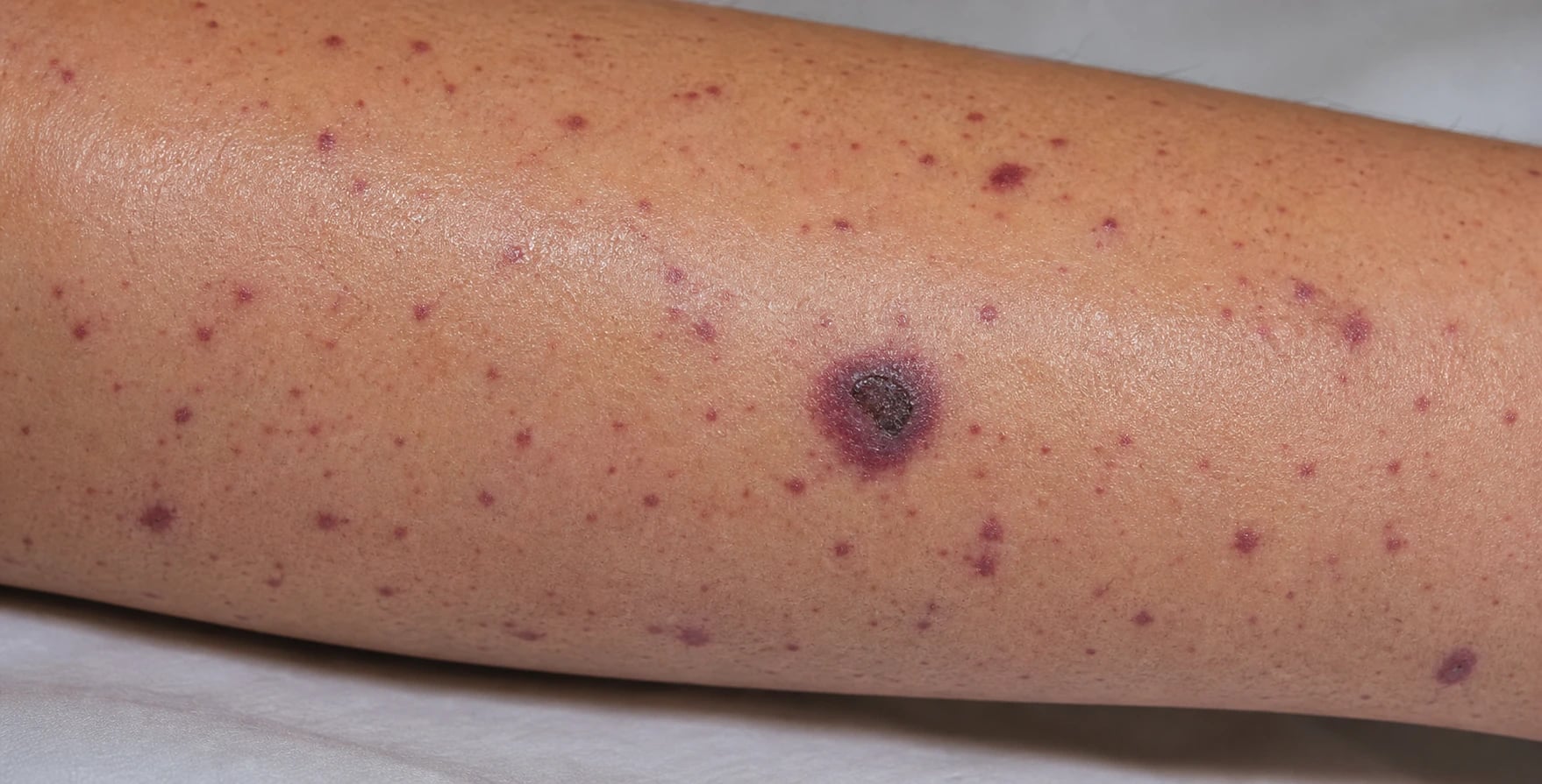
Meningococcal disease is the spectrum of invasive illness caused by Neisseria meningitidis, a gram-negative, oxidase-positive diplococcus that is carried asymptomatically in the human nasopharynx and transmitted through respiratory droplets and close (kissing) contact.[2][6] Although the organism can cause isolated arthritis, pneumonia, pericarditis or conjunctivitis, the two dominant — and overlapping — phenotypes are meningococcal meningitis (meningeal inflammation, often without a rash) and meningococcal septicaemia (systemic bacteraemia with the characteristic rash ± septic shock), the latter conventionally called meningococcaemia. Roughly half of patients show both together.[5]
The dermatologist's importance in this disease is singular: the non-blanching petechial/purpuric rash is the earliest reliable clue that a febrile, unwell patient has meningococcaemia rather than a viral illness, and its recognition triggers an antibiotic that must be given within the hour. The most feared dermatological phenotype is purpura fulminans, a disseminated intravascular coagulation (DIC)-driven cutaneous microvascular thrombosis that produces skin necrosis, limb gangrene and amputation.[3][7] A rare, indolent form — chronic meningococcaemia — runs over weeks with low-grade fever, arthralgia and an evanescent rash, and is easily missed.[2]
[1]Classification
Meningococcal disease is classified in two intersecting axes — clinical syndrome and capsular serogroup — because both determine management and prevention. [1]
Clinically, disease is divided into meningitis alone (best prognosis), septicaemia with or without purpura fulminans (worst prognosis), and the mixed picture (~50%). A small proportion present as chronic meningococcaemia, and occasional patients have focal disease (arthritis, pericarditis, pneumonia, conjunctivitis, urethritis).[2][5]
Twelve capsular serogroups are recognised; six are clinically important (A, B, C, W135, X, Y). The capsule is the key virulence factor and the principal vaccine antigen, so serogroup matters for epidemiology, outbreaks and vaccine choice.[4]
Serogroup B
- #1 cause in developed/temperate countries
- Polysialic acid capsule mimics human neural tissue → poorly immunogenic
- Needs protein-based vaccines (Bexsero/4CMenB, Trumenba)
- Dominant in UK, Europe, Americas, Australasia
Serogroup A
- Historically the epidemic strain of the sub-Saharan 'meningitis belt'
- Conjugate vaccine (MenAfriVac) has dramatically reduced belt disease
- Hajj-associated outbreaks pre-vaccination
Serogroup C
- #1 in UK before MenC conjugate vaccine introduction (1999)
- Near-eliminated in vaccinated populations via herd immunity
- Still circulates in unvaccinated groups
Serogroup W135
- Rose after the 2000 Hajj outbreak
- Higher case-fatality and atypical presentations (joint, GI)
- Covered by MenACWY conjugate vaccine
Serogroup Y
- Increasing in the Americas and parts of Europe
- More common in older adults
- Covered by MenACWY conjugate vaccine
Serogroup X
- Outbreaks reported in the African meningitis belt
- No widely available conjugate vaccine yet
- Surveillance important
Epidemiology & Risk Factors
Meningococcal disease is global but geographically patterned. The highest endemic burden sits in the sub-Saharan African meningitis belt — a swath from Senegal to Ethiopia — where dry-season epidemics, historically driven by serogroup A and increasingly by W135, C and X, can reach attack rates of 1000 per 100,000.[4] Outside the belt, incidence in high-income countries is low (around 0.5–1 per 100,000 annually) but punctuated by outbreaks in semi-closed communities — university dormitories, military barracks, the Hajj pilgrimage, and refugee camps.[2][4]
Age distribution is bimodal: a large peak in children under 5 years (peak under 1) and a second peak in teenagers and young adults aged 15–19, who have the highest nasopharyngeal carriage rates (up to 30–50% in some cohorts). Carriage in the general population is ~10%.[2]
The decisive host risk factors are defects in meningococcal clearance, particularly the terminal complement pathway. Deficiency of C5, C6, C7, C8 or C9 — which together form the membrane attack complex essential for lysing N. meningitidis — raises the risk of invasive disease 7,000- to 10,000-fold and predisposes to recurrent episodes.[11] Properdin deficiency (X-linked) and factor D deficiency cause particularly fulminant disease. The monoclonal anti-C5 antibodies eculizumab and ravulizumab (used for paroxysmal nocturnal haemoglobinuria, atypical HUS and some refractory myasthenia/ANCA-vasculitis indications) pharmacologically reproduce this defect and carry a labelled warning for meningococcal infection.[11] Functional or anatomical asplenia (sickle-cell disease, post-splenectomy, congenital asplenia), HIV infection, alcohol misuse, smoking, passive smoke exposure in children, concurrent viral upper respiratory infection, overcrowding, and mass gatherings all increase risk.[2][4]
Pathophysiology
Understanding the cascade explains the rash, the shock, and why antibiotics must be given before any test.[3][6]
1. Colonisation. N. meningitidis colonises the nasopharyngeal mucosa using type IV pili and the Opa/Opc adhesins, which bind epithelial CD66/CEA receptors. Most carriers are asymptomatic; invasion is the exception, favoured by viral co-infection (mucosal damage), smoking, low mucosal IgA, and host susceptibility.[6]
2. Invasion and bacteraemia. The organism traverses the mucosal barrier (aided by its IgA1 protease, which cleaves secretory IgA) and enters the bloodstream. The antiphagocytic capsular polysaccharide and factor-H-binding protein (fHbp) (which recruits complement factor H to down-regulate opsonisation) allow it to evade innate immunity.[6]
3. Endotoxin-driven cytokine storm. N. meningitidis sheds outer-membrane lipooligosaccharide (LOS, the meningococcal endotoxin) as membrane "blebs". LOS binds the TLR4/MD-2/CD14 complex on monocytes, macrophages and endothelial cells, triggering release of TNF-α, IL-1, IL-6 and IL-8. The resulting cytokine storm drives the septic phenotype: fever, endothelial leak, vasodilatation, myocardial depression and shock.[3]
4. Disseminated intravascular coagulation and purpura fulminans. LOS and cytokine-activated endothelium express tissue factor, generating thrombin; simultaneously protein C, protein S and antithrombin are consumed and the protein C pathway is dysregulated. The net effect is widespread microvascular thrombosis with consumption of platelets and clotting factors — DIC. In the skin, dermal capillary and venule thrombosis produces haemorrhagic infarction: the lesions progress from petechiae to purpura to ecchymoses to necrotic black eschars (purpura fulminans), and in the limbs to gangrene requiring amputation.[3][7]
5. Meningeal invasion. Bacteraemic organisms cross the blood–brain barrier at the choroid plexus, seeding the subarachnoid space where complement and immunoglobulin are scarce and phagocytosis is inefficient. Meningeal inflammation produces neck stiffness, raised intracranial pressure and the CSF findings of bacterial meningitis.[5]
6. Adrenal haemorrhage (Waterhouse-Friderichsen). DIC within the richly vascular adrenal cortex causes bilateral adrenal haemorrhage, acute adrenal insufficiency and refractory vasodilatory shock — the classic Waterhouse-Friderichsen syndrome of fulminant meningococcaemia.[1]
Clinical Presentation
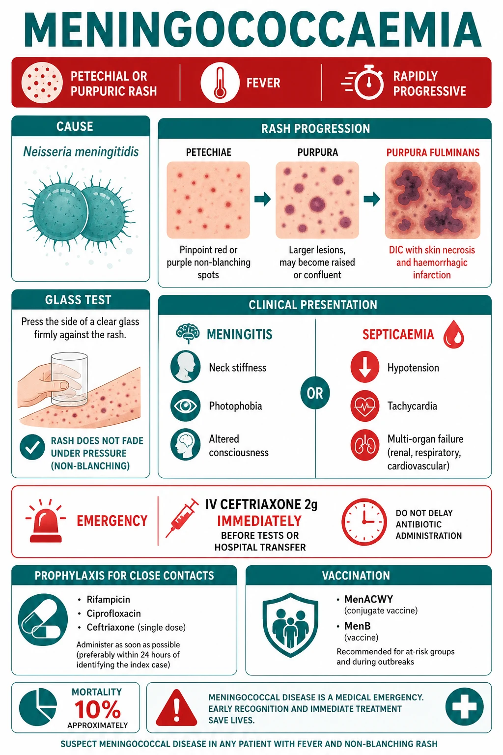
The disease evolves over hours, and the earliest phase is notoriously non-specific, masquerading as a viral illness — which is why a high index of suspicion and a deliberate search for the rash is essential.[1][2]
Fever, malaise, sore throat, myalgia, nausea, headache — indistinguishable from influenza or a viral upper respiratory infection. A blanching maculopapular rash is present early in up to a third of cases and may mislead.
Petechiae appear (1–2 mm red-purple macules), typically first on the trunk and lower limbs, spreading rapidly and becoming confluent. Crucially they are non-blanching (positive glass test).
Lesions enlarge to purpura and ecchymoses; in fulminant cases areas of skin become dusky, then necrotic black eschars — purpura fulminans. Limb ischaemia, mottling and cold peripheries signal DIC.
Tachycardia, hypotension unresponsive to fluids, oliguria, confusion, respiratory distress, ARDS. Meningeal signs may coexist (mixed picture).
The rash — dermatological hallmark
The rash is the single most important physical sign. It begins as petechiae (pinpoint non-blanching macules), evolves into purpura (larger confluent areas) and, in severe cases, ecchymoses with central necrosis. Distribution favours the trunk and lower limbs but any site including mucous membranes and conjunctivae can be involved. The glass test — pressing a clear glass or tumbler firmly against the lesion and watching for blanching — is positive: meningococcal lesions do not fade because they represent extravasated blood and microvascular thrombosis rather than inflammatory vasodilatation.[1][10]
[1]Meningococcal meningitis (~50%)
When meningeal seeding dominates, the patient has the classic triad of fever, neck stiffness and altered mental status (all three in roughly half; at least two in nearly all). Headache is severe and diffuse; photophobia and Kernig and Brudzinski signs are present. The rash may be absent, sparse or prominent. Seizures, focal neurology and decreasing conscious level indicate raised intracranial pressure or complications (cerebral oedema, venous sinus thrombosis, empyema).[5]
Meningococcal septicaemia / meningococcaemia (~30–40%)
The septicaemic phenotype is the most dangerous. High fever, cold peripheries with mottled or ashen skin, tachycardia, tachypnoea, hypotension and oliguria reflect septic shock and evolving multi-organ failure. Purpura fulminans is DIC made visible: large areas of purpuric skin become necrotic, digits and limbs become ischaemic, and amputation may become unavoidable. Waterhouse-Friderichsen syndrome — bilateral adrenal haemorrhage with refractory shock — may supervene.[1][3][7]
Chronic meningococcaemia (rare)
A distinct, indolent phenotype lasting weeks: intermittent low-grade fever, arthralgia/arthritis, myalgia, and an evanescent maculopapular or petechial rash that waxes and wanes. Blood cultures or PCR are positive but the illness mimics gonococcaemia, Henoch-Schönlein purpura or a viral exanthem. Untreated it can seed the meninges or progress to fulminant septicaemia, so it is treated as invasive disease.[2]
Differential Diagnosis
The differential of fever plus a petechial/purpuric or vasculitic rash is broad, and a febrile patient with a non-blanching rash is managed empirically as meningococcaemia until proven otherwise while the other possibilities are considered in parallel.[1][10]
Meningococcaemia
- Rapidly progressive petechiae→purpura
- Shock, DIC, multi-organ failure over hours
- Gram-negative diplococci on Gram stain/biopsy
- PCR positive for N. meningitidis
Other viral exanthems (enterovirus, measles, dengue, viral haemorrhagic fevers)
- Petechiae in dengue/VHF with thrombocytopenia
- Slower tempo (days), prodromal features
- Travel/exposure history
- Normal CSF; serology/PCR diagnostic
Henoch-Schönlein purpura (IgA vasculitis)
- Palpable purpura on lower limbs/buttocks
- Abdominal pain, arthritis, haematuria
- Children, subacute course over days–weeks
- Normal platelets and coagulation; no shock
Thrombocytopenia (ITP, leukaemia, TTP/HUS, aplastic)
- Isolated thrombocytopenia on FBC
- No fever/sepsis in ITP; blast cells in leukaemia
- TTP: microangiopathic anaemia, neuro, renal
- Blood film and marrow diagnostic
Other sepsis with DIC (pneumococcal, staphylococcal, post-varicella)
- Purpura fulminans after chickenpox (Staph)
- Pneumococcal bacteraemia in asplenia
- Different organism on culture/PCR
- Source (pneumonia, skin) on examination/imaging
Other vasculitides (PAN, urticarial/cryoglobulinaemic)
- Subacute, livedo, nodules, ulcers
- Associated autoimmune disease, hepatitis B/C
- Biopsy: vessel-wall inflammation not DIC
- Complement/ANCA/criooglobulins guide
The bedside discriminator is tempo and systemic toxicity: meningococcaemia evolves over hours with mounting shock and DIC; the vasculitides and thrombocytopenias run over days to weeks. A non-blanching rash in a febrile, unwell patient is nevertheless treated as meningococcaemia while the work-up proceeds.[1]
Clinical & Bedside Assessment
A focused, rapid assessment runs in parallel with the first antibiotic, never in series with it.[1][5]
Rash. Examine the whole skin in good light, including conjunctivae, palate, ears, soles and dependent areas. Characterise each element: petechia (under 3 mm), purpura (over 3 mm), ecchymosis (over 1 cm), necrotic/eschar. Perform the glass test. Look for areas of dusky, map-like purpura with grey-black centres signalling purpura fulminans, and for mottling or cold, ischaemic digits indicating limb-threatening DIC. [1]
Meningeal signs. Kernig sign — with the patient supine and hip flexed at 90°, the knee cannot be passively extended without pain or resistance. Brudzinski sign — passive flexion of the neck produces involuntary flexion of the hips and knees. Jolt accentuation — the baseline headache worsens on rapid horizontal rotation of the head two to three times per second — has high sensitivity for meningeal irritation. Head-holding, tripoding and photophobia are supportive.[5]
Septic screen. Vital signs (temperature, heart rate, respiratory rate, blood pressure, capillary refill, oxygen saturation), conscious level (GCS), and signs of shock (mottled or cold skin, prolonged capillary refill, weak pulses, oliguria, hypotension — a late sign in children). Identify any source (otitis, sinusitis, pneumonia) and look for stigmata of risk (splenectomy scar, Hickman line, known complement deficiency, eculizumab).[1]
Skin biopsy / lesion scrape. A Gram stain of a petechial scraping or a punch biopsy of an early purpural lesion can show gram-negative diplococci within neutrophils — a rapid bedside confirmation that does not delay treatment.[1]
Histopathology
The histopathology of the skin lesion mirrors the underlying vascular catastrophe and is itself diagnostic when culture is negative or the patient is pre-treated.[3][7]
Early petechial lesion. A punch biopsy shows dermal capillaries and post-capillary venules occluded by fibrin thrombi, with extravasated red cells and a neutrophil-predominant infiltrate around and within vessel walls — a pattern variously described as thrombotic and leucocytoclastic. Unlike classical small-vessel vasculitis, the dominant event is intravascular thrombosis rather than immune-complex vessel-wall necrosis, reflecting the DIC substrate. Gram stain may reveal gram-negative diplococci within endothelial cells and neutrophils.[3]
Purpura fulminans. The fully evolved lesion shows full-thickness dermal and often subcutaneous infarction with diffuse microvascular thrombosis, haemorrhage, and a Confluent necrotic epidermis that detaches as a black eschar. A sharp border often separates necrotic from viable skin. Adnexal structures, subcutaneous fat and even muscle may be infarcted, explaining the depth of tissue loss that necessitates debridement and grafting.[7]
Adrenals (Waterhouse-Friderichsen). At post mortem the adrenal glands are enlarged, boggy and uniformly haemorrhagic, with cortical-cell necrosis and preserved medulla — the anatomical basis for acute mineralocorticoid and glucocorticoid failure layered on top of septic shock.[1]
Why histology matters for the dermatologist. A biopsy of an atypical lesion can settle the differential between meningococcal purpura, post-infectious (IgA) vasculitis and thrombotic microangiopathy: thrombosis without immune-complex deposition favours infection-associated purpura fulminans, while IgA deposition on immunofluorescence favours Henoch-Schönlein purpura. Gram stain and PCR on biopsy tissue confirm N. meningitidis even after antibiotics have sterilised the blood.[3][7]
Investigations
Investigations confirm the diagnosis but never delay the first antibiotic. The hierarchy is: treat, then culture, then confirm by PCR.[5]
Blood culture
- Draw BEFORE antibiotics if possible, but never delay antibiotics for it
- Gold standard for isolation and susceptibility
- Positive in a majority of septicaemic cases
- Negative if pre-treated
PCR (blood + CSF)
- Detects N. meningitidis DNA (ctrA, sodC targets)
- Remains positive after antibiotics started
- High sensitivity and specificity
- Also serogroups the strain for public health
Lumbar puncture / CSF
- Gram-negative diplococci, neutrophil pleocytosis, high protein, low glucose
- DEFER if raised ICP, coagulopathy, or septic shock
- PCR on CSF if antibiotic-pretreated
- Opening pressure raised
Rapid antigen / bedside tests
- Latex agglutination (less used)
- Bedside/molecular PCR platforms
- Useful when LP deferred or culture negative
- Not a substitute for blood culture + PCR
Supportive bloods
- FBC (leucocytosis or leucopenia, thrombocytopenia)
- Coagulation (PT, APTT, fibrinogen, D-dimer — DIC)
- U&E, LFTs, CRP, lactate (severity), glucose, blood gas
Other
- Throat swab for carriage
- Skin biopsy Gram stain/culture of lesion
- CT head before LP if focal neurology / raised ICP
- CXR if respiratory source suspected
CSF in meningococcal meningitis
The CSF pattern is that of pyogenic bacterial meningitis: raised opening pressure; cloudy fluid; neutrophilic pleocytosis (typically 100–5,000 cells/µL); raised protein (over 1 g/L or 100 mg/dL); low glucose (under 2.2 mmol/L or under 40% of simultaneous blood glucose); and gram-negative diplococci inside neutrophils on Gram stain. Culture grows N. meningitidis; PCR is positive even after antibiotics.[5]
When to defer the lumbar puncture
LP is deferred — and empirical antibiotics plus a CT head given instead — in the presence of signs of raised intracranial pressure (decreasing GCS, focal neurology, seizures, papilloedema, Cushing's triad), coagulopathy (platelet count under 100 × 10⁹/L, INR over 1.4, or known DIC), or septic shock in which positioning and sedation are unsafe. Antibiotics are never withheld to obtain CSF; PCR later confirms the organism.[5]
Management — Resuscitation
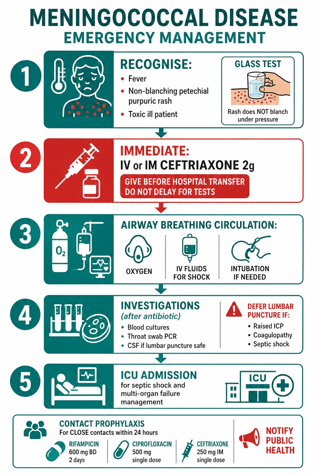
Meningococcal disease is a time-critical emergency. The resuscitation bundle runs in parallel: recognise, give antibiotics immediately, secure ABC, investigate, escalate.[1][5]
Step 1 — Recognise
Fever with a non-blanching petechial/purpuric rash in an unwell patient is meningococcaemia until proven otherwise. The glass test confirms non-blanching. Do not wait for a classical picture — early blanching maculopapular rashes and even absent rash still warrant treatment if suspicion is high.[1]
Step 2 — Immediate antibiotic (before any test or transfer)
- Pre-hospital (community): IV or IM benzylpenicillin 2.4 g (adult); 1.2 g if under 12 years; in infants give 300 mg/kg. Give at once when meningococcal disease is suspected, before hospital transfer.[1]
- Hospital (first hour): IV ceftriaxone 2 g (adult) or cefotaxime 2 g immediately on admission; do not wait for blood cultures, LP, or imaging. Ceftriaxone covers penicillin-resistant strains and achieves reliable CSF levels.[5]
- Why speed: mortality rises by ~7% for each hour of antibiotic delay, the central justification for pre-hospital penicillin policies.[5]
Step 3 — ABC and supportive care
- Airway/Breathing: high-flow oxygen; intubate and ventilate if comatose (GCS under 8), rapidly deteriorating, or in respiratory failure (lung-protective ventilation).
- Circulation: establish IV/IO access; for septic shock give crystalloid boluses — 20 mL/kg in children, 500 mL aliquots in adults — repeated to restore perfusion, with frequent reassessment. Begin noradrenaline early for fluid-refractory shock, targeting MAP at least 65 mmHg; adrenaline if myocardial depression predominates. Correct hypoglycaemia, hypocalcaemia and acidosis.[1]
- Raised ICP: head-up 30°, normoxia, normocapnia, normoglycaemia; mannitol or hypertonic saline if herniating.
- Isolation: droplet precautions for the first 24 h of antibiotic therapy.[5]
Fluid resuscitation in detail. Use balanced crystalloid (Hartmann's or Plasma-Lyte) over 0.9% saline to avoid hyperchloraemic acidosis; give aliquots of 500 mL in adults and 10–20 mL/kg in children, reassessing perfusion, liver size, breath sounds and capillary refill after each bolus. In children, avoid the liberal bolus strategy disproved by the FEAST trial in resource-limited settings — give cautiously and watch for pulmonary oedema, especially where intensive-care monitoring is limited. Aim for the Surviving Sepsis targets: MAP at least 65 mmHg, lactate normalising, urine output over 0.5 mL/kg/h, and normal mental state. Add human albumin for refractory shock or large-volume resuscitation. Start noradrenaline first-line for vasodilatory shock; add vasopressin or switch to adrenaline if myocardial depression predominates (cold shock). Invasive arterial and central venous pressure monitoring guides escalation. If purpura fulminans with a coagulopathy is present, transfuse FFP, cryoprecipitate and platelets to keep INR and APTT near normal, fibrinogen over 1.5 g/L and platelets over 50, before any procedure or LP.[3][7]
Step 4 — Investigations (after the first antibiotic)
Blood cultures (drawn before antibiotics if achievable without delay), blood and CSF PCR for N. meningitidis, FBC, coagulation/DIC screen, U&E, LFTs, CRP, lactate, glucose and blood gas. LP is performed only if safe; otherwise defer and rely on PCR.[5]
Step 5 — Escalate and protect contacts
Admit to ICU for shock, coma, purpura fulminans or respiratory failure. Begin contact prophylaxis within 24 h for household, kissing and secretion-exposed contacts, and notify public health.[2]
[1]Management — Definitive & Stepwise
Directed antibiotic therapy
Once N. meningitidis is confirmed, directed therapy runs for 7 days.[5]
- Ceftriaxone 2 g IV twice daily (or cefotaxime 2 g IV q6h) — drug of choice; excellent CSF penetration, single or twice-daily dosing, safe in penicillin allergy.[5]
- Penicillin allergy: chloramphenicol 25 mg/kg IV q6h for 7 days (monitor for marrow suppression; avoid in late pregnancy and neonates); where feasible, penicillin desensitisation under specialist cover is reasonable because cephalosporin cross-reactivity is low with modern agents.[8]
- Adjunctive dexamethasone 0.15 mg/kg (10 mg adult) IV q6h for 4 days, given with or just before the first antibiotic dose, is of greatest benefit in pneumococcal meningitis; its role in isolated meningococcal disease is limited, but it is commonly administered empirically until the organism is identified.[5]
Ceftriaxone (drug of choice)
- 2 g IV BD (adult); 80–100 mg/kg/day (child)
- 7 days
- High CSF penetration; covers resistant strains
- Avoid in neonatal jaundice (biliary sludging)
Cefotaxime (alternative)
- 2 g IV q6h
- Equivalent to ceftriaxone
- Preferred in neonates and pregnancy
- Same 7-day course
Chloramphenicol (penicillin-allergic)
- 25 mg/kg IV q6h
- Monitor blood count (marrow suppression)
- Avoid in late pregnancy and neonates
- Good CSF penetration
Benzylpenicillin (once susceptible)
- 2.4 g IV q4h (adult); 50 mg/kg (child)
- Only when isolate penicillin-susceptible
- Rising intermediate resistance globally
- Cheap; historical first-line
Purpura fulminans — DIC and skin/limb salvage
Purpura fulminans is managed in ICU with a haematology and surgical team:[3][7]
- DIC support: fresh-frozen plasma, cryoprecipitate (to keep fibrinogen above 1.5 g/L), and platelet transfusion to keep platelets above 50 × 10⁹/L in active bleeding or before procedures. Heparin and protein C concentrate are used in selected centres; activated protein C (drotrecogin alfa) has been withdrawn and is not recommended. Treat the underlying infection decisively.[7]
- Perfusion: vasopressors to maintain limb perfusion, normoglycaemia and normothermia; avoid over-constriction where it worsens digital ischaemia.
- Surgical: urgent fasciotomy for compartment syndrome, debridement of necrotic skin, and amputation for irreversible limb or digit ischaemia — often staged once demarcation is clear. Skin grafting and reconstructive surgery follow in rehabilitation.[7]
- Adrenal support: hydrocortisone 200 mg/24 h IV (stress-dose) if Waterhouse-Friderichsen with refractory shock; fluids and vasopressors continue.[1]
Chemoprophylaxis of close contacts
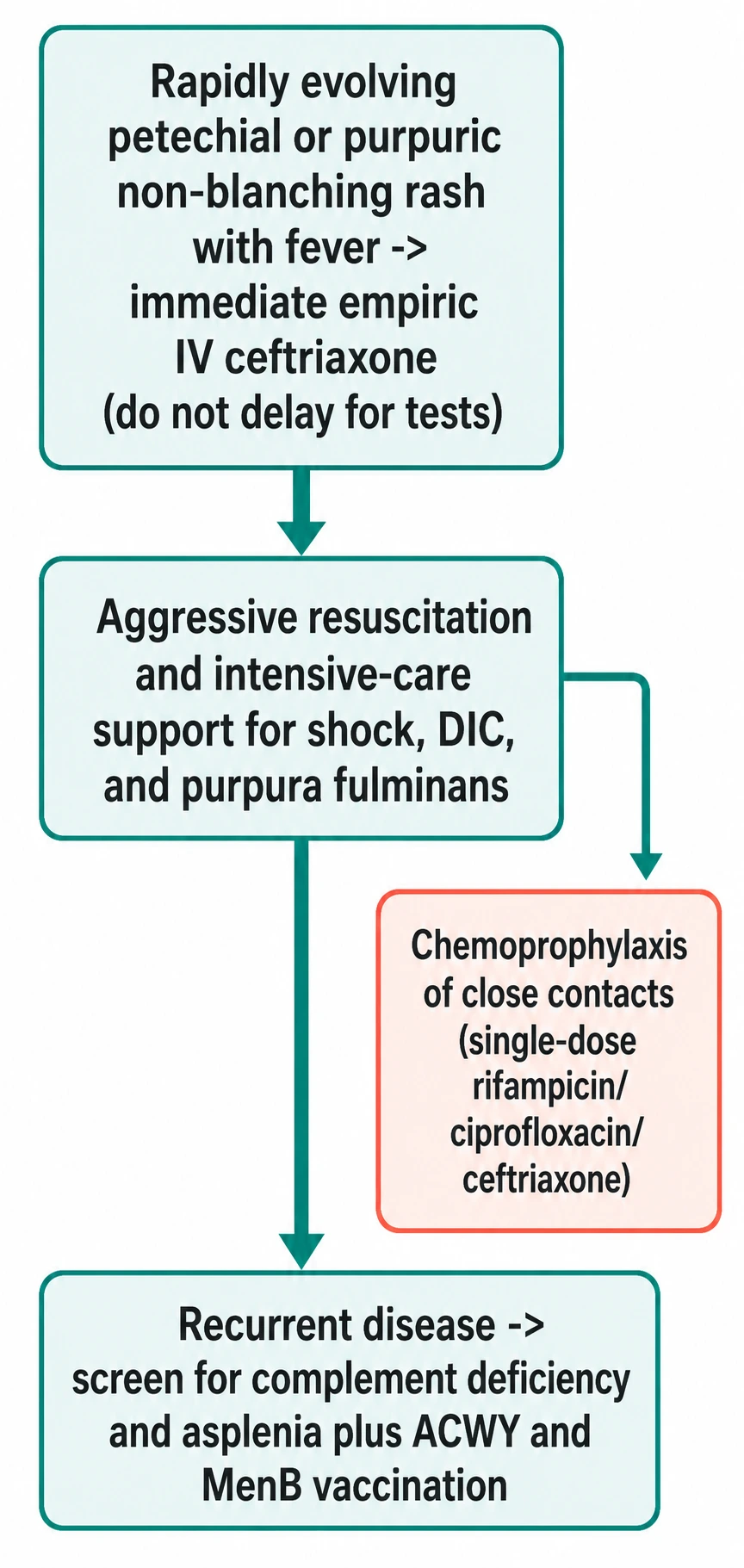
Carriage eradication protects contacts, who are at up to 1,000-fold higher risk than the baseline population in the days after exposure. Give prophylaxis within 24 h (effective up to 14 days) to household contacts, kissing contacts, and healthcare workers exposed to respiratory secretions (e.g., intubation without a face mask, mouth-to-mouth resuscitation).[2][5]
Rifampicin
- 600 mg PO BD for 2 days (adult)
- Child: 10 mg/kg BD; neonate 5 mg/kg BD
- Enzyme inducer — reduces OCP/anticoagulant levels
- Orange-red secretions/urine; contact lenses stained
Ciprofloxacin (adult first-line)
- 500 mg PO single dose
- Preferred in adults and mass prophylaxis
- Avoid in pregnancy and children where alternatives exist
- Single dose improves adherence
Ceftriaxone (pregnancy / first-line in some guidelines)
- 250 mg IM single dose (adult); 125 mg (child)
- Safe in pregnancy and children
- Single dose; no enzyme interaction
- Preferred when oral agents unsuitable
The index case also receives chemoprophylaxis before discharge if treated with an agent that does not eradicate carriage (e.g., benzylpenicillin) — ceftriaxone itself eradicates carriage.[2]
Vaccination
Vaccination is the cornerstone of prevention and has reshaped serogroup epidemiology.[4][9]
- MenACWY (quadrivalent conjugate vaccine) — against serogroups A, C, W135 and Y; conjugation to a carrier protein produces T-cell-dependent memory and reduces carriage, generating herd immunity. The UK MenC programme (from 1999) and the shift to MenACWY (from 2015, in response to rising W135) drove dramatic declines in those serogroups.[4]
- MenB (4CMenB, Bexsero; or MenB-FHbp, Trumenba) — the serogroup B polysialic-acid capsule is poorly immunogenic (it mimics human neural cell-adhesion molecules), so MenB vaccines use subcapsular proteins (factor-H-binding protein, NadA, NHBA, PorA). 4CMenB entered universal infant schedules in 2015 and is effective against the dominant serogroup in developed countries.[9]
- MenA (MenAfriVac and conjugate equivalents) — mass campaigns across the African meningitis belt using MenA conjugate vaccine have nearly eliminated serogroup A epidemics there.[4]
- Serogroup X has no widely licensed conjugate vaccine and remains a surveillance priority in Africa.
ANZ (ATAGI / New Zealand): MenB (Bexsero) and MenACWY (Nimenrix) funded under the National Immunisation Program for infants and adolescents; indigenous and high-risk groups prioritised.
Sub-Saharan Africa / India / ICMR: MenA conjugate (MenAfriVac) in the meningitis belt; MenACWY required for Hajj pilgrims; MenB availability and routine infant use limited by cost. Outbreak response combines vaccination and chemoprophylaxis.
Public health and outbreak control
Every case of confirmed or suspected meningococcal disease is notifiable to public health. The unit of prevention is the close contact: household members, kissing contacts, and healthcare workers exposed to respiratory secretions during airway manoeuvres. All receive chemoprophylaxis within 24 h of the index case's onset. Clustering — two or more linked cases in a school, barracks or dormitory within a short window — triggers enhanced surveillance, wider prophylaxis of the defined at-risk group, and (depending on the serogroup) vaccination: MenACWY for A/C/W/Y clusters, MenB for serogroup B clusters. Travellers to the Hajj and to the sub-Saharan meningitis belt require a recent MenACWY certificate. Long-term, the epidemiologist's tools are conjugate vaccination of adolescents (to interrupt carriage), infant MenB programmes, and laboratory surveillance that serogroups every isolate so that vaccine-escape strains are detected early.[4][2]
Specific Subtypes & Scenarios
Purpura fulminans
The most catastrophic phenotype: DIC-driven cutaneous and visceral microvascular thrombosis with rapidly spreading purpuric skin necrosis, limb ischaemia, and multi-organ failure. Mortality is 30–50%; survivors frequently need amputation and skin grafting and bear major scarring. Management combines aggressive antibiotic therapy, DIC support (FFP, cryoprecipitate, platelets), ICU resuscitation, and early surgical involvement.[3][7]
Waterhouse-Friderichsen syndrome
Bilateral adrenal haemorrhage in fulminant meningococcaemia produces acute adrenal insufficiency layered on septic shock — refractory hypotension, abdominal and back pain, hyponatraemia and hyperkalaemia. Treat with stress-dose hydrocortisone 200 mg/24 h IV in addition to fluids and vasopressors, while continuing antibiotics.[1]
Chronic meningococcaemia
Weeks of intermittent low-grade fever, arthralgia, myalgia and an evanescent rash (maculopapular, petechial or nodular) — blood culture or PCR is positive. It mimics gonococcaemia, subacute endocarditis and IgA vasculitis. Treat as invasive disease with ceftriaxone; untreated it may seed the meninges or escalate to fulminant septicaemia.[2]
Serogroup-specific patterns
- W135 disease (post-Hajj strains) tends to be more fulminant and to present atypically (joint, gastrointestinal).
- Serogroup Y more often affects older adults and may present as pneumonia.
- Serogroup A remains the epidemic strain of the meningitis belt, now much reduced by MenAfriVac.[4]
Complications & Pitfalls
Death
- ~10% overall case-fatality
- Up to 40% in fulminant septicaemia
- Rises ~7%/h with antibiotic delay
Limb and skin loss
- Amputation of digits/limbs from purpura fulminans
- Extensive skin scarring requiring grafting
- Contractures and rehabilitation needs
Hearing loss
- Sensorineural hearing loss in ~10%
- Reason for dexamethasone and audiology follow-up
- Commonest long-term neurological sequel
Neurological
- Seizures, focal deficits, cognitive impairment
- Stroke from vasculitis/venous sinus thrombosis
- Cranial nerve palsies (III, IV, VI, VII, VIII)
Organ failure
- Renal failure (ATN from shock)
- ARDS, myocardial depression
- Adrenal insufficiency (Waterhouse-Friderichsen)
Psychological
- PTSD and depression in survivors and families
- Major impact on quality of life
- Needs long-term support
Classic pitfalls: delaying the first antibiotic for blood cultures, LP, CT or hospital transfer; dismissing an early blanching maculopapular rash as viral; performing an LP in coagulopathy or raised ICP; forgetting chemoprophylaxis of household/kissing contacts within 24 h; under-resuscitating septic shock; and missing atypical presentations (sore throat only, arthritis, isolated pneumonia) — especially in asplenic, complement-deficient or eculizumab-treated patients.[1][5]
Comorbidities
Meningococcal disease is both a consequence and a cause of comorbidity. On the risk side, the host conditions that permit invasion are themselves the comorbidities to screen for: terminal complement (C5–C9) and properdin deficiency (test with a CH50/AP50 assay in any patient with recurrent or familial disease, or a first episode with an unusual serogroup), functional or anatomical asplenia (sickle-cell disease, post-splenectomy, congenital), HIV, and the anti-C5 monoclonals eculizumab and ravulizumab.[11] Identifying one of these changes long-term management — vaccination, standing emergency antibiotics, and patient education — and may prompt family screening for complement deficiency.[11]
On the consequence side, survivors acquire lasting morbidity that must be actively managed: sensorineural hearing loss (universal audiology referral), chronic adrenal insufficiency after Waterhouse-Friderichsen (short Synacthen testing where persistent hypotension, fatigue or electrolyte disturbance suggests it), renal impairment after ATN, post-ICU critical-illness weakness and neuropathy, amputation-related and skin-graft disability, and psychological morbidity (PTSD, anxiety, depression) in survivors and their families. A structured post-discharge follow-up — audiology, endocrine assessment where indicated, rehabilitation, psychology and (for amputees) prosthetics — is part of definitive care, not an optional add-on.[1][7]
Prognosis & Disposition
Overall mortality is ~10%, rising to 30–50% in purpura fulminans and up to 40% in septicaemic shock. Meningitis without septicaemia carries the best prognosis. Poor prognostic markers are a rapidly progressive purpura, shock at presentation, low GCS, high bacterial load (low PCR cycle threshold), thrombocytopenia, low fibrinogen, high lactate, renal failure and age extremes.[1][3]
About 20% of survivors have long-term sequelae: sensorineural hearing loss, amputation, skin scarring, neurological and cognitive deficit, and psychological trauma. Audiology before discharge is mandatory.[1]
Disposition: ICU for shock, coma, purpura fulminans or respiratory failure; the ward once stable and afebrile on directed therapy; discharge after completing or arranging the antibiotic course, audiology, and a rehabilitation plan for amputees and grafted patients. Public-health follow-up coordinates contact prophylaxis and any outbreak vaccination.[2]
Special Populations
- Children: weight-based dosing — ceftriaxone 80–100 mg/kg/day IV (avoid in jaundiced neonates — use cefotaxime); benzylpenicillin 50 mg/kg pre-hospital; fluid boluses of 20 mL/kg crystalloid for shock with frequent reassessment; lower threshold for intubation and ICU.[1]
- Pregnancy: ceftriaxone is safe and effective; chloramphenicol avoided in late pregnancy. Chemoprophylaxis in pregnancy = ceftriaxone 250 mg IM single dose (avoid ciprofloxacin/rifampicin where alternatives exist). Meningococcal disease may be more severe in pregnancy.[2]
- Asplenia / complement deficiency / eculizumab or ravulizumab: lifelong increased risk. Maintain up-to-date MenACWY + MenB vaccination, supply a standing emergency antibiotic (e.g., amoxicillin) for the patient to take at first sign of fever, and educate about early presentation. On exposure, give prophylaxis regardless of vaccination status.[11]
- Immunocompromised / HIV: broader empirical cover, atypical and severe course, lower threshold for ICU.
- Mass gatherings (Hajj, dormitories, military): MenACWY vaccination required; outbreak control combines vaccination, chemoprophylaxis of close contacts and enhanced surveillance.[4]
- Neonate: rare but severe; transmit via maternal carriage or nosocomially. Use cefotaxime rather than ceftriaxone (avoid biliary sludging and kernicterus), weight-based penicillin, and a low threshold for intubation and ICU. Group B streptococcus and E. coli remain the dominant neonatal meningitis organisms, so empirical cover should address all three until culture/PCR clarifies.[1]
- Elderly: atypical presentations — confusion and falls without fever or neck stiffness — delay diagnosis; comorbidity and immunosenescence worsen outcome; lower threshold to treat empirically and to image before LP.[5]
Evidence, Guidelines & Regional Differences
- Pre-hospital penicillin — UK general-practitioner policy of giving IV/IM benzylpenicillin before transfer underpins the first-hour antibiotic bundle and is supported by observational evidence of reduced mortality.[1][5]
- 4CMenB (Bexsero) — a multi-component, protein-based vaccine that overcame the poor immunogenicity of the serogroup B capsule; a decade of real-world data shows effectiveness against infant MenB disease.[9]
- MenACWY conjugate and MenC — conjugate vaccines reduce carriage and confer herd immunity; the UK MenC programme (1999) and the 2015 shift to MenACWY in response to rising W135 produced steep declines.[4]
- Surviving Sepsis / NICE / ANZ — the hour-1 bundle (antibiotics, lactate, cultures, fluids, vasopressors) applies directly to meningococcal septicaemia.[5]
- Antibiotic resistance — penicillin intermediate-susceptibility and, rarely, ciprofloxacin resistance have emerged in N. meningitidis; surveillance guides local prophylaxis and therapy choices.[8]
- Complement-directed therapy risk — eculizumab/ravulizumab carry a boxed warning for meningococcal infection; vaccinate (MenACWY + MenB) and give prophylaxis, yet infections still occur.[11]
- Controversies: benefit of dexamethasone in isolated meningococcal meningitis (limited versus pneumococcal); role of heparin and protein C in purpura fulminans; and whether activated protein C has any role after its withdrawal.[3][7]
Exam Pearls
MENINGOCOCCUS
A 19-year-old university student has fever, myalgia and a rapidly spreading non-blanching purpuric rash with cold peripheries. What is the single most important first action, and what should NOT delay it?
Give IV/IM benzylpenicillin 2.4 g (or IV ceftriaxone 2 g) immediately, before blood cultures, lumbar puncture, CT, or hospital transfer. Recognise this as meningococcal septicaemia with purpura fulminans; activate ICU and prepare for shock resuscitation. Mortality rises ~7% per hour of antibiotic delay.
Red Flags
Exam application bank (NEET-PG / INICET)
One-line answer
Meningococcaemia = Neisseria meningitidis septicaemia presenting with petechial/purpuric non-blanching rash + fever ± meningitis. Rapidly progressive; mortality 10%. Glass test (rash does NOT fade under pressure). Purpura fulminans (DIC → skin necrosis → gangrene → amputation). EMERGENCY: IV/IM benzylpenicillin 2.4 g pre-hospital OR IV ceftriaxone 2 g immediately on admission (before tests or transfer). May present as meningitis (neck stiffness, photophobia, altered consciousness) or septicaemia (shock, multi-organ failure) or both. Prophylaxis: rifampicin/ciprofloxacin/ceftriaxone for close contacts. Vaccination: MenACWY and MenB. [1]
Worked stems (answer without another resource)
Stem 1 — Classic presentation. Map symptoms to mechanism; name the first investigation and first treatment step with dose/route if drug therapy is standard. [1]
Stem 2 — Unstable / complicated. List red flags that force immediate resuscitation, theatre, ICU, antidote, or reperfusion — and what you do in the first 15 minutes. [1]
Stem 3 — Atypical group. Elderly, pregnancy, child, or immunocompromised: how presentation and thresholds change. [1]
Stem 4 — Differential trap. Name the three closest mimics and one discriminator for each. [1]
Stem 5 — Disposition. Who goes home with safety-netting, who is admitted, who needs HDU/ICU/theatre, and what follow-up is mandatory. [1]
Rapid viva checklist
- Definition + classification
- Pathophysiology chain
- Bedside signs / criteria
- Score with exact components (if any)
- Emergency bundle
- Definitive therapy with doses
- Complications of disease and of treatment
- Special populations
- Guideline/trial name if classic
- Three exam traps
Coverage self-check
If you cannot answer any stem above from this page alone, re-read the matching section — the page is intended to be self-sufficient for final-prof and NEET-PG/INICET questions on Meningococcaemia.
[1]References
- [1]Rajapaksa S, Starr M. Meningococcal sepsis Aust Fam Physician, 2010.PMID 20485712
- [2]Vaz LE. Meningococcal Disease Pediatr Rev, 2017.PMID 28364047
- [3]Lécuyer H, Borgel D, Nassif X, et al. Pathogenesis of meningococcal purpura fulminans Pathog Dis, 2017.PMID 28334263
- [4]Parikh SR, Campbell H, Bettinger JA, et al. The everchanging epidemiology of meningococcal disease worldwide and the potential for prevention through vaccination J Infect, 2020.PMID 32504737
- [5]van de Beek D, Brouwer M, Hasbun R, et al. Community-acquired bacterial meningitis Nat Rev Dis Primers, 2016.PMID 27808261
- [6]Pizza M, Rappuoli R. Neisseria meningitidis: pathogenesis and immunity Curr Opin Microbiol, 2015.PMID 25461575
- [7]Bendapudi PK, Losman JA. How I diagnose and treat acute infection-associated purpura fulminans Blood, 2025.PMID 39786416
- [8]Rodriguez E, Tzeng Y-L, Berry I, et al. Progression of antibiotic resistance in Neisseria meningitidis Clin Microbiol Rev, 2025.PMID 39887238
- [9]Abitbol V, Martinón-Torres F, Taha MK, et al. 4CMenB journey to the 10-year anniversary and beyond Hum Vaccin Immunother, 2024.PMID 38976659
- [10]Castro MCR, Ramos-E-Silva M. The rash with mucosal ulceration Clin Dermatol, 2020.PMID 32197747
- [11]Leon J, LeStang MB, Sberro-Soussan R, et al. Complement-driven hemolytic uremic syndrome Am J Hematol, 2023.PMID 36683290