Dermatology · Medicine
Merkel cell carcinoma
Also known as Merkel cell carcinoma (MCC) · Primary neuroendocrine carcinoma of the skin · Trabecular carcinoma · Toker tumour
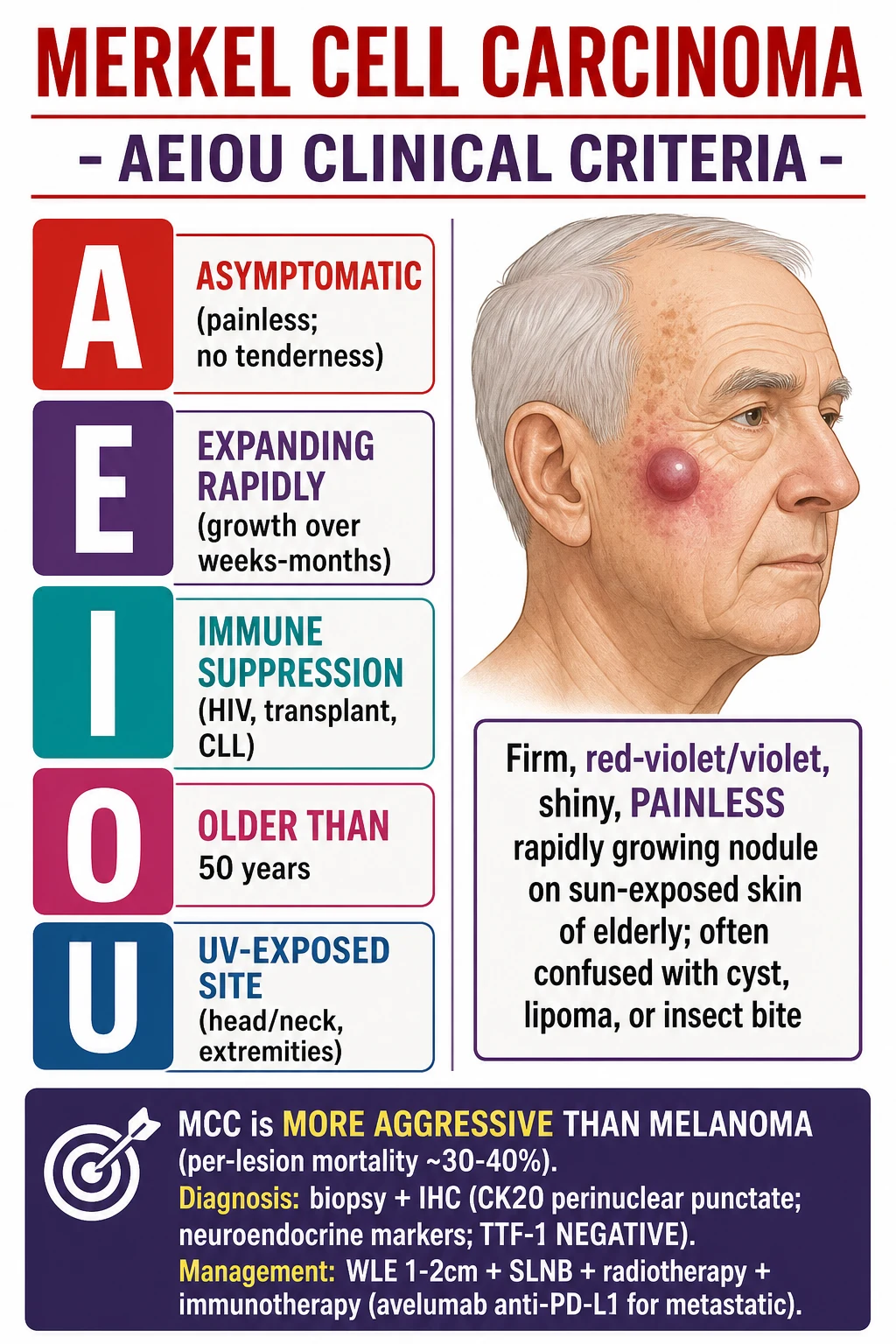
Merkel cell carcinoma (MCC) is an aggressive neuroendocrine carcinoma of the skin. Clinical AEIOU criteria: Asymptomatic (painless), Expanding rapidly, Immune suppression, Older than 50, UV-exposed site. Histology shows small round blue cells; CK20 perinuclear punctate staining is pathognomonic and TTF-1 is negative, distinguishing MCC from small cell lung carcinoma. Management is surgical (WLE or Mohs) plus sentinel lymph node biopsy and adjuvant radiotherapy for high-risk disease; metastatic disease is treated first-line with anti-PD-1/PD-L1 immunotherapy (avelumab, pembrolizumab, nivolumab).
On this page & tools
Your progress
Saved locally on this device.
Exam tags
Red flags
Definition and classification
Merkel cell carcinoma (MCC) is an aggressive primary cutaneous neuroendocrine carcinoma derived from Merkel cells, which are specialised epidermal mechanoreceptors involved in light-touch discrimination. The tumour is composed of small round blue cells with neuroendocrine differentiation, expresses epithelial and neuroendocrine immunohistochemical markers, and has a propensity for early lymphatic and haematogenous spread.[2][6]
Within the 2022 WHO classification of neuroendocrine neoplasms, MCC is classified as a high-grade poorly differentiated neuroendocrine carcinoma of cutaneous origin. Unlike the well-differentiated neuroendocrine tumours of the gastrointestinal tract and lung, MCC is by definition high-grade and aggressive. The terminology distinguishes MCC from other small round blue cell tumours and from extrapulmonary poorly differentiated neuroendocrine carcinomas that may metastasise to the skin.[1]
Historically, MCC has been known as trabecular carcinoma, Toker tumour (now reserved for a separate benign or very-low-grade nipple/areolar lesion), and primary neuroendocrine carcinoma of the skin. Some older texts used the term APUDoma of skin. Histological growth patterns are described as trabecular, intermediate or diffuse (most common), and small cell. These patterns are of descriptive interest only; they do not independently alter prognosis because all behave aggressively.[6]
A critical distinction is primary cutaneous MCC versus cutaneous metastasis of small cell lung carcinoma (SCLC). Both are small round blue cell neuroendocrine carcinomas, but MCC is CK20 positive in a perinuclear punctate (dot-like) pattern and TTF-1 negative, whereas metastatic SCLC is CK20 negative and TTF-1 positive. This distinction is essential because the primary site, staging work-up, and systemic management differ profoundly.[6]
Rarely, MCC may present as in-situ or microinvasive disease confined to the epidermis (stage 0a or Tis concept), but the vast majority of cases are invasive at diagnosis. The term "Merkel cell carcinoma in situ" should not be used loosely because most lesions with epidermal involvement also show dermal invasion on deeper sections.[5]
Epidemiology and risk factors
MCC is rare but its incidence is rising. In the United States SEER-18 registry the age-adjusted incidence is approximately 0.7 per 100,000 person-years, with a 6 to 8 percent annual increase over the past two decades. Australia, particularly Queensland, reports the highest documented incidence worldwide at 1.6 to 2.0 per 100,000, attributed to high ambient ultraviolet exposure, a fair-skinned population, and active skin cancer surveillance.[2][6]
The demographic profile is distinctive: elderly, fair-skinned (Fitzpatrick phototypes I and II), male, and chronically sun-damaged. The median age at diagnosis is approximately 75 years, and fewer than 5 percent of cases occur before age 50. Males are affected roughly twice as often as females. The tumour is uncommon in deeply pigmented populations, but when it occurs it tends to present at a more advanced stage and with worse outcomes because of delayed recognition.[4][6]
The major risk factors are chronic ultraviolet exposure, immunosuppression, and Merkel cell polyomavirus carriage. Chronic UV exposure is the strongest attributable risk factor overall. Immunosuppressive states increase risk dramatically: HIV infection with low CD4 counts, solid organ transplantation (10 to 24-fold increased risk), and chronic lymphocytic leukaemia (CLL, 16 to 30-fold increased risk). Other associations include prior PUVA therapy, ciclosporin, arsenic exposure, and chronic immunosuppression for autoimmune disease.[3][4]
The mechanism by which CLL increases MCC risk is multifactorial. CLL produces a profound defect in T-cell and natural-killer-cell function, and many patients receive additional immunosuppressive therapy such as fludarabine or Bruton tyrosine kinase inhibitors. The defective immune surveillance is particularly important for clearing MCPyV-infected or transformed cells, allowing clonal expansion of MCC. In transplant recipients, the risk is amplified by the duration and intensity of immunosuppression and by older age at transplantation.[4]
Clinical features that raise suspicion for MCC
The AEIOU criteria are a widely taught clinical recognition tool. They are highly sensitive: a patient with a painless, rapidly growing, red-violet nodule on sun-exposed skin in an older or immunosuppressed individual should be considered to have MCC until proven otherwise. The acronym should not be used to exclude MCC when only some features are present, but it is an excellent prompt for urgent biopsy.[2][6]
Pathophysiology
MCC arises through two distinct, mutually exclusive molecular pathways that converge on the inactivation of the retinoblastoma (RB1) tumour suppressor and the acquisition of a neuroendocrine phenotype.[3][4]
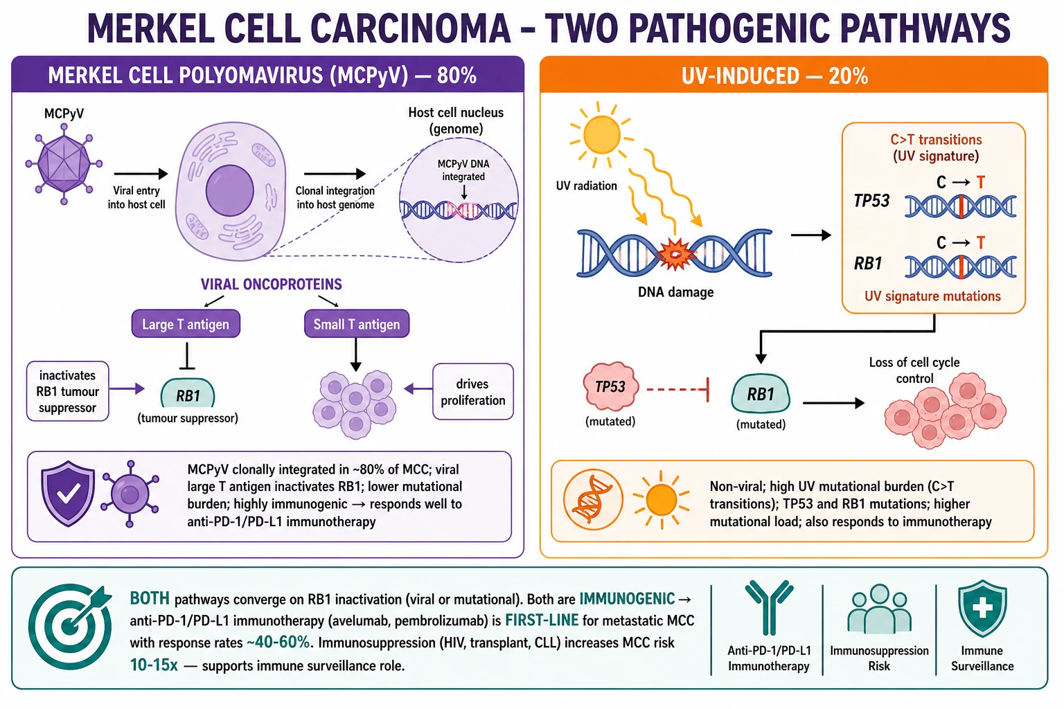
Merkel cell polyomavirus (MCPyV)-driven pathway (approximately 80% of cases): MCPyV is a small, non-enveloped, double-stranded DNA polyomavirus with a 5.4 kb circular genome. It was discovered in 2008 by Feng and colleagues using digital transcriptome subtraction of MCC tumour tissue. In viral-positive MCC, the viral genome is clonally integrated into the host genome, a finding that proves MCPyV integration is an early, transforming event rather than a passenger infection. The viral large T antigen binds and functionally inactivates RB1, releasing E2F transcription factors and driving uncontrolled G1-to-S cell-cycle progression. The viral small T antigen stabilises large T, inhibits protein phosphatase 2A, activates cap-dependent translation through 4E-BP1, and contributes to the transformed phenotype. MCPyV-positive tumours generally have a lower UV-type mutational burden and express viral oncoproteins that are foreign to the immune system, making them highly immunogenic.[6]
UV-induced, virus-negative pathway (approximately 20% of cases): These tumours lack detectable MCPyV DNA and instead show a high burden of UV-signature mutations, particularly C→T transitions at dipyrimidine sites. Recurrent mutations include TP53, RB1, NOTCH1, KMT2D, and other chromatin modifiers. These tumours behave more like UV-driven keratinocyte carcinomas at the molecular level, often arising on chronically sun-damaged skin, and may have a worse prognosis than MCPyV-positive tumours in some cohorts. Because they carry many neoantigens from UV-induced mutations, they are also immunogenic and respond to immune checkpoint inhibitors.[3][4]
Both pathways converge on RB1 loss of function. This convergence explains why MCC is exquisitely sensitive to agents that restore immune control: in MCPyV-positive tumours the immune system recognises viral large T and small T antigens, while in virus-negative tumours it recognises UV-induced neoantigens. The strong immunogenicity of both subtypes underpins the high response rates to anti-PD-1 and anti-PD-L1 antibodies.[4]
MCC spreads first via lymphatics to the regional draining basin. Sentinel lymph node biopsy identifies occult nodal disease in approximately 30 percent of patients with clinically negative nodes. Haematogenous dissemination occurs later to the lung, liver, bone, and brain. In-transit and satellite metastases may appear as small nodules between the primary site and the draining nodal basin and usually herald regional nodal involvement.[5][6]
Clinical presentation
The typical MCC lesion is a firm, dome-shaped, shiny, red-violet to flesh-coloured, painless papule or nodule measuring 0.5 to 2 cm at presentation. It is often mistaken clinically for a cyst, lipoma, arthropod bite, basal cell carcinoma, or vascular lesion. The key feature that separates it from most other benign or inflammatory lesions is that it is painless and expanding rapidly. The overlying skin may be thin and telangiectatic, and ulceration is less common than in squamous cell carcinoma but can occur in advanced lesions.[2][6]
Approximately half of all MCCs arise on the head and neck, about 40 percent on the extremities (especially the forearm and hand), and about 10 percent on the trunk. Rare sites include the buttocks, genitalia, periocular skin, lip, and vulva. Mucosal MCC is uncommon and generally behaves more aggressively. Because the head and neck are common sites, MCC may first be noticed by hairdressers, patients, or family members rather than by the patient themselves, leading to diagnostic delay.[2]
In the elderly and frail patient, MCC often presents late as a bleeding or ulcerated large nodule with satellite metastases. The lesion may be dismissed as a bruise, cyst, or age-related skin change. In patients with CLL, the tumour is typically a rapidly growing blue-red nodule on the head, neck, or upper trunk, and the course is particularly aggressive. In solid organ transplant recipients, MCC may arise at atypical sites, show multiple primary lesions, and run a more aggressive course because of ongoing immunosuppression. In HIV infection, risk rises as the CD4 count falls below 200 cells per microlitre, and patients tend to present at a younger age.[4]
Occasionally, MCC presents as an unknown primary with a nodal or visceral metastasis and regression of the cutaneous primary. Rarely, MCC arises in previously irradiated or chronically inflamed skin, analogous to Marjolin-type squamous cell carcinoma. In-transit and satellite metastases are small skin-coloured or pink nodules between the primary site and the nodal basin; their presence strongly suggests nodal involvement and upstages the patient.[5]
Why does the lesion look red-violet?
The red-violet colour of MCC reflects a highly vascular dermal tumour with thin overlying epidermis. The shiny surface is due to the dome-shaped elevation and the thin epidermis stretched over the nodule. The lack of tenderness and the rapid growth rate together should raise suspicion for MCC rather than a benign cyst or vascular malformation.
Differential diagnosis
The differential diagnosis of a rapidly growing red-violet or skin-coloured nodule on sun-exposed skin of an elderly patient is broad. MCC must be distinguished from common benign and malignant skin lesions, as well as from cutaneous metastases of internal malignancy.[5][6]
Basal cell carcinoma usually grows slowly, has a pearly appearance with surface telangiectasia, and may show central ulceration. On immunohistochemistry it is BerEP4 positive, BCL2 positive, and CK20 negative. Squamous cell carcinoma is more often keratotic or ulcerated, may be tender, and is CK5/6 and p40 positive with CK20 negativity. Amelanotic melanoma can mimic MCC closely clinically, but it is S100, SOX10, HMB45, and melan-A positive. Cutaneous lymphoma, particularly B-cell lymphoma, can resemble small round blue cell tumours on haematoxylin and eosin staining, but it is CD45 positive and shows B-cell or T-cell lineage markers. Atypical fibroxanthoma occurs in sun-damaged skin of elderly patients and is CD10, CD68, and vimentin positive with negative cytokeratins and S100. Dermatofibroma is composed of spindle cells, is Factor XIIIa positive, and is indolent. Glomus tumour is typically painful, smooth-muscle actin positive, and vascular. Adnexal tumours such as porocarcinoma may show CK7 and CK20 positivity and EMA positivity, requiring clinical correlation.[5][6]
The most important differential diagnosis in histopathology is cutaneous metastasis of small cell lung carcinoma (SCLC). Both are small round blue cell neuroendocrine carcinomas. MCC is CK20 positive with perinuclear dot-like staining and TTF-1 negative, whereas SCLC is CK20 negative and TTF-1 positive. In any patient with small round blue cell histology and no clear primary skin lesion, a chest computed tomography scan is mandatory to exclude a lung primary. MCPyV large T antigen staining is also helpful because it is positive in MCC and negative in SCLC. Neuroendocrine markers (chromogranin, synaptophysin, CD56, NSE) are positive in both and therefore do not discriminate.[6]
Serum antibody to MCPyV oncoproteins (viral capsid and large T antigen) has been investigated as a biomarker. A rising titre correlates with tumour burden and may herald recurrence, but it is not yet used as a standalone diagnostic test in routine clinical practice.[4]
Clinical and bedside assessment
Assessment of a suspected MCC begins with a focused skin examination. The lesion should be inspected for colour, surface characteristics, ulceration, and telangiectasia, and palpated to confirm firmness and depth. The draining nodal basin should be examined carefully: cervical and supraclavicular nodes for head and neck lesions, axillary and epitrochlear nodes for upper-limb lesions, and inguinal and popliteal nodes for lower-limb lesions. The epitrochlear and popliteal nodes are often overlooked but are clinically significant when palpable, even if only 1 cm in size. A palpable node in this setting should prompt urgent fine-needle aspiration.[5]
The skin around the primary lesion should be surveyed for satellite and in-transit metastases, which appear as small skin-coloured or pink nodules between the primary and the nodal basin. A full skin examination is required because patients may have additional skin cancers, photodamage, or a second primary MCC. The lesion should be measured and photographed for documentation and for comparison at follow-up. Tenderness should be assessed: MCC is typically non-tender, so tenderness should prompt consideration of an inflamed cyst, abscess, or glomus tumour.[5]
An ABCDE-style assessment adapted for non-pigmented lesions is useful: Asymmetry, irregular Border, Colour (red-violet or erythematous), Diameter (the 6 mm rule does not apply to nodules; any rapidly growing bump warrants biopsy), and Evolution (rapid growth over weeks). The presence of immune suppression, advanced age, or a UV-exposed site should lower the threshold for biopsy.[6]
In immunosuppressed patients, the clinical picture may be atypical. CLL patients may present with small, firm, red-violet nodules that grow rapidly; HIV patients may have atypical distributions; and transplant recipients may have more aggressive ulceration and a higher risk of multiple primaries. These patients should be referred urgently for multidisciplinary management.[4]
Investigations and staging
The diagnosis of MCC requires a full-thickness biopsy with adequate dermal tissue. For small lesions, an excisional biopsy with 1 to 2 mm margins is ideal. For larger or anatomically challenging lesions, a punch biopsy of 4 mm or more through the centre of the lesion is acceptable. A superficial shave biopsy may miss the diagnosis if it does not sample the dermal component, so deep shave or punch biopsy is preferred. The biopsy should be sent for histology and immunohistochemistry.[5][6]
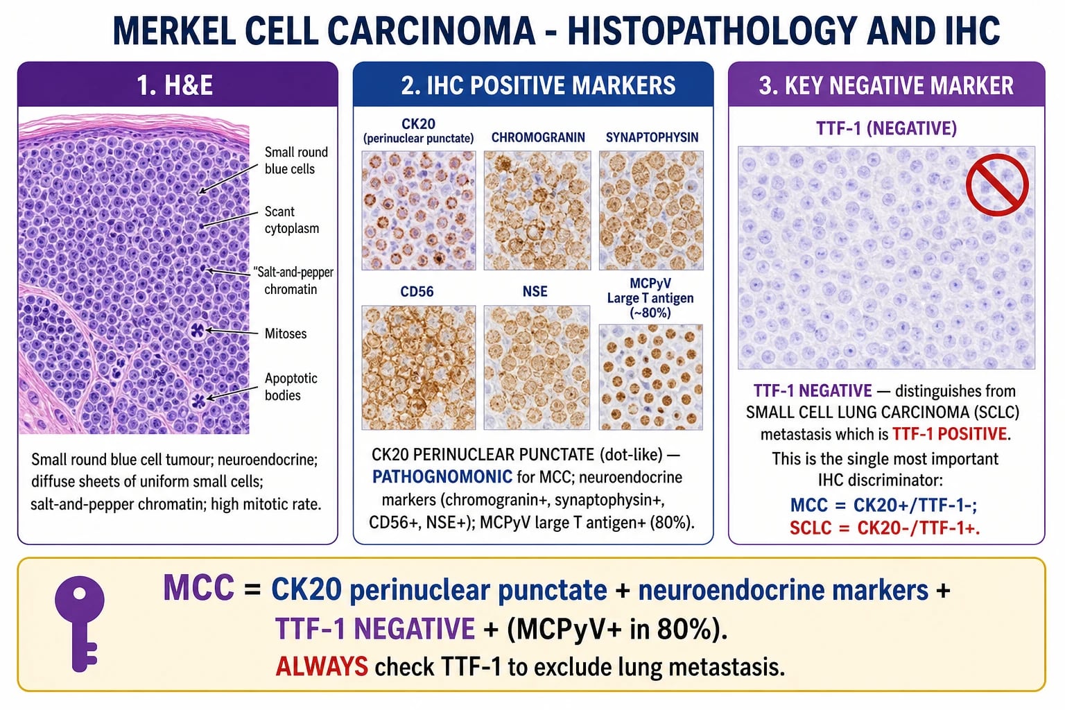
The diagnostic immunohistochemical panel is: [1]
- CK20 — perinuclear punctate (dot-like) positivity is pathognomonic for MCC. Diffuse cytoplasmic CK20 staining is not characteristic.
- Neuroendocrine markers — chromogranin A, synaptophysin, CD56 (NCAM), and neuron-specific enolase (NSE) are usually positive.
- MCPyV large T antigen — positive in approximately 80% of cases (CM2B4 antibody); helps confirm the viral pathway and distinguish MCC from SCLC.
- TTF-1 — negative in MCC; positive in most SCLCs.
- CDX2, CEA, CD45/LCA — usually negative in MCC, helping exclude intestinal metastasis, adenocarcinoma, and lymphoma respectively.[6]
| Marker | MCC | SCLC metastasis |
|---|---|---|
| CK20 | Positive (perinuclear punctate) | Negative |
| TTF-1 | Negative | Positive |
| Neuroendocrine markers | Positive | Positive |
| MCPyV large T antigen | Positive in ~80% | Negative |
| Clinical context | Cutaneous primary on sun-exposed skin | Often known lung primary; chest imaging abnormal |
Staging uses the AJCC 8th edition (2017) TNM system for MCC: [1]
- T stage: T1 = tumour 2 cm or less in greatest dimension; T2 = more than 2 cm but not more than 5 cm; T3 = more than 5 cm; T4 = invasion of deep extradermal structures such as fascia, muscle, cartilage, or bone.
- N stage: N0 = no regional nodal metastasis; N1a = microscopic nodal metastasis detected by sentinel lymph node biopsy but not clinically apparent; N1b = macroscopic nodal metastasis clinically or radiologically detected; N2 = in-transit metastasis without nodal metastasis; N3 = in-transit or satellite metastasis with nodal metastasis.
- M stage: M0 = no distant metastasis; M1a = distant metastasis to skin, soft tissue, or distant lymph nodes; M1b = metastasis to lung; M1c = metastasis to other viscera.[5]
Prognostic stage groups are: Stage 0 (Tis N0 M0), Stage I (T1 N0 M0), Stage IIA (T2-3 N0 M0), Stage IIB (T4 N0 M0), Stage IIIA (T1-3 N1a M0 or in-transit without nodal disease), Stage IIIB (T1-4 N1b-N2 M0), and Stage IV (any T N3 M0 or any T any N M1).[5]
Sentinel lymph node biopsy (SLNB) is the cornerstone of nodal staging. In clinically node-negative patients, SLNB upstages approximately 30 percent to node-positive disease and changes management. It is ideally performed at the same time as definitive excision of the primary tumour, using dual mapping with lymphoscintigraphy and blue dye or radiocolloid and fluorescent dye.[5][6]
Imaging for staging includes contrast-enhanced CT of the chest, abdomen, and pelvis at baseline. The lung and liver are the most common sites of distant metastasis. 18F-FDG PET-CT is useful for detecting occult nodal or distant disease, particularly when CT findings are equivocal or the primary is high-risk. Brain MRI is reserved for patients with neurological symptoms. Serum MCPyV antibody testing may be used as a baseline and surveillance biomarker in centres with access, with rising titres suggesting recurrence.[5]
The NCCN 2024 risk stratification categorises patients as low risk (primary 1 cm or less, node negative, no lymphovascular invasion), intermediate risk (1 to 2 cm, node negative), and high risk (any node positive, more than 2 cm, lymphovascular invasion, or immunosuppression). Adjuvant therapy recommendations are tailored to this risk profile. ESMO-EURACAN 2024 similarly recommends systemic staging with CT or PET-CT, mandatory SLNB for T1 or greater disease, and multidisciplinary referral at all stages.[5]
Management of urgent presentations
Acute presentations of MCC are uncommon but require immediate attention. A bleeding or ulcerated primary MCC should be managed with direct pressure, alginate or haemostatic dressing, and urgent biopsy if not already performed. Analgesia and tetanus cover are provided if needed. The lesion should be photographed for multidisciplinary referral and for documentation of baseline appearance. Rarely, surgical haemostasis is required in the emergency department.[5]
A fungating MCC in a transplant recipient should be biopsied urgently. Immunosuppression should not be reduced abruptly without multidisciplinary consultation because this may precipitate graft rejection or loss. The transplant team, dermatologist, medical oncologist, and radiation oncologist should jointly decide on any modification of immunosuppression, which may include conversion to an mTOR inhibitor such as sirolimus or everolimus to reduce MCC risk while preserving graft function.[5]
Suspected spinal cord compression from vertebral metastasis is an oncological emergency. Management follows standard principles: high-dose dexamethasone, urgent MRI of the whole spine, and urgent radiotherapy. Surgical decompression is considered if there is progressive motor deficit, spinal instability, or a single compressive lesion in a patient with good performance status. MCC is highly radiosensitive, so radiotherapy is often effective for rapid palliation.[5]
Tumour lysis syndrome is rare with MCC because the tumour burden is usually smaller than in bulky lymphoma or leukaemia, but it can occur in disseminated disease or after rapid response to immunotherapy. Prophylaxis includes aggressive hydration, allopurinol, and rasburicase for high-risk patients, with careful electrolyte monitoring.[5]
Definitive and stepwise management
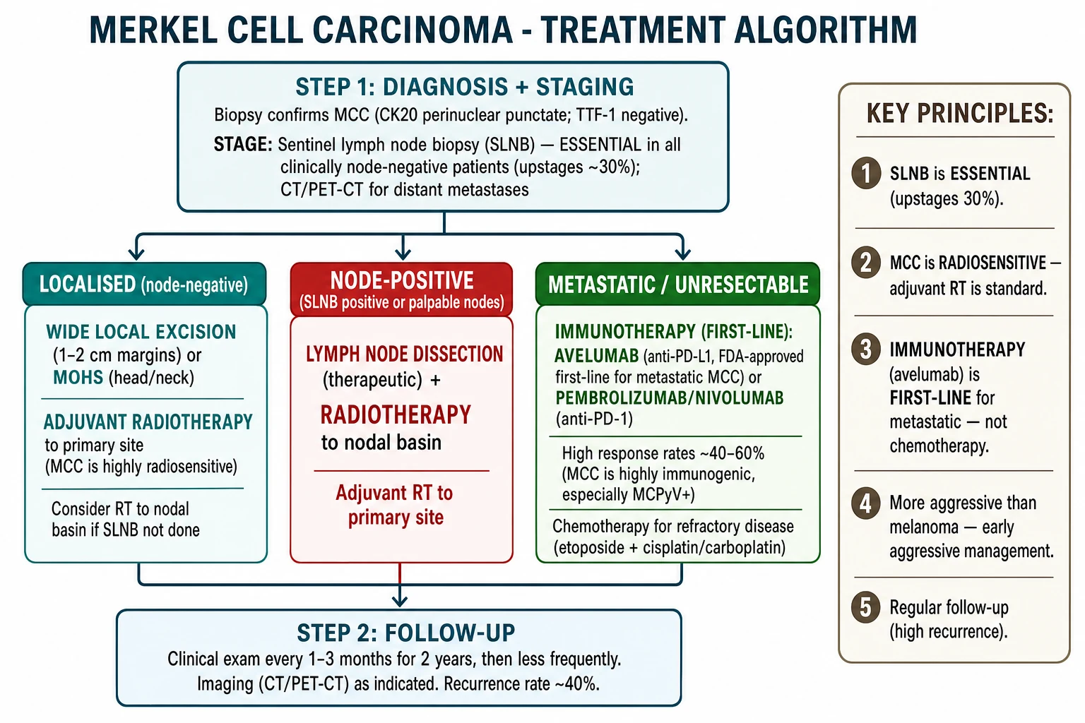
Management of MCC is multidisciplinary and depends on stage. The standard approach for localised disease is surgical excision with adequate margins and sentinel lymph node biopsy, followed by adjuvant radiotherapy for high-risk features. Metastatic disease is treated with systemic immunotherapy as first-line therapy; chemotherapy is reserved for immunotherapy-refractory disease or urgent palliation.[5][6]
Surgery
Wide local excision (WLE) is the standard primary treatment. The NCCN recommends 1 cm clinical margins for tumours 1 cm or less and 2 cm clinical margins for tumours larger than 1 cm, with excision down to the deep fascia. For tumours on the head and neck, eyelid, lip, nose, or ear, achieving these margins can be functionally or cosmetically challenging. In these sites, Mohs micrographic surgery is an acceptable alternative, allowing complete circumferential peripheral and deep margin assessment while preserving uninvolved tissue. The goal is a histologically clear margin; when Mohs is used, a narrow peripheral margin is acceptable if the deep margin is clear.[5][6]
Sentinel lymph node biopsy
SLNB is mandatory for clinically node-negative patients with T1 or greater disease according to NCCN and ESMO-EURACAN guidelines. It should be performed at the same time as wide local excision. The technique uses dual mapping to identify the sentinel node(s) and allows histological examination of the node with immunohistochemistry. A positive SLNB upstages the patient and guides further management.[5]
Management of a positive sentinel lymph node
For patients with a positive SLNB, current practice includes completion therapeutic lymph node dissection (TLND) or adjuvant radiotherapy to the nodal basin, or both. NCCN 2024 favours nodal basin radiotherapy with or without completion dissection; ESMO-EURACAN 2024 similarly recommends nodal basin radiotherapy for positive SLNB. The choice depends on patient factors, the number of positive nodes, the presence of extracapsular extension, and morbidity concerns such as lymphoedema.[5]
Radiotherapy
MCC is highly radiosensitive. Adjuvant radiotherapy is recommended for the primary site when margins are close or positive, the tumour is larger than 1 cm, there is lymphovascular invasion, the primary is on the head or neck, or the patient is immunosuppressed. The usual dose to the primary site is 50 to 66 Gy in 2 Gy fractions. The nodal basin receives 50 to 66 Gy if SLNB is positive or if TLND is not performed. For patients who are not surgical candidates, definitive radiotherapy alone at 60 to 66 Gy in 2 to 2.5 Gy fractions over 5 to 6 weeks can provide excellent local control.[5][6]
Immunotherapy for metastatic disease
Immune checkpoint inhibitors are the first-line treatment for metastatic MCC because they produce durable responses far superior to chemotherapy. The principal agents are:[4][5]
- Avelumab (anti-PD-L1), 800 mg intravenously every 2 weeks — FDA and EMA approved for metastatic MCC based on the JAVELIN Merkel 200 trial, which showed durable responses in first-line and previously treated patients. In the first-line cohort, the objective response rate was approximately 62 percent, with most responses durable at 6 months or longer.[7]
- Pembrolizumab (anti-PD-1), 200 mg intravenously every 3 weeks — approved for metastatic MCC based on KEYNOTE-017, in which first-line pembrolizumab produced an objective response rate of 56 percent in patients with advanced disease, with responses in both MCPyV-positive and virus-negative tumours.[8]
- Nivolumab (anti-PD-1), 240 mg every 2 weeks or 480 mg every 4 weeks — also active in metastatic MCC, and evaluated in the neoadjuvant setting.
Second-line options after progression on anti-PD-1 or anti-PD-L1 include nivolumab plus ipilimumab and investigational combinations such as cabozantinib plus nivolumab and ipilimumab. Response rates are lower and toxicity higher with dual immunotherapy, so patient selection is important. The optimal duration of immunotherapy and the role of maintenance therapy remain under investigation.[5]
Chemotherapy
Chemotherapy is now reserved for patients who are refractory to or ineligible for immunotherapy, or for urgent disease control when immunotherapy is not appropriate. The standard regimen is etoposide 100 mg per square metre on days 1 to 3 plus cisplatin or carboplatin every 3 weeks for 4 to 6 cycles. Topotecan is an alternative. Response rates are modest, approximately 20 to 30 percent, and progression-free survival is short, with a median of 3 to 4 months. Chemotherapy does not produce durable responses and has been demoted from first-line use by international guidelines.[5][6]
Adjuvant and neoadjuvant immunotherapy
Adjuvant nivolumab has shown improved disease-free survival compared with observation in the phase 2 ADMEC-O trial. In this study, patients with completely resected MCC were randomised to nivolumab 480 mg every 4 weeks for 1 year or observation. The 12-month and 24-month disease-free survival rates were higher with nivolumab, supporting its clinical feasibility in this setting, although overall survival data were immature. The phase 3 ADAM trial (adjuvant avelumab) and STAMP trial (adjuvant pembrolizumab) have completed accrual and results are awaited.[9]
Neoadjuvant nivolumab was evaluated in the CheckMate 358 trial. Patients with resectable MCC received two doses of nivolumab before planned surgery. Approximately 47 percent achieved a pathological complete response, and 54 percent had a radiographic tumour reduction of 30 percent or more. Recurrence-free survival correlated with pathological response. Neoadjuvant immunotherapy is an emerging strategy that may reduce recurrence risk in high-risk resectable MCC.[10]
Follow-up
MCC has a high recurrence rate, with up to 40 percent of patients experiencing recurrence, most within the first 2 years. Follow-up therefore is intensive: clinical examination every 3 to 6 months for 2 years, then every 6 to 12 months until 5 years. Patients are taught self-examination and are encouraged to report new lesions promptly. In stage III or IV disease, surveillance imaging with CT or PET-CT is performed every 6 to 12 months for the first 3 years. Serum MCPyV antibody titres may be trended in patients whose tumours were antibody-positive at baseline.[5]
Specific subtypes and scenarios
MCPyV-positive versus virus-negative MCC: Viral-positive MCC accounts for approximately 80 percent of cases in cooler climates, has a lower UV-type mutational burden, and generally shows robust responses to immune checkpoint inhibitors. Virus-negative MCC is more common in regions with very high UV exposure, has a higher mutational burden, a molecular profile closer to keratinocyte carcinoma, and may have a worse prognosis in some studies. Both subtypes are treated identically by stage, although clinical trials increasingly stratify by viral status.[3][4]
MCC in transplant recipients: These patients have the worst prognosis among MCC cohorts. They often develop multiple primary lesions, and immune checkpoint inhibitors carry a substantial risk of graft rejection, particularly in kidney, heart, and lung transplant recipients. Management requires close collaboration with the transplant team. Strategies may include reduction or conversion of immunosuppression to an mTOR inhibitor, with or without immunotherapy on a case-by-case basis. The risk-benefit discussion must be individualised and documented.[4]
MCC in HIV: Risk increases with a CD4 count below 200 cells per microlitre. Patients tend to be younger and may have more aggressive disease. Immune checkpoint inhibitors can be used with appropriate antiretroviral therapy, and opportunistic infection prophylaxis should be considered. ART optimisation before systemic therapy is important.[4]
MCC in CLL: Patients with CLL have a 16 to 30-fold increased risk of MCC. The B-cell immunosuppression of CLL and prior chemo-immunotherapy compound the defect in immune surveillance. Prognosis is worse than in CLL-free patients. Management of the CLL may need to be modified to allow effective immunotherapy.[4]
Primary nodal MCC (unknown primary): Rarely, MCC presents with isolated nodal or visceral disease and no identifiable cutaneous primary. The behaviour is similar to cutaneous MCC. Management is nodal dissection plus adjuvant radiotherapy, with systemic staging. Prognosis is poorer than for localised cutaneous disease.[5]
MCC in children and adolescents: Fewer than 1 percent of cases occur in children. The diagnosis should be made with caution because other small round blue cell tumours, congenital neurocristopathies, and lymphoma can mimic MCC. Outcomes in small series appear better than in adults, but long-term data are limited.[5]
MCC on mucous membranes: MCC of the lip, eyelid, vulva, or other mucosal sites is uncommon and tends to be more aggressive. Margins may be narrow because of anatomical constraints, and the threshold for adjuvant radiotherapy is lower.[5]
Complications and pitfalls
Local complications include bleeding, ulceration, superinfection, local recurrence, and in-transit or satellite metastases. Approximately 40 percent of recurrences are local or in-transit. Regional complications include recurrence in the nodal basin, nodal rupture, and lymphoedema after lymph node dissection. Distant complications include liver metastasis, lung metastasis, bone metastasis with pathological fracture, and brain metastasis with mass effect or seizures. Leptomeningeal disease is rare and has a dismal prognosis.[5]
Paraneoplastic associations are uncommon but important. Ectopic ACTH secretion can cause Cushing syndrome, parathyroid hormone-related peptide (PTHrP) can cause hypercalcaemia, and a rare Lambert-Eaton myasthenic syndrome has been described, related to neuronal autoimmunity. These should be considered in patients with suggestive symptoms.[5]
Treatment complications include postoperative bleeding, wound dehiscence, and infection. Radiotherapy can cause skin atrophy, telangiectasia, and lymphoedema. Immune checkpoint inhibitors cause immune-related adverse events (irAEs) such as colitis, hepatitis, pneumonitis, thyroiditis, hypophysitis, adrenal insufficiency, and myocarditis. Myocarditis is rare but potentially lethal. Recognition and early treatment with corticosteroids and withholding the offending agent are essential.[5]
Diagnostic pitfalls include assuming the lesion is a benign cyst or lipoma, missing the dermal component on a superficial shave biopsy, failing to perform CK20 staining, not recognising the perinuclear dot-like pattern as pathognomonic, and omitting TTF-1, which can lead to misdiagnosis of MCC as metastatic SCLC. Treatment pitfalls include omitting SLNB, taking inadequate surgical margins, omitting adjuvant radiotherapy in high-risk primaries, and switching prematurely from immunotherapy to chemotherapy before allowing sufficient time for response.[5]
[1]Prognosis and disposition
MCC is one of the most aggressive skin cancers, with a per-lesion mortality higher than that of melanoma. Prognosis is strongly stage-dependent.[2][5]
Five-year overall survival by AJCC 8th edition stage is approximately: [1]
- Stage I (T1 N0 M0): 55 to 79 percent, depending on the cohort.
- Stage IIA-IIB (T2-4 N0 M0): 30 to 60 percent.
- Stage III (node-positive): 30 to 50 percent.
- Stage IV (distant metastasis): 14 to 25 percent. [1]
Overall, approximately 50 percent of patients with localised disease survive 5 years, dropping sharply with regional and distant disease. The overall recurrence rate is approximately 40 percent, with most recurrences occurring within the first 2 years. Independent adverse prognostic factors include primary site on the head or neck or trunk, tumour size greater than 2 cm, lymphovascular invasion, immunosuppression, MCPyV-negative status in some cohorts, positive SLNB, and depth greater than 6 mm or invasion beyond the subcutis.[5]

Follow-up is intensive and lifelong. After definitive treatment, patients are examined clinically every 3 to 6 months for 2 years and then every 6 to 12 months until 5 years. Imaging is reserved for high-risk disease and for symptomatic patients. Serum MCPyV antibody titres may be useful for surveillance in patients with antibody-positive tumours. Patient education is essential: new lesions, new lumps, or neurological symptoms should prompt urgent review.[5]
Palliative care referral is appropriate for patients with symptomatic metastatic disease, declining performance status despite immunotherapy, leptomeningeal disease, uncontrolled pain, or bleeding. Early involvement of palliative care can improve symptom control and quality of life, even when disease-modifying therapy is ongoing.[5]
Special populations
Elderly and frail patients: Older age is the norm in MCC, but frailty limits treatment options. For small primaries in patients who are not surgical candidates, definitive radiotherapy alone may be preferred over extensive surgery. Geriatric assessment helps identify patients who will tolerate immunotherapy and those in whom treatment intensity should be reduced. A 75-year-old patient may still benefit from standard care if performance status is good, whereas a 90-year-old with significant comorbidity may be best served by local radiotherapy or best supportive care.[5]
Pregnancy: MCC in pregnancy is rare. Staging with MRI without gadolinium is preferred; CT should be avoided if possible. Surgery is safest in the second trimester. Radiotherapy and immunotherapy are generally deferred until after delivery because of fetal risk. PD-1 and PD-L1 inhibitors cross the placenta and can cause fetal immune toxicity. Management must be individualised by a multidisciplinary team including oncology, dermatology, obstetrics, and neonatology.[5]
Childhood and adolescence: MCC is rare in children. When it occurs, it may be associated with congenital immunodeficiency or MCPyV infection. The diagnosis should be confirmed by expert pathology because other small round blue cell tumours can mimic MCC. Outcomes in small series are better than in adults, but long-term data are limited.[5]
Transplant recipients: MCC in this population is high-risk. The transplant team must be involved from the outset. Options include reducing immunosuppression, converting to an mTOR inhibitor, and carefully selected immunotherapy with informed consent regarding graft rejection risk. Localised disease may be treated with surgery and radiotherapy, avoiding systemic immunotherapy when possible.[4]
HIV and other immunosuppression: Opportunistic screening for skin cancers is recommended in patients older than 50 years with HIV. ART optimisation before systemic therapy is important. Immune checkpoint inhibitors are generally feasible with effective ART. Opportunistic infection prophylaxis should be considered according to CD4 count.[4]
MCC on chronically inflamed or irradiated skin: These lesions are more aggressive. Even with clear margins, adjuvant radiotherapy should be considered because the local microenvironment favours recurrence.[5]
Evidence, guidelines, and regional differences
Landmark trials have transformed MCC management. JAVELIN Merkel 200 established avelumab as first-line and later-line therapy for metastatic MCC, with durable responses. KEYNOTE-017 established pembrolizumab as an effective first-line agent. CheckMate 358 demonstrated the feasibility and efficacy of neoadjuvant nivolumab. ADMEC-O provided the first randomised evidence for adjuvant nivolumab in resected MCC. Phase 3 trials of adjuvant avelumab (ADAM) and pembrolizumab (STAMP) are ongoing and results are awaited.[7][8][9][10]
Regional guidelines differ slightly in emphasis. NCCN 2024 (United States) recommends broad use of adjuvant radiotherapy for high-risk primaries and nodal basins, with completion lymph node dissection or observation after positive SLNB. ESMO-EURACAN 2024 reserves adjuvant radiotherapy for positive margins, positive nodes, and immunosuppression, noting that omission of radiotherapy in low-risk disease may not compromise overall survival. Both guidelines recommend avelumab, pembrolizumab, or nivolumab as first-line systemic therapy for metastatic disease. NICE UK (NG12) focuses on rapid recognition and referral of suspicious skin lesions, including MCC under the umbrella of rapidly growing lesions. Cancer Council Australia guidelines align with international standards but place additional emphasis on UV prevention and high incidence in fair-skinned populations. ICMR India captures MCC under rare cutaneous neoplasms in its non-melanoma skin cancer guidance.[5]
Current controversies include whether to omit completion lymph node dissection after positive SLNB (NCCN 2024 allows observation plus nodal radiotherapy), the role of neoadjuvant immunotherapy before surgery, the optimal duration of adjuvant immunotherapy, and the management of immunotherapy-refractory disease. Chemotherapy has been demoted from first-line because of short response duration and inferior durability compared with immune checkpoint inhibitors.[5]
Serum MCPyV antibody titres are an emerging biomarker. Rising titres correlate with tumour burden and recurrence, and serial testing may become part of routine surveillance. However, the test is not yet standardised worldwide and should be interpreted alongside clinical and imaging findings.[4]
Exam pearls
[1]Red flags
Exam application bank (NEET-PG / INICET)
One-line answer
Merkel cell carcinoma (MCC) is an aggressive neuroendocrine carcinoma of the skin. Clinical AEIOU criteria: Asymptomatic (painless), Expanding rapidly, Immune suppression, Older than 50, UV-exposed site. Histology shows small round blue cells; CK20 perinuclear punctate staining is pathognomonic and TTF-1 is negative, distinguishing MCC from small cell lung carcinoma. Management is surgical (WLE or Mohs) plus sentinel lymph node biopsy and adjuvant radiotherapy for high-risk disease; metastatic disease is treated first-line with anti-PD-1/PD-L1 immunotherapy (avelumab, pembrolizumab, nivolumab).
Worked stems (answer without another resource)
Stem 1 — Classic presentation. Map symptoms to mechanism; name the first investigation and first treatment step with dose/route if drug therapy is standard. [1]
Stem 2 — Unstable / complicated. List red flags that force immediate resuscitation, theatre, ICU, antidote, or reperfusion — and what you do in the first 15 minutes. [1]
Stem 3 — Atypical group. Elderly, pregnancy, child, or immunocompromised: how presentation and thresholds change. [1]
Stem 4 — Differential trap. Name the three closest mimics and one discriminator for each. [1]
Stem 5 — Disposition. Who goes home with safety-netting, who is admitted, who needs HDU/ICU/theatre, and what follow-up is mandatory. [1]
Rapid viva checklist
- Definition + classification
- Pathophysiology chain
- Bedside signs / criteria
- Score with exact components (if any)
- Emergency bundle
- Definitive therapy with doses
- Complications of disease and of treatment
- Special populations
- Guideline/trial name if classic
- Three exam traps
Coverage self-check
If you cannot answer any stem above from this page alone, re-read the matching section — the page is intended to be self-sufficient for final-prof and NEET-PG/INICET questions on Merkel cell carcinoma.
[1]References
- [1]Rindi G, Mete O, Uccella S, et al. Overview of the 2022 WHO Classification of Neuroendocrine Neoplasms Endocr Pathol, 2022.PMID 35294740
- [2]Strong J, Hallaert P, Brownell I. Merkel Cell Carcinoma Hematol Oncol Clin North Am, 2024.PMID 39060119
- [3]Hernandez LE, Mohsin N, Yaghi M, et al. Merkel cell carcinoma: An updated review of pathogenesis, diagnosis, and treatment options Dermatol Ther, 2022.PMID 34967084
- [4]Becker JC, Stang A, Schrama D, et al. Merkel Cell Carcinoma: Integrating Epidemiology, Immunology, and Therapeutic Updates Am J Clin Dermatol, 2024.PMID 38649621
- [5]Lugowska I, Becker JC, Ascierto PA, et al. Merkel-cell carcinoma: ESMO-EURACAN Clinical Practice Guideline for diagnosis, treatment and follow-up ESMO Open, 2024.PMID 38796285
- [6]Becker JC, Stang A, DeCaprio JA, et al. Merkel cell carcinoma Nat Rev Dis Primers, 2017.PMID 29072302
- [7]D'Angelo SP, Russell J, Lebbé C, et al. Efficacy and Safety of First-line Avelumab Treatment in Patients With Stage IV Metastatic Merkel Cell Carcinoma: A Preplanned Interim Analysis of a Clinical Trial JAMA Oncol, 2018.PMID 29566106
- [8]Nghiem PT, Bhatia S, Lipson EJ, et al. PD-1 Blockade with Pembrolizumab in Advanced Merkel-Cell Carcinoma N Engl J Med, 2016.PMID 27093365
- [9]Becker JC, Ugurel S, Leiter U, et al. Adjuvant immunotherapy with nivolumab versus observation in completely resected Merkel cell carcinoma (ADMEC-O): disease-free survival results from a randomised, open-label, phase 2 trial Lancet, 2023.PMID 37451295
- [10]Topalian SL, Bhatia S, Amin A, et al. Neoadjuvant Nivolumab for Patients With Resectable Merkel Cell Carcinoma in the CheckMate 358 Trial J Clin Oncol, 2020.PMID 32324435