Dermatology · Medicine
Miliaria / heat rash
Also known as Miliaria · Heat rash · Prickly heat · Sweat rash · Sudamina
Miliaria is a common, self-limiting disorder of eccrine sweat gland obstruction in which retained sweat leaks into the surrounding tissue at a level determined by the depth of duct obstruction — producing three classic subtypes: miliaria crystallina (superficial, stratum corneum, clear vesicles), miliaria rubra / prickly heat (mid-epidermal, itchy red papules), and miliaria profunda (dermo-epidermal, flesh-coloured papules with risk of hypohidrosis and heat exhaustion). Triggers are heat, humidity, occlusion, fever and intensive-care incubation. Fellowship-level assessment demands mastery of the level-of-obstruction → morphology correlation, the neonatal vs adult presentations, secondary infection (miliaria pustulosa), and the rare but life-threatening thermoregulatory failure of extensive profunda in tropical residents.
On this page & tools
Your progress
Saved locally on this device.
Exam tags
Red flags
Definition & Classification
Miliaria is an acquired, usually self-limiting disorder of the eccrine sweat gland in which obstruction of the sweat duct at a defined anatomical level causes retained sweat to extravasate into the surrounding tissue, producing a morphology that is predictable from the depth of obstruction. Three classical subtypes are recognised, plus a pustular variant:[4]
| Subtype | Level of obstruction | Morphology | Itch | Typical host |
|---|---|---|---|---|
| Miliaria crystallina (sudamina) | Stratum corneum (subcorneal) | 1–2 mm clear, thin-roofed, flaccid vesicles, easily ruptured | None | Neonates; febrile/bedbound adults |
| Miliaria rubra (prickly heat) | Mid-epidermis (intraepidermal spinous layer) | 1–3 mm erythematous papules and papulovesicles on an erythematous base | Intense ('prickly') | Any age in hot/humid climate; occluded skin |
| Miliaria profunda | Dermo-epidermal junction / papillary dermis | 1–3 mm flesh-coloured, firm papules; no surrounding erythema | Usually none | Tropical residents after repeated rubra |
| Miliaria pustulosa | Mid-epidermis + secondary infection | Pustules on a rubra background | Itchy/painful | Occluded, immunocompromised, or ICU patients |
Miliaria subtypes from shallow to deep: C-R-P
C-R-P
Stratum corneum (shallowest) -> clear, dew-drop vesicles on non-erythematous base; non-pruritic; neonatal/febrile form
Mid-epidermis -> erythematous papules and papulovesicles with INTENSE pricking itch on an erythematous base; commonest in all ages
Dermo-epidermal junction (deepest) -> flesh-coloured firm papules without erythema; non-pruritic; hypohidrosis -> heat stroke risk
Epidemiology & Risk Factors
- Ubiquitous in hot, humid climates; affects all ages but peaks in neonates (immature eccrine ducts) and in bedbound/ICU adults under radiant warmers or occlusive dressings.[1]
- Up to ~4% of neonates develop miliaria (predominantly crystallina) in the first weeks of life; rubra predominates beyond infancy. Reported series from tropical regions put the lifetime prevalence of prickly heat among children and outdoor workers as high as 30–40% during peak summer.[3]
- Sex distribution: roughly equal in neonates; adult rubra and profunda are reported more frequently in men, reflecting occupational exposure (military, construction, agriculture) rather than a biological sex bias. There is no clear racial predisposition, although tropical climate is the dominant driver.
- Risk factor triad: heat + humidity + occlusion (tight clothing, dressings, casts, incubators, prolonged bed rest). A fourth sub-factor — heavy emollient use or polymer-occlusive dressings (e.g. petrolatum, hydrocolloid, occlusive wound films) — is increasingly recognised, especially in postoperative and ICU cohorts.
- Fever of any cause predisposes (miliaria crystallina crops in febrile infants and acutely febrile adults); vigorous exercise, hyperhidrosis, sauna/steam-room exposure, prolonged PPE use and military field exercises in hot weather are well-described precipitants. The COVID-19 era brought a notable rise in front-line and indoor-worker miliaria driven by prolonged N95/PPE wear; case series documented 'mask-ne' and trunk rubra triggered by continuous PPE shifts.
- Tropical residence is the dominant risk for miliaria profunda, which is otherwise rare.[5]
- Special-risk populations (full list): neonates (immature eccrine ducts); obese patients (skin folds, sweating); military recruits and athletes; ICU, NICU and post-operative patients under radiant warmers, occlusive dressings, or warming blankets; transplant recipients on calcineurin inhibitors (cyclosporine, tacrolimus) who have reduced sweating and altered keratinocyte turnover; workers in foundries, kitchens and laundries; soldiers on deployment to hot-arid climates. The single largest outbreak report comes from military field exercises where up to 75% of recruits develop rubra within the first fortnight.
- Time course in adults: symptoms typically appear within hours of heat exposure and resolve within 24–72 h of removal of the trigger; profunda takes days to weeks and recurs with each heat wave. Miliaria is seasonal in temperate zones (May–September in the northern hemisphere) but non-seasonal in tropical latitudes.
- Global warming and rising ambient temperatures have shifted the geographic range of chronic rubra/profunda into previously temperate regions (southern Europe, southern United States, southeast Australia); recrudescence in endemic Gulf and South-East Asia settings is well documented.
Miliaria at a glance
Pathophysiology
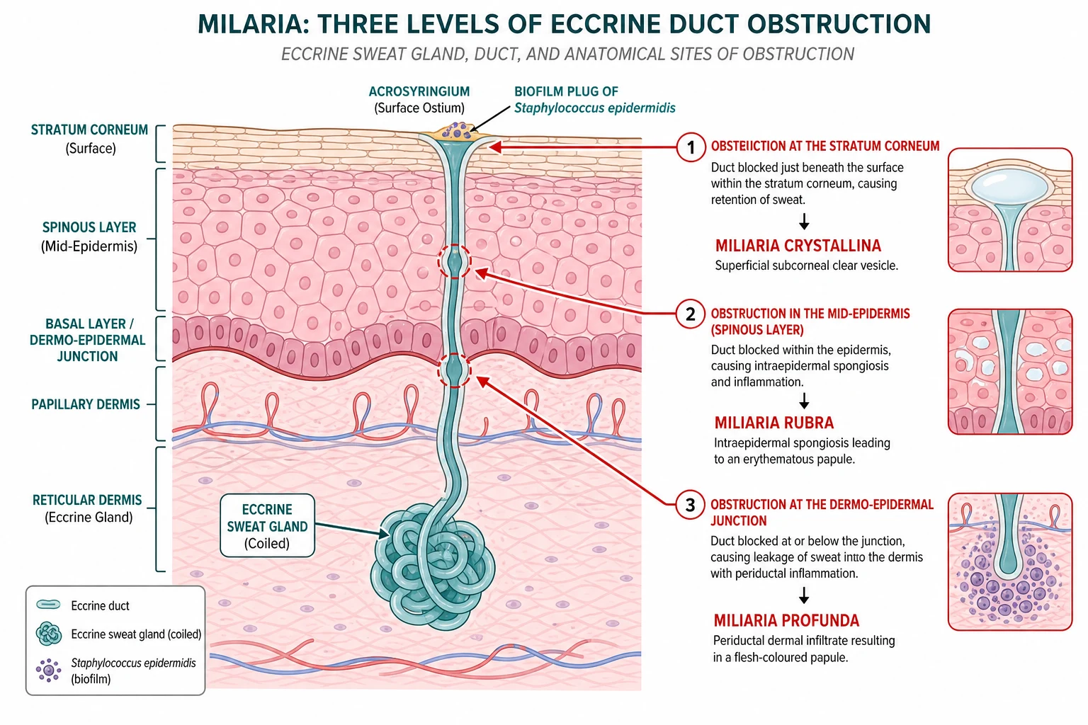
Duct obstruction and extravasation
Obstruction of the intra-epidermal sweat duct prevents sweat reaching the surface. Pressure builds until the duct ruptures proximal to the block, extravasating sweat into the surrounding keratinocytes. The clinical lesion sits at the level of that rupture.[4]
- Crystallina — obstruction at the stratum corneum; superficial subcorneal vesicle of clear sweat.
- Rubra — obstruction in the mid-epidermis; sweat leaks into the spinous layer with surrounding spongiosis and inflammation → erythema and itch.
- Profunda — obstruction at the dermo-epidermal junction; sweat leaks into the papillary dermis with minimal epidermal change → flesh-coloured papule. [1]
The bacterial biofilm
A copious glycocalyx (biofilm) produced by coagulase-negative staphylococci (Staphylococcus epidermidis) colonising the acrosyringium is the best-characterised obstructive mechanism. The biofilm occludes the ostium and triggers focal inflammation; this explains why occluded, over-hydrated stratum corneum (macerated by sweating) is the prerequisite, and why antibacterial measures and de-occlusion resolve the rash.[1]
Hypohidrosis in profunda
Repeated cycles of rubra cause progressive periductal fibrosis and ductal disruption. Once a critical mass of glands is knocked out, sweating falls (hypohidrosis) and thermoregulation is impaired, setting up the rare but dangerous heat-exhaustion phenotype.[5]
Clinical Presentation
Cutaneous findings by subtype
Miliaria crystallina. Crops of tiny (1–2 mm), clear, dew-drop vesicles on a non-erythematous base, easily ruptured by clothing or a finger. Never itchy. Trunk, face, neck and intertriginous areas in neonates; trunk and occluded sites in adults.[2]
Miliaria rubra (prickly heat). Erythematous, 1–3 mm papules and papulovesicles on an erythematous base, with an intense pricking/prickling itch. Symmetrical on the trunk, neck, upper chest, axillae, groin, waistband and flexures — wherever sweat is trapped. Classically flares within minutes of heat exposure and settles with cooling. [1]
Miliaria profunda. Flesh-coloured, firm, 1–3 mm papules without surrounding erythema, on the trunk and limbs, emerging minutes after sweating starts and persisting for an hour or more after cooling. Because it is not itchy, patients often present instead with the consequences — heat intolerance, fatigue, dizziness. [1]
Miliaria crystallina
- Stratum corneum obstruction; subcorneal vesicle without inflammation
- 1-2 mm clear dew-drop vesicles on non-erythematous base, easily ruptured
- Non-pruritic; trunk, face, neck, intertrigines in neonates
- Often follows fever (crystallina = neonatal/febrile form)
- Resolves within 2-3 days of cooling and de-occlusion; no pharmacotherapy needed
Miliaria rubra (prickly heat)
- Mid-epidermis obstruction; intra-epidermal spongiosis + lymphocytic infiltrate
- 1-3 mm erythematous papules/papulovesicles on an erythematous base
- INTENSE prickling itch; trunk, neck, chest, axillae, groin, waistband, flexures
- Most common subtype across all ages; flares within minutes of heat exposure
- Resolves within days of de-occlusion; calamine ± short-course hydrocortisone for severe itch
Miliaria profunda
- Dermo-epidermal junction obstruction; periductal dermal lymphocytic/neutrophilic infiltrate
- 1-3 mm flesh-coloured, firm papules WITHOUT surrounding erythema
- Usually NON-pruritic; trunk and limbs; emerges during sweating
- Followed by periductal fibrosis -> hypohidrosis/anhidrosis and heat intolerance
- The DANGEROUS one: tropical residents, occupational morbidity, heat stroke risk
Miliaria pustulosa
- Rubra + secondary bacterial colonisation (S. aureus / S. epidermidis)
- Superficial pustules on a rubra background; itchy or painful
- Occluded, immunocompromised, ICU, neonatal, or hyperhidrosis patients
- Add antistaphylococcal therapy (topical mupirocin/fusidic acid or oral flucloxacillin)
- Cooling and de-occlusion alone will NOT suffice
Miliaria pustulosa. Superficial pustules superimposed on a rubra background; implies secondary bacterial colonisation and is seen in occluded/immunocompromised hosts. [1]
Distribution and natural history
- Onset within hours to days of heat/occlusive exposure; resolution within days of removing the trigger for crystallina and rubra; profunda may take weeks and recurs with each heat exposure.
- Systemic symptoms are absent unless heat illness supervenes (profunda) or secondary infection develops (pustulosa).
Differential Diagnosis
Mimics of miliaria cluster around four pitfalls: an itchy eruption in flexures (atopic/contact/folliculitis), a febrile neonatal pustular rash (erythema toxicum, transient neonatal pustular melanosis, neonatal cephalic pustulosis), a widespread symmetrical eruption in adults (viral exanthem, drug eruption), and a deep-seated papular eruption in tropical workers (folliculitis, papular urticaria, sarcoidosis). The decision rules below distinguish them reliably: [1]
Viral exanthem
- Systemic features dominate — prodromal fever, coryza, cough, myalgia, Koplik spots (measles)
- Cephalocaudal spread with mucous membrane involvement; palms/soles often spared
- Itch is variable and not prickly; lesions are blanching macules/papules, NOT heat-triggered
- Coxsackie, measles, EBV, parvovirus B19, enterovirus and dengue are the typical causes in returning travellers
Drug eruption (morbilliform)
- Temporal link to a new drug (7-14 days for first exposure, 1-3 days on re-challenge); eosinophilia in ~30%
- Symmetric morbilliform eruption that spares the face and flexures in classic cases; no heat trigger
- Resolves 1-2 weeks after drug withdrawal; rechallenge reproduces it — distinguishing from miliaria
Folliculitis / furunculosis
- Follicular-based pustules centred on a hair shaft; positive bacterial/fungal culture
- Not purely heat-triggered; common in hot/humid climates and in occluded skin but lesions are pilosebaceous
Atopic / contact dermatitis
- Chronic, relapsing, intensely itchy and lichenified; spares eccrine-distribution sites
- Personal/family atopy history; positive patch test (contact); flexural accentuation
- NOT transiently relieved by cooling alone; responds to emollients and topical steroids over weeks
Erythema toxicum neonatorum
- Erythematous macules with central pale papule/pustule; eosinophils on Wright-stained smear
- Present within 24-72 h of birth; resolves spontaneously in 5-7 days; no heat trigger
- Differs from neonatal miliaria crystallina which begins AFTER birth with sweating/fever
Transient neonatal pustular melanosis
- Present AT BIRTH (not triggered); superficial vesiculopustules with neutrophilic smear
- Lesions rupture leaving a hyperpigmented macule with a collarette of scale; resolves over weeks-months
- Affects any skin phototype; common in darker-skinned neonates
Papular urticaria
- Chronic, recurrent, grouped urticarial papules with central punctum (insect bite reaction)
- Children 2-10 y; exposed sites (forearms, legs); NOT sweat-distribution; intensely itchy
| Mimic | Distinguishing features |
|---|---|
| Viral exanthem (enterovirus, measles) | Systemic features, prodrome, Koplik spots, caudal spread; not itchy-prickly |
| Drug eruption | Temporal link to new drug; eosinophilia; no predilection for occluded flexures |
| Folliculitis | Follicular-based pustules; culture positive; not triggered purely by heat |
| Erythema toxicum neonatorum | Erythematous macules with central pale papule/pustule; eosinophilic smear; fades in days |
| Transient neonatal pustular melanosis | Present at birth; pigmented macule as pustule resolves; no heat trigger |
| Atopic dermatitis (infants) | Chronic, flexural, dry skin; personal/family atopy |
| Acrodermatitis enteropathica / scabies | Distribution and burrows (scabies); peri-orificial/acral (zinc deficiency) |
| Subcorneal pustular dermatosis (Sneddon-Wilkinson) | Annular/circular flaccid pustules in flexures of older adults; not heat-triggered |
Histopathology
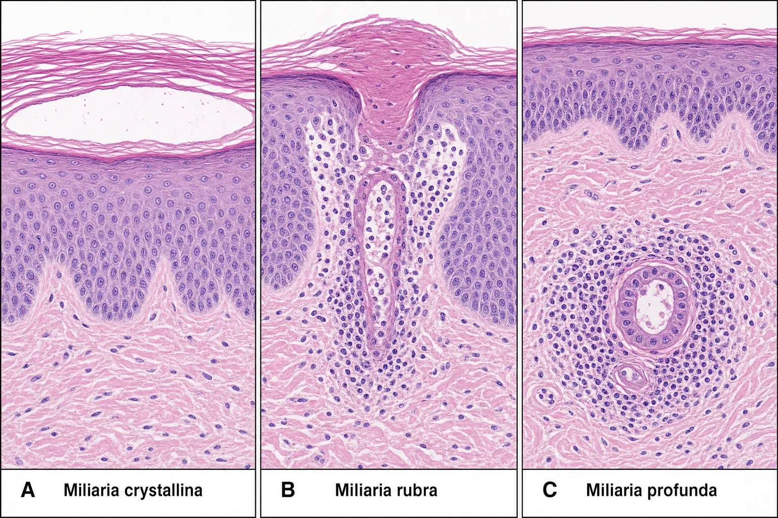
- Crystallina: subcorneal, intra- or subcorneal vesicle without inflammation.
- Rubra: intra-epidermal spongiosis with a lymphocytic infiltrate around and within the sweat duct at the spinous layer.
- Profunda: normal or near-normal epidermis; periductal (eccrine) neutrophilic and lymphocytic infiltrate in the papillary dermis.
- Biopsy is almost never required — the diagnosis is clinical; biopsy only if a blistering disorder or autoinflammatory cause is genuinely suspected. [1]
Clinical & Bedside Assessment
- Diagnosis is clinical. No formal severity score exists for miliaria.
- Practical severity classification (clinic / exam use):
- Mild — limited area, no itch impact, no systemic features.
- Moderate — widespread, symptomatic itch interfering with sleep.
- Severe — extensive profunda with heat intolerance, secondary infection, or pustulosa.
- Assess for heat illness in profunda: core temperature, hydration, orthostatic symptoms.
- Assess for secondary infection in pustulosa: surrounding cellulitis, lymphangitis, fever. [1]
Investigations [1]
Diagnosis is clinical and does not require investigations. A directed history (heat/occlusion trigger, distribution, itch, prior episodes) plus targeted examination is sufficient in >95% of cases. [1]
- Skin swab for bacterial culture — only for pustulosa with spreading cellulitis, recalcitrant rash, or risk factors for MRSA (recent hospitalisation, prior MRSA, military field training, contact sports); not for simple rubra.
- Skin biopsy — only if a blistering disorder, autoinflammatory pustular dermatosis, Hailey-Hailey, or subcorneal pustular dermatosis (Sneddon-Wilkinson) is genuinely suspected, or if histology would change management (e.g. suspected GvHD in stem-cell transplant). The histology of miliaria is largely non-specific; biopsy is overused.
- Wright-stained smear of pustule contents — distinguishes eosinophilic (erythema toxicum, neonatal cephalic pustulosis) from neutrophilic (transient neonatal pustular melanosis, pustulosa, bacterial infection) vesicles in equivocal neonatal cases.
- Dermoscopy (entomodermoscopy) — increasingly described as an adjunct: crystallina shows a sharply demarcated oval clear structure with a 'dewdrop on a rose petal' pattern, rubra shows pink-to-red globules around the acrosyringium, and profunda shows ill-defined structureless pale areas corresponding to periductal oedema. Use is supportive, not diagnostic.
- Bloods — not required for typical crystallina or rubra. Order a basic work-up (FBC, U&E, CRP, CK, LFTs, coagulation, ABG) in any patient who has collapsed in heat or is suspected of heat stroke, especially with extensive miliaria profunda.
- Quantitative sudomotor axon reflex testing (QSART) or thermoregulatory sweat testing — research-grade tools that document the distribution and severity of hypohidrosis/anhidrosis in patients with chronic profunda; useful to confirm post-miliarial hypohidrosis but rarely needed in routine care.
- Modified starch-iodine or Quinizarin sweat printing — visualise the distribution of sweating and confirm the abrupt cut-off of anhidrosis over previously affected areas; again, research and complex-clinic territory.
- Reconsider the diagnosis in a neonate with persistent or atypical 'miliaria' — and consider bacterial/viral PCR, KOH and Tzanck smear before settling on the diagnosis.
- Heat-stroke workup (if heat illness suspected) — FBC, U&E, CK, urinalysis for myoglobin, LFTs, coagulation, lactate, ABG; serial monitoring over 48–72 h for rhabdomyolysis, AKI, DIC and hepatic injury. [1]
Management — General Measures & Cooling
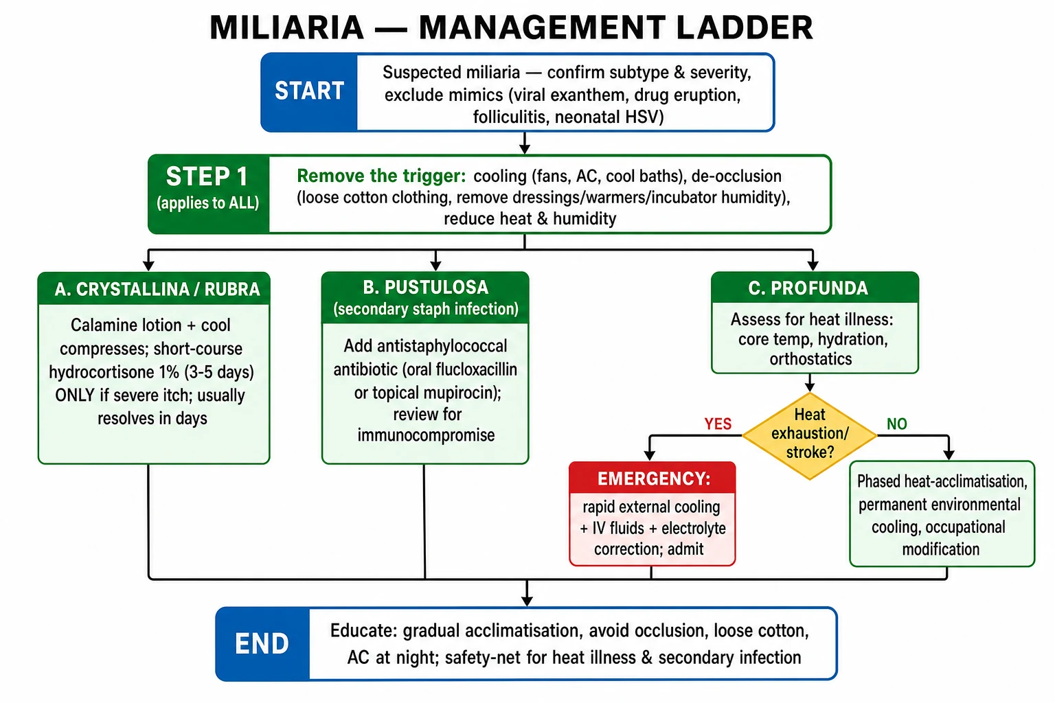
The cornerstone is removal of the obstructive trigger:[4]
- Cool the patient — air conditioning, fans, cool baths/showers, cool compresses.
- De-occlude — loose, breathable cotton clothing; remove or shorten use of occlusive dressings, casts, or warmers; ventilate incubators.
- Avoid heat and humidity — environmental control is curative; air-conditioned sleep is often the single most effective intervention.
- Reduce sweating — avoid strenuous exercise in heat; acclimatise gradually to tropical climates.
- Skin care — gentle cleansing; avoid heavy emollients that may further occlude duct ostia. [1]
Most crystallina and rubra resolve within days of de-occlusion without pharmacotherapy. [1]
Management — Pharmacotherapy
Pharmacotherapy in miliaria is reserved for itch control in inflamed rubra and for confirmed secondary bacterial infection (pustulosa). The cornerstone is still trigger removal; do not over-treat what a fan and a loose cotton shirt will cure. [1]
Symptomatic and anti-inflammatory
- Calamine lotion and cool compresses — first-line symptomatic itch relief in rubra; safe in pregnancy and in infants older than 6 months.
- Low-potency topical corticosteroid (e.g. hydrocortisone 1% for 3–5 days) — reserved for severely itchy/inflamed rubra; higher potencies and prolonged courses are not justified, are not steroid-sparing, and may worsen sweating-rate dysregulation. [1]
Hydrocortisone 1% cream
Topical corticosteroid — short-course rescue for severe rubra itch
Dose
Apply a thin film to affected areas twice daily for 3–5 days
Antistaphylococcal therapy — for pustulosa and secondary infection
- Topical antibiotics — mupirocin 2% ointment or fusidic acid 2% ointment applied 2–3 times daily for 7 days — first-line for localised pustulosa or impetiginised rubra.
- Oral antistaphylococcal agents — flucloxacillin (or erythromycin/clarithromycin in penicillin-allergy) — used when pustulosa is widespread, when cellulitis is present, or when topical therapy has failed. Treat for 7 days in adults; review earlier in infants. [1]
Mupirocin 2% ointment
Topical antibiotic — first-line for localised pustulosa/impetiginised miliaria
Dose
Apply thin layer to affected areas 2–3 times daily for 7 days
Fusidic acid 2% cream/ointment
Topical antibiotic — alternative to mupirocin for localised pustulosa
Dose
Apply 2–3 times daily for 7 days
Flucloxacillin
Oral anti-staphylococcal penicillin — first-line systemic therapy for widespread pustulosa or cellulitis
Dose
Adult: 500 mg to 1 g four times daily for 7 days. Child 1 month–18 years: 12.5–25 mg/kg (max 500 mg–1 g) four times daily for 7 days
Adjuncts and 'no role' list
- Topical anhydrous lanolin has historical support for neonatal crystallina by softening obstructive keratin plugs; not routinely needed and may itself occlude.
- Antiseptics (chlorhexidine wash, povidone–iodine scrub) are useful adjuncts when there is clinical infection; they are not curative on their own.
- No role for oral antifungals, oral antibiotics prophylactically (only treat confirmed infection), oral corticosteroids, or systemic antihistamines as primary therapy; oral antihistamines may be considered for severe nocturnal itch in adults with profound rubra but should not be relied upon as monotherapy. [1]
Management — Special Sites & Scenarios
- Neonates in incubators: reduce incubator temperature/humidity to the minimum compatible with thermoneutrality; avoid over-swaddling.
- ICU/bedbound adults under radiant warmers: lower warmer output, turn regularly, change occlusive dressings, use breathable drapes.
- Tropical residents with profunda: phased heat-acclimatisation, permanent environmental cooling (fans, AC at night), occupational modification; treat any heat exhaustion emergently.
- Pustulosa: de-occlude + antistaphylococcal therapy + cooling. [1]
Complications, Emergencies & Pitfalls
- Secondary bacterial infection (impetiginisation, cellulitis) of rubra/pustulosa — the commonest complication; needs antimicrobial therapy. Look for surrounding cellulitis, lymphangitic streaking, fever, or systemic upset.
- Heat exhaustion / heat stroke from extensive profunda hypohidrosis — the dangerous, rare complication; presents with hyperthermia (>40°C core), tachycardia, hypotension, hot dry skin, confusion, seizure or multi-organ failure. The driver is a critical mass of destroyed eccrine ducts producing anhidrosis; morbidity is high if cooling is delayed beyond 30 minutes.[5]
- Sleep disturbance / secondary excoriation in itchy rubra, which in children can disrupt feeding and weight gain.
- Folliculitis and furunculosis secondary to scratching, particularly in adolescents and in adult men in hot climates.
- Miliaria-induced anhidrotic heat exhaustion (postmiliarial hypohidrosis): chronic anhidrosis of the affected areas persists for weeks after the rash clears, leading to compensatory hyperhidrosis elsewhere and a sense of persistent heat intolerance even after cooling measures are instituted.
Heat-stroke emergency pathway for extensive profunda
Any patient with widespread miliaria profunda who collapses in heat should be presumed to have heat stroke until proven otherwise. The cooling window is narrow and outcomes worsen with every minute of delay above a core temperature of 40°C. [1]
Heat-stroke emergency pathway (suspect profound anhidrosis from extensive miliaria profunda)
Recognise and call for help
Core temp ≥ 40 °C, hot dry skin (or profound anhidrosis on the affected areas), CNS dysfunction (confusion, ataxia, seizure, coma), hypotension. Move the patient to shade; remove occlusive clothing.
Rapid external cooling — cold-water immersion or evaporative + ice packs
Cold-water immersion to neck is the gold standard if available (cool to < 39 °C within 30 min). If impractical, use evaporative cooling (tepid water spray + fans) plus ice packs to neck, axillae and groins. Monitor core temperature continuously (rectal or oesophageal probe).
Airway, breathing, circulation
High-flow O₂, two large-bore IV cannulae, IV isotonic crystalloid (0.9% saline or Ringer's lactate, 1 L bolus, repeat to haemodynamic endpoints). Treat seizures with IV benzodiazepines; correct hypoglycaemia, hypokalaemia and metabolic acidosis. AVOID antipyretics — they do not work in heat stroke and may worsen hepatic/renal injury.
Treat secondary infection if present
If associated pustulosa, cellulitis or bacteraemia is suspected, send blood cultures, wound swab and start IV empiric anti-staphylococcal therapy (e.g. flucloxacillin 1–2 g IV q6h or equivalent). Cooling and antibiotics are run in parallel, not sequentially.
Disposition and cooling-target
ICU/HDU admission. Cool to a target core of < 39 °C, then 'permissive slowing' to 38.5 °C to avoid overshoot. Watch for rhabdomyolysis (CK, urinalysis for myoglobin), DIC, AKI and hepatic injury over the next 48–72 h. Document profound anhidrosis on the affected skin as the diagnostic clue linking the event to miliaria profunda.
Classic pitfalls in diagnosis and management
[1]Special Populations
Neonates
Crystallina is the neonatal default — sweat ducts are immature and are easily blocked by even mild hyperthermia (over-bundling, phototherapy, radiant warmers, incubators with high humidity). It is benign and self-limiting; review swaddling/incubator settings and ambient temperature; reduce humidity to 40–60% when feasible. Exclude erythema toxicum neonatorum and transient neonatal pustular melanosis — both present at or near birth, are not heat-triggered, and behave differently on Wright-stained smears (eosinophils vs neutrophils). Miliaria rubra in infants must be distinguished from neonatal cephalic pustulosis (Malassezia-driven, no heat trigger), infantile seborrhoeic dermatitis (greasy yellow scale on scalp/axillae) and erythema toxicum. [1]
Neonatal miliaria crystallina — typical timing in an incubator/baby with fever
Pregnancy
No change in approach: cooling and de-occlusion are first-line and are safe at any gestational age; topical hydrocortisone 1% is acceptable short-course. Heat exposure and prickly heat are uncomfortable in pregnancy; topical anhydrous lanolin and breathable cotton are simple adjuncts. Avoid prolonged or potent topical steroids, and avoid systemic antibiotics unless pustulosa is confirmed. [1]
Elderly and bedbound
Focus on turning, de-occlusion and thermal regulation. Watch for secondary infection (impetiginisation, candidal intertrigo, cellulitis). Beware dual diagnoses — scabies, drug eruption and contact dermatitis from incontinence pads can coexist with miliaria. Maintain room temperature ≤ 22 °C where feasible. [1]
Immunocompromised and transplant recipients
Higher risk of pustulosa and atypical, more severe presentation; lower threshold for skin swab, blood culture, and antimicrobial therapy. Solid-organ transplant recipients on calcineurin inhibitors (cyclosporine, tacrolimus) have reduced sweating and altered keratinocyte turnover, occasionally producing anhidrosis and a miliaria-like pruritic eruption that improves with dose reduction; graft-versus-host disease in stem-cell transplant may mimic widespread rubra and needs biopsy. [1]
Athletes, military recruits, occupational groups
Phased heat acclimatisation (10–14 days), scheduled hydration, breathable kit, scheduled cooling breaks, and access to showers are the evidence-based controls for occupational rubra. Academies and deployment training programmes have near-eliminated severe outbreaks with structured acclimatisation protocols. [1]
Comorbidities
- No specific comorbidity screening is required for typical miliaria.
- Recurrent/profunda in a non-tropical host should prompt consideration of hyperhidrosis, febrile illness, occlusive dressing/cast, or ICU warming protocol rather than an intrinsic disease. [1]
Prognosis & Follow-Up
- Excellent for crystallina and rubra: resolves in days once the trigger is removed; recurrence is prevented by ongoing environmental control.
- Profunda recurs with each heat exposure and may persist for weeks; heat intolerance can be disabling in tropical workers.
- No routine follow-up is required for straightforward cases; review only if heat illness, secondary infection, or diagnostic uncertainty. [1]
Evidence, Guidelines & Regional Differences
- Evidence base is largely observational and expert opinion; high-quality RCTs are scarce because miliaria is benign, self-limiting and ethically hard to placebo-control. Most modern recommendations derive from dermatology textbook consensus, neonatal/paediatric reviews and occupational heat-illness literature.[1][3]
- Regional differences are stark: in tropical countries and the Gulf states, miliaria rubra and profunda are major occupational morbidities in military recruits, construction and agriculture workers, and a recognised contributor to heat illness; in temperate climates, miliaria is predominantly a neonatal, ICU or summer phenomenon. Prickly heat (rubra) is the leading heat-related dermatosis in deployed soldiers during the Gulf and Afghanistan campaigns, with documented impacts on operational readiness.
- Landmark evidence — heat acclimatisation reduces incidence: classic military studies (notably from the 1940s US Army Desert Testing Programme and corroborated by the Israeli Defense Forces acclimatisation data) showed that structured 10–14 day heat acclimatisation reduces rubra incidence by 60–80% in recruits.
- Antibacterial skin cleansers (chlorhexidine, triclosan-containing washes) modestly shorten rubra duration in controlled trials of military recruits; the effect size is small and cooling/de-occlusion remain the dominant intervention.
- No disease-specific international guideline exists; recommendations reflect dermatology textbook consensus. Management ladders are taken from neonatal/paediatric reviews and adult occupational-medicine protocols.
- Australian & NZ (RACP/ACD), British (BAD) and American (AAD) dermatology consensus statements uniformly recommend conservative management first; the BAD and AAD position papers emphasise avoiding potent topical steroids and avoiding antibiotics unless pustulosa or impetiginisation is confirmed.
Heat acclimatisation in military recruits
US Army / IDF Desert Field Studies (1940s–1980s) and modern meta-analyses
Prospective cohort / field-controlled study in conscripts deployed to hot-arid climates; structured 10–14 day progressive heat exposure with scheduled hydration and graded aerobic work
Key finding
Structured acclimatisation reduces the incidence of miliaria rubra in new recruits by 60–80% and reduces concurrent heat-illness events by a similar order of magnitude; benefits persist for 1–3 months after re-deployment.
Practice change
Mandatory pre-deployment heat acclimatisation protocols are now standard across NATO, IDF, ADF and other modern military forces — the single most effective population-level intervention for occupational miliaria.
Red Flags
Exam application bank (NEET-PG / INICET)
One-line answer
Miliaria is a common, self-limiting disorder of eccrine sweat gland obstruction in which retained sweat leaks into the surrounding tissue at a level determined by the depth of duct obstruction — producing three classic subtypes: miliaria crystallina (superficial, stratum corneum, clear vesicles), miliaria rubra / prickly heat (mid-epidermal, itchy red papules), and miliaria profunda (dermo-epidermal, flesh-coloured papules with risk of hypohidrosis and heat exhaustion). Triggers are heat, humidity, occlusion, fever and intensive-care incubation. Fellowship-level assessment demands mastery of the level-of-obstruction → morphology correlation, the neonatal vs adult presentations, secondary infection (miliaria pustulosa), and the rare but life-threatening thermoregulatory failure of extensive profunda in tropical residents.
Worked stems (answer without another resource)
Stem 1 — Classic presentation. Map symptoms to mechanism; name the first investigation and first treatment step with dose/route if drug therapy is standard. [1]
Stem 2 — Unstable / complicated. List red flags that force immediate resuscitation, theatre, ICU, antidote, or reperfusion — and what you do in the first 15 minutes. [1]
Stem 3 — Atypical group. Elderly, pregnancy, child, or immunocompromised: how presentation and thresholds change. [1]
Stem 4 — Differential trap. Name the three closest mimics and one discriminator for each. [1]
Stem 5 — Disposition. Who goes home with safety-netting, who is admitted, who needs HDU/ICU/theatre, and what follow-up is mandatory. [1]
Rapid viva checklist
- Definition + classification
- Pathophysiology chain
- Bedside signs / criteria
- Score with exact components (if any)
- Emergency bundle
- Definitive therapy with doses
- Complications of disease and of treatment
- Special populations
- Guideline/trial name if classic
- Three exam traps
Coverage self-check
If you cannot answer any stem above from this page alone, re-read the matching section — the page is intended to be self-sufficient for final-prof and NEET-PG/INICET questions on Miliaria / heat rash.
[1]References
- [1]O'Connor NR, McLaughlin MR, Ham P. Newborn skin: Part I. Common rashes Am Fam Physician, 2008.PMID 18236822
- [2]Palaniappan V, Sadhasivamohan A, Sankarapandian J, et al. Miliaria crystallina Clin Exp Dermatol, 2023.PMID 36692206
- [3]Chadha A, Jahnke M. Common Neonatal Rashes Pediatr Ann, 2019.PMID 30653638
- [4]Feng E, Janniger CK. Miliaria Cutis, 1995.PMID 7796612
- [5]Kirk JF, Wilson BB, Chun W, et al. Miliaria profunda J Am Acad Dermatol, 1996.PMID 8912605