Dermatology · Medicine
Mongolian spot / congenital dermal melanocytosis
Also known as Congenital dermal melanocytosis · Dermal melanocytosis · Sacral blue-grey macule of infancy · Congenital dermal melanocytosis (historical Mongolian spot)
Multi-board congenital dermal melanocytosis (historical Mongolian spot): sacral blue-grey macule present at birth, dermal melanocyte arrest and Tyndall colour, usual childhood fading, spectrum with nevus of Ota/Ito, bruise and child-protection differential framed carefully, extensive extrasacral patterns and lysosomal storage disease associations, documentation and reassurance-first management with selective cosmetic options.
On this page & tools
Your progress
Saved locally on this device.
Exam tags
Red flags
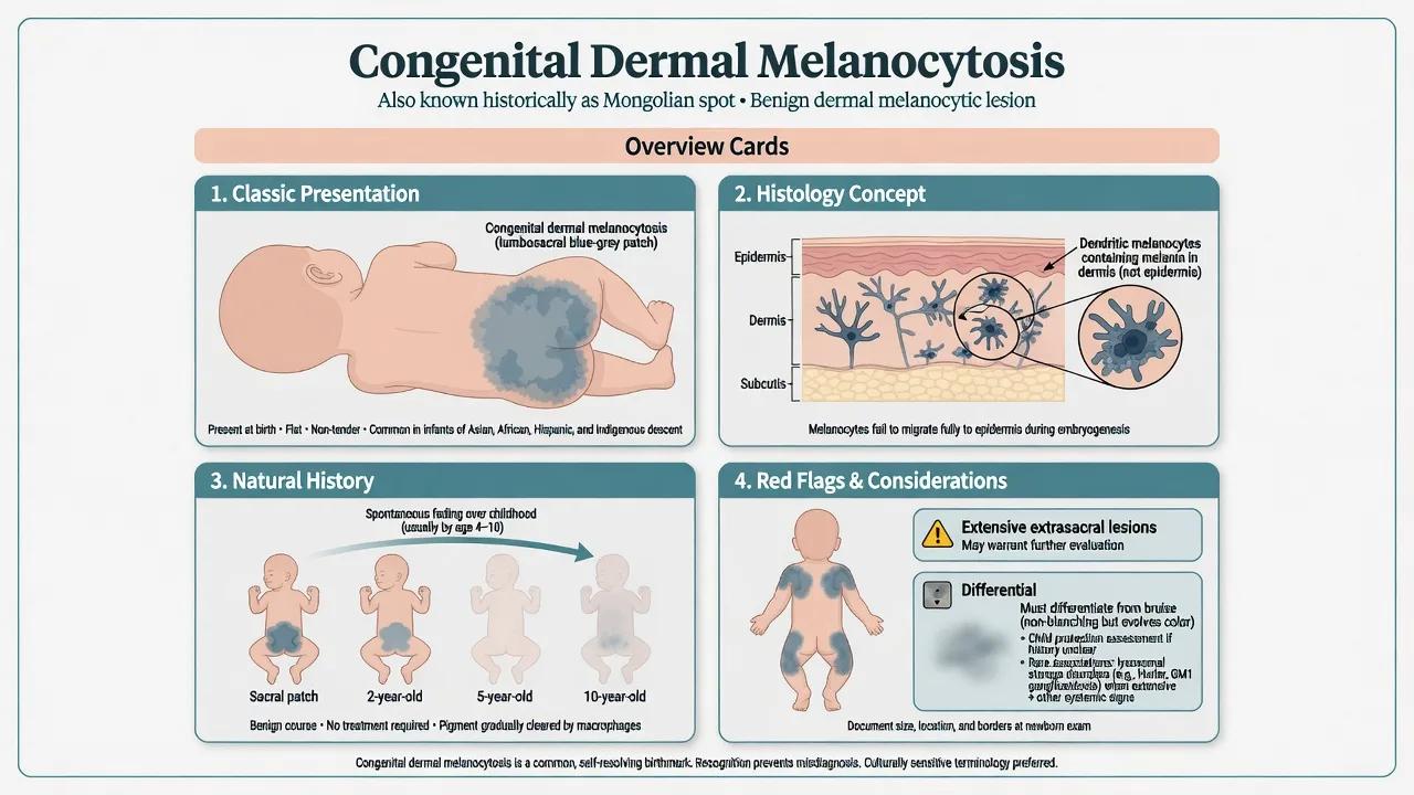
Overview & Definition
Congenital dermal melanocytosis (CDM) is a benign developmental collection of melanocytes in the dermis producing a blue-grey macule or patch. The historical eponym Mongolian spot remains widely recognised in exams but is increasingly replaced by descriptive terminology because it is imprecise and culturally loaded.[1][4][5] Classic lesions are present at birth, commonest on the lumbosacral/gluteal skin, and are a staple of neonatal dermatology teaching.[8]
Classification
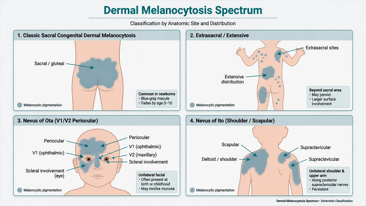
Classic sacral CDM
Extrasacral / extensive
Nevus of Ota
Nevus of Ito
Rare variants
Epidemiology & Risk Factors
Classic sacral CDM is common worldwide and is particularly frequent in infants of Asian, African, Hispanic, and Indigenous ancestry; lower but non-zero rates occur in other groups.[1] Nearly all true CDM is congenital or appears in early infancy. Extensive extrasacral disease is less common and is the subset linked in the literature to storage-disease associations when additional clinical clues exist.[2][3]
Pathophysiology
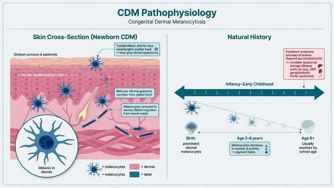
Neural-crest melanocytes that fail to complete epidermal migration remain in the dermis. Deep dermal melanin appears blue-grey clinically (optical depth / Tyndall-type effect). Over childhood, dermal melanocytes typically diminish and classic sacral patches fade.[1][5] Extensive dermal melanocytosis coexisting with storage disorders has been hypothesised to reflect abnormal melanocyte–matrix interactions in those diseases.[2]
Clinical Presentation
Classic CDM
Homogeneous flat blue-grey or blue-black macule/patch over sacrum or buttocks; non-tender; present at birth; no scale or induration.[1][5]
Natural history
Progressive lightening over years is expected for classic lesions; many are barely visible by school age, though residual pigment can persist.[1]
Extensive / extrasacral
Multiple sites (back, shoulders, limbs) or unusually large patches; may fade more slowly or persist; document carefully and examine for other findings.[2][6]
Spectrum lesions
Nevus of Ota — unilateral facial/periocular blue-grey pigment ± sclera. Nevus of Ito — shoulder girdle. These usually persist and are managed differently from fading sacral CDM.[6]
Differential Diagnosis
| Finding | Prefer | Key distinguisher |
|---|---|---|
| Blue-grey sacrum from birth | Classic CDM | Stable colour; no day-to-day bruise evolution |
| Multi-coloured patch evolving over days | Bruise | Colour sequence, history of trauma, resolution weeks |
| Unilateral periocular + sclera | Nevus of Ota | Site and persistence |
| Shoulder/scapular blue patch | Nevus of Ito | Distribution |
| Extensive CDM + organomegaly/delay | Storage disease context | Not CDM alone — systemic work-up |
Bruise versus CDM is a high-stakes paediatric differential: CDM is present from birth and does not march through the classic bruise colour stages; bruises appear after trauma and evolve over days to weeks. Safeguarding decisions require the whole history and exam, not colour alone.[7]
Clinical & Bedside Assessment
- Confirm presence from birth (parental history, newborn notes).
- Map site(s), measure, photograph with consent.
- Survey entire skin for extrasacral lesions.
- If extensive/persistent, review growth, development, organomegaly, coarse features.
- For periocular pigment, check sclera and consider ophthalmology. [1]
Investigations
None for classic sacral CDM. Biopsy is almost never required when the clinic is classic. Extensive dermal melanocytosis plus independent systemic clues may justify metabolic/genetic pathways informed by paediatric specialty teams; CDM alone is not a diagnosis of storage disease.[2][3]
Management — Resuscitation
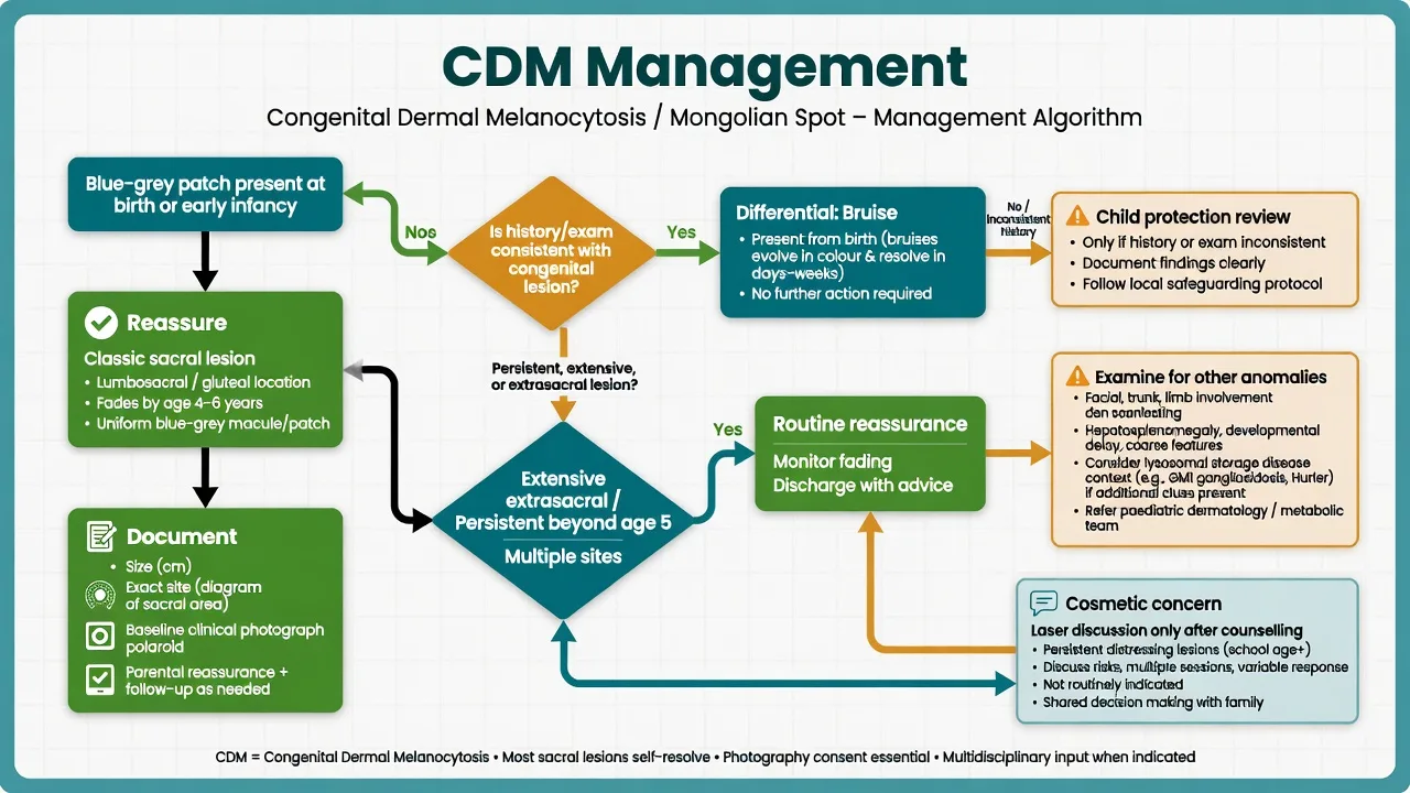
No acute dermatologic resuscitation for classic CDM. Escalate safeguarding only when the overall picture is inconsistent with a congenital lesion.[7]
Management — Definitive & Stepwise
Classic sacral CDM: reassurance about benign nature and expected fading; document in the record so later clinicians do not misread residual pigment as trauma.[1][8]
Extensive / persistent disease: full clinical context; paediatric dermatology; storage-disease consideration only with additional clues.[2][3]
Cosmetic residual pigment: discuss lasers (often Q-switched or related pigment lasers in contemporary practice) only after counselling on incomplete response, multiple sessions, and risks — not routine for fading infancy lesions.[6]
Specific Subtypes & Scenarios
Nursery documentation prevents later false trauma labels.[7]
Halo CDM is a rare morphological variant noted in case reports.[9]
Nevus of Ota needs ocular surface awareness and long-term cosmetic/ophthalmic planning rather than “it will definitely fade like a sacral spot.”[6]
Complications & Pitfalls
- Calling every blue patch on a dark-skinned infant “always abuse” or, conversely, missing true trauma by overcalling CDM.[7]
- Biopsying classic sacral CDM.
- Ignoring extensive extrasacral pigment when the child has other storage-disease signs.[2]
- Using pejorative or imprecise eponyms when descriptive language is clearer.[4]
Prognosis & Disposition
Excellent for classic sacral CDM — most fade substantially. Persistent extrasacral and Ota/Ito lesions need individualised follow-up and cosmetic counselling.[1][6]
Special Populations
Newborns: examine and chart. School-age children: residual sacral pigment may still be visible. Adolescents seeking laser: shared decision-making after natural fading window. [1]
Evidence, Guidelines & Regional Differences
Descriptive reviews remain the main teaching base for classic CDM.[1] Arch Dermatol association work links extensive dermal melanocytosis with selected lysosomal storage diseases in the appropriate multi-feature context.[2] Terminology papers argue for descriptive renaming.[4] Forensic/paediatric literature highlights bruise–CDM diagnostic difficulty.[7]
Exam Pearls
BLUE pearls
- Dermal depth → blue clinical colour.[1]
- Not an evolving bruise rainbow.[7]
- Document once, spare the family later trauma of misinterpretation.[8]
- Ota/Ito are cousins, not identical natural histories.[6]
Exam application bank (NEET-PG / INICET)
One-line answer
Multi-board congenital dermal melanocytosis (historical Mongolian spot): sacral blue-grey macule present at birth, dermal melanocyte arrest and Tyndall colour, usual childhood fading, spectrum with nevus of Ota/Ito, bruise and child-protection differential framed carefully, extensive extrasacral patterns and lysosomal storage disease associations, documentation and reassurance-first management with selective cosmetic options.
Worked stems (answer without another resource)
Stem 1 — Classic presentation. Map symptoms to mechanism; name the first investigation and first treatment step with dose/route if drug therapy is standard. [1]
Stem 2 — Unstable / complicated. List red flags that force immediate resuscitation, theatre, ICU, antidote, or reperfusion — and what you do in the first 15 minutes. [1]
Stem 3 — Atypical group. Elderly, pregnancy, child, or immunocompromised: how presentation and thresholds change. [1]
Stem 4 — Differential trap. Name the three closest mimics and one discriminator for each. [1]
Stem 5 — Disposition. Who goes home with safety-netting, who is admitted, who needs HDU/ICU/theatre, and what follow-up is mandatory. [1]
Rapid viva checklist
- Definition + classification
- Pathophysiology chain
- Bedside signs / criteria
- Score with exact components (if any)
- Emergency bundle
- Definitive therapy with doses
- Complications of disease and of treatment
- Special populations
- Guideline/trial name if classic
- Three exam traps
Coverage self-check
If you cannot answer any stem above from this page alone, re-read the matching section — the page is intended to be self-sufficient for final-prof and NEET-PG/INICET questions on Mongolian spot / congenital dermal melanocytosis.
Expanded exam teaching (depth pass)
Clinical reasoning
For Mongolian spot / congenital dermal melanocytosis, examiners test whether you can prioritise life threats, choose the right first test, and give specific therapy (agent, dose, route, timing). Generic phrases without numbers score poorly.
Mechanism → feature map
Build a short chain: cause → pathophysiologic intermediate → clinical feature → complication. Every major symptom in the classic vignette should sit on that chain.
Investigation strategy
- Bedside/first-line tests that change immediate management
- Confirmatory or staging tests
- What a normal result does not exclude
- When not to delay treatment for imaging (unstable patient)
Management ladder
- Resuscitation / ABC / sepsis or haemorrhage bundle as relevant
- Specific antidote / procedure / antimicrobial / reperfusion / surgery
- Supportive care and monitoring targets
- Definitive long-term therapy and secondary prevention
- Disposition and safety-net advice
Special populations
Always prepare one line each for children, pregnancy, elderly, renal/hepatic impairment, and immunocompromised patients when the topic allows.
Pitfalls that fail candidates
- Treating the number not the patient
- Missing pregnancy status when relevant
- Imaging before stabilisation
- Wrong empiric cover or wrong antidote timing
- Incomplete counselling on recurrence, adherence, or red-flag return
Multi-board congenital dermal melanocytosis (historical Mongolian spot): sacral blue-grey macule present at birth, dermal melanocyte arrest and Tyndall colour, usual childhood fading, spectrum with nevus of Ota/Ito, bruise and child-protection differential framed carefully, extensive extrasacral patterns and lysosomal storage disease associations, documentation and reassurance-first management with selective cosmetic options. [1]
Structured revision sheet
Must-know numbers and names
List every score, size threshold, dose, and time window from this topic on a blank page from memory, then check against the sections above.
Three classic MCQ angles
- Most likely diagnosis given a vignette
- Next best step in management
- Most appropriate investigation
Three classic SAQ angles
- Pathophysiology in five steps
- Management algorithm with doses
- Complications and prevention
Clinical station flow
Greet → focused history → targeted exam → investigations → explain diagnosis → emergency care → definitive plan → safety-net / follow-up → answer examiner questions on mechanism and pitfalls.
Exam anchors
References
- [1]Gupta D, Thappa DM. Mongolian spots Indian J Dermatol Venereol Leprol, 2013.PMID 23760316
- [2]Hanson M, Lupski JR, Hicks J, et al. Association of dermal melanocytosis with lysosomal storage disease: clinical features and hypotheses regarding pathogenesis Arch Dermatol, 2003.PMID 12873889
- [3]Tran MC, Lam JM. Cutaneous Manifestations of Mucopolysaccharidoses Pediatr Dermatol, 2016.PMID 27601403
- [4]Yale S, Tekiner H, Yale ES. Reimagining the Terms Mongolian Spot and Sign Cureus, 2021.PMID 35036226
- [5]Zhu J, Zhu Y, Lin X. Dermatology Images: Congenital dermal melanocytosis J Am Acad Dermatol, 2026.PMID 42364768
- [6]Zhu J, Cen Q, Chang R, et al. Patchy Dermal Melanocytosis: Differential Diagnosis and Management J Cosmet Dermatol, 2025.PMID 39485055
- [7]Rzepczyk S, Świderski P, Rusek D, et al. The so-called Mongolian spots and suspected child abuse - difficulties in differential diagnosis Arch Med Sadowej Kryminol, 2024.PMID 40366721
- [8]Mallory SB. Neonatal skin disorders Pediatr Clin North Am, 1991.PMID 1870904
- [9]Bart BJ, Olson CL. Congenital halo mongolian spot J Am Acad Dermatol, 1991.PMID 1810989