Dermatology · Medicine
Morphea (localized scleroderma)
Also known as Morphea · Localized scleroderma · Circumscribed scleroderma · Linear scleroderma · En coup de sabre
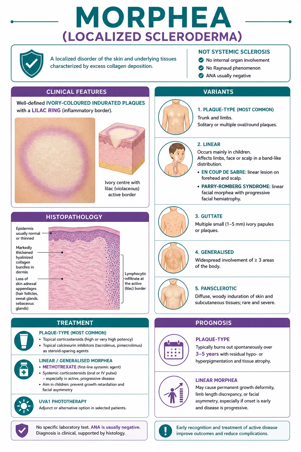
Morphea (localized scleroderma) is a fibrosing inflammatory disorder confined to the skin and subcutaneous tissue WITHOUT internal organ involvement, distinguishing it absolutely from systemic sclerosis. Presents as ivory-coloured indurated plaques with a violaceous lilac ring (plaque-type, most common in adults) or as a linear band on a limb or the face (linear morphea, most common in children). Linear facial disease includes en coup de sabre (forehead sabre-cut) and Parry-Romberg syndrome (hemifacial atrophy), which carry risk of growth deformity and eye and CNS involvement. NO Raynaud, NO SSc-specific antibodies (anti-Scl-70, anti-centromere negative), NORMAL nailfold capillaroscopy. Treatment: topical corticosteroids, calcipotriol, or calcineurin inhibitors for limited plaque-type; phototherapy (UVA1 or narrowband UVB) for widespread plaque-type; methotrexate plus corticosteroids first-line systemic for active linear, generalised, or deep disease. Plaque-type burns out over 3 to 5 years; linear may cause permanent deformity in growing children. Morphea does NOT progress to systemic sclerosis.
On this page & tools
Your progress
Saved locally on this device.
Exam tags
Red flags
Overview & Definition
Morphea, also called localized (or localised) scleroderma or circumscribed scleroderma, is a fibrosing inflammatory disorder of the skin in which excessive collagen deposition produces circumscribed areas of cutaneous induration and, in some subtypes, sclerosis of the underlying subcutaneous fat, fascia, muscle, and bone. The defining and most heavily examined feature is that the disease process stops at the skin and its adnexal structures — it does not produce the widespread microvascular and visceral fibrosis of systemic sclerosis.[1][3]
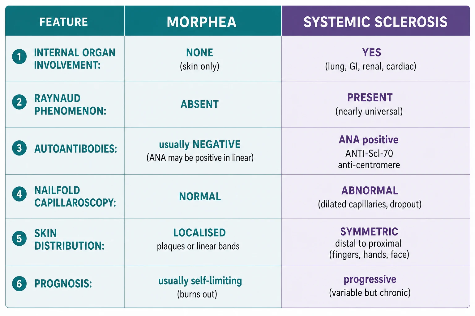
The distinction from systemic sclerosis is absolute and is the single most rewarding discriminator in exams. Morphea is not systemic sclerosis confined to the skin; it is a biologically distinct entity. Three bedside facts confirm it: there is no Raynaud phenomenon, the nailfold capillaroscopy is normal, and the systemic-sclerosis-specific antibodies (anti-Scl-70/topoisomerase I and anti-centromere) are negative.[4] Morphea does not progress to systemic sclerosis, and patients can be reassured on this point. The disease is rare, runs a self-limiting course in its commonest (plaque) form, but can be permanently disfiguring in its linear form in growing children, where it earns its place as a dermatologic near-emergency.[1][6]
Classification
Morphea is classified by the depth of tissue involved and the morphology and distribution of the lesions. The widely used schema recognises five principal subtypes, of which plaque-type is by far the commonest in adults and linear the commonest and most consequential in children.[1][3]
Plaque-type morphea (commonest, ~80%)
Plaque-type morphea is the prototype. One to a handful of oval, well-demarcated plaques, usually between 1 and 20 cm across, appear on the trunk (especially the waist, abdomen, and chest) and the proximal limbs. The active lesion evolves through three morphological zones that an examiner will ask you to describe in order:[1][3]
- An outer lilac (violaceous) inflammatory ring — the active, expanding edge, where lymphocytic inflammation is maximal.
- A central ivory or yellowish indurated zone — where the dermis has become sclerotic and bound down.
- Eventually atrophy and post-inflammatory hyperpigmentation as the lesion burns out. [1]
The lesions may be single or multiple, are usually asymptomatic but can itch, and over months to years soften to leave a brownish, atrophic patch. [1]
Linear morphea (commonest in children, most consequential)
Linear morphea presents as a band of sclerosis along the lines of Blaschko, most often on a limb (where it may run from distal to proximal and cross joints) or on the face and scalp. It is the commonest subtype in children (roughly two-thirds of paediatric cases) and is the subtype most likely to cause permanent deformity because it can involve underlying muscle, fascia, and bone, and in a growing child it can arrest growth of the affected part.[1][6]
Two named facial variants are heavily examined: [1]
- En coup de sabre (literally "stroke of a sabre") — a depressed, linear, paramedian band on the forehead, sometimes extending into the frontal scalp and the upper cheek. The depression can involve the underlying frontal bone, and the lesion may be associated with ocular involvement (uveitis, lid abnormalities) and central nervous system involvement (seizures, headache, intracerebral calcification).[1]
- Parry-Romberg syndrome — progressive hemifacial atrophy, with loss of subcutaneous fat, dermis, muscle, and eventually bone on one side of the face. It overlaps with en coup de sabre and produces marked cosmetic and dental asymmetry.[4]
Limb linear morphea produces joint contractures, limb length discrepancy, and muscle atrophy as it crosses joints and growth plates. [1]
Generalised morphea
Generalised morphea is defined as four or more large plaques across two or more anatomical sites. The plaques may coalesce, and the disease burden is sufficient to cause joint contractures and restriction of movement. It is rarer than plaque-type but more disabling, and it is one of the indications for systemic therapy.[1]
Bullous morphea
Tense bullae arise within or over sclerotic plaques, thought to reflect lymphatic obstruction by the dermal fibrosis. It is a rare morphological variant rather than a separate disease. [1]
Deep (subcutaneous) morphea
Deep morphea involves the subcutis, fascia, and even muscle and bone, sometimes with relative sparing of the overlying skin. When sclerosis is circumferential around a limb or the trunk it is termed pansclerotic or disabling morphea and can restrict chest-wall expansion and limb mobility.[3]
Related/borderline entities
Guttate morphea describes small, porcelain-white, slightly atrophic papules on the upper trunk — some authorities regard it as an overlap with lichen sclerosus. Atrophoderma of Pasini and Pierini shows passive dermal depression without active induration and is regarded by most as a burnt-out or abortive variant of morphea.[3][4]
Epidemiology & Risk Factors
Morphea is uncommon. Reliable population data are sparse because the disease is rare and under-reported, but incidence is estimated at 0.3 to 3 per 100,000 population per year, with plaque-type the commonest subtype overall.[1]
The disease shows a female predominance of roughly 2.6 to 3:1. There is a characteristic bimodal age distribution: plaque-type morphea peaks in adults aged 20 to 50 years, while linear morphea peaks in childhood, between roughly 5 and 10 years.[1][6]
Proposed triggers are mostly local and mechanical rather than systemic. Recognised associations include preceding localised trauma, surgery, radiation therapy (radiation-induced morphea is a well-described phenomenon at the treatment field), vaccination, and certain infections. In European cohorts an association with Borrelia burgdorferi infection has been reported on serological, molecular, and epidemiological grounds; this association is not reproduced in UK or North American series and remains controversial (see Pathophysiology and Evidence).[1][3]
There is a familial autoimmune tendency: patients with morphea report personal and family histories of autoimmune disease more often than controls, but morphea itself is largely sporadic and shows no Mendelian pattern.[1]
Pathophysiology
Morphea is fundamentally a regional fibrotic reaction in which an autoimmune/inflammatory insult activates dermal fibroblasts to overproduce collagen. The cascade has five linked steps, and although the precise trigger is unknown the downstream biology is well characterised and overlaps with — but is anatomically confined relative to — systemic sclerosis.[1][4]
- Vascular endothelial injury. An initial (often unrecognised) insult to the dermal microvasculature produces endothelial activation and the expression of adhesion molecules.
- Autoimmune inflammatory infiltrate. CD4-positive T lymphocytes, along with macrophages and occasionally eosinophils, accumulate perivascularly and at the advancing lilac edge. The T-helper response is skewed toward Th2, with secretion of interleukin-4 (IL-4) and interleukin-13 (IL-13), cytokines that prime fibroblasts for collagen production.
- Fibroblast activation. Inflammatory mediators, above all transforming growth factor beta (TGF-beta) acting through the Smad 2/3 signalling pathway, convert quiescent dermal fibroblasts into myofibroblasts. Connective tissue growth factor (CTGF/CCN2) and platelet-derived growth factor amplify the response.
- Excess collagen deposition. Activated myofibroblasts overproduce type I and type III collagen and other extracellular matrix proteins. The dermis thickens, collagen bundles become homogenised and hyalinised, and adnexal structures (hair follicles, sweat glands) are engulfed and lost.
- Sclerosis with atrophy. As the inflammatory drive fades, the sclerotic plaque remains bound down, then softens over years into an atrophic, hyperpigmented patch.[1][4]
The Borrelia question
In parts of Europe, particularly Germany and Austria, Borrelia burgdorferi has been implicated in a subset of morphea cases on the basis of serology, PCR of lesional skin, and the demonstration of oligoclonal T cells. The hypothesis is that a localised borrelial infection seeds a chronic inflammatory fibrosis. The association is not found in the UK or North America, and even in Europe the evidence is inconsistent; routine antibiotic therapy directed at Lyme disease is not standard outside proven infection. The link is therefore an exam-worthy curiosity rather than a management driver.[1][3]
Why morphea stays in the skin
The fibrotic process in morphea is driven by regionally dysregulated fibroblasts (which may follow developmental Blaschko-line mosaicism, explaining the linear distribution) rather than the systemic microvascular vasculopathy of systemic sclerosis. This regional, sometimes mosaic, distribution is why the disease does not cause Raynaud, does not produce nailfold capillary dropout, and does not fibrose internal organs.[1]
Clinical Presentation
Plaque-type morphea
The classic presentation is an asymptomatic or mildly pruritic oval plaque on the trunk or proximal limb. The patient (usually an adult woman) first notices a pink or violaceous patch that enlarges centrifugally. Over weeks the centre becomes ivory-white, waxy, and indurated while the advancing edge remains lilac-coloured — the lilac ring. The plaque is bound down to underlying tissue, the overlying hair is lost, and sweating is reduced. Over 3 to 5 years the induration softens, leaving a wrinkled, hyperpigmented, atrophic patch. Lesions may be single or multiple, and a few patients develop new lesions sequentially over years.[1][3]
Linear morphea
Linear morphea dominates paediatric practice. A linear band of induration appears on a limb, follows the lines of Blaschko, and may run from a digit proximally across the elbow or knee to the trunk. As it crosses joints it causes flexion contractures, and as it involves the deep tissues and growth plates it produces muscle atrophy and limb length discrepancy — the deformities that make early systemic therapy imperative in children.[1][6]
Facial linear morphea has two overlapping forms: [1]
- En coup de sabre — a depressed linear band on the forehead, usually paramedian, sometimes reaching the scalp (where it causes scarring alopecia), cheek, or upper lip. The depression may involve the underlying frontal bone, and the lesion is associated with ipsilateral uveitis (which can be sight-threatening and asymptomatic) and central nervous system involvement (seizures, headache, and characteristic intracerebral calcification on imaging).[1]
- Parry-Romberg syndrome — progressive hemifacial atrophy with loss of subcutaneous fat, dermis, muscle, and bone, producing striking facial asymmetry, dental malocclusion, and ocular displacement.[4]
Guttate, generalised, bullous, and deep variants
Guttate morphea presents as multiple small porcelain-white papules on the upper trunk. Generalised morphea presents with multiple large plaques across several sites, sometimes with coalescence and joint contractures. Bullous morphea shows tense bullae over sclerotic plaques. Deep or pansclerotic morphea produces circumferential sclerosis of a limb or the trunk, restricting movement and, in the trunk, respiratory excursion.[1][3]
Differential Diagnosis
The differential splits into two examiner questions: (a) is this morphea or systemic sclerosis? and (b) which other scleroderma-like disorder is it? The first is the high-yield discriminator; the second is a named list of mimics.[4]
Morphea versus systemic sclerosis
| Feature | Morphea | Systemic sclerosis |
|---|---|---|
| Distribution | Localised plaques or linear bands | Symmetric, distal-to-proximal (sclerodactyly) |
| Face | May be involved (en coup de sabre) | Microstomia, perioral rhagades, beak-like nose |
| Raynaud | Absent | Present in over 90 percent |
| Nailfold capillaroscopy | Normal | Giant capillaries, microhaemorrhages, dropout |
| Internal organs | None | Lung (ILD), GI, renal, cardiac |
| ANA | Negative in ~50-60 percent; anti-histone in linear | Positive ~95 percent |
| SSc-specific antibodies | Negative (anti-Scl-70, anti-centromere) | Positive (anti-Scl-70, anti-centromere, anti-RNA polymerase III) |
| Course | Self-limiting (plaque-type); may deform (linear) | Progressive, multisystem |
The four bedside discriminators worth memorising are: no Raynaud, normal nailfold capillaroscopy, no SSc-specific antibody, no internal organ involvement.[4]
Other scleroderma-like disorders
Clinical & Bedside Assessment
The diagnosis of morphea is clinical, confirmed by biopsy when the morphology is atypical. The focused examination has four goals: confirm the morphology, exclude systemic sclerosis, stage subtype and activity, and detect the complications of linear disease.[1][3]
Examine the lesion itself for the three concentric zones — the active lilac ring, the ivory indurated centre, and the atrophic hyperpigmented periphery. Palpate to confirm that the plaque is bound down to underlying tissue and has lost adnexal structures (hair loss, reduced sweating). Photograph the lesion with a ruler for serial monitoring. [1]
Exclude systemic sclerosis at the bedside. Examine the hands for sclerodactyly, digital pitting scars, and ischaemic ulcers (all absent in morphea). Perform nailfold capillaroscopy with a drop of immersion oil and an ophthalmoscope or dermatoscope — in morphea the capillary loops are normal and regular, in contrast to the giant capillaries, microhaemorrhages, and dropout of systemic sclerosis. Ask explicitly about Raynaud (absent in morphea).[4]
Stage subtype and activity. Determine whether the disease is plaque-type, linear, generalised, bullous, or deep, and whether lesions are actively inflammatory (lilac edge, warmth, expansion) or burnt out (atrophic, hyperpigmented). The modified Localized Scleroderma Skin Severity Index (mLoSSI) scores disease activity at each anatomical site across three domains, each graded 0 to 3: new lesion or extension (N), erythema (E), and skin thickness (T). A rising mLoSSI signals active disease warranting systemic therapy; a flat or falling mLoSSI signals burn-out.[9]
Detect complications of linear disease. In a child with linear limb disease, measure and record limb length and circumference and joint range of motion at every visit for growth-discrepancy surveillance. In facial linear disease (en coup de sabre or Parry-Romberg), assess facial symmetry, lid position, and ocular motility, and arrange ophthalmology review (uveitis is frequently asymptomatic) and neurology review with neuroimaging if there are seizures, headache, or focal signs.[1][6]
Investigations
Morphea is a clinical diagnosis. Investigations serve three purposes: confirm the diagnosis histologically when needed, exclude systemic sclerosis and mimics, and monitor disease activity.[1]
Histopathology
A punch biopsy through the active lilac edge is diagnostic when the clinical picture is unclear. The hallmark findings are:[3]
- Thickened, homogenised (sclerotic, hyalinised) collagen bundles in the reticular dermis.
- Loss of adnexal structures — hair follicles, arrector pili, and sweat glands are engulfed and destroyed.
- A lymphocytic inflammatory infiltrate at the active lilac border, periadnexal and perivascular.
- In deep variants, sclerosis extends into the subcutaneous septae, fascia, and muscle. [1]
Early lesions may show more inflammation than sclerosis; late lesions show dense sclerosis with little inflammation and prominent atrophy. [1]
Laboratory tests
- ANA — positive in roughly 40 to 50 percent, more often in linear and generalised disease. A positive ANA does not indicate systemic sclerosis.
- Anti-histone antibodies — positive in about 50 percent of linear and generalised morphea, the antibody most associated with this disease.[1]
- Anti-Scl-70 (anti-topoisomerase I) and anti-centromere — negative; their presence argues for systemic sclerosis, not morphea.
- Borrelia burgdorferi serology and PCR — considered in European endemic regions when the history is suggestive; rarely positive and not part of routine workup elsewhere.[3]
- Baseline full blood count, electrolytes, liver and renal function, and inflammatory markers — to screen for overlapping autoimmune disease and to provide a baseline before systemic therapy.
Imaging and monitoring tools
- Clinical photography with a ruler — the single most useful monitoring tool.
- Infrared thermography — active inflammation produces a hot lilac edge; a cooling lesion is burning out.
- 20 MHz ultrasound — quantifies dermal thickness objectively and is increasingly used in trials and specialist clinics.
- MRI — indicated when deep or subfascial involvement is suspected (linear, pansclerotic) and in facial disease to assess bone, muscle, and intracranial involvement.[1]
- Modified Localized Scleroderma Skin Severity Index (mLoSSI) and the Localized Scleroderma Skin Damage Index (LoSDI) — the validated paired activity and damage scores used to follow treatment response.[9]
Management
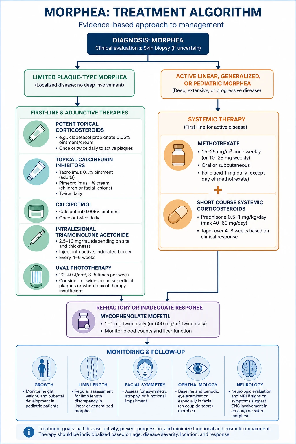
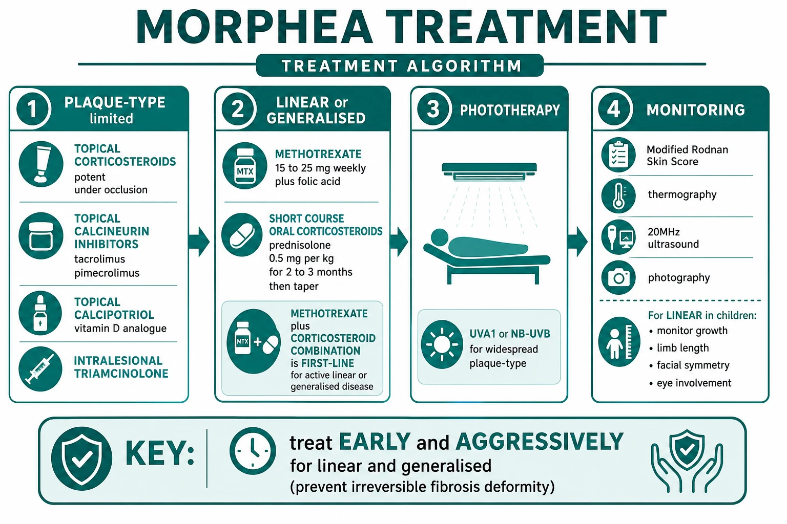
There is no cure for morphea, and the evidence base is acknowledged to be thin — the Cochrane review of interventions found few randomised data and relied heavily on expert consensus.[5] Therapy is therefore stratified by subtype and activity. The central principle is that burnt-out plaque-type disease needs little or no treatment, while active linear disease in a growing child needs prompt systemic therapy to prevent irreversible deformity.
Step 1 — Topical therapy for limited plaque-type disease
For one or a few superficial plaque-type lesions, topical therapy is first-line:[1][3]
- Super-potent topical corticosteroid — clobetasol propionate 0.05% ointment or cream applied once daily, usually for up to 4 weeks then tapered, to settle the inflammatory lilac edge.
- Topical calcipotriol — a vitamin D analogue, calcipotriol 0.005% ointment applied twice daily, which downregulates fibroblast collagen synthesis; often combined with a topical steroid.
- Topical calcineurin inhibitor — tacrolimus 0.1% ointment or pimecrolimus 1% cream, particularly useful for facial and intertriginous sites where a super-potent steroid is undesirable.
- Intralesional triamcinolone — for a stubborn thick plaque, 5 to 10 mg/mL injected into the lesion. [1]
Step 2 — Phototherapy for widespread superficial plaque-type disease
When plaque-type disease is extensive but superficial, phototherapy is the treatment of choice. Both UVA1 (medium or high dose) and narrowband UVB (311 nm) are effective; UVA1 has the stronger evidence base, particularly from German and European centres. A typical regimen is three sessions per week for several weeks, softening induration and fading the lilac edge.[1][3]
Step 3 — Systemic therapy for active linear, generalised, or deep disease
Methotrexate combined with systemic corticosteroids is first-line for active linear, generalised, and deep morphea.[1][7]
- Methotrexate — 15 mg once weekly, oral or subcutaneous, with folic acid 5 mg once weekly (given on a non-methotrexate day). Methotrexate is the disease-modifying backbone and is continued for many months to years.
- Systemic corticosteroid — prednisolone approximately 1 mg/kg/day (maximum around 60 mg/day) tapered over 2 to 3 months, OR pulse intravenous methylprednisolone (for example 20 to 30 mg/kg/day on three consecutive days each month for one to three months). The corticosteroid is a bridge to control the inflammatory lilac phase while methotrexate takes effect.
- Mycophenolate mofetil — for methotrexate-resistant or methotrexate-intolerant disease, particularly juvenile localized scleroderma, mycophenolate mofetil is the established second-line agent.[8]
When to escalate from topical to systemic therapy — LINEAR
Step 4 — Physiotherapy and reconstruction
Physiotherapy and occupational therapy are essential adjuncts in linear disease crossing joints, both to prevent and to treat contractures. Reconstructive surgery — tissue expansion, autologous fat grafting, scar revision, and orthognathic correction of facial asymmetry — is undertaken only when the disease has been inactive for at least 1 to 2 years, because operating on active morphea risks reactivating and worsening the fibrosis.[1]
Monitoring
Throughout treatment, monitor with the mLoSSI, serial clinical photography, infrared thermography, and (in children) limb length and circumference and joint range of motion. In facial linear disease maintain ophthalmology surveillance (uveitis) and arrange neuroimaging if neurological symptoms arise. Reassure the patient at every visit that morphea does not progress to systemic sclerosis.[1][6]
Specific Subtypes & Scenarios
Plaque-type morphea in adults
This is the bread-and-butter scenario. The lesion is identified, the diagnosis is confirmed clinically (with biopsy if needed), systemic sclerosis is excluded, and limited disease is treated topically or with phototherapy. The natural history is self-limiting over 3 to 5 years, and the patient is reassured that there is no risk of internal organ involvement and no progression to systemic sclerosis.[1][3]
Linear morphea in children (juvenile localized scleroderma)
Juvenile localized scleroderma is the commonest paediatric rheumatic skin disease after juvenile psoriasis and is dominated by the linear subtype (about two-thirds of cases). It is the scenario in which morphea causes the most harm: a band of sclerosis crossing a growth plate can arrest growth of the limb, producing permanent limb length discrepancy and contracture; a band on the face can produce lifelong asymmetry and can involve the eye and brain. Management is therefore multidisciplinary — paediatric dermatology, paediatric rheumatology, physiotherapy and occupational therapy, ophthalmology, and (for facial disease) maxillofacial surgery and neurology — and first-line systemic therapy with methotrexate plus corticosteroids is started early for any active lesion.[6][7][8]
En coup de sabre and Parry-Romberg syndrome
Facial linear morphea demands particular vigilance for ocular involvement (anterior and intermediate uveitis, which can be silent and sight-threatening) and central nervous system involvement (seizures, headache, and characteristic intracerebral calcification on imaging). Every patient with en coup de sabre should have a baseline ophthalmology review and a low threshold for neuroimaging. Reconstruction of the facial deformity (fat grafting, tissue expansion, orthognathic surgery) is deferred until the disease has been inactive for 1 to 2 years.[1][4]
Generalised, bullous, and deep/pansclerotic morphea
Generalised morphea is managed as for linear disease, with systemic therapy when active. Bullous morphea is treated by controlling the underlying plaque-type disease. Deep and pansclerotic morphea are disabling, restrict chest-wall expansion and limb mobility, and warrant aggressive systemic therapy plus physiotherapy.[1][3]
Complications & Pitfalls
The complications of morphea are determined almost entirely by subtype. Plaque-type disease leaves only cosmetic residua (atrophy, hyperpigmentation). Linear disease in a growing child produces the serious complications:[1][6]
- Joint contractures where a linear band crosses a joint.
- Limb length discrepancy and growth retardation where it crosses a growth plate.
- Facial asymmetry (Parry-Romberg) and scarring alopecia of the scalp.
- Ocular involvement (uveitis) in en coup de sabre, which may be asymptomatic and sight-threatening.
- Neurological involvement (seizures, headache, intracerebral calcification) in en coup de sabre.
- Cosmetic and psychological burden, especially with facial and extensive disease.
- Pruritus in active plaque-type disease. [1]
Exam application bank (NEET-PG / INICET)
One-line answer
Morphea (localized scleroderma) is a fibrosing inflammatory disorder confined to the skin and subcutaneous tissue WITHOUT internal organ involvement, distinguishing it absolutely from systemic sclerosis. Presents as ivory-coloured indurated plaques with a violaceous lilac ring (plaque-type, most common in adults) or as a linear band on a limb or the face (linear morphea, most common in children). Linear facial disease includes en coup de sabre (forehead sabre-cut) and Parry-Romberg syndrome (hemifacial atrophy), which carry risk of growth deformity and eye and CNS involvement. NO Raynaud, NO SSc-specific antibodies (anti-Scl-70, anti-centromere negative), NORMAL nailfold capillaroscopy. Treatment: topical corticosteroids, calcipotriol, or calcineurin inhibitors for limited plaque-type; phototherapy (UVA1 or narrowband UVB) for widespread plaque-type; methotrexate plus corticosteroids fir
Worked stems (answer without another resource)
Stem 1 — Classic presentation. Map symptoms to mechanism; name the first investigation and first treatment step with dose/route if drug therapy is standard. [1]
Stem 2 — Unstable / complicated. List red flags that force immediate resuscitation, theatre, ICU, antidote, or reperfusion — and what you do in the first 15 minutes. [1]
Stem 3 — Atypical group. Elderly, pregnancy, child, or immunocompromised: how presentation and thresholds change. [1]
Stem 4 — Differential trap. Name the three closest mimics and one discriminator for each. [1]
Stem 5 — Disposition. Who goes home with safety-netting, who is admitted, who needs HDU/ICU/theatre, and what follow-up is mandatory. [1]
Rapid viva checklist
- Definition + classification
- Pathophysiology chain
- Bedside signs / criteria
- Score with exact components (if any)
- Emergency bundle
- Definitive therapy with doses
- Complications of disease and of treatment
- Special populations
- Guideline/trial name if classic
- Three exam traps
Coverage self-check
If you cannot answer any stem above from this page alone, re-read the matching section — the page is intended to be self-sufficient for final-prof and NEET-PG/INICET questions on Morphea (localized scleroderma).
The classic pitfalls are: (1) mislabelling morphea as systemic sclerosis, provoking unnecessary anxiety and a battery of visceral investigations; (2) missing the eye and CNS complications of en coup de sabre because they can be silent; (3) delaying methotrexate in active linear disease in a growing child, allowing irreversible deformity; and (4) treating burnt-out, inactive plaque-type disease aggressively with systemic agents that cannot reverse established atrophy. The mLoSSI and thermography distinguish active from burnt-out disease and prevent the last error.[1][9]
Prognosis & Disposition
Plaque-type morphea is self-limiting. Each lesion evolves through inflammation to sclerosis to atrophy over roughly 3 to 5 years, and the patient is left with a brownish, atrophic patch. New lesions may appear sequentially over years, but the disease ultimately burns out. There is no mortality from plaque-type morphea.[3]
Linear morphea is progressive in children and can cause permanent deformity if untreated — limb length discrepancy, joint contractures, and facial asymmetry. Early aggressive systemic therapy with methotrexate plus corticosteroids substantially improves the outcome. Even with treatment, residual deformity may require later reconstructive surgery.[1][6]
The reassurance that examiners reward is that morphea does NOT progress to systemic sclerosis. The two are biologically distinct, and a patient with morphea has no increased risk of subsequently developing systemic sclerosis.[4]
Disposition. Limited plaque-type disease is managed in dermatology outpatients with topical therapy and phototherapy. Linear, generalised, and deep disease, and all juvenile localized scleroderma, are managed by a multidisciplinary team — dermatology and paediatric rheumatology, physiotherapy and occupational therapy, ophthalmology for facial disease, and neurology and maxillofacial surgery where indicated. Admission is rarely required; the urgency is the timely initiation of systemic therapy for active linear disease in a child.[1][6]
Special Populations
Children (juvenile localized scleroderma)
The paediatric population is where morphea does its damage. About two-thirds of paediatric cases are linear. Methotrexate is dosed at 10 to 15 mg/m2 once weekly (typically 15 mg subcutaneous or oral in school-age children) with folic acid, and is monitored with full blood count and liver function. The child needs school and psychological support because visible deformity, especially facial, is stigmatising. Growth, limb length, and joint range of motion are measured at every visit. Methotrexate is teratogenic, and adolescent girls need contraceptive counselling.[6][8]
Pregnancy
Methotrexate is absolutely contraindicated in pregnancy (teratogenic — abortifacient and a cause of fetal methotrexate syndrome) and must be discontinued at least three months before conception in both women and men. A woman with active morphea planning pregnancy is transitioned to a pregnancy-safe regimen (topical therapy, phototherapy) under specialist guidance. Morphea itself does not appear to adversely affect fertility or pregnancy outcome, and disease often improves during pregnancy.[1]
Cosmetic and psychological burden
Adults with facial or extensive plaque-type disease carry a significant cosmetic and psychological burden. The hyperpigmentation and atrophy of burnt-out lesions are cosmetically conspicuous, and facial linear disease can be disfiguring. Camouflage cosmetics, psychological support, and — once disease is inactive — reconstructive and laser techniques all have a role.[1]
Evidence, Guidelines & Regional Differences
The evidence base for morphea treatment is acknowledged to be thin. The Cochrane review of interventions for morphea (Albuquerque et al, 2019) found few randomised trials of adequate size, and concluded that most recommendations rest on expert consensus and observational data rather than high-quality randomised evidence.[5]
The strongest evidence is for methotrexate combined with corticosteroids in active linear, generalised, and deep disease. The single-centre prospective longitudinal study by Torok and Arkachaisri established the regimen (methotrexate weekly plus a tapering corticosteroid course) as the standard of care, and it is endorsed by juvenile localized scleroderma treatment consensus on both sides of the Atlantic.[7] For methotrexate-resistant juvenile disease, mycophenolate mofetil is supported by the Padua paediatric rheumatology experience.[8]
UVA1 phototherapy has its strongest evidence base in German and European centres, where UVA1 lamps are widely available; in the UK and North America narrowband UVB is more commonly used because the equipment is ubiquitous and the evidence for plaque-type disease is comparable.[1][3]
The Borrelia burgdorferi association remains a regional controversy: implicated in some European cohorts and not in UK or North American series. Antibiotic therapy directed at Lyme disease is not standard outside proven infection.[1]
Disease activity and damage are quantified by the validated mLoSSI and LoSDI instruments (Arkachaisri et al), which have brought rigour to both trials and routine monitoring.[9]
Exam Pearls
Define morphea and state the four bedside features that distinguish it from systemic sclerosis.
Morphea (localized scleroderma) is a fibrosing inflammatory disorder confined to the skin and subcutaneous tissue. The four bedside discriminators from systemic sclerosis are: (1) no Raynaud phenomenon, (2) normal nailfold capillaroscopy, (3) no internal organ involvement, and (4) negative systemic-sclerosis-specific antibodies (anti-Scl-70 and anti-centromere). ANA may be positive (40 to 50 percent) and anti-histone antibodies in about 50 percent of linear/generalised disease, but the SSc-specific antibodies are negative.[1][4]
A 7-year-old girl has a linear indurated band crossing her right elbow and a shorter lesion on her forehead. What subtype is this, what are the principal risks, and what is first-line management?
This is linear morphea with en coup de sabre (facial component). The principal risks are joint contracture and growth arrest of the limb at the elbow crossing, limb length discrepancy, facial asymmetry, ocular involvement (uveitis — often silent and sight-threatening) and central nervous system involvement (seizures, headache) from the forehead lesion. First-line management is systemic methotrexate 15 mg once weekly with folic acid plus a tapering course of oral prednisolone (about 1 mg/kg/day) or pulse intravenous methylprednisolone, with physiotherapy for the contracture, baseline ophthalmology review, and a low threshold for neuroimaging. Reconstructive surgery is deferred until disease has been inactive for 1 to 2 years.[1][6][7]
Describe the histology of morphea and the one feature that distinguishes it from lichen sclerosus on biopsy.
Morphea shows thickened, homogenised (sclerotic, hyalinised) collagen bundles in the reticular dermis, with loss of adnexal structures (hair follicles, sweat glands) and a lymphocytic inflammatory infiltrate at the active lilac border. Lichen sclerosus, by contrast, shows epidermal atrophy with a hyalinised band in the upper (papillary) dermis and follicular plugging, with relative sparing of the deeper dermis. The distinguishing feature is that lichen sclerosus affects the upper dermis beneath an atrophic epidermis, whereas morphea affects the deeper reticular dermis with thickened collagen bundles and loss of appendages.[3]
Why is morphea managed differently from systemic sclerosis, and what is the role of UVA1 phototherapy?
Morphea is a regional skin fibrosis without the systemic microvasculopathy of systemic sclerosis, so management targets the skin and subcutis rather than internal organs — topical corticosteroids, calcipotriol, calcineurin inhibitors, phototherapy, and (for active/deep disease) methotrexate plus corticosteroids. Systemic-sclerosis-type vasodilators, immunosuppression for organ-threatening disease, and ACE-inhibitor renal-crisis protocols have no role in morphea. UVA1 phototherapy softens superficial plaque-type induration and fades the inflammatory lilac edge by downregulating dermal fibroblast activity and T-cell-driven inflammation; it is the treatment of choice for widespread but superficial plaque-type disease, with narrowband UVB an acceptable alternative where UVA1 lamps are unavailable.[1][3]
Morphea rewards the candidate who can hold the line between two errors: over-treating burnt-out plaque-type disease that needs only reassurance, and under-treating active linear disease in a growing child that needs prompt systemic therapy. Master the four bedside discriminators from systemic sclerosis, recognise the lilac-ringed ivory plaque, and escalate linear disease early, and the topic is yours.[1]
References
- [1]Papara C, De Luca DA, Bieber K, Vorobyev A, et al. Morphea: The 2023 update Front Med (Lausanne), 2023.PMID 36860340
- [2]Wenzel D, Haddadi NS, Afshari K, Richmond JM, et al. Upcoming treatments for morphea Immun Inflamm Dis, 2021.PMID 34272836
- [3]Careta MF, Romiti R. Localized scleroderma: clinical spectrum and therapeutic update An Bras Dermatol, 2015.PMID 25672301
- [4]Ferrelli C, Gasparini G, Parodi A, Cozzani E, et al. Cutaneous Manifestations of Scleroderma and Scleroderma-Like Disorders: a Comprehensive Review Clin Rev Allergy Immunol, 2017.PMID 28712039
- [5]Albuquerque JVG, Andriolo BN, Vasconcellos MR, Civile VT, et al. Interventions for morphea Cochrane Database Syst Rev, 2019.PMID 31309547
- [6]Kaushik A, Mahajan R, De D, Handa S. Paediatric morphoea: a holistic review. Part 2: diagnosis, measures of disease activity, management and natural history Clin Exp Dermatol, 2020.PMID 32449205
- [7]Torok KS, Arkachaisri T. Methotrexate and corticosteroids in the treatment of localized scleroderma: a standardized prospective longitudinal single-center study J Rheumatol, 2012.PMID 22247357
- [8]Martini G, Saggioro L, Culpo R, et al. Mycophenolate mofetil for methotrexate-resistant juvenile localized scleroderma Rheumatology (Oxford), 2021.PMID 32978631
- [9]Arkachaisri T, Vilaiyuk S, Torok KS, Medsger TA Jr. Development and initial validation of the localized scleroderma skin damage index and physician global assessment of disease damage: a proof-of-concept study Rheumatology (Oxford), 2010.PMID 20008472
- [10]Errichetti E, Stinco G. Dermoscopy in General Dermatology: A Practical Overview Dermatol Ther (Heidelb), 2016.PMID 27613297