Dermatology · Medicine
Nappy dermatitis (diaper dermatitis)
Also known as Nappy dermatitis · Diaper dermatitis · Napkin dermatitis · Irritant napkin dermatitis · Candidal napkin dermatitis · Seborrhoeic napkin dermatitis · Granuloma gluteale infantum
Nappy dermatitis is an irritant contact dermatitis of the nappy area from prolonged contact with urine and faeces (ammonia, faecal enzymes, moisture, friction), presenting as confluent erythema sparing the skin folds (unlike candidal or seborrhoeic variants), complicated by secondary candidiasis (satellite pustules) or bacterial infection, and managed by frequent nappy changes, barrier creams (zinc oxide), mild topical corticosteroids for short courses if inflamed, and antifungal if candidal. Fellowship-level assessment demands mastery of the irritant vs candidal vs seborrhoeic patterns, the 'fold sparing' sign, differential diagnoses including napkin psoriasis, Langerhans cell histiocytosis, and acrodermatitis enteropathica, and the prevention strategies.
On this page & tools
Your progress
Saved locally on this device.
Practise this topic
Exam tags
Red flags
Overview
Nappy dermatitis (diaper dermatitis, napkin dermatitis) is the commonest dermatosis of infancy, affecting up to 50% of infants at some point during the nappy-wearing period (peak incidence 9–12 months). It is primarily an irritant contact dermatitis caused by prolonged skin contact with urine and faeces under the occlusive environment of the nappy. The hallmark clinical sign is confluent erythema on convex surfaces that spares the skin folds (the "fold sparing" sign), which distinguishes irritant nappy dermatitis from candidal or seborrhoeic variants that involve the folds. Management centres on prevention (frequent nappy changes, barrier creams) with topical therapies for established rash.[1][2][5]
Epidemiology
- Prevalence: affects up to 50% of infants during the first year of life; peak incidence at 9–12 months (corresponding to dietary diversification and sitting/crawling increasing friction).[2]
- Risk factors: infrequent nappy changes, diarrhoea, broad-spectrum antibiotics (disrupt skin microbiome), exclusive formula feeding (more alkaline faeces than breast-fed), introduction of solid foods, sitting/crawling stage (increased friction and moisture), use of cloth nappies (less absorbent than disposables), and underlying skin barrier defects (atopic dermatitis).[7]
- Gender: equal in M and F infants (though some studies report slight female predominance).
- Microbiome: disruption of the normal skin microbiome (decreased Staphylococcus epidermidis, increased Candida albicans) contributes to inflammation and secondary infection.[7]
Pathophysiology
The nappy area is an occluded, warm, moist environment that disrupts the stratum corneum barrier through multiple mechanisms:[5][6]
- Ammonia: urea in urine is hydrolysed by faecal urease-producing bacteria (e.g., Proteus, Bacteroides) to ammonia, which raises the skin pH from its normal acidic mantle (pH 4.5–5.5) to an alkaline pH. Alkaline pH activates faecal proteases and lipases that digest stratum corneum proteins and lipids, compromising barrier integrity.
- Moisture: prolonged hydration of the stratum corneum causes maceration and increased permeability to irritants.
- Friction: mechanical abrasion from the nappy and the infant's movement further damages the barrier.
- Faecal enzymes: proteases, lipases, and bile salts in faeces directly digest the stratum corneum; breast-fed infants have more acidic, lower-enzyme faeces than formula-fed infants (explaining lower incidence in breast-fed babies).
- Microbial overgrowth: the warm, moist, alkaline environment favours Candida albicans colonisation (present in 40–80% of severe or persistent cases) and bacterial superinfection (S. aureus, Streptococcus).[7][11]
The net effect is an irritant contact dermatitis (non-immunological, in contrast to allergic contact dermatitis) with epidermal spongiosis and dermal inflammation. [1]
Clinical patterns
1. Irritant nappy dermatitis (commonest, ~80%)
- Distribution: convex surfaces — buttocks, lower abdomen, pubic area, inner thighs, genitalia, and upper buttock cleft.
- Key sign: FOLD SPARING — the inguinal creases, gluteal cleft, and thigh folds are spared because they are protected from direct contact with urine/faeces (the irritant is in the wet nappy pad, which contacts the convex surfaces, not the recessed folds).
- Morphology: confluent, shiny, glazed erythema with mild scaling; may progress to erosions, papules, and vesicopustules in severe cases.
- Symptoms: infant is irritable but otherwise well; no systemic symptoms.[5][6]
2. Candidal nappy dermatitis (~20–40% of persistent cases)
- Bright-red, beefy, confluent erythema that INVOLVES the folds (inguinal creases, gluteal cleft — the opposite of irritant).
- Satellite lesions: discrete pustules and erythematous papules scattered beyond the main margin — the hallmark of candidal infection.
- Often follows a course of broad-spectrum antibiotics (which eliminate competing bacterial flora) or oral thrush.
- May involve the perianal area (from GI colonisation).[11][12]
3. Seborrhoeic napkin dermatitis
- Yellow, greasy scales on an erythematous base affecting the folds AND other seborrhoeic sites: scalp (cradle cap), eyebrows, retroauricular folds, neck, axillae.
- Less symptomatic than irritant; infant is not uncomfortable.
- Prognosis: self-limiting over weeks to months.[6]
4. Allergic contact dermatitis (uncommon)
- Less common than irritant; caused by fragrances, preservatives, dyes in nappy materials, wipes, or topical products (e.g., lanolin, balsam of Peru, methylisothiazolinone).
- Sharply demarcated rash corresponding to the contact area; may have vesicles and pruritus.
- Identified by patch testing. [1]
5. Bacterial secondary infection
- Impetiginisation: honey-coloured crusts, pustules, surrounding erythema/cellulitis.
- Perianal streptococcal dermatitis: well-demarcated, bright-red perianal erythema caused by Group A β-haemolytic Streptococcus; may cause anal fissuring and pain on defecation. Treat with oral penicillin/cephalosporin. [1]
6. Granuloma gluteale infantum
- A rare complication of severe nappy dermatitis (often with potent topical steroid use under occlusion): reddish-bluish, granulomatous nodules on the convex surfaces of the buttocks and genitalia.
- Histology: pseudo-carcinomatous hyperplasia with dermal granulomatous inflammation.
- Self-resolving over weeks to months after withdrawal of the causative agent and barrier restoration. [1]
7. Jacquet's erosive dermatitis
- Severe variant: well-demarcated, punched-out erosions and ulcers with raised borders on the perianal and perineal skin; associated with severe diarrhoea and prolonged contact. [1]
8. Psoriasiform napkin dermatitis
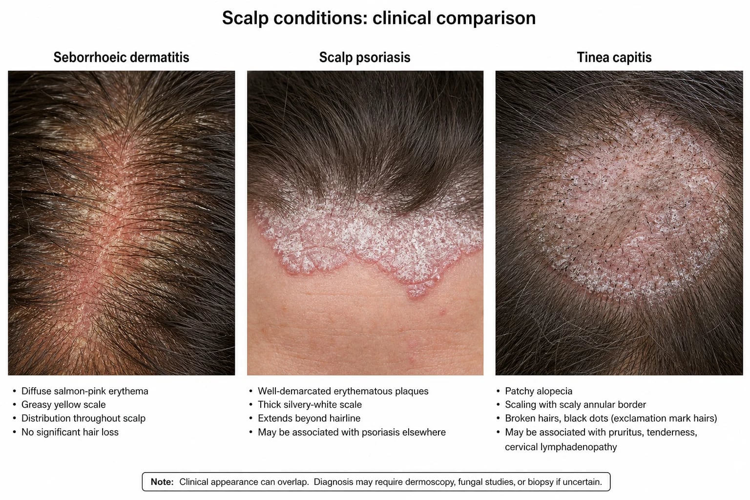
- Well-demarcated, salmon-pink plaques with overlying silvery scale involving the convex surfaces and occasionally the folds.
- Important to recognise because of the risk of progression to generalised pustular psoriasis (von Zumbusch); family history of psoriasis is a clue.
- May respond to mild topical corticosteroid + barrier, but tends to be more refractory than irritant dermatitis.[3]
Irritant (80% of cases)
- **Convex surfaces only** — buttocks, lower abdomen, inner thighs, genitalia; **FOLD SPARING** is the diagnostic hallmark
- **Confluent, glazed, shiny erythema** with mild scaling; papules, erosions, and vesicopustules in severe cases
- Mechanism: **ammonia from urea (faecal urease) → alkaline pH → activated faecal proteases/lipases → barrier damage**
- Treatment: prevention + barrier; mild hydrocortisone 1% for short course if inflamed; never potent steroids
Candidal (20-40% of persistent)
- **Bright-red, beefy plaques** involving the **FOLDS** (opposite of irritant); **satellite pustules and papules** scattered beyond the main margin
- Often follows **broad-spectrum antibiotics** or coexists with **oral thrush**; perianal involvement from GI reservoir
- KOH microscopy: **budding yeast and pseudohyphae**; swab culture if refractory
- Treatment: **topical azole (clotrimazole 1% / miconazole 2% / ketoconazole 2%) or nystatin BID for 7-14 days**; oral nystatin if oral thrush
Seborrhoeic
- **Yellow, greasy scales** in **folds AND seborrhoeic sites** — scalp (cradle cap), eyebrows, retroauricular folds, neck, axillae
- **Non-itchy**; infant is comfortable; presents in the first 1-2 months of life
- Self-limiting; emollients and gentle cleansing usually suffice; mild hydrocortisone + antifungal combination if persistent
Bacterial superinfection
- **Impetiginisation** (honey-coloured crusts, pustules) or **perianal streptococcal dermatitis** (well-demarcated bright-red perianal erythema, pain on defecation)
- Group A β-haemolytic *Streptococcus* in perianal disease; *S. aureus* in impetiginised irritant rash
- Treatment: **flucloxacillin** for staph; **phenoxymethylpenicillin (penicillin V) 10 days** for perianal streptococcal
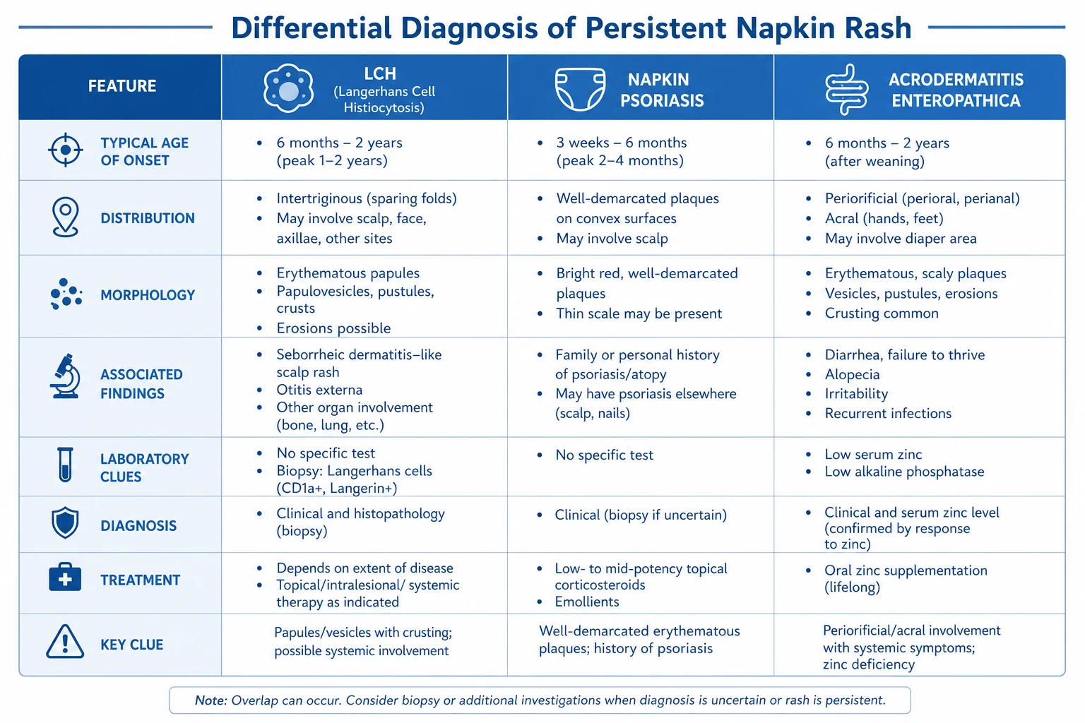
Differential diagnosis of persistent napkin rash
| Condition | Key features | Distinguishing clue |
|---|---|---|
| Napkin psoriasis | Well-demarcated, salmon-pink plaques with silvery scale on convex surfaces ± folds | May progress to generalised pustular psoriasis; family history of psoriasis |
| Atopic dermatitis | Usually SPARES the nappy area (moisture is protective) | Eczema on face, flexures; family history of atopy; pruritus |
| Seborrhoeic dermatitis | Yellow greasy scales in folds + scalp (cradle cap) | Non-itchy; other seborrhoeic sites |
| Langerhans cell histiocytosis (LCH) | Refractory erosive napkin rash, petechiae/purpura, skin-fold involvement | Biopsy (CD1a+/Langerin+ histiocytes); multi-system involvement; serious |
| Acrodermatitis enteropathica | Periorificial + acral dermatitis + diarrhoea + alopecia | Zinc deficiency (SLC39A4); low serum zinc; responds to zinc supplementation |
| Candidiasis | Bright-red, satellite pustules, folds involved | KOH microscopy; follows antibiotics |
| Allergic contact dermatitis | Sharply demarcated, pruritic, vesicles | Patch testing; exposure to allergen |
| Immunodeficiency (e.g., Hyper-IgE, Wiskott-Aldrich, HIV) | Refractory seborrhoeic-like or candidal rash + recurrent infections | Immune workup; failure to thrive |
Causes of REFRACTORY napkin rash — mnemonic: PERSIST
PERSIST
Well-demarcated salmon-pink plaques; risk of generalised pustular psoriasis
Periorificial + acral + diarrhoea + alopecia; low serum zinc; responds to zinc
Persistent candida after antibiotics; perianal streptococcal dermatitis often missed
Yellow greasy scales in folds + cradle cap; usually self-limiting
Refractory candidal / seborrhoeic rash + recurrent infections + failure to thrive
Petechiae / purpura in refractory rash; biopsy CD1a+ / Langerin+; multi-system workup
Reddish-bluish granulomatous nodules from potent steroids under occlusion
Nappy rash mimickers — deep dive
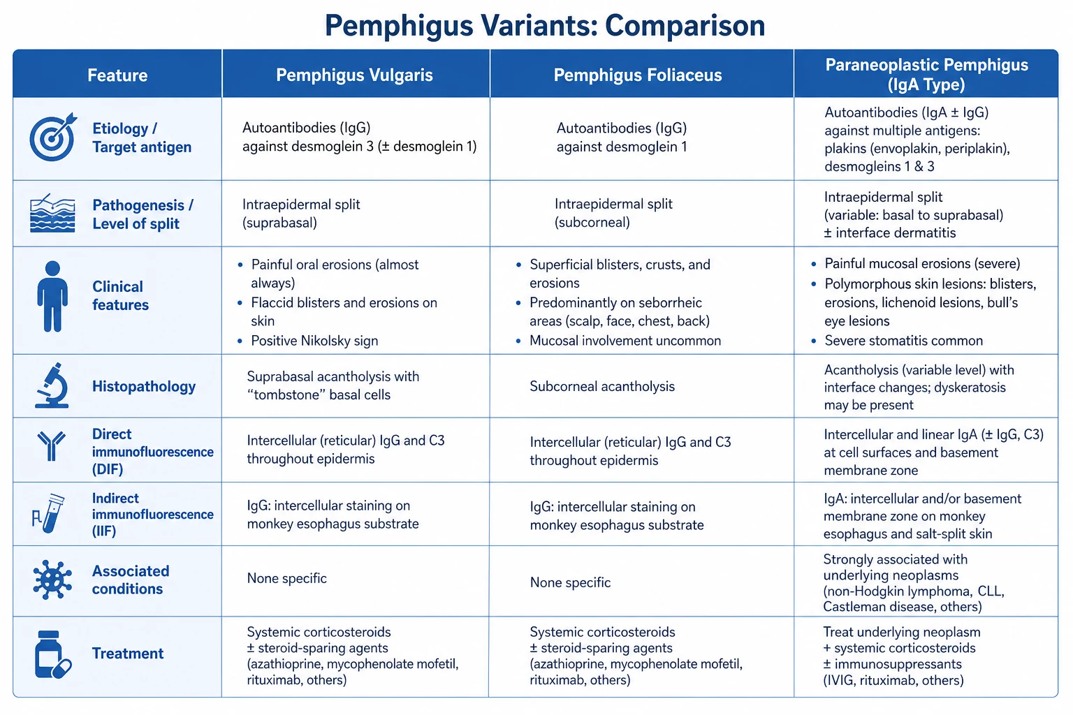
The most common cause of nappy rash is irritant contact dermatitis, but in any infant with a nappy eruption that fails to respond to standard barrier + mild steroid + antifungal therapy within 7–10 days, the differential must extend well beyond irritant/candidal/bacterial napkin dermatitis. Below is a deep dive into the most clinically important mimickers, each of which has a distinct diagnostic handle that should not be missed.[3]
1. Napkin psoriasis (psoriasiform napkin dermatitis)
- Morphology: sharply demarcated, bright-red (salmon-pink), glistening plaques with a thin overlying scale. Unlike classic adult psoriasis, the scale is often minimal in the nappy area because the moist occluded environment macerates the silvery scale.
- Distribution: favours the convex surfaces, but unlike simple irritant dermatitis it frequently involves the folds (inguinal creases and gluteal cleft) — the opposite of the typical irritant pattern.
- Course: persistent beyond 2 weeks despite barrier cream and topical hydrocortisone. May extend beyond the nappy to the trunk and scalp. Family history of psoriasis is supportive.
- Risk: a subset progresses to generalised pustular psoriasis (von Zumbusch) — a dermatological emergency with fever, leucocytosis and sterile pustules.
- Diagnostic clue: sharp demarcation + involvement of folds + family history; biopsy shows regular acanthosis, parakeratosis and neutrophils in the stratum corneum (Munro microabscesses).[3]
- Treatment: short course of mild topical corticosteroid (hydrocortisone 1%) for the inflamed plaques; topical calcipotriol is generally avoided under 6 years because of calcium-disruption risk; referral for systemic therapy (acitretin, methotrexate, ciclosporin, biologics) if progression to pustular psoriasis.
2. Langerhans cell histiocytosis (LCH) — "the great mimicker"
- Morphology: refractory erosive napkin rash with petechiae, purpura, haemorrhagic crusts and discrete pink-to-skin-coloured papules scattered on the scalp, face, trunk and perineum; intertriginous fissuring is common.
- Why it matters: cutaneous LCH in infants under 12 months has a high risk of progression to multi-system LCH (bone, liver, spleen, bone marrow, pituitary → diabetes insipidus). It is the single most important diagnosis not to miss in a refractory napkin rash.
- Diagnostic clue: any napkin rash lasting >2 weeks despite appropriate therapy, with petechiae/purpura or extra-nappy papules, demands a 3- or 4-mm punch biopsy from a fresh papule.
- Histology & immunohistochemistry: dermal infiltrate of CD1a+, CD207 (Langerin)+, S100+ histiocytes with reniform (coffee-bean) nuclei; Birbeck granules ("tennis-racket" organelles) on electron microscopy.
- Workup once LCH confirmed: full blood count, LFTs, coagulation, skeletal survey (looking for lytic lesions, especially skull), urine osmolality (DI), chest imaging; referral to paediatric haematology-oncology for risk stratification and treatment (observation vs vinblastine + prednisolone vs targeted BRAF/MEK inhibitor for refractory disease).[9]
3. Allergic contact dermatitis (methylisothiazolinone and other wipe allergens)
- Morphology: sharply demarcated, erythematous, often vesicular rash corresponding to the area of contact with the offending product; commonly affects the buttocks, perianal area, genitals, and sometimes the face (the parent's hand touches the face after a wipe change).
- Why it matters: in the 2010s an "epidemic" of contact allergy occurred due to the introduction of methylisothiazolinone (MI) and methylchloroisothiazolinone (MCI) as standalone preservatives in baby wipes, wet wipes, leave-on cosmetics and detergents. Affected infants may have been treated repeatedly with barrier creams and topical steroids to no avail.
- Diagnostic clue: a napkin rash that only began after a change in brand of wipes or that flares within hours of wiping; vesicles and weeping rather than glazed erythema; patch testing is confirmatory.
- Common allergens in the nappy region: methylisothiazolinone (MI), methylchloroisothiazolinone (MCI), fragrance mixes I and II, balsam of Peru, lanolin, cocamidopropyl betaine, propylene glycol, neomycin (if a topical antibiotic has been used).
- Management: strict avoidance of all isothiazolinone-containing wipes and leave-on products; switch to plain water + soft cloth or wipes labelled "free from MI/MCI/parabens/fragrance"; topical hydrocortisone 1% for 5–7 days if inflamed.[3]
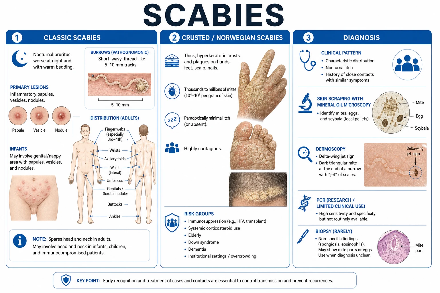
4. Infantile scabies
- Morphology: a polymorphous eruption of papules, vesicles, pustules and inflammatory nodules on the napkin area, palms, soles, scalp, face and trunk. Burrows (tiny wavy thread-like lines) may be visible but are easily obscured by secondary eczematisation.
- Why it matters: scabies is a great mimicker of napkin dermatitis in infants because the polymorphic eruption often overlaps with irritant, candidal and even LCH patterns. The napkin area is in fact one of the most heavily infested sites in infants.
- Diagnostic clues: intense nocturnal irritability in the infant; family contacts with similar itch (parents, siblings — often with classic interdigital burrows); involvement of palms and soles (rarely affected in adult scabies); papules on the trunk and axillae.
- Diagnosis: dermoscopy of a burrow shows the delta-wing jetliner sign (a dark triangular mite head); microscopy of skin scrapings in 10% KOH reveals the Sarcoptes scabiei mite, eggs and faecal pellets.
- Management: permethrin 5% cream applied from the neck down (in infants, also to the scalp/face avoiding the periorbital area) for 8–12 hours, repeated after 7 days; ivermectin 200 µg/kg PO single dose is an alternative for refractory cases or large households; treat all household contacts simultaneously; launder bedding/clothing at ≥50 °C. [1]
5. Bullous impetigo
- Morphology: flaccid, fluid-filled bullae containing straw-coloured or turbid fluid that rupture rapidly to leave a moist denuded area with a thin varnish-like crust. In the napkin area the bullae are easily macerated and may present as shiny erosions with a peripheral collarette of scale.
- Why it matters: bullous impetigo is caused by exfoliative-toxin-producing Staphylococcus aureus (phage group II, types 71 and 55). The exfoliative toxins are serine proteases that cleave desmoglein-1 in the superficial epidermis, producing a localised scalded-skin appearance. It is the localised counterpart of Staphylococcal Scalded Skin Syndrome (SSSS), where the toxin disseminates.
- Diagnostic clue: rapid appearance of flaccid blisters and shiny erosions with surrounding erythema; bacterial swab of fluid grows S. aureus; infants lack protective antibodies to exfoliative toxins and have immature renal clearance, so are particularly susceptible.
- Management: topical fusidic acid or mupirocin for localised disease; oral flucloxacillin (or clarithromycin if penicillin-allergic) for extensive or recurrent disease; swabs for MRSA if risk factors (recent hospitalisation, household MRSA). [1]
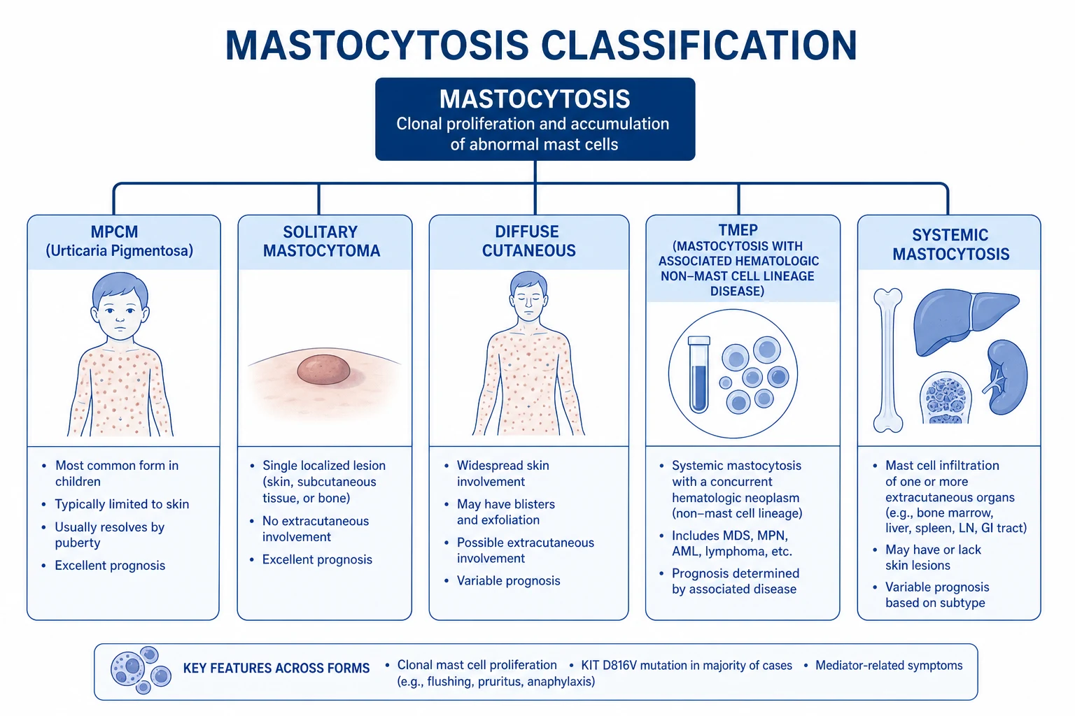
6. Bullous mastocytosis (diffuse cutaneous mastocytosis)
- Morphology: diffuse infiltration of the skin by mast cells presenting as thickened, doughy, yellowish-orange or "pebbly" skin with widespread tense bullae and erosions triggered by friction, hot baths, crying, or rubbing the nappy area. The Darier sign (urtication on stroking) is positive.
- Why it matters: bullous mastocytosis in infants can mimic scalded skin syndrome, epidermolysis bullosa, severe irritant napkin dermatitis and acrodermatitis enteropathica. Massive mast-cell degranulation can cause flushing, hypotension, diarrhoea and shock ("mast cell activation"), which is a medical emergency.
- Diagnostic clues: positive Darier sign (wheal-and-flare within minutes of stroking an intact lesion); elevated serum tryptase (often >20 ng/mL); KIT D816V mutation analysis from peripheral blood.
- Histology: dense dermal infiltrate of mast cells (toluidine blue or tryptase stain); in children the mutation profile includes KIT D816V in approximately one-third and other activating KIT mutations in the remainder.
- Management: avoid triggers (heat, friction, NSAIDs, opiates, alcohol); oral antihistamines (H1 ± H2) for symptom control; injectable epinephrine at the bedside if systemic symptoms; consider midostaurin or masitinib for severe systemic disease; most paediatric cases improve by puberty. [1]
7. Kawasaki disease — perineal erythema and desquamation
- Morphology: confluent, "boiled-lobster" erythema of the perineum and napkin area appearing in the first 2–5 days of fever, followed in some infants by fine desquamation of the perineum and finger/toe tips in the subacute phase. The rash often extends beyond the napkin to the trunk and extremities.
- Why it matters: perineal erythema is a supportive (non-diagnostic) feature of Kawasaki disease present in up to 67% of affected infants. Recognition in a febrile infant with a "diaper rash" that fails to settle prompts early echocardiography and treatment with IVIG + high-dose aspirin, dramatically reducing the risk of coronary artery aneurysms.
- Diagnostic clue: any febrile infant with a non-resolving napkin rash + bilateral non-purulent conjunctivitis + red cracked lips + erythematous extremities + cervical lymphadenopathy should trigger Kawasaki consideration, even if fewer than 4 of 5 classic criteria are met (incomplete KD).
- Distinction from simple napkin dermatitis: perineal erythema of KD is bright-red, confluent, often with sharp demarcation at the diaper line, and accompanied by desquamation in the subacute phase; the child is febrile and unwell.
- Management: paediatric urgent referral + echocardiogram + IVIG 2 g/kg + high-dose aspirin; the perineal rash itself requires only bland emollient. [1]
8. Nutritional deficiency dermatitis (acrodermatitis enteropathica)
- Morphology: well-demarcated, erythematous, scaly and sometimes vesiculobullous or pustular plaques in a strikingly periorificial (perioral, perianal, perigenital) and acral (hands, feet) distribution — the distribution is the diagnostic clue, not the morphology, which can mimic severe irritant, candidal or even psoriasiform napkin dermatitis.
- Why it matters: acrodermatitis enteropathica (AE) is the classic genetic form — autosomal recessive loss-of-function mutations in SLC39A4 (chromosome 8q24.3), encoding the ZIP4 intestinal zinc transporter — leading to severe systemic zinc deficiency. Acquired zinc deficiency (premature infants, malabsorption, prolonged TPN without trace elements, breast milk with low zinc, cystic fibrosis, Crohn's disease) can produce an identical picture. Both present with the classic triad of dermatitis + diarrhoea + alopecia (the full triad is present in only ~20%, so absence does not exclude the diagnosis).
- Diagnostic clues: periorificial AND acral involvement (often the giveaway); irritability, apathy, failure to thrive, photophobia, delayed wound healing, paronychia, alopecia; low serum zinc (often ~50% of normal; alkaline phosphatase is zinc-dependent and is also low); confirm with SLC39A4 genetic testing for the hereditary form.
- Treatment: lifelong oral zinc supplementation (zinc sulfate 3 mg/kg/day of elemental zinc); clinical response is often rapid (days to weeks) and may precede the rise in serum zinc; monitor serum copper because prolonged high-dose zinc can cause copper deficiency; the genetic form requires lifelong therapy, the acquired form responds to the underlying cause plus zinc.[10]
Diagnostic algorithm for the refractory nappy rash
When a napkin rash fails to settle with standard prevention + barrier + topical steroid/antifungal within 7–10 days, a structured diagnostic approach is required. The following algorithm is the consultant-level sequence used in paediatric dermatology: [1]
Step-by-step diagnostic workup of refractory napkin rash
Self-test: name the two investigations that confirm LCH and the two that confirm acrodermatitis enteropathica
LCH: (1) 3-4 mm punch biopsy of a fresh papule showing dermal CD1a+, CD207 (Langerin)+, S100+ histiocytes with reniform nuclei (and Birbeck granules on EM); (2) skeletal survey + urine osmolality + LFTs to stage as single-system vs multi-system. Acrodermatitis enteropathica: (1) serum zinc (low; alkaline phosphatase is also low); (2) SLC39A4 genetic testing for the hereditary form — or resolution within 7 days of empirical zinc supplementation 3 mg/kg/day elemental zinc.
Prevention strategies — evidence-based, layered
Prevention is the cornerstone of nappy dermatitis management and is more effective than any topical therapy. The literature supports a layered approach combining skin contact reduction, barrier restoration, microbiome support and caregiver education.[4][8]
Skin-contact reduction
- **Change nappies frequently** — at least every **2–3 hours** during the day and **once overnight**; change **immediately** after soiling (urine or stool).
- Use **superabsorbent disposable nappies** with a breathable outer layer; these keep the skin surface drier than cloth nappies and significantly reduce irritant dermatitis severity.
- **Avoid plastic over-pants** that increase occlusion and trap humidity; use loose clothing over the nappy.
- Allow **nappy-free time** of 30–60 minutes twice daily to permit air-drying and barrier recovery.
Barrier restoration
- Apply a **thick barrier cream at every change** — **zinc oxide 10–40% in petrolatum** is the gold standard for moderate-to-severe dermatitis; **petrolatum 100%** (Vaseline) is adequate for prevention in low-risk infants.
- Barrier creams should be applied **after gentle cleansing** and **before** the fresh nappy is fitted (the barrier sits between skin and the next insult).
- For infants with established dermatitis, **zinc oxide + petrolatum combination products** (e.g., Sudocrem, Desitin) provide an occlusive plus an astringent effect.
- **Avoid talcum powder** — no proven benefit and significant **inhalation risk** (pulmonary talc granulomatosis).
Cleansing and microbiome
- Use **warm water + soft cloth or cotton wool** as the default; if wipes are needed, choose **fragrance-free, alcohol-free, MI/MCI-free formulations**.
- **Avoid alkaline soaps** that raise skin pH and activate faecal enzymes; a **pH 4.5–5.5 cleanser** is preferable.
- Support the **skin microbiome**: exclusive **breastfeeding** for the first 4–6 months (more acidic faeces, lower enzyme activity, lower nappy dermatitis incidence).
- If broad-spectrum antibiotics have been given, consider **probiotic supplementation** (e.g., *Lactobacillus reuteri*) and watch for secondary candidiasis.
Caregiver education & red-flag recognition
- Educate parents about **normal nappy dermatitis** (mild, settles with prevention in 3–7 days) vs **red flags** (petechiae, purpura, bullae, vesicles, papules beyond the nappy, persistent fever, failure to thrive, persistent diarrhoea, alopecia).
- Provide a **written nappy-care plan** at the 6-week immunisation visit: change frequency, barrier cream choice, when to seek review.
- **Avoid over-the-counter potent steroids** (clobetasol, betamethasone dipropionate) without medical advice — occlusion dramatically increases systemic absorption and the risk of **granuloma gluteale infantum** and **iatrogenic Cushing syndrome**.
- **Re-evaluate any napkin rash persisting beyond 7 days** despite correct prevention — this is the threshold for in-person review and consideration of biopsy.
Investigations
- Diagnosis is primarily clinical — history (onset, triggers, feeding, antibiotics) and examination (distribution, fold sparing vs involvement, satellite lesions).
- Skin swab (bacterial and fungal culture): if pustular, crusted, or refractory — to identify secondary S. aureus, Streptococcus, or Candida.
- KOH microscopy: if candidal infection suspected — pseudohyphae and budding yeast.
- Skin biopsy: if rash is refractory to appropriate treatment for >2 weeks or if petechiae, purpura, or ulceration are present — to exclude LCH, psoriasis, or malignancy. LCH shows CD1a+/Langerin+ histiocytes with Birbeck granules on electron microscopy.[3][9]
- Serum zinc level: if acrodermatitis enteropathica suspected (low zinc; alkaline phosphatase is also zinc-dependent and may be low).[10]
- Complete blood count and immune workup: if immunodeficiency suspected.
Management
Prevention (the cornerstone)
- Frequent nappy changes — at least every 2–3 hours and immediately after bowel movements; allow nappy-free time between changes.[4][8]
- Super-absorbent disposable nappies — preferable to cloth (draw moisture away from skin); avoid plastic pants (increase occlusion).[4]
- Gentle cleansing — use warm water and soft cloth/cotton wool; avoid alkaline soaps and alcohol-based wipes. Pat dry, do not rub.
- Barrier cream at every change — zinc oxide (10–40%) or petrolatum (Vaseline) to protect the skin from moisture and irritants.[4][8]
- Avoid talcum powder — inhalation risk, no proven benefit.
- Breastfeeding continued if possible (lower faecal pH, fewer enzymes).[5]
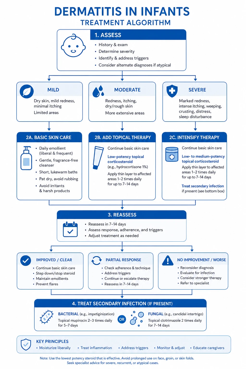
Treatment (by clinical pattern)
Irritant nappy dermatitis
- Step 1: maximise prevention (above).
- Step 2: apply barrier cream (zinc oxide 10–40%, petrolatum, or dimethicone-containing products) thickly at every change.
- Step 3: a short course of mild topical corticosteroid (hydrocortisone 1% once daily for 3–5 days) if inflammation is severe. NEVER use potent or fluorinated steroids in the nappy area — the occlusion dramatically increases systemic absorption (risk of granuloma gluteale infantum, skin atrophy, and HPA axis suppression).[4]
Candidal nappy dermatitis
- Topical antifungal: clotrimazole 1%, miconazole 2%, ketoconazole 2%, or nystatin applied BID to the affected area for 7–14 days after rash clears.
- Combine with a barrier cream (apply antifungal first, allow to dry, then barrier on top).
- Oral nystatin if oral thrush coexists (GI reservoir of Candida).
- Check for and treat any predisposing factors (recent antibiotics, diabetes).[11][12]
Seborrhoeic napkin dermatitis
- Usually mild and self-limiting. Emollients and gentle cleansing suffice. If persistent, a short course of mild topical corticosteroid + antifungal combination (e.g., hydrocortisone + clotrimazole) may be used. [1]
Bacterial infection
- Impetigo/cellulitis: oral flucloxacillin (or cephalexin if MRSA-unlikely; clindamycin if MRSA suspected).
- Perianal streptococcal dermatitis: oral phenoxymethylpenicillin (penicillin V) for 10 days. [1]
When to combine steroid + antifungal
A combination product (hydrocortisone 1% + clotrimazole 1% + miconazole) may be used for mixed irritant + candidal inflammation for a short course (5–7 days maximum) to avoid steroid atrophy in the occluded nappy area.[4]
Complications
- Secondary candidiasis (most common complication).
- Bacterial superinfection (impetigo, cellulitis, abscess).
- Granuloma gluteale infantum (granulomatous nodules from prolonged potent steroid use).
- Jacquet's erosive dermatitis (deep erosions/ulcers from severe, untreated dermatitis).
- Psycho-social impact on parents/caregivers (distress, sleep disruption). [1]
Prognosis
- Self-limiting with appropriate management; resolves within days to weeks.
- Recurrence common until the child is toilet-trained.
- No long-term sequelae in the vast majority; scarring only in severe neglected cases with secondary infection. [1]
NAPPY 4 - the 4 most important differentials in persistent napkin rash
Most common; flexures = candidal; convex surfaces = irritant; treat with barrier + appropriate topical
Recurrent crops of pruritic vesicles/pustules on hands, feet, diaper; onset 2-12 months; self-limiting by 2-3 years; dapsone for severe
Sharply demarcated red plaques; persistent beyond 2 weeks; napkin sparing; family history; treat with mild topical steroid
Bright red sharply demarcated perianal erythema; GABHS culture; oral penicillin V 10 days
Persistent beyond 2 weeks despite treatment + skin-coloured papules + hepatosplenomegaly = biopsy
Red flags [1]
[1]Exam pearls
[1]Red flags
Exam application bank (NEET-PG / INICET)
One-line answer
Nappy dermatitis is an irritant contact dermatitis of the nappy area from prolonged contact with urine and faeces (ammonia, faecal enzymes, moisture, friction), presenting as confluent erythema sparing the skin folds (unlike candidal or seborrhoeic variants), complicated by secondary candidiasis (satellite pustules) or bacterial infection, and managed by frequent nappy changes, barrier creams (zinc oxide), mild topical corticosteroids for short courses if inflamed, and antifungal if candidal. Fellowship-level assessment demands mastery of the irritant vs candidal vs seborrhoeic patterns, the 'fold sparing' sign, differential diagnoses including napkin psoriasis, Langerhans cell histiocytosis, and acrodermatitis enteropathica, and the prevention strategies.
Worked stems (answer without another resource)
Stem 1 — Classic presentation. Map symptoms to mechanism; name the first investigation and first treatment step with dose/route if drug therapy is standard. [1]
Stem 2 — Unstable / complicated. List red flags that force immediate resuscitation, theatre, ICU, antidote, or reperfusion — and what you do in the first 15 minutes. [1]
Stem 3 — Atypical group. Elderly, pregnancy, child, or immunocompromised: how presentation and thresholds change. [1]
Stem 4 — Differential trap. Name the three closest mimics and one discriminator for each. [1]
Stem 5 — Disposition. Who goes home with safety-netting, who is admitted, who needs HDU/ICU/theatre, and what follow-up is mandatory. [1]
Rapid viva checklist
- Definition + classification
- Pathophysiology chain
- Bedside signs / criteria
- Score with exact components (if any)
- Emergency bundle
- Definitive therapy with doses
- Complications of disease and of treatment
- Special populations
- Guideline/trial name if classic
- Three exam traps
Coverage self-check
If you cannot answer any stem above from this page alone, re-read the matching section — the page is intended to be self-sufficient for final-prof and NEET-PG/INICET questions on Nappy dermatitis (diaper dermatitis).
[1]References
- [1]Chiriac A, Wollina U. Diaper dermatitis-a narrative review of clinical presentation, subtypes, and treatment Wien Med Wochenschr, 2024.PMID 37861874
- [2]Helms LE, Burrows HL, et al. Diaper Dermatitis Pediatr Rev, 2021.PMID 33386307
- [3]Fölster-Holst R. Differential diagnoses of diaper dermatitis Pediatr Dermatol, 2018.PMID 29596730
- [4]Blume-Peytavi U, Kanti V, et al. Prevention and treatment of diaper dermatitis Pediatr Dermatol, 2018.PMID 29596731
- [5]Atherton DJ. Understanding irritant napkin dermatitis Int J Dermatol, 2016.PMID 27311779
- [6]Tüzün Y, Wolf R, Bağlam S, et al. Diaper (napkin) dermatitis: A fold (intertriginous) dermatosis Clin Dermatol, 2015.PMID 26051065
- [7]Hertiš Petek T, Petek M, Petek T, et al. Emerging Links between Microbiome Composition and Skin Immunology in Diaper Dermatitis: A Narrative Review Children (Basel), 2022.PMID 35053737
- [8]Octarica SG, Ellistasari EY, Dewi AK, et al. Preventive and curative approaches to diaper dermatitis in children: a systematic review Acta Dermatovenerol Alp Pannonica Adriat, 2025.PMID 41014074
- [9]Lee LW, Azfar RS, Yan AC, et al. A 4-month-old boy with diaper dermatitis. Langerhans cell histiocytosis Pediatr Ann, 2008.PMID 18488977
- [10]Maverakis E, Fung MA, Lynch PJ, et al. Acrodermatitis enteropathica and an overview of zinc metabolism J Am Acad Dermatol, 2007.PMID 17190629
- [11]Bonifaz A, Rojas R, Tirado-Sánchez A, et al. Superficial Mycoses Associated with Diaper Dermatitis Mycopathologia, 2016.PMID 27193417
- [12]Taudorf EH, Jemec GBE, Hay RJ, et al. Cutaneous candidiasis - an evidence-based review of topical and systemic treatments to inform clinical practice J Eur Acad Dermatol Venereol, 2019.PMID 31287594