Dermatology · Medicine
Necrobiosis lipoidica
Also known as Necrobiosis lipoidica · Necrobiosis lipoidica diabeticorum · NLD · NL · Oppenheim-Urbach disease
Necrobiosis lipoidica is a chronic granulomatous dermatosis of collagen degeneration, classically producing yellow-brown atrophic telangiectatic plaques with a violaceous rim on the anterior shins. It is strongly associated with diabetes mellitus but does not reliably improve with glycaemic control. Diagnosis is clinical when typical and biopsy shows palisading or layered granulomas around necrobiotic collagen with plasma cells and vascular change. Management centres on smoking cessation, trauma avoidance, topical or intralesional corticosteroid to the active rim, tacrolimus or phototherapy for steroid-sparing control, antiplatelet or pentoxifylline in selected cases, biologics for refractory disease, and meticulous ulcer care.
On this page & tools
Your progress
Saved locally on this device.
Exam tags
Red flags
Overview & Definition
Necrobiosis lipoidica is a palisading granulomatous dermatosis in which damaged collagen in the dermis becomes the centre of a chronic histiocytic inflammatory reaction. The word necrobiosis means degeneration of connective tissue rather than infection or true necrosis, and lipoidica reflects the yellow lipid-rich clinical colour that develops in mature plaques. The older term necrobiosis lipoidica diabeticorum is still tested, but it is too narrow because NL also occurs in people without diabetes; the modern term is simply necrobiosis lipoidica.[1][2]
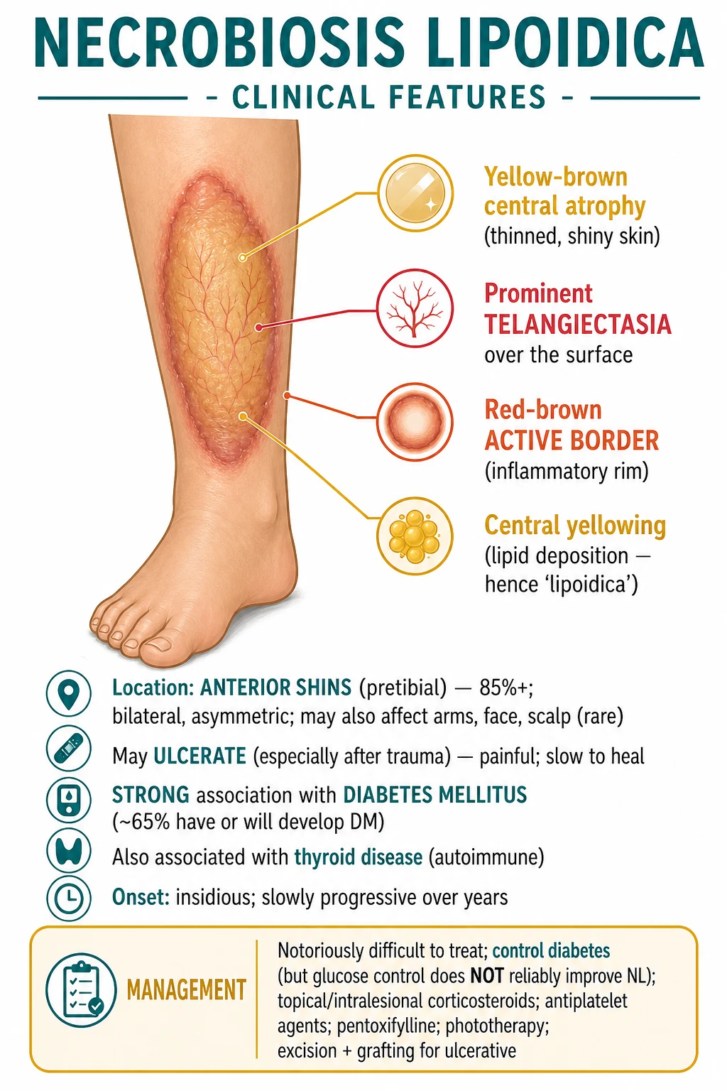
The classic lesion is easy to remember because it has three layers. The centre is yellow-brown, waxy, shiny and atrophic because the dermis has thinned. The surface shows prominent telangiectasia because the atrophic epidermis reveals superficial vessels. The edge is red-brown or violaceous and slightly raised because active granulomatous inflammation sits at the advancing border. The usual site is the anterior shin, often bilaterally and asymmetrically, although lesions can occur on the ankles, feet, thighs, arms, hands, trunk, face or scalp.[1][8]
Classification
NL is not usually classified by a formal staging score. For exams and bedside decisions, classify it by activity, extent, ulceration, and clinical context, because these determine whether observation, topical anti-inflammatory therapy, biopsy, systemic escalation or wound care is needed.[3]
Early inflammatory NL
- Small red-brown papules or plaques, often mistaken for granuloma annulare or eczema
- Active raised rim is more conspicuous than central atrophy
- Best window for anti-inflammatory treatment because irreversible atrophy has not yet dominated
- Biopsy if site or morphology is atypical
Mature atrophic plaque
- Yellow-brown waxy centre with cigarette-paper atrophy
- Prominent surface telangiectasia and a red-brown or violaceous rim
- Usually pretibial; often bilateral but asymmetric
- Treat the active edge, not the inactive atrophic centre
Ulcerative NL
- Erosion or ulcer within a fragile plaque, usually after minor trauma
- Pain, exudate and delayed healing become the main clinical problem
- Requires wound assessment, vascular assessment, infection assessment and biopsy of suspicious chronic edges
- Escalation often needs dermatology plus wound-care team input
Atypical or extratibial NL
- Lesions on arms, trunk, scalp or face, or nodular rather than plaque morphology
- Lower threshold for biopsy because sarcoidosis, granuloma annulare, morphea, xanthoma and infection can mimic it
- Ask specifically about diabetes, smoking, thyroid disease and previous trauma
- Histology must be interpreted with the clinical site
A second useful axis is systemic association. The commonest teaching association is diabetes mellitus, especially type 1 diabetes in younger patients and type 2 diabetes in older patients, but NL can precede diabetes, follow diabetes by years, or occur without diabetes. Autoimmune thyroid disease is a recognised comorbidity. Smoking and microvascular disease worsen ulcer risk and healing even when they do not prove causation.[1][5]
Epidemiology & Risk Factors
Necrobiosis lipoidica is uncommon. In diabetes clinics it is a rare cutaneous complication, classically quoted around a fraction of one percent of diabetic patients, while among patients who already have NL, published series report a variable but strong diabetes association. The safest exam phrasing is: NL is strongly associated with diabetes mellitus, but most people with diabetes never develop NL, and not every person with NL has diabetes.[1]
Risk factors and associations to ask for are diabetes mellitus, impaired glucose tolerance, family history of diabetes, smoking, peripheral vascular disease, venous insufficiency, obesity, thyroid disease, dyslipidaemia and repeated shin trauma. Smoking is particularly important because it compounds microvascular injury and wound-healing failure; cessation is therefore not cosmetic advice but disease-modifying risk reduction for ulcerative NL.[1][3]
Women are affected more often than men in many clinical series, and onset is commonly in young to middle adult life, but the disease spans childhood to older age. Paediatric NL is uncommon and should prompt the same careful search for diabetes, family history, trauma and atypical differentials; treatment choices are narrower because long courses of potent topical steroids, phototherapy logistics and systemic immunomodulation carry age-specific risks.[7]
The examiner trap is to say that tight glycaemic control treats NL. Glycaemic optimisation is essential for overall diabetic health and for wound healing, but NL lesions often persist despite good HbA1c and can appear in non-diabetic patients. Therefore, diabetes control is a comorbidity intervention, not a reliable cutaneous cure.[1][3]
Pathophysiology
The pathogenesis is best explained as an interaction between microvascular injury, immune-mediated granulomatous inflammation, and connective-tissue degeneration. Diabetic microangiopathy, endothelial damage and vessel-wall thickening may reduce dermal perfusion. Damaged collagen then behaves as an inflammatory focus, attracting histiocytes that arrange themselves in palisades around zones of necrobiotic collagen. Plasma cells, lymphocytes and thickened vessels are common histological clues.[1][2]
From shin trauma to a chronic NL plaque
Microvascular susceptibility
Diabetes, smoking, venous disease or local vascular fragility reduce the reserve of pretibial skin, an area already exposed to knocks and low soft-tissue cushioning.
Collagen degeneration
Dermal collagen becomes altered and necrobiotic. This is degeneration, not bacterial necrosis.
Palisading granulomatous response
Histiocytes, multinucleate giant cells, lymphocytes and plasma cells collect around the damaged collagen in layered horizontal zones.
Atrophy and telangiectasia
Chronic dermal loss and epidermal thinning produce the shiny depressed centre; superficial vessels become visible as telangiectasia.
Ulceration
A thin atrophic plaque cannot absorb trauma; small knocks can break the surface and create a chronic ulcer.
The yellow-brown colour is partly optical and partly structural: thinned epidermis, dermal collagen degeneration, lipid-laden change, haemoglobin breakdown, and visible vessels combine to give the waxy ochre centre. The violaceous rim marks active inflammation at the edge. This is why intralesional steroid belongs in the active border; injecting the atrophic centre risks worsening atrophy without treating the driver.[2][3]
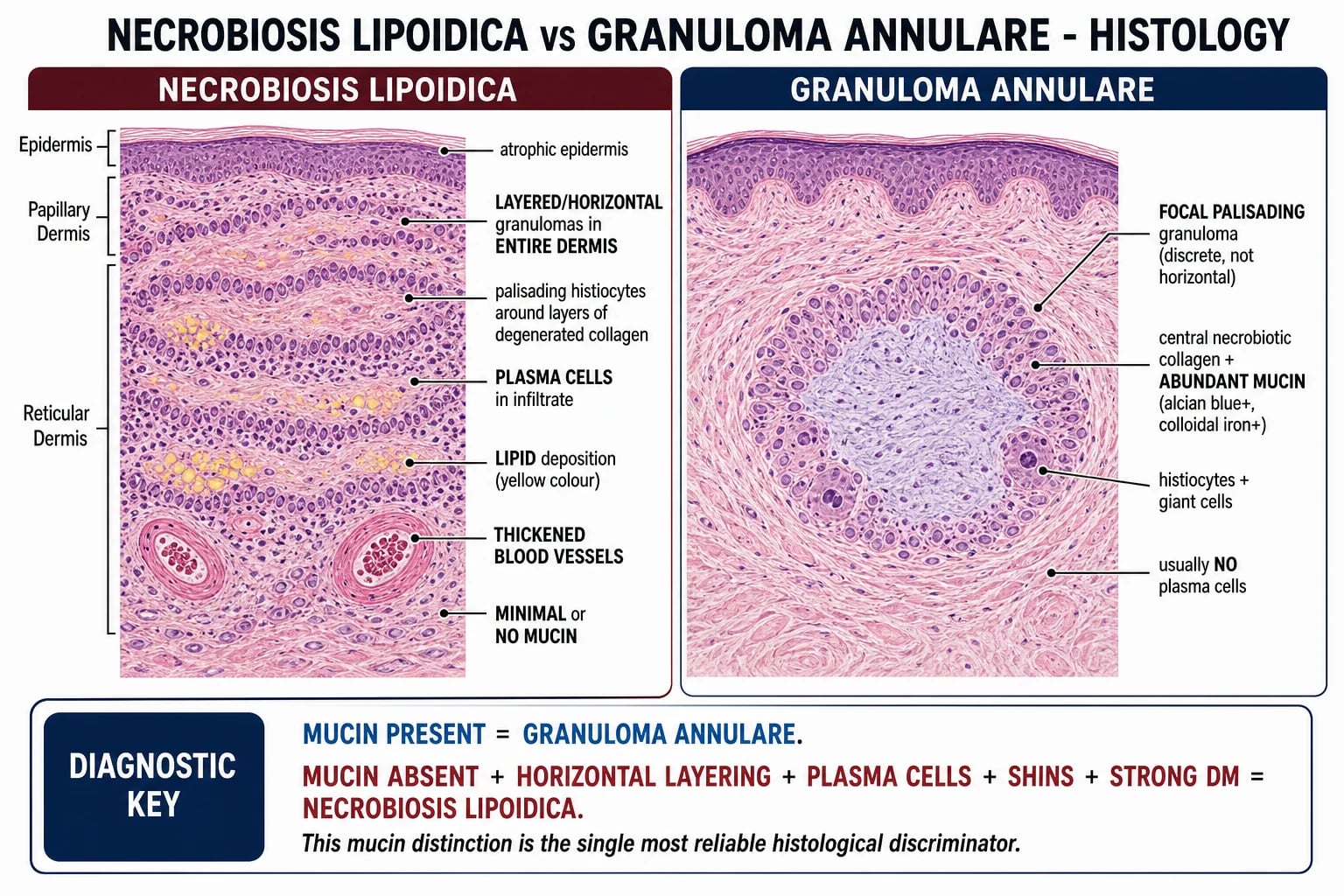
NL overlaps histologically with granuloma annulare because both are necrobiotic granulomatous dermatoses, but their architecture differs. NL usually has broader, tiered, horizontal zones of necrobiosis through the dermis and sometimes into subcutis, more plasma cells, and more vascular wall thickening. Granuloma annulare tends to have smaller focal palisading granulomas with abundant dermal mucin. The diagnosis is therefore clinicopathological: the pathologist needs to know that the biopsy came from a yellow atrophic pretibial plaque.[2]
Clinical Presentation
The usual patient notices one or more slowly enlarging plaques on the anterior shins. Early lesions may be red-brown papules or plaques. Over months to years, the centre becomes yellow, waxy, shiny, depressed and atrophic, while the border remains red-brown or violaceous. Telangiectasia becomes prominent over the centre. Lesions are often bilateral but not mirror-image symmetrical.[1][8]
Symptoms vary. Many plaques are asymptomatic and cosmetically distressing. Others are tender, itchy, dysaesthetic or painful, especially when ulcerated. Ulceration can occur after minor trauma such as shaving, scratching, a knock against furniture, adhesive dressings, compression friction or scratching from pruritus. Once an ulcer forms, pain, exudate, crusting, secondary infection and delayed healing dominate the consultation.[3][6]
Atypical presentations are examinable because they prevent pattern-recognition errors. In children, NL is rare but may be a presenting clue to type 1 diabetes or familial metabolic risk. In immunosuppressed patients, granulomatous infections and atypical mycobacterial disease must be excluded before escalating immunomodulation. In pregnant patients, avoid assuming every shin plaque is pregnancy-related stasis or trauma; biopsy can be performed if malignancy, infection or an atypical granulomatous dermatosis is a concern, while most elective systemic therapies are deferred.[7]
Dermoscopy can support recognition. Reported features include a yellow-orange or yellow-brown structureless background, white scar-like areas and a network of linear, serpentine or branching telangiectatic vessels. Dermoscopy is not required to diagnose a classic plaque, but it helps distinguish NL from eczema, psoriasis, tinea, morphea, granuloma annulare and vascular lesions, and it guides biopsy to the active border rather than the burnt-out centre.[4]
Differential Diagnosis
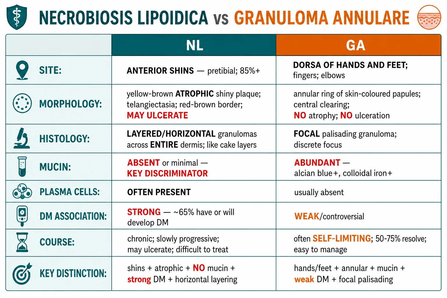
The differential diagnosis is built from four questions: Is it pretibial? Is it atrophic? Is it telangiectatic? Is it granulomatous on biopsy? Conditions that share one answer but not all four are the mimics. The highest-yield mimics are pretibial myxoedema, granuloma annulare, sarcoidosis and stasis dermatitis.[2][5]
Pretibial plaques — distinguishing the exam differentials
Pretibial myxoedema is the most tempting wrong answer because both are shin conditions with yellow-waxy colour. The discriminator is texture: myxoedema is indurated, oedematous, non-pitting and mucinous, often with Graves ophthalmopathy; NL is thinned, atrophic, shiny, telangiectatic and ulcer-prone. If a shin plaque is thick and swollen rather than thin and depressed, think thyroid dermopathy.[5]
Granuloma annulare is the histology trap. It is also a palisading granulomatous disease, and it may have necrobiotic collagen, but it usually appears as non-atrophic annular papules on the hands and feet and contains abundant mucin on biopsy. The presence of mucin alone is not an absolute rule, but mucin-rich focal palisading granulomas in a non-atrophic hand lesion strongly favours granuloma annulare, while horizontal granulomas with plasma cells in a pretibial atrophic plaque favours NL.[2]
Sarcoidosis is the granuloma not to miss. It may present with plaques on the legs, scar infiltration or lupus pernio, and it may coexist with systemic disease. Biopsy shows naked non-caseating granulomas rather than necrobiotic palisading granulomas, and the patient needs respiratory, ocular and systemic assessment if sarcoidosis is suspected.[2]
Stasis dermatitis matters because it changes management. A venous eczema plaque may surround or coexist with NL, especially in older patients with oedema. If venous disease is present, compression can improve ulcer healing, but it must be introduced only after arterial supply is checked clinically and, when indicated, by ankle-brachial index.[6]
Clinical & Bedside Assessment
Assessment begins with a deliberate skin description: number, site, symmetry, size, shape, colour, border, surface, atrophy, telangiectasia, scale, induration, ulceration, exudate and tenderness. Palpate the plaque gently. NL feels thin and atrophic centrally with an active raised edge; pretibial myxoedema feels thick and oedematous; morphea feels bound down and sclerotic.[3]
The bedside examination must include the feet and vascular system, not just the plaque. Check dorsalis pedis and posterior tibial pulses, capillary refill, temperature, neuropathy with a 10 g monofilament if the patient has diabetes, oedema, varicosities, lipodermatosclerosis and signs of infection. These findings determine ulcer risk and whether compression or debridement is safe.[1][6]
Ask for duration, change, pain, itch, trauma, shaving or adhesive injury, previous ulceration, diabetes history, HbA1c trend, smoking, vascular disease, thyroid symptoms, pregnancy possibility, drug history, immunosuppression, systemic symptoms, ocular symptoms and respiratory symptoms. The history should also ask what the patient wants: pain relief, ulcer healing, cosmetic improvement, reassurance about diabetes, or prevention of progression. NL treatment is often slow and imperfect, so expectation-setting is part of the assessment.[3]
Focused clinic assessment
Describe the plaque
Pretibial site, yellow-brown atrophic centre, telangiectasia, violaceous rim, ulcer or no ulcer.
Decide if typical
Classic bilateral shin plaque in a diabetic patient may be diagnosed clinically; atypical site, nodularity, ulcer edge change or diagnostic doubt needs biopsy.
Assess systemic context
HbA1c or fasting glucose, diabetes complications, smoking, thyroid disease, venous oedema, peripheral arterial disease and neuropathy.
Assess ulcer risk
Trauma exposure, footwear, shaving, scratching, adhesive dressings, vascular supply, oedema and infection.
Set treatment goal
Arrest active border, prevent trauma, heal ulcer, improve pain, reduce cosmetic distress; do not promise rapid clearance.
Investigations
A classic NL plaque can be diagnosed clinically, but investigations answer four exam questions: Is there diabetes or thyroid disease? Is the diagnosis secure? Is an ulcer complicated by vascular disease, infection or malignancy? Is it safe to use immunomodulatory treatment?[1][3]
Investigation plan
Histology is the key confirmatory investigation. The pathologist looks for palisading or layered granulomatous inflammation, zones of necrobiotic collagen, histiocytes and giant cells, plasma cells, thickened blood vessels, and relatively little mucin compared with granuloma annulare. The pattern may extend through the full dermis and sometimes into subcutaneous tissue. Special stains may be used to exclude infection when the presentation is atypical or the patient is immunosuppressed.[2][3]
Dermoscopy is supportive rather than mandatory. A yellow-orange or yellow-brown background, white scar-like areas and linear branching vessels support NL, but biopsy remains the answer when the lesion is atypical, nodular, rapidly changing, ulcerated in an unusual way, or not responding as expected.[4]
Management — Resuscitation
NL is usually an outpatient chronic dermatosis, so there is no routine resuscitation bundle. The time-critical management scenario is an ulcerated, infected, ischaemic or malignant-looking plaque. In that situation, treat it as a lower-limb ulcer with dermatology-specific differentials rather than as a cosmetic rash.[6]
Urgent ulcerated NL bundle
Check systemic illness
Fever, spreading cellulitis, tachycardia, hypotension, confusion or severe pain needs same-day medical assessment and sepsis-aware care.
Protect the wound
Clean with saline, use a non-adherent dressing, avoid adhesive trauma on atrophic skin, provide analgesia and offload repeated knocks.
Assess perfusion and oedema
Pulses, capillary refill, neuropathy, venous oedema and ankle-brachial index when compression is planned.
Identify infection correctly
Swab only when there is spreading erythema, warmth, purulence, malodour, increased pain or systemic features; colonisation alone does not need antibiotics.
Biopsy danger signs
Raised, rolled, bleeding, hyperkeratotic or non-healing ulcer edge needs biopsy to exclude squamous cell carcinoma.
Pain must be taken seriously. NL ulcers can be painful because the thin plaque exposes nerves and because venous oedema, infection or ischaemia may coexist. Escalating pain, necrosis, black eschar, disproportionate tenderness or systemic toxicity should trigger reconsideration of vasculitis, infection, pyoderma gangrenosum, arterial disease or malignancy rather than simply "bad NL".[6]
Management — Definitive & Stepwise
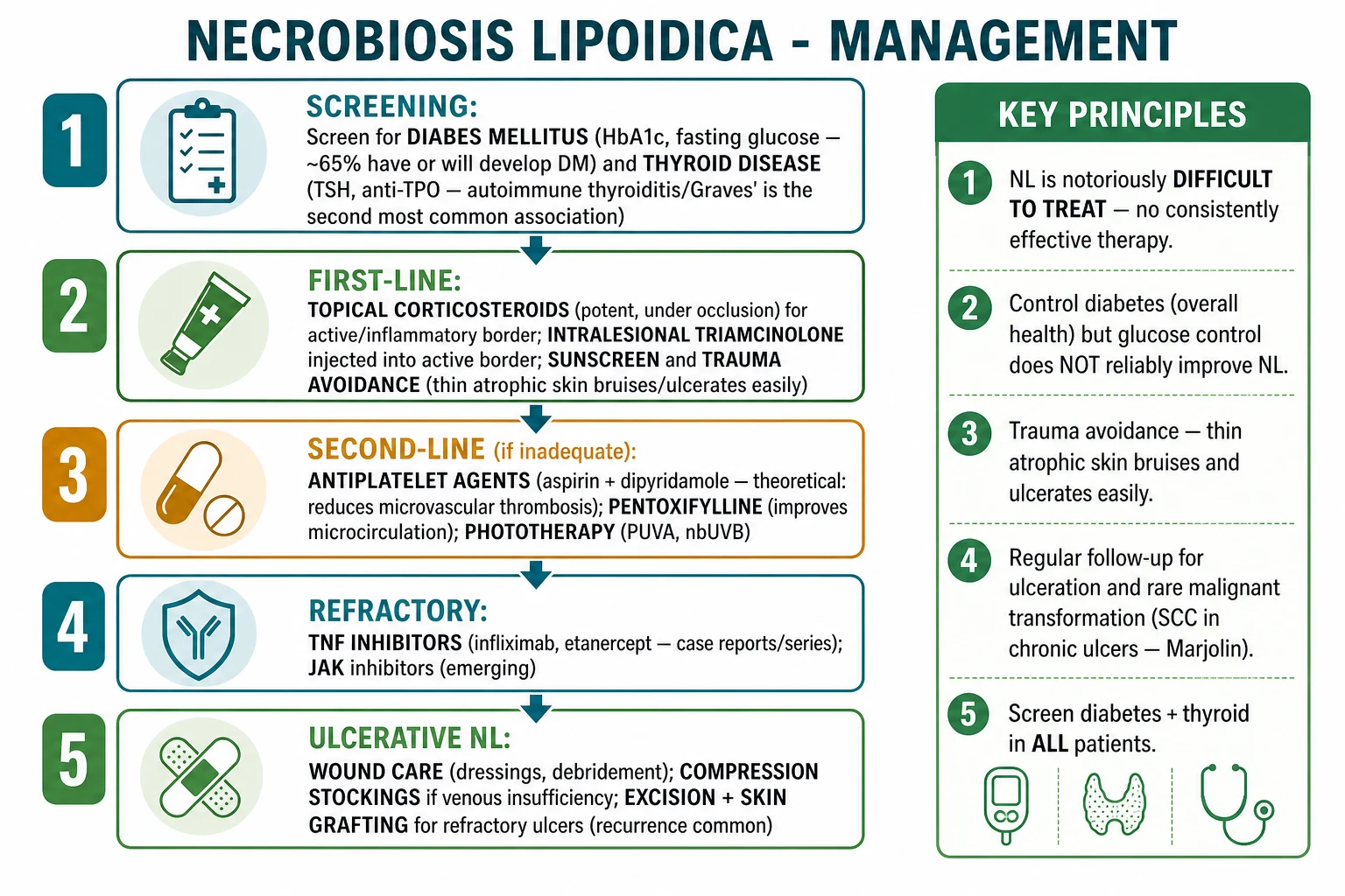
Treatment is difficult because mature NL contains irreversible dermal atrophy as well as active inflammation. The realistic goal is to stop progression, reduce inflammation at the border, prevent ulceration, heal ulcers and improve symptoms, not to promise complete cosmetic normalisation. Management should be layered: education and risk reduction for every patient, topical or intralesional therapy for active lesions, steroid-sparing options when atrophy or chronicity limits steroids, phototherapy or systemic therapy for extensive disease, and wound-care escalation for ulcers.[3]
Stepwise management ladder
Foundation for every patient
Explain chronicity; stop smoking; protect shins from trauma; avoid shaving cuts and adhesive stripping; optimise diabetes, lipids, blood pressure and thyroid disease; photograph and measure the plaque.
Active non-ulcerated border
Potent topical corticosteroid to the raised rim for a short course, or intralesional triamcinolone into the active rim when local expertise is available.
Steroid-sparing local therapy
Tacrolimus ointment can be used on thin atrophic plaques where further steroid atrophy is a concern, especially for maintenance or sensitive sites.
Widespread or refractory non-ulcerated disease
Consider phototherapy, pentoxifylline or antiplatelet therapy after discussing limited evidence and contraindications.
Refractory ulcerative or progressive disease
Specialist options include systemic immunomodulation or biologic therapy such as TNF-alpha inhibition; screen for infection first and monitor carefully.
Non-healing ulcer
Structured wound care, compression if venous disease and arterial supply allow, biopsy suspicious edge, and selected excision with grafting only after careful multidisciplinary review.
Common treatment options used in practice
The key technical point is where to apply treatment. Potent topical steroid and intralesional steroid are aimed at the raised inflammatory rim, not the paper-thin centre. Long unsupervised steroid use over the centre can worsen atrophy and ulcer risk. Tacrolimus is useful as a steroid-sparing option because it does not thin the skin, although burning and irritation can limit adherence.[3][7]
Smoking cessation should be stated as a treatment, not a lifestyle afterthought. Use a brief intervention, offer nicotine replacement or varenicline according to local practice, and explain the reason: smoking worsens microvascular perfusion and wound healing, the very vulnerabilities that make NL ulcerate. Trauma avoidance is equally practical: shin guards for high-risk work or sport, careful moisturising, avoiding adhesive tapes on plaques, using electric clippers rather than shaving over lesions, and prompt dressing of minor erosions.[3][6]
Wound care for ulcerated NL is systematic. Cleanse gently, use non-adherent dressings, maintain moisture balance with foam, hydrofiber or hydrocolloid dressings according to exudate, protect periwound atrophic skin, treat oedema with compression only when arterial supply is adequate, offload repeated trauma, and treat true cellulitis with appropriate antibiotics. Avoid repeated traumatic procedures that enlarge the defect. Conservative debridement can remove nonviable slough when perfusion is adequate, but aggressive debridement without reassessing the diagnosis can be harmful if the ulcer is actually pyoderma gangrenosum or vasculitis.[6]
Surgery is not first-line for ordinary plaques because recurrence, graft failure and poor healing are possible. Excision with split-thickness skin grafting is reserved for selected refractory ulcers or disabling localized disease after vascular supply, diabetes, smoking, venous oedema and infection have been optimised. Even then, counsel about recurrence at the graft edge or donor-site issues.[3][6]
Specific Subtypes & Scenarios
Non-ulcerated active pretibial NL is the common outpatient scenario. Document baseline size and photograph, screen for diabetes and thyroid disease, advise smoking cessation and trauma avoidance, and treat the active edge with a time-limited potent topical steroid or intralesional steroid. Review after 6 to 12 weeks. If the lesion is stable and asymptomatic, observation with protection is reasonable because treatment toxicity can exceed benefit.[3]
Mature inactive atrophic plaques are managed differently. If the rim is not inflamed and the centre is thin, repeated steroid escalation is unlikely to reverse atrophy and may worsen fragility. Focus on emollients, protective clothing, cosmetic camouflage if desired, monitoring for ulceration, and risk-factor management. This is a common viva point: do not inject a steroid into the thinnest part of the plaque.[3]
Ulcerated NL requires a dual diagnosis: it is both NL and a chronic lower-limb ulcer. Assess vascular supply, venous oedema, neuropathy, infection, pain and trauma. Biopsy any suspicious raised, rolled, bleeding or hyperkeratotic edge. Use a wound-care plan with non-adherent dressings and compression if venous disease is present and arterial assessment permits. Escalate to dermatology or tissue-viability services if the ulcer is enlarging, very painful, infected, or not improving.[6][9]
Paediatric NL is rare, and the differential includes granuloma annulare, sarcoidosis, morphea and traumatic scarring. Screen carefully for type 1 diabetes symptoms and family history. Use the minimum effective topical treatment and avoid creating steroid atrophy in growing skin. Phototherapy and systemic therapy require specialist assessment and family counselling.[7]
Extratibial NL on the arms, trunk, scalp or face should not be dismissed as classic until biopsy supports it. The differential changes with the site: sarcoidosis and granuloma annulare on the arms, morphea and xanthoma on the trunk, scarring alopecia or discoid lupus on the scalp, and vascular or granulomatous mimics on the face. The more atypical the site, the more the diagnosis must be clinicopathological.[2][3]
NL in a patient without known diabetes should trigger screening but not labelling. Tell the patient that NL is associated with diabetes but does not prove diabetes. Check HbA1c or fasting glucose, repeat according to risk and local diabetes-screening intervals, and screen for thyroid disease when symptoms, family history or clinical clues are present.[1][5]
Complications & Pitfalls
The main complication is ulceration. Ulceration is clinically important because it causes pain, infection risk, scarring, delayed healing and repeated healthcare visits. It often begins after minor trauma to the atrophic centre. Once ulcerated, NL behaves like a chronic lower-limb wound, and progress depends on perfusion, venous oedema, neuropathy, smoking, glycaemic status, wound care and avoidance of further trauma.[3][6]
Secondary infection can complicate an ulcer, but colonisation is common and does not by itself justify antibiotics. Treat cellulitis when there is spreading erythema, warmth, swelling, purulence, increasing pain, lymphangitis or systemic features. If the ulcer is painful out of proportion, necrotic or rapidly expanding, reconsider the diagnosis and assess for vasculitis, arterial disease, pyoderma gangrenosum or deep infection.[6]
Squamous cell carcinoma arising in chronic NL ulcers is rare but examinable because missing it is serious. The warning signs are a chronic non-healing ulcer with a raised, rolled, indurated, bleeding, hyperkeratotic or rapidly enlarging edge. Biopsy is mandatory; do not keep changing dressings for months without tissue diagnosis when the edge has changed.[9]
Psychological morbidity is another complication. Shin plaques are visible, chronic and often cosmetically distressing. Patients may hide their legs, avoid sport or social situations, or feel blamed because of the diabetes association. A good management plan acknowledges this, offers realistic expectations, and discusses camouflage or support when appearance is the major concern.[3]
Prognosis & Disposition
NL is chronic and often slowly progressive. Some plaques stabilise, some fluctuate, and some persist for years. Complete spontaneous resolution can occur but is not predictable, and residual atrophy or pigmentation may remain. Mature plaques rarely return to normal skin because dermal collagen and elastic support have been lost.[1][3]
Prognosis is best when lesions are small, non-ulcerated, protected from trauma, and treated while the rim is active rather than after widespread atrophy. Prognosis is worse with ulceration, smoking, venous disease, arterial disease, neuropathy, repeated trauma, infection and poor wound-care access. Diabetes itself matters most through vascular and neuropathic complications rather than through a direct HbA1c-to-plaque relationship.[1][6]
Most patients can be managed as outpatients. Refer to dermatology when the diagnosis is uncertain, the lesion is atypical or extratibial, biopsy is needed, ulceration occurs, the plaque is rapidly progressive, pain is significant, first-line therapy fails, or systemic therapy is being considered. Refer urgently or arrange same-day review for spreading cellulitis, systemic illness, critical limb ischaemia signs, severe pain, or a suspicious ulcer edge.[3][6]
Safety-net advice should be explicit: return if a plaque breaks down, becomes rapidly painful, develops pus or spreading redness, bleeds, forms a raised hard edge, enlarges quickly, or if new symptoms of diabetes or thyroid disease develop. Advise protective dressings or clothing for activities that expose the shin to knocks.[3]
Special Populations
Diabetes mellitus. Check HbA1c or fasting glucose at baseline and coordinate diabetic foot and vascular risk care. Examine for neuropathy and pulses, because reduced sensation allows unnoticed trauma and poor perfusion delays healing. Explain that diabetes optimisation helps overall health and ulcer healing, but NL plaques may persist despite excellent control.[1]
Pregnancy. Most NL care in pregnancy is conservative: trauma avoidance, emollients, non-adherent dressings if erosions occur, and careful diabetes care. Use potent topical steroids only when needed and for short courses, avoiding large areas and prolonged occlusion. Defer elective phototherapy decisions and systemic agents to dermatology and obstetrics; biopsy remains appropriate when malignancy, infection or atypical disease is a concern.[3]
Children and adolescents. NL in childhood is uncommon and can be psychologically difficult because lesions are visible. Screen for type 1 diabetes symptoms, check family history, and avoid long unsupervised potent steroid courses. Growth, school activity, sports trauma and adherence to dressings should be included in the plan.[7]
Older adults. Older patients are more likely to have venous insufficiency, arterial disease, neuropathy, anticoagulant use and skin fragility. Do not assume a leg ulcer in NL is only inflammatory; assess vascular disease and malignancy, and be cautious with compression unless arterial supply is adequate.[6]
Immunosuppressed patients. Atypical infection, deep fungal disease, mycobacterial infection and malignancy enter the differential. Biopsy with appropriate stains and cultures should precede systemic immunosuppression when the morphology is not classic. Screen for TB, hepatitis and HIV before biologic therapy.[2][3]
Patients taking anticoagulants or antiplatelets. Antiplatelet therapy has been used historically for NL, but bleeding risk matters. Do not add aspirin or dipyridamole casually to a patient already anticoagulated or with high bleeding risk; coordinate with the prescriber and choose local or wound-directed options when safer.[3]
Evidence, Guidelines & Regional Differences
The evidence base for NL therapy is limited. Much of treatment is built from case reports, small series, pathophysiological rationale and expert guidance rather than large randomised trials. This is why guidelines emphasise individualised goals, risk-factor modification, local therapy for active inflammation, wound care for ulcers, and cautious escalation for refractory disease.[3]
Evidence reality check
Guideline synthesis and case-series evidence
Population: Patients with necrobiosis lipoidica, including non-ulcerated, ulcerated and paediatric disease
Key finding
Variable response; no universally reliable therapy; ulcer care and trauma prevention remain central
Practice change
Do not present a single drug as curative. State the goal, evidence limitation, contraindications and escalation trigger.
Global teaching is consistent on the core points: recognise the pretibial atrophic telangiectatic plaque, screen for diabetes, protect against trauma, treat active inflammation at the rim, biopsy atypical or suspicious ulcers, and use wound-care principles for ulcerated disease.[1][3]
ANZ practice mirrors the global approach: dermatologist confirmation for atypical disease, diabetes and cardiovascular risk management in primary care, careful wound-care referral for ulceration, and specialist-only biologic escalation for refractory ulcerative disease.[3][6]
The controversies are practical. First, whether microangiopathy or immune dysregulation is primary remains unresolved. Second, antiplatelet therapy and pentoxifylline are biologically plausible but not consistently effective. Third, biologics such as TNF-alpha inhibitors may help selected refractory cases, but evidence is too limited for routine first-line use. Fourth, surgery can close a selected ulcer but can fail or recur if vascular, venous, smoking and trauma factors remain.[2][3]
Follow-up, monitoring and counselling
Follow-up should be structured around the activity of the rim and the integrity of the centre, not simply around whether the plaque still exists. At each review, record lesion size, photograph the plaque with consent, mark whether the border is still raised or violaceous, document symptoms, and ask specifically about new erosions, bleeding, adhesive injury, shaving cuts, scratching, sports trauma and work-related knocks. If a potent topical steroid has been used, check for steroid atrophy, telangiectatic worsening, purpura and striae, and stop or step down once the active rim has flattened. If intralesional triamcinolone was used, inspect for focal depressions and dyspigmentation at injection sites.[3]
A practical review interval is 6 to 12 weeks after starting local therapy, sooner if there is ulceration, pain or rapid change. Stable non-ulcerated plaques can then be reviewed less frequently with clear safety-net instructions. Ulcerated NL needs closer review until the wound trajectory is clear: measure length, width and depth, record exudate, pain and periwound maceration, and reassess vascular supply and oedema if healing stalls. A chronic ulcer that does not shrink despite appropriate care should not simply receive another dressing prescription; revisit the differential, optimise pressure and venous factors, and biopsy suspicious tissue.[6][9]
Counselling should prevent two harms. The first is false reassurance: patients need to know that the plaque is benign but fragile, and that ulceration, infection or a changing ulcer edge needs prompt review. The second is false blame: because NL is linked to diabetes, patients often assume poor control caused the lesion. Explain that diabetes and microvascular disease are important associations and affect wound healing, but NL is not a simple report card for HbA1c and may persist despite excellent control. That message improves trust and adherence to protective measures.[1][3]
Before starting a systemic or biologic option, write down the indication in concrete terms: progressive active border despite adequate local therapy, extensive symptomatic plaques, recurrent ulceration, or a non-healing ulcer after vascular and wound factors have been addressed. Baseline monitoring should match the drug, but biologic escalation specifically requires screening for latent tuberculosis, hepatitis B and C, HIV where appropriate, vaccination review and counselling about infection risk. Document that the evidence is limited and that the goal is reduced activity or ulcer healing, not guaranteed cosmetic clearance.[3]
For MBBS exams, this follow-up logic is often the difference between a list of treatments and a safe management answer. A complete answer says what to treat, where to treat, what to avoid, when to biopsy, when to refer, and what to tell the patient to watch for. In NL, those verbs are: protect, stop smoking, screen, treat the rim, spare the atrophic centre, dress ulcers gently, assess vessels, biopsy danger edges and escalate only when the risk-benefit balance is clear.[3][6]
Exam Pearls
NL SHIN
SHIN
Pretibial anterior shins are the classic site; bilateral but asymmetric plaques are common.
Horizontal or layered palisading granulomas around necrobiotic collagen, with plasma cells and vascular thickening.
Strong diabetes association, but good glycaemic control does not reliably clear established NL.
Trauma can ulcerate the atrophic plaque; biopsy suspicious chronic edges for SCC.
Stem diagnosis
- Young or middle-aged woman, diabetes or impaired glucose tolerance
- Anterior shin plaque
- Yellow-brown waxy atrophic centre
- Telangiectasia
- Violaceous red-brown active border
Histology
- Palisading or layered horizontal granulomas
- Necrobiotic collagen
- Plasma cells
- Thickened vessels
- Less mucin than granuloma annulare
Differentials
- Pretibial myxoedema: Graves, non-pitting mucinous induration
- Granuloma annulare: hands/feet, annular papules, mucin
- Sarcoid: naked granulomas and systemic disease
- Stasis dermatitis: oedema, scale, venous signs
Management
- Smoking cessation and trauma avoidance
- Screen HbA1c and thyroid disease
- Potent topical steroid or intralesional triamcinolone to active rim
- Tacrolimus, phototherapy, pentoxifylline or antiplatelet options in selected refractory cases
- TNF-alpha inhibitor only for severe refractory disease
- Structured wound care for ulcers
Do not miss
- Ulceration is the major morbidity
- Glycaemic control does not reliably improve plaques
- Biopsy changing chronic ulcer edge for SCC
- Do not inject steroid into atrophic centre
“Yellow-brown atrophic telangiectatic pretibial plaque with violaceous rim, diabetes association, palisading necrobiotic granulomas, ulcer risk and difficult treatment.”
Embedded SAQ — shin plaque in diabetes
10 minutes · 10 marks
A 38-year-old woman with type 1 diabetes has bilateral yellow-brown shiny plaques on the anterior shins. The plaques have central atrophy, prominent telangiectasia and a violaceous rim. One plaque has a shallow erosion after minor trauma.
“”
Exam application bank (NEET-PG / INICET)
One-line answer
Necrobiosis lipoidica is a chronic granulomatous dermatosis of collagen degeneration, classically producing yellow-brown atrophic telangiectatic plaques with a violaceous rim on the anterior shins. It is strongly associated with diabetes mellitus but does not reliably improve with glycaemic control. Diagnosis is clinical when typical and biopsy shows palisading or layered granulomas around necrobiotic collagen with plasma cells and vascular change. Management centres on smoking cessation, trauma avoidance, topical or intralesional corticosteroid to the active rim, tacrolimus or phototherapy for steroid-sparing control, antiplatelet or pentoxifylline in selected cases, biologics for refractory disease, and meticulous ulcer care.
Worked stems (answer without another resource)
Stem 1 — Classic presentation. Map symptoms to mechanism; name the first investigation and first treatment step with dose/route if drug therapy is standard. [1]
Stem 2 — Unstable / complicated. List red flags that force immediate resuscitation, theatre, ICU, antidote, or reperfusion — and what you do in the first 15 minutes. [1]
Stem 3 — Atypical group. Elderly, pregnancy, child, or immunocompromised: how presentation and thresholds change. [1]
Stem 4 — Differential trap. Name the three closest mimics and one discriminator for each. [1]
Stem 5 — Disposition. Who goes home with safety-netting, who is admitted, who needs HDU/ICU/theatre, and what follow-up is mandatory. [1]
Rapid viva checklist
- Definition + classification
- Pathophysiology chain
- Bedside signs / criteria
- Score with exact components (if any)
- Emergency bundle
- Definitive therapy with doses
- Complications of disease and of treatment
- Special populations
- Guideline/trial name if classic
- Three exam traps
Coverage self-check
If you cannot answer any stem above from this page alone, re-read the matching section — the page is intended to be self-sufficient for final-prof and NEET-PG/INICET questions on Necrobiosis lipoidica.
References
- [1]Lima AL, Illing T, Schliemann S, et al. Cutaneous Manifestations of Diabetes Mellitus: A Review Am J Clin Dermatol, 2017.PMID 28374407
- [2]Terziroli Beretta-Piccoli B, Mainetti C, Peeters MA, et al. Cutaneous Granulomatosis: a Comprehensive Review Clin Rev Allergy Immunol, 2018.PMID 29352388
- [3]Erfurt-Berge C, Renner R, Peckruhn M, et al. S1-Guideline for diagnosis and therapy of necrobiosis lipoidica J Dtsch Dermatol Ges, 2026.PMID 41420334
- [4]Errichetti E, Stinco G. Dermoscopy in General Dermatology: A Practical Overview Dermatol Ther (Heidelb), 2016.PMID 27613297
- [5]Lause M, Kamboj A, Fernandez Faith E. Dermatologic manifestations of endocrine disorders Transl Pediatr, 2017.PMID 29184811
- [6]Dissemond J, Placke JM, Moelleken M, et al. The Differential Diagnosis of Leg Ulcers Dtsch Arztebl Int, 2024.PMID 39115274
- [7]Schiefer-Niederkorn A, Sadoghi B, Binder B. Necrobiosis lipoidica in childhood: a review of literature with emphasis on therapy J Dtsch Dermatol Ges, 2023.PMID 37401158
- [8]Liu MJ, Li J. Necrobiosis Lipoidica N Engl J Med, 2024.PMID 38169491
- [9]Vasari L, Simetić L, Kuna SK, et al. Cutaneous Squamous Cell Carcinoma Developing in Necrobiosis Lipoidica - A Case Report with Literature Review Dermatol Pract Concept, 2026.PMID 41912178