Dermatology · Medicine
Necrotising fasciitis
Also known as Necrotising fasciitis · Flesh-eating disease · Necrotising soft tissue infection · Fournier's gangrene
Necrotising fasciitis is a rapidly progressive, life-threatening necrotising infection of the deep fascia and subcutaneous tissue that constitutes a SURGICAL EMERGENCY. Classified as Type I (polymicrobial), Type II (monomicrobial Group A Streptococcus ± Staphylococcus aureus — the 'flesh-eating disease'), or Type III (marine — Vibrio vulnificus). Clinically characterised by severe pain out of proportion to the visible signs, rapid progression from erythema to purple-grey discoloration, haemorrhagic bullae, necrosis and crepitus, with systemic toxicity progressing to septic shock. Diagnosis is CLINICAL — the LRINEC score (≥6 high risk) supports but does not replace clinical judgment, and imaging must NOT delay surgical exploration. Management requires URGENT SURGICAL DEBRIDEMENT (the definitive treatment — to healthy bleeding tissue, repeated daily), broad-spectrum IV antibiotics (piperacillin-tazobactam/carbapenem + clindamycin + vancomycin), clindamycin for toxin suppression, IVIG for streptococcal toxic shock, and ICU support. Mortality is 20-40%, increasing exponentially with delay in surgery. Fellowship-level assessment demands mastery of the clinical features (pain out of proportion), the LRINEC score and its limitations, the imperative of urgent surgical debridement (not delayed for imaging), the antibiotic regimen with clindamycin rationale, and the special case of Fournier's gangrene.
On this page & tools
Your progress
Saved locally on this device.
Exam tags
Red flags
Necrotising fasciitis — the time-critical numbers
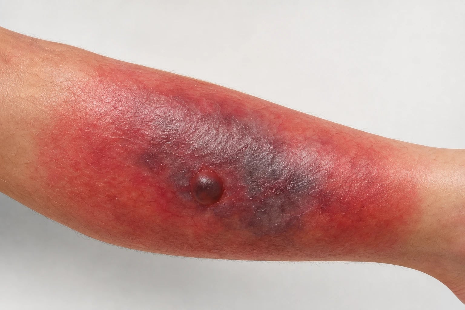
Definition & Classification
Necrotising fasciitis is a rapidly progressive, life-threatening necrotising infection of the deep fascia and subcutaneous tissue, producing tissue destruction, systemic toxicity and a high mortality. It is a surgical emergency — the definitive treatment is urgent surgical debridement.[1][4] The term was popularised in the 1950s by Wilson and is preferred today to older labels such as "streptococcal gangrene" or "hospital gangrene"; "flesh-eating disease" remains a media-friendly synonym reserved almost exclusively for monomicrobial Type II disease.
Three microbiological patterns are recognised — Type I, Type II and Type III — and one eponymous variant, Fournier's gangrene.[1][10]
| Type | Microbiology | Host | Key features |
|---|---|---|---|
| Type I (polymicrobial) | Mixed aerobes + anaerobes (Group A Strep, Staph, Enterobacteriaceae, Bacteroides, Clostridia) | Immunocompromised, diabetic, post-surgical, perianal | Commonest type (~70–80%); gas-forming; perineal (Fournier's); slower onset than Type II |
| Type II (monomicrobial) | Group A Streptococcus (Streptococcus pyogenes) ± Staph. aureus (often MRSA) | Previously healthy host, IVDU, post-trauma/varicella | "Flesh-eating disease"; streptococcal toxic shock; very rapid progression |
| Type III (marine / freshwater) | Vibrio vulnificus (seawater), Aeromonas hydrophila (freshwater), rarely Edwardsiella | Chronic liver disease, fish-handlers, freshwater wounds | Haemorrhagic bullae; sepsis; mortality over 50% in cirrhotic patients |
| Fournier's gangrene (perineal Type I) | Mixed aerobes + anaerobes | Perianal, urogenital, immunocompromised | Perineum, scrotum, genitalia; urgent urology + general surgery |
The classification matters because each type predicts a different organism, a different host profile, a different empiric antibiotic bundle and a different prognosis.[1][10] Fournier's gangrene is eponymous because the perineum is anatomically unforgiving: superficial fascial planes communicate with the abdominal wall, the thigh and the retroperitoneum, allowing infection to track rapidly into the pelvis and the perinephric space.[8][9]
The four TYPEs of necrotising fasciitis — TGIF (one hint per letter)
TGIF
Mixed aerobes + anaerobes; diabetic / immunocompromised host; gas-forming; commonest
Group A Streptococcus (S. pyogenes) ± Staph aureus; previously healthy host; 'flesh-eating'
Vibrio vulnificus (sea), Aeromonas (fresh); chronic liver disease; haemorrhagic bullae
Perineum, scrotum, genitalia; urology + general surgery; colostomy / cystostomy often needed
Epidemiology & Risk Factors
Incidence: approximately 0.3–1.5 cases per 100,000 population per year in developed settings, with cluster rates rising sharply during outbreaks of invasive group A streptococcal (iGAS) disease and after natural disasters.[1][4] Mortality remains 20–40% despite modern care; in low-resource settings and in the elderly it can exceed 50%.
Host risk factors that the examiner wants named: [1]
| Risk factor | Why it matters |
|---|---|
| Diabetes mellitus | Commonest single comorbidity (present in ~50% of cases); impairs neutrophil function and microvascular perfusion |
| Immunosuppression (HIV, transplant, chemotherapy, biologics, steroids) | Reduces host containment; atypical organisms |
| Peripheral vascular disease | Limits antibiotic delivery; impairs wound healing after debridement |
| Chronic liver disease / alcohol excess | Strongest driver of fulminant Vibrio vulnificus septicaemia |
| Chronic kidney disease | Uraemic immune dysfunction; higher mortality |
| Intravenous drug use (IVDU) | Skin and injection-site portal; MRSA, oral flora, polymicrobial Type I |
| Recent surgery / trauma / burn | Direct breach of skin; postoperative NF may be missed as "wound infection" |
| NSAID use | May mask early pain (the very sign that would have triggered surgery) |
| Obesity / malnutrition | Altered fascial blood supply; delayed presentation |
Other antecedent events include insect bites, varicella (in children),[11] perianal abscess or fissure, dental abscess with cervical spread, and intramuscular injection. In many cases no portal of entry is identified — these "idiopathic" cases tend to have higher mortality because diagnosis is delayed.[4]
A delay in surgical debridement beyond 12–24 hours from admission dramatically increases mortality; one meta-analysis puts the increase at roughly 9-fold.[5]
Pathophysiology
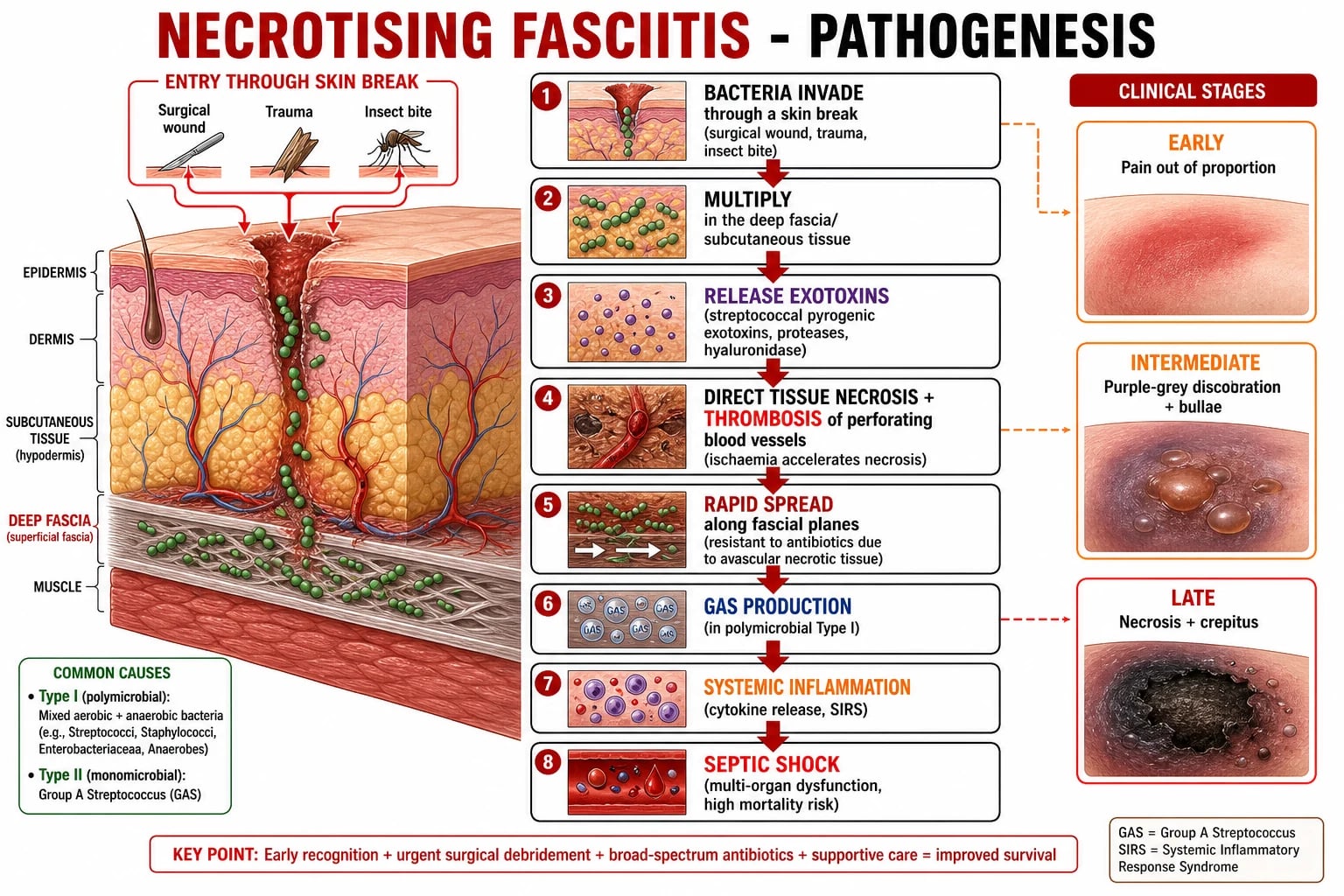
Necrotising fasciitis is best understood as the convergence of a bacterial invasion and a destructive host response along the deep fascia. The sequence is reproducible.[1][10]
- Portal of entry. Bacteria reach the subcutis through any breach in the skin — surgical wound, blunt trauma, fissure, IV injection site. In Type III the portal is often a freshwater or saltwater wound in a host with chronic liver disease. In Fournier's gangrene the portal is perianal, periurethral or perigenital.[8][9]
- Avascular niche. The deep fascia is a relatively avascular, poorly defended plane; once bacteria reach it they multiply rapidly beyond the reach of circulating neutrophils.
- Exotoxin and enzyme release. Group A Streptococcus (Type II) releases streptococcal pyrogenic exotoxins (Spe A, B, C), streptolysins, hyaluronidase and M-protein, which produce direct tissue destruction and which act as superantigens that bypass antigen-presentation to trigger massive T-cell activation and a cytokine storm.[10] In Type I, mixed aerobes and anaerobes synergise — facultative bacteria consume oxygen and lower redox potential, allowing obligate anaerobes to thrive; clostridial species produce gas (CO₂, H₂, H₂S), accounting for crepitus.[1]
- Microvascular thrombosis. The crucial lesion is thrombosis of perforating cutaneous vessels, mediated by bacterial exotoxins and inflammatory cytokines. The result is a progressive ischaemia of the overlying skin and an antibiotic-impermeable compartment — even IV antibiotics cannot reach bacteria locked inside the necrotic, thrombosed tissue. This is why surgery is mandatory.[1][4]
- Fascial spread. Bacterial hyaluronidase and proteases dissolve connective-tissue barriers; infection tracks the superficial fascia at centimetres per hour, producing the characteristic rapid spread.
- Systemic sepsis. The combination of cytokine storm, bacterial superantigen activity and bacteraemia produces septic shock, DIC, acute respiratory distress syndrome (ARDS) and acute kidney injury within hours.[10]
Type I
- Polymicrobial synergy — aerobes consume O₂, anaerobes ferment
- Gas from clostridial metabolism produces crepitus
- Slower evolution (48–72 h); polymicrobial cover needed
- Host is typically diabetic or immunocompromised
Type II
- Monomicrobial GAS — driven by exotoxin + superantigen
- Most rapid progression (hours); previously healthy host
- Strongest indication for clindamycin (toxin suppression) and IVIG
- STSS frequently co-exists (hypotension + multi-organ failure)
Type III
- Marine (Vibrio) or freshwater (Aeromonas) portals
- Catastrophic in chronic liver disease (50–80% mortality)
- Haemorrhagic bullae and rapid bulla-to-necrosis progression
- Empiric doxycycline + ceftriaxone added to standard bundle
Fournier's
- Perineal / scrotal Type I — anatomically distinct, eponymous
- Source control needs urology + colorectal + general / plastic surgery
- Diverting colostomy or cystostomy often required
- Mortality 20–40%; higher with sepsis at presentation
Clinical Presentation
The hallmark is severe pain OUT OF PROPORTION to the visible skin changes — this is the earliest and most important clinical sign.[4][5]
Clinical progression (the disease clock): [1]
| Stage | Skin signs | Systemic | Bacteriology |
|---|---|---|---|
| Early (0–24 h) | Erythema, warmth, oedema, severe tenderness out of proportion to visible erythema | Fever, malaise, normal BP early | Type I: polymicrobial; Type II: GAS |
| Intermediate (24–48 h) | Purple-grey discoloration; bullae (serous → haemorrhagic); skin becomes anaesthetic (nerve necrosis) — the paradoxical pain decrease | Tachycardia, hypotension, lactate rising | Polymicrobial or GAS + Staph |
| Late (>48 h) | Necrosis, crepitus (gas), skin sloughing, rapid centrifugal spread | Septic shock, multi-organ failure, altered consciousness | Often mixed; mortality climbs sharply |
Other clinical clues that the bedside examiner looks for: hardness / woody induration of the subcutaneous tissue (the examiner can often feel that the skin itself feels "fixed" to deep fascia, unlike cellulitis); rapid progression (hours rather than days); systemic toxicity disproportionate to visible infection; skin anaesthesia over the infected area (nerve destruction — paradoxically, the pain may decrease as sensory nerves die, and the patient may then be erroneously reassured); bullae that are haemorrhagic, not serous; and an advancing edge that crosses fascial boundaries rather than lymphatic territories.[5]
Bedside & Clinical Assessment
The bedside examination is the diagnostic test. A structured five-step assessment avoids missing late disease.[5]
- Look. Mark the advancing erythematous edge (a surgical marker pen on intact skin at the periphery). Re-mark and re-examine every hour or document the spread with serial photographs. Asymmetry between the two limbs, a purple-grey patch within the erythema or any haemorrhagic bulla is concerning.
- Feel. Palpate for woody induration of the subcutis (the deep soft tissues feel "stuck" to the fascia, unlike the boggy oedema of cellulitis). Check for crepitus (the crackling of subcutaneous gas). Test sensation within and just outside the lesion — early anaesthesia over the area is a key clue.
- Probe — "finger test". Under analgesia and aseptic technique, a 2 cm incision is made down to fascia. Lack of bleeding, dull-grey fascia and effortless passage of a finger along the tissue planes (loss of tissue resistance) confirms necrotising fasciitis intra-operatively.[5]
- Sample. Aspirate any bulla fluid for Gram stain and culture. Send blood cultures before empiric antibiotics. Tissue from debridement goes to microbiology and histopathology.
- Search for source and shock. Examine the perineum (Fournier's), look for injection marks (IVDU) and check for varicella lesions in children.[11] Document blood pressure, lactate, urine output, mental state and capillary refill — these drive the sepsis bundle.
Investigations
- Diagnosis is CLINICAL — the most important "test" is clinical suspicion based on pain out of proportion, rapid progression and systemic toxicity.[5][7]
- LRINEC score — supports the diagnosis but does NOT replace clinical judgment.[6]
- Imaging (CT/MRI): may show fascial gas, fluid, oedema and tissue necrosis — useful if available rapidly and diagnosis is uncertain, but must NOT delay surgical exploration.[7] Plain radiographs have poor sensitivity for early gas; CT is the imaging study of choice when time permits.
- Surgical exploration (fingers test): a small incision down to fascia — if the tissue does not bleed, the fascia is greyish and dull, and a finger passes easily through the planes (loss of tissue resistance), the diagnosis is confirmed.[5] Tissue samples are sent for microbiology and histopathology.
- Blood cultures, tissue culture from debridement — identify the organism and guide antibiotic therapy.
- Adjunct labs: lactate, creatinine kinase (CK), electrolytes, coagulation screen, full blood count, glucose, HbA1c, liver function and arterial blood gas. A rising lactate, falling platelet count, rising CK and worsening metabolic acidosis are markers of advancing disease.
LRINEC Score
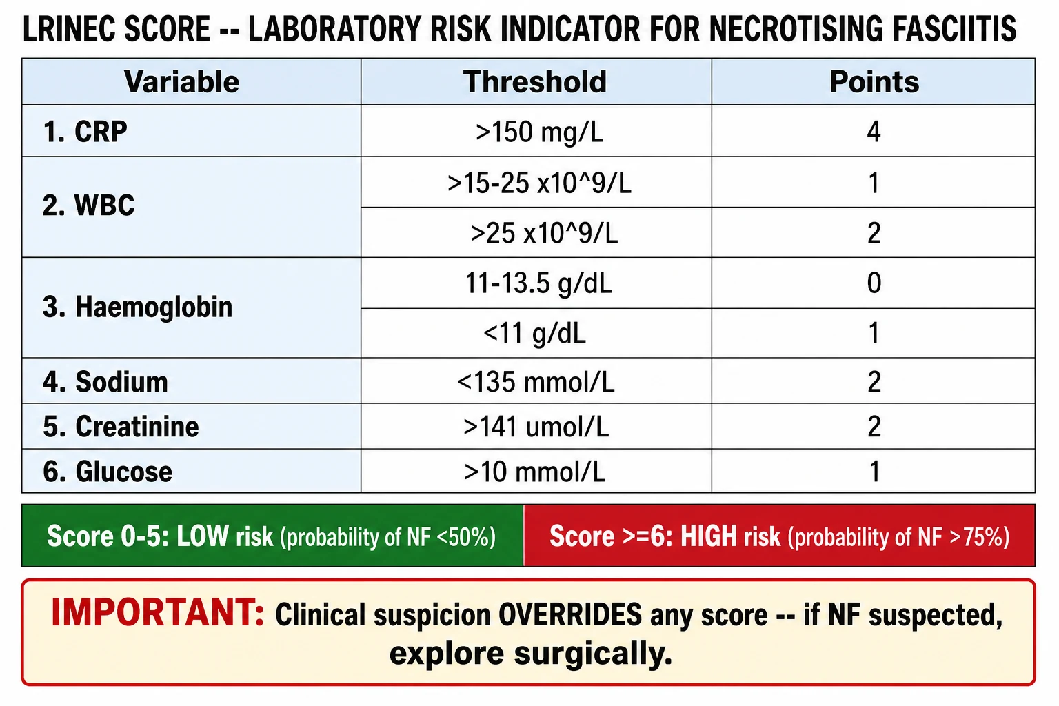
The LRINEC (Laboratory Risk Indicator for Necrotising Fasciitis) score uses routine blood tests:[6][7]
| Variable | Threshold | Points |
|---|---|---|
| CRP | >150 mg/L | 4 |
| WBC | 15–25 × 10⁹/L | 1 |
| >25 × 10⁹/L | 2 | |
| Haemoglobin | 11–13.5 g/dL | 0 |
| less than 11 g/dL | 1 | |
| Sodium | less than 135 mmol/L | 2 |
| Creatinine | >141 µmol/L | 2 |
| Glucose | >10 mmol/L (about 180 mg/dL) | 1 |
Interpretation: Score ≤5 = low risk (probability of NF less than 50%); 6–7 = intermediate; ≥6 = high risk (probability >75%) in the original derivation.[6] Independent meta-analysis confirms moderate sensitivity and specificity (around 60–80%) and confirms what the bedside clinician already knows — the LRINEC is not a rule-out test.[7] A low score does not exclude NF if clinical suspicion is high — explore surgically.
Differential Diagnosis
| Mimic | Distinguishing features |
|---|---|
| Cellulitis / erysipelas | Responds to antibiotics; pain is less severe; no systemic toxicity disproportionate to visible signs; LRINEC low; tissue planes intact on finger test |
| Gas gangrene (clostridial myonecrosis) | Muscle involvement (not just fascia); severe systemic toxicity; post-traumatic; gas on imaging from the outset |
| Staphylococcal scalded skin syndrome (SSSS) | Children; superficial peeling (Granzyme B-mediated cleavage of desmoglein-1); no deep necrosis, no crepitus |
| Severe dermal drug reaction (SJS / TEN) | Recent drug history; mucosal involvement; no crepitus; full-thickness epidermal detachment rather than fascial necrosis |
| Calciphylaxis | Chronic kidney disease; painful livedo reticularis → necrosis; not infectious; biopsy shows medial calcification |
| Pyoderma gangrenosum | Ulcerative with violaceous undermined edges; sterile; associated with IBD / haematological disease |
| Deep vein thrombosis | Unilateral leg swelling; pain is cramping / pressure; no erythema progression across fascial planes; Doppler positive |
| Toxic shock syndrome (without NF) | Diffuse erythematous macular rash → desquamation; tampon / device source; hypotension and multi-organ failure |
Microbiology in Depth
The microbiological pattern changes empiric cover and prognosis. [1]
| Pattern | Commonest organisms | Empiric implication |
|---|---|---|
| Type I polymicrobial (70–80%) | Group A Strep, Group C/G Strep, Staph aureus (MSSA / MRSA), Enterobacteriaceae (E. coli, Klebsiella, Enterobacter), Bacteroides, Clostridia, Peptostreptococcus | Broad gram-positive + gram-negative + anaerobic cover: piperacillin-tazobactam or meropenem plus vancomycin plus clindamycin |
| Type II monomicrobial (15–25%) | Group A Strep (S. pyogenes, M1, M3, M12 serotypes commonly); ± community MRSA | Penicillin + clindamycin; add vancomycin if MRSA prevalence high |
| Type III marine / freshwater (less than 10%) | Vibrio vulnificus, Aeromonas hydrophila, Edwardsiella tarda, Shewanella | Add doxycycline or a third-generation cephalosporin to the bundle |
| Special / atypical (immunocompromised) | Pseudomonas, Mucorales, non-tuberculous mycobacteria, Nocardia | Broaden cover (antifungal, anti-pseudomonal), involve infectious diseases |
Toxin considerations. Group A streptococci release Spe A/B/C superantigens which bypass MHC-II restricted antigen presentation and cause massive polyclonal T-cell activation. Clindamycin is added not because of bacterial killing (it is bacteriostatic) but because of its suppression of ribosomal toxin synthesis.[2][10] Beta-lactams alone remain partly effective against GAS cell wall synthesis but do not stop exotoxin release — hence the mortality-reducing effect of clindamycin in invasive β-haemolytic streptococcal infection demonstrated by Babiker et al. (Lancet Infectious Diseases 2021).[2]
In MRSA-colonised or high-prevalence IVDU populations, MRSA cover (vancomycin, linezolid or daptomycin) must be included empirically until culture data return. Daptomycin is inactivated by surfactant and should not be used when there is pulmonary co-infection. [1]
Management
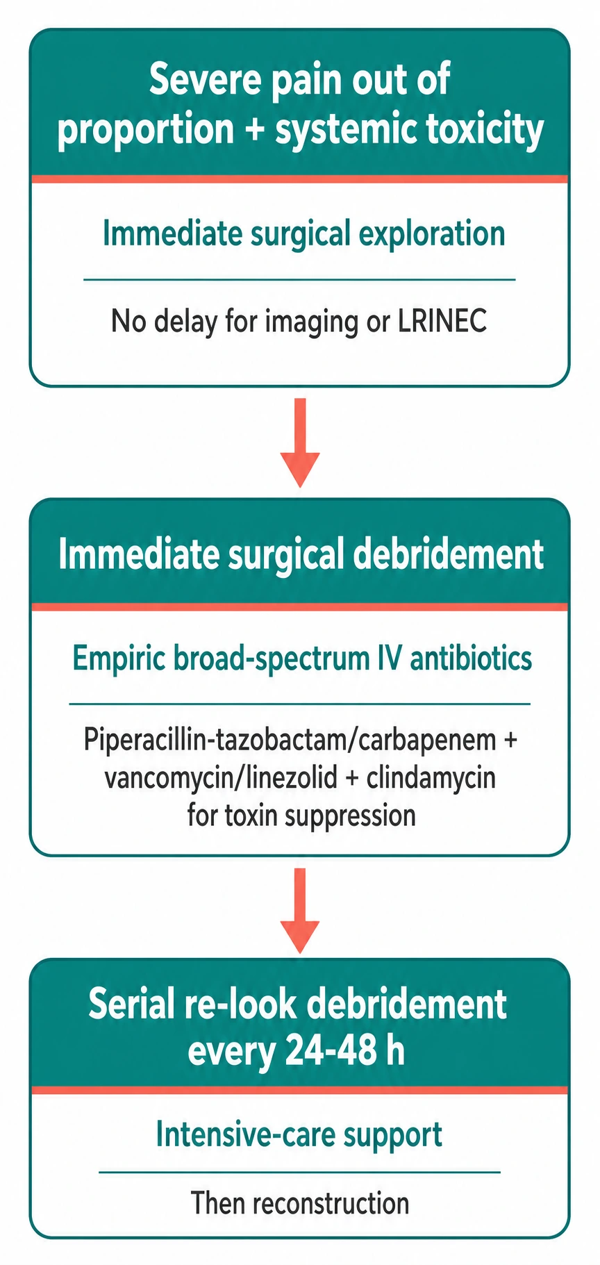
Management runs in three parallel tracks: the resuscitation track (ICU), the definitive track (operating theatre), and the microbiological track (blood and tissue cultures). All three start within minutes of the diagnosis.[1][3][4]
Resuscitation (runs in parallel with surgery)
- Airway, Breathing, Circulation — high-flow oxygen; early tracheal intubation if mental state or shock threatens the airway; wide-bore IV access; arterial line; urinary catheter; nasogastric tube.
- Aggressive fluid resuscitation with balanced crystalloid (Hartmann's or Plasma-Lyte); early norepinephrine if MAP less than 65 mmHg after fluid challenge; vasopressin second line; inotropic support with dobutamine if cardiogenic element is suspected.
- Bloods before antibiotics: lactate, FBC, U&E, LFT, coagulation, CK, blood gas, blood culture × 2, lactate dehydrogenase, haptoglobin if haemolysis suspected, HbA1c, save serum for atypical serology.
- Empiric IV antibiotics within 1 hour — do not delay first dose for imaging or transfer.
- Stress ulcer and VTE prophylaxis; tetanus update if portal is wound-related; analgesia (often requires IV opioid infusion). [1]
Surgical debridement — the definitive treatment
- Urgent surgical debridement to healthy bleeding tissue is the single most important intervention. Do not delay for imaging, blood results or transfer if the patient is deteriorating.[4][5]
- Wide excision of all necrotic fascia, subcutaneous fat and overlying skin — until viable, bleeding tissue is reached. The surgeon must be prepared to excise dramatically more than the visible skin lesion.
- Repeat ("second-look") debridement every 24 hours until no further necrosis is found. Even with complete initial debridement, residual tissue declares itself over the next 24–48 h; second-look within 24 h is mandatory and a third look at 48 h is common.
- Wounds are left open and managed with negative-pressure wound therapy (VAC) after infection control.[3]
- Reconstruction (split-skin graft, fasciocutaneous or free flap) is planned once the wound is clean and the patient is haemodynamically stable — typically days to weeks after the last debridement.[9]
Antibiotics — the empiric bundle
- Broad-spectrum IV antibiotics started immediately:[1][3]
- Piperacillin-tazobactam 4.5 g IV 6-hourly OR carbapenem (meropenem 1 g IV 8-hourly) — covers the polymicrobial Type I spectrum including anaerobes.
- Clindamycin 600–900 mg IV 8-hourly — mandatory adjunct for any streptococcal cover; inhibits bacterial ribosomal toxin synthesis and suppresses exotoxin and M-protein production, reducing mortality in invasive GAS disease.[2]
- Vancomycin 25–30 mg/kg loading then AUC-guided dosing OR linezolid 600 mg IV 12-hourly — for MRSA cover until culture.
- Adjust to culture results when available — de-escalate as soon as organisms and sensitivities allow.
- In Type III / marine / chronic liver disease exposures, add doxycycline 100 mg IV 12-hourly or ceftriaxone 2 g IV daily for Vibrio/Aeromonas cover.
- Duration: IV antibiotics until infection is controlled (10–14 days minimum, often longer), followed by oral step-down guided by clinical response and surgical findings. Continue until second-look debridements find no further necrosis and the patient is afebrile, off vasopressors and improving.
Adjunctive therapy
- IVIG (intravenous immunoglobulin) — neutralises streptococcal and staphylococcal superantigens and provides opsonising antibody; used in streptococcal toxic shock syndrome (Type II NF with shock) where the cytokine storm drives mortality. Dosing is typically 1–2 g/kg on day 1 then 0.4 g/kg daily for up to 5 days.[10][12]
- Hyperbaric oxygen (HBO) — adjunctive; some retrospective series suggest reduced mortality and amputation in polymicrobial Type I disease, but it must never delay surgery, and availability is limited.[12]
- Supportive: ICU admission; mechanical ventilation for ARDS; renal replacement therapy for AKI; transfusion and coagulation-factor support for DIC; nutritional support (enteral feeding, high protein); physiotherapy from day 1 to prevent contractures; pressure-area care; psychological support for the patient and family.[1]
Special Populations
Special POPULATIONS who change NF management
POP-FACS
Cover S. aureus + GAS; aggressive debridement; reconstructive scar burden; consider IVIG
Atypical presentation; high mortality; ceiling-of-care discussion early
Puerperal Type II GAS; sepsis bundle modified (fluid caution in pre-eclampsia)
Urology + general/plastic; colostomy or cystostomy; check anorectal + urogenital source
Add MRSA cover; unusual portals (injection site); social-discharge planning
Vibrio vulnificus risk; doxy/ceftriaxone; very high mortality
Atypical organisms; broaden empiric cover; early ID consult
Fournier's gangrene is the most examined special population. Source control requires diverting colostomy (perianal source), suprapubic cystostomy (urethral source), wide perineal debridement and, frequently, orchiectomy only when the tunica albuginea itself is necrotic — the testes are often spared because their blood supply is independent of the affected fascial planes. Mortality 20–40% even in expert centres.[8][9]
Children develop NF most commonly after varicella (chickenpox lesions become secondarily infected with GAS or S. aureus), after burns, or post-operatively. Progression is rapid; aggressive debridement plus IVIG produces the best outcomes. Mortality remains around 5–10% in paediatric series but is higher with delay.[11]
The immunocompromised patient (neutropenia, transplant, HIV with low CD4, chronic steroids) presents with atypical organisms including Pseudomonas, Mucorales, and non-tuberculous mycobacteria; broaden empiric cover and involve infectious diseases early.[1]
Cirrhotic and chronic-liver-disease patients exposed to seawater or raw shellfish are at risk of fulminant Vibrio vulnificus septicaemia with haemorrhagic bullae and shock; mortality exceeds 50% and empirical doxycycline or ceftriaxone should be added. [1]
Complications & Pitfalls
Complications of the disease: [1]
- Multi-organ failure (ARDS, AKI requiring renal replacement therapy, DIC, hepatic failure) — the commonest cause of death.
- Amputation — required in 15–25% of limb NF when debridement cannot salvage the extremity or when revascularisation is impossible.
- Septic thromboembolism — pulmonary septic emboli and metastatic abscesses.
- Sepsis-related secondary squamous-cell carcinoma developing in chronic scars (Marjolin-like) — long-term follow-up is required.
- Extensive scarring, contractures, lymphoedema — drive the need for skin-graft and flap reconstruction, compression garments and physiotherapy.
- Psychological sequelae — PTSD, body-image disturbance, prolonged rehabilitation. [1]
Diagnostic pitfalls: [1]
- Mis-labelling as cellulitis — the commonest error. The triad of pain out of proportion, rapid progression and disproportionate systemic toxicity is the diagnostic anchor.[5]
- False reassurance from a normal LRINEC — a low score does not exclude NF when clinical suspicion is high.[6][7]
- Missing Fournier's gangrene — a perianal abscess that becomes necrotic isNF until proven otherwise.
- Missing Type III exposure history — Vibrio vulnificus in cirrhotic shellfish handlers.
- Confusing NF with staphylococcal scalded skin syndrome in children — SSSS is superficial, not deep.
Management pitfalls: [1]
- Delay for imaging — CT must not delay surgical exploration.
- Omitting clindamycin — beta-lactam monotherapy does not suppress toxin synthesis.[2]
- Inadequate empiric MRSA cover in IVDU or MRSA-prevalent settings.
- No second-look debridement within 24 hours — residual necrosis is the rule, not the exception.
- Unrealistic limb-sparing attempts when amputation is life-saving.
- Daptomycin in suspected pulmonary co-infection — inactivated by surfactant.
- Under-resuscitation — NF causes profound capillary leak and vasoplegic shock.
Prognosis & Disposition
- Overall mortality 20–40% despite treatment; in Type III / chronic liver disease, mortality exceeds 50%. Each hour of surgical delay beyond the first 24 h measurably increases mortality.[1][4][5]
- Independent prognostic factors: age, comorbidity burden (especially diabetes, immunosuppression, chronic liver disease), APACHE II / SOFA score at presentation, time-to-debridement, bacteraemia, extent of body-surface area involved and the need for amputation.
- Disposition after the acute phase: ICU step-down to a surgical ward once vasopressors, ventilation and renal support are no longer needed; wound care (VAC therapy, dressing changes, skin-graft and flap reconstruction), antibiotic step-down to oral therapy guided by cultures and clinical response, physiotherapy and rehabilitation, diabetes and vascular risk-factor optimisation, and psychological support.
- Discharge counselling must include warning signs (new fever, expanding redness, wound discharge, systemic symptoms), wound-care plan, follow-up with plastics/reconstructive surgery, and vaccination update (tetanus, influenza, pneumococcal in chronic disease).
Evidence, Guidelines & Controversies
- IDSA 2014 (updated) Practice Guidelines for the Diagnosis and Management of Skin and Soft-Tissue Infections are the cornerstone reference; they standardise the antibiotic bundle, the surgical-imperative principle and the role of IVIG in streptococcal toxic shock. The Surgical Infection Society and BOAST (UK) add regional implementation detail.[1]
- LRINEC systematic review and meta-analysis (Bechar 2017, Fernando 2019) confirm moderate sensitivity and specificity; clinical suspicion must override the score.[6][7]
- Clindamycin effectiveness in invasive β-haemolytic streptococcal infection (Babiker 2021, Lancet Infectious Diseases) showed reduced mortality when added to β-lactam therapy — a rare high-quality observational study in this field.[2]
- Controversies. IVIG's evidence base is mostly observational and meta-analyses are mixed; a 2018 Cochrane review found no clear mortality benefit, but most experts continue to use it in streptococcal toxic shock because the cytokine-storm physiology is biologically compelling and the side-effect profile is favourable.[10][12] Hyperbaric oxygen has enthusiastic proponents and limited controlled evidence; the consensus is that HBO, when available, is an adjunct to surgery but cannot be allowed to delay the operation.[12] The optimal debridement timing ("within 1 hour" versus "within 6 hours") is debated but every published cohort shows that earlier is better.[4][5]
Prevention
- Prompt, complete treatment of community skin infections — cellulitis, infected eczema, tinea pedis with bacterial superinfection, and infected animal bites.
- Wound-care discipline in diabetics and the immunocompromised — daily inspection, early review for any wound that becomes more painful or expands, antibiotic prophylaxis when indicated (e.g., compound fractures, bites).
- Vaccination update — annual influenza, pneumococcal and varicella (in non-immune children and contacts), COVID-19.
- Avoid NSAIDs in the early hours of a skin infection — they mask the pain that should be driving urgent review.
- IVDU harm reduction — clean injecting equipment, rotation of sites, awareness of rapidly spreading injection-site pain.
- Shellfish / seawater exposure counselling for cirrhotic and chronic-liver-disease patients. [1]
Exam application bank (NEET-PG / INICET)
One-line answer
Necrotising fasciitis is a rapidly progressive, life-threatening necrotising infection of the deep fascia and subcutaneous tissue that constitutes a SURGICAL EMERGENCY. Classified as Type I (polymicrobial), Type II (monomicrobial Group A Streptococcus ± Staphylococcus aureus — the 'flesh-eating disease'), or Type III (marine — Vibrio vulnificus). Clinically characterised by severe pain out of proportion to the visible signs, rapid progression from erythema to purple-grey discoloration, haemorrhagic bullae, necrosis and crepitus, with systemic toxicity progressing to septic shock. Diagnosis is CLINICAL — the LRINEC score (≥6 high risk) supports but does not replace clinical judgment, and imaging must NOT delay surgical exploration. Management requires URGENT SURGICAL DEBRIDEMENT (the definitive treatment — to healthy bleeding tissue, repeated daily), broad-spectrum IV antibiotics (piperac
Worked stems (answer without another resource)
Stem 1 — Classic presentation. Map symptoms to mechanism; name the first investigation and first treatment step with dose/route if drug therapy is standard. [1]
Stem 2 — Unstable / complicated. List red flags that force immediate resuscitation, theatre, ICU, antidote, or reperfusion — and what you do in the first 15 minutes. [1]
Stem 3 — Atypical group. Elderly, pregnancy, child, or immunocompromised: how presentation and thresholds change. [1]
Stem 4 — Differential trap. Name the three closest mimics and one discriminator for each. [1]
Stem 5 — Disposition. Who goes home with safety-netting, who is admitted, who needs HDU/ICU/theatre, and what follow-up is mandatory. [1]
Rapid viva checklist
- Definition + classification
- Pathophysiology chain
- Bedside signs / criteria
- Score with exact components (if any)
- Emergency bundle
- Definitive therapy with doses
- Complications of disease and of treatment
- Special populations
- Guideline/trial name if classic
- Three exam traps
Coverage self-check
If you cannot answer any stem above from this page alone, re-read the matching section — the page is intended to be self-sufficient for final-prof and NEET-PG/INICET questions on Necrotising fasciitis.
[1]References
- [1]Stevens DL, Bryant AE, Goldstein EJ. Necrotizing Soft Tissue Infections Infect Dis Clin North Am, 2021.PMID 33303335
- [2]Babiker A, Li X, Lai YL, et al. Effectiveness of adjunctive clindamycin in β-lactam antibiotic-treated patients with invasive β-haemolytic streptococcal infections in US hospitals: a retrospective multicentre cohort study Lancet Infect Dis, 2021.PMID 33333013
- [3]Saeed K, Esposito S, Gould I, et al. Hot topics in necrotising skin and soft tissue infections Int J Antimicrob Agents, 2018.PMID 29501822
- [4]Hasham S, Matteucci P, Stanley PR, et al. Necrotising fasciitis BMJ, 2005.PMID 15817551
- [5]Goh T, Goh LG, Ang CH, et al. Early diagnosis of necrotizing fasciitis Br J Surg, 2014.PMID 24338771
- [6]Bechar J, Sepehripour S, Hardwicke J, et al. Laboratory risk indicator for necrotising fasciitis (LRINEC) score for the assessment of early necrotising fasciitis: a systematic review of the literature Ann R Coll Surg Engl, 2017.PMID 28462647
- [7]Fernando SM, Tran A, Cheng W, et al. Necrotizing Soft Tissue Infection: Diagnostic Accuracy of Physical Examination, Imaging, and LRINEC Score: A Systematic Review and Meta-Analysis Ann Surg, 2019.PMID 29672405
- [8]Singh A, Ahmed K, Aydin A, et al. Fournier's gangrene. A clinical review Arch Ital Urol Androl, 2016.PMID 27711086
- [9]Kopechek KJ, Patel HV, Koch GE. Modern Management of Fournier's Gangrene Curr Urol Rep, 2025.PMID 40455358
- [10]Stevens DL, Bryant AE. Severe Group A Streptococcal Infections 2016.PMID 26866227
- [11]Schröder A, Gerin A, Firth GB, et al. A systematic review of necrotising fasciitis in children from its first description in 1930 to 2018 BMC Infect Dis, 2019.PMID 30975101
- [12]Young MH, Engleberg NC, Mulla ZD, et al. Therapies for necrotising fasciitis Expert Opin Biol Ther, 2006.PMID 16436041