Dermatology · Medicine
Nutrition and the skin
Also known as Nutrition and the skin · Nutritional dermatology · Cutaneous manifestations of nutritional deficiency · Micronutrient skin disease
Micronutrient deficiency produces a characteristic cutaneous signature for almost every essential vitamin and trace element. This topic covers the nutrient-skin axis at the level of keratinocyte differentiation, sebocyte lipogenesis, melanocyte function, and wound biology; the named deficiency syndromes (scorbutic rosary, pellagra / Casal's necklace, acrodermatitis enteropathica, phrynoderma, kwashiorkor flaky-paint dermatosis, Menkes kinky hair disease, Keshan cardiomyopathy); the four prototypes of nutrient-driven skin disease — follicular, periorificial, photodistributed, and mucocutaneous; refeeding syndrome with thiamine-first resuscitation; and evidence-based nutritional therapy in acne, psoriasis, atopic dermatitis and hair loss.
On this page & tools
Your progress
Saved locally on this device.
Exam tags
Red flags

Definition
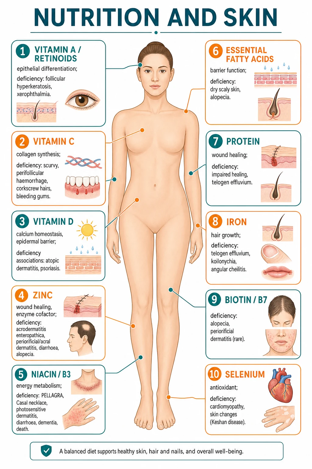
Nutritional dermatology is the study of cutaneous manifestations of nutrient deficiency, excess, or imbalance. The skin is a structural, metabolic, and immunological interface whose appearance depends on the adequacy of vitamins, trace elements, essential fatty acids, and protein for keratinocyte differentiation, sebocyte lipogenesis, melanocyte pigment synthesis, hair matrix proliferation, collagen crosslinking, and wound healing. The cutaneous findings often precede systemic features and may make the diagnosis at the bedside. Inversely, certain cutaneous therapies (long-term retinoids, nicotinamide, methotrexate, biologics) and chronic dermatoses themselves (erythroderma, severe atopic dermatitis, exfoliative dermatitis, Darier disease, bullous disorders) deplete specific micronutrients and must be supplemented. [1]
Nutritional skin disease is organised into four clinical prototypes that localise the nutritional defect: follicular (vitamin A phrynoderma, vitamin C corkscrew hairs), periorificial and acral (zinc acrodermatitis enteropathica, biotin deficiency), photodistributed (niacin pellagra, Hartnup disease), and mucocutaneous (riboflavin angular cheilitis, B12 glossitis, scurvy bleeding gums). Recognition of the cutaneous pattern is the first clinical step; the second is to test for and replace the missing nutrient while treating the underlying cause. [1]
Refeeding syndrome — the iatrogenic nutritional emergency
Refeeding syndrome risk factors
Quick numbers for the examiner
Pathophysiology — the nutrient-skin axis
The skin is a metabolically active organ with high cell turnover (keratinocyte transit time 28 days), continuous lipid synthesis (stratum corneum lamellar bodies), rapid hair matrix proliferation (the fastest dividing cell population in the body), melanocyte pigment synthesis, and perpetual collagen remodelling by dermal fibroblasts. Each of these processes depends on specific micronutrients, and the deficiency phenotype localises to the process that fails first. Vitamin C is the cofactor for prolyl and lysyl hydroxylases that cross-link collagen; failure produces the scorbutic triad of perifollicular haemorrhage, corkscrew hairs, and gum bleeding. Niacin and riboflavin generate the redox currency (NAD, FAD) for keratinocyte energy metabolism, so their deficiency produces epithelial atrophy and the mucocutaneous syndrome. Zinc is the cofactor for more than 300 zinc-finger transcription factors and metalloenzymes (DNA and RNA polymerases, matrix metalloproteinases, superoxide dismutase), so its deficiency abolishes epithelial turnover and wound re-epithelialisation, producing the acral and periorificial dermatitis of acrodermatitis enteropathica. Vitamin A is the ligand for RAR and RXR nuclear receptors that drive keratinocyte terminal differentiation; deficiency causes the follicular hyperkeratosis of phrynoderma. Iron is required by ribonucleotide reductase (the rate-limiting enzyme of DNA synthesis) in rapidly dividing hair matrix cells; deficiency shortens the anagen phase and produces telogen effluvium and koilonychia. [1]
Four clinical prototypes organise nutritional skin disease: [1]
- Follicular pattern — plugging of the hair follicle by retained keratin and sebum produces rough, sandpaper-like skin. Causes: vitamin A (phrynoderma), vitamin C (corkscrew hairs with perifollicular haemorrhage), vitamin B2 (riboflavin), essential fatty acid deficiency, and chronic sunlight exposure (lichen spinulosus). [1]
Clinical features — recognising the nutritional pattern
The cutaneous pattern of nutritional deficiency depends on which epidermal or dermal process fails first. Four prototypes dominate the clinical landscape. [1]
Follicular pattern — rough, sandpaper-like skin with discrete keratotic follicular plugs. The pathology is failure of normal keratinocyte desquamation around the hair follicle. Vitamin A deficiency (phrynoderma, also called toad skin — extensor surfaces, thighs, posterior upper arms; vitamin C deficiency (perifollicular haemorrhage with corkscrew hairs on shins and forearms), riboflavin deficiency (follicular hyperkeratosis with seborrhoeic dermatitis-like facial involvement), and essential fatty acid deficiency (sandy, bran-like scaling) all produce this pattern. Lichen spinulosus, keratosis pilaris atrophicans, and pityriasis rubra pilaris are histological differential diagnoses. [1]
Periorificial and acral pattern — clusters of itchy or painful erythema, vesicles, pustules, erosions and crusting around the mouth, nostrils, eyes, genitalia, hands and feet, with characteristic sparing of the vermilion border of the lip (2 to 5 mm rim of clinically normal skin between the rash and the lip). The differential diagnosis includes acrodermatitis enteropathica (zinc), multiple carboxylase deficiency (biotin), necrolytic migratory erythema (glucagonoma — a paraneoplastic mimic of zinc deficiency), essential fatty acid deficiency, glucagonoma syndrome, and severe atopic dermatitis. Candidal superinfection is common because of the impaired epithelial and immune barrier. [1]
Photodistributed pattern — painful erythema, vesicles and bullae, sometimes bullae with subepidermal separation, crusting and desquamation, followed by hyperpigmentation, strictly localised to sun-exposed areas (face in a butterfly distribution, the V-shaped neck — Casal necklace — dorsal hands and feet — pellagrous gauntlet and pellagrous boot — and the anterior shin in those wearing shorts). The submental triangle, inframammary folds, and the skin under a watch strap are characteristically spared because they have not been exposed. The differential diagnosis includes pellagra, Hartnup disease, drug-induced pseudopellagra (isoniazid, hydralazine, D penicillamine), chronic cutaneous lupus, polymorphous light eruption, erythropoietic protoporphyria, and xeroderma pigmentosum. Niacin skin biopsy shows non-specific epidermal necrosis with subepidermal blisters and vasodilation. [1]
Mucocutaneous and appendageal pattern — tongue and lip changes are the giveaway. A smooth, depapillated magenta or beefy-red tongue (magenta glossitis in riboflavin deficiency, Hunter glossitis in B12), angular cheilitis with fissuring and maceration at the corners of the mouth (B2, B6, B12, folate, iron), atrophic glossitis (B12, folate, iron), generalised xerosis (EFA, vitamin A, thyroid), brittle nails with koilonychia (iron), yellow nail syndrome (vitamin E), diffuse telogen or anagen effluvium (iron, zinc, biotin, protein, vitamin D), and the brown flag sign of the hair in alternating-banded protein malnutrition. [1]
Neonatal and infantile presentors that warrant urgent nutritional attention include acrodermatitis enteropathica (4 to 6 weeks after weaning with breastfeeding, earlier if formula is not zinc-fortified), vitamin K deficiency bleeding (neonates with prolonged cholestasis or breastfed infants not supplemented), Menkes disease (neonates with kinky hair and hypotonia), vitamin B12 deficiency (maternal deficiency or vegan diet — developmental regression), biotinidase deficiency (3 to 6 months, seizures plus dermatitis plus alopecia), and Hartnup disease (post-weaning pellagra-like rash). [1]
Investigations — confirming the deficit
Investigation strategy is layered: screen for the most likely deficiency given the clinical pattern, confirm with a specific assay, treat, and reassess the response. Many dermatology patients have a coexisting micronutrient deficit that worsens their skin disease; a single presentation may warrant five or six targeted tests. [1]
Bedside tests. Full mucocutaneous examination (skin, hair, nails, oral cavity, perineum, periumbilical area) and growth charting in children (height, weight, mid-upper arm circumference, head circumference) are part of the standard approach. Wickham striae look for lichen planus; corkscrew hairs and perifollicular haemorrhage look for scurvy; Bitot spots in the temporal conjunctiva look for vitamin A; magenta tongue and cheilosis for riboflavin. [1]
First-line blood tests. [1]
- Full blood count and blood film (macrocytes in B12 and folate, hypersegmented neutrophils, microcytic hypochromic cells in iron, pancytopenia in copper deficiency).
- C-reactive protein (interprets ferritin and zinc which are acute-phase reactants).
- Iron studies with ferritin.
- Serum vitamin B12, folate, and (where B12 is borderline) methylmalonic acid and homocysteine.
- Serum 25-hydroxyvitamin D.
- Liver function tests, albumin, total protein and pre-albumin for nutritional reserve.
- Serum zinc, alkaline phosphatase (zinc-dependent; functional marker of zinc status), and serum copper if zinc deficiency suspected.
- Serum vitamins A, E, K (in high-risk groups) and selenium if endemic deficiency suspected.
- Whole blood thiamine (transketolase activation assay) in alcohol use disorder before parenteral thiamine. [1]
Second-line and specialised tests. [1]
- Urinary organic acids — diagnostic for biotinidase deficiency (3-methylcrotonylglycine), methylmalonic acid in B12, and homocysteine elevation.
- Plasma tryptophan level in suspected Hartnup disease.
- Skin biopsy with H&E and direct immunofluorescence where needed — exclude porphyria cutanea tarda (subepidermal blister with festooning, eosinophilic PAS-positive vessel wall thickening), necrolytic migratory erythema (zinc deficiency mimic of glucagonoma), and epidermolysis bullosa.
- Genetic tests: ATP7A (Menkes), SLC39A4 (acrodermatitis enteropathica), SLC6A19 (Hartnup), SLC19A2 (thiamine-responsive basal ganglia disease), and biotinidase (BTD).
- Trial of therapy — many nutritional conditions (pellagra, scurvy, niacin deficiency) respond dramatically within 48 to 72 hours and the response is itself a useful diagnostic confirmation. [1]
Avoid over-interpretation of borderline results. Serum zinc falls during acute inflammation (acute-phase reactant), serum ferritin rises in inflammation (masking iron deficiency), serum vitamin B12 can be falsely normal in the early phase of deficiency, and 25-hydroxyvitamin D is influenced by sun exposure. Always interpret an assay in the context of the clinical picture. [1]
Complications — when deficiency becomes chronic
Untreated or late-treated nutritional skin disease carries a constellation of systemic, infectious, and developmental complications: [1]
Wound healing failure. Vitamin C, zinc, protein and iron deficiency all produce chronic non-healing wounds, dehiscence of previously healed scars, and pressure ulceration. Vitamin C-deficient patients demonstrate wound breakdown in the original site of trauma — sometimes decades later. [1]
Infectious complications. Cutaneous candidiasis and bacterial superinfection of any crusted dermatitis is the rule in zinc, biotin, and protein deficiency. Vitamin A deficiency impairs innate immunity (cathelicidin, mucociliary clearance, secretory IgA) and predisposes to measles-associated morbidity in children. Iron, B12 and folate deficiency impair neutrophil oxidative burst and lymphocyte function. [1]
Ocular and neurological complications. Vitamin A deficiency culminates in keratomalacia and irreversible corneal scarring blindness if untreated. Vitamin B12 deficiency causes subacute combined degeneration with permanent paraparesis. Niacin and EFA deficiency can cause cognitive decline. Vitamin E deficiency produces spinocerebellar ataxia and irreversible peripheral neuropathy. [1]
Endocrine and growth complications. Zinc, vitamin A and protein deficiency impair growth hormone axis and cause stunting in children. Menkes disease causes neurodegeneration in the first year of life, and prognosis depends on early treatment. B12 deficiency in pregnancy causes neural tube defects and infant developmental delay. [1]
Ongoing deficiency syndrome. Chronic alcoholism, post-bariatric surgery, chronic pancreatitis, IBD and dialysis patients remain at high risk of combined micronutrient deficiency for life. Long-term multivitamin replacement, annual screening, and liaison with dietetics are mandatory. [1]
Refeeding syndrome. The most common iatrogenic complication of correcting nutritional skin disease is refeeding syndrome — discussed in detail below. Mortality is 5 to 10 percent of those with hypophosphataemia; with appropriate thiamine-first feeding protocols, mortality falls dramatically. [1]
Management — replacement therapy principles
Management of nutritional skin disease is built on five pillars: [1]
1. Recognise the pattern and confirm the deficiency. Pattern recognition + biochemistry + clinical response. Replace the missing nutrient with adequate, weight-appropriate doses. Avoid the trap of partial replacement (just iron for fatigue) that ignores coexisting zinc or vitamin B12 deficiency. [1]
2. Treat the underlying cause. Recognition of the precipitating mechanism is essential. Iron deficiency in an H. pylori-positive patient requires eradication. Pellagra in an alcoholic patient requires sustained abstinence plus nicotinamide plus B6. Zinc deficiency in Crohn disease requires disease control, lifelong zinc replacement, and routine monitoring. Acrodermatitis enteropathica is genetic and requires lifelong zinc supplementation regardless of intake. [1]
3. Replace in adequate doses, by the right route, and for long enough. See the doses table above. Replace for at least one cycle of immune or epithelial turnover (28 days for epidermis, 4 months for iron stores, longer for neurological recovery in B12). Where oral absorption is compromised (short bowel, TPN, severe malabsorption), use parenteral (intramuscular hydroxocobalamin for B12, intravenous iron for iron deficiency, intravenous trace elements for zinc and copper in TPN). [1]
4. Arrange monitoring. Repeat blood tests every 3 to 12 months in patients on long-term replacement (iron ferritin, zinc, alkaline phosphatase, B12, vitamin D, LFTs). Monitor growth, weight, school or work attendance, and skin photography in chronic conditions. Liaise with dietetics for dietary counseling. [1]
Key Nutrients and Their Skin Roles
Vitamin A (retinoids)
Vitamin A is the umbrella term for retinol, retinal, retinoic acid and the provitamin carotenoids (beta-carotene). Vitamin A is the ligand for the retinoic acid receptor (RAR) and retinoid X receptor (RXR) nuclear transcription factors that drive keratinocyte terminal differentiation, hair follicle cycling, sebocyte lipogenesis, and melanocyte pigment transfer. In the epidermis, retinoic acid downregulates keratin 5 and keratin 1, suppresses proliferation in the basal layer, and induces differentiation in the suprabasal compartment. In the sebaceous gland, isotretinoin (13-cis retinoic acid) shrinks the sebaceous gland and reduces sebum production by 90% within 6 weeks, which is the basis of acne therapy. [1]
Deficiency produces the textbook chain of events: night blindness (failure of retinal rhodopsin regeneration), Bitot spots (keratin debris in the conjunctival fornix), conjunctival and corneal xerosis (dryness), keratomalacia (corneal ulceration and scarring — leading global cause of preventable paediatric blindness in vitamin-A-deficient populations, especially South Asia and sub-Saharan Africa), follicular hyperkeratosis (phrynoderma — rough, sandpaper-like plugs of keratin in hair follicles on extensor surfaces and thighs, also called toad skin), generalised xerosis (dry scaly skin), and increased susceptibility to infection (failure of epithelial barrier and innate immunity).[8]
Causes include fat malabsorption (cystic fibrosis, chronic pancreatitis, cholestatic liver disease, bariatric surgery), strict vegan diets without fortified foods, and refugee populations with limited access to dairy and eggs. Treatment of deficiency is high-dose oral vitamin A (200,000 IU on day 1, day 2 and 14 in children over 12 months; lower doses in younger infants to avoid toxicity), with retinol preferred over beta-carotene for severe deficiency. Toxicity (hypervitaminosis A) is the mirror image — dry scaly skin, alopecia, hepatotoxicity, pseudotumor cerebri, and teratogenicity in the first trimester.[8]
Vitamin B2 (riboflavin)
Riboflavin is the precursor of flavin mononucleotide (FMN) and flavin adenine dinucleotide (FAD), the coenzymes for the electron transport chain (Complex I and II), fatty acid beta oxidation, glutathione reductase, and xanthine oxidase. The skin and cornea have high flavin requirements because of rapid epithelial turnover.[9] Riboflavin deficiency produces the classic mucocutaneous triad: angular stomatitis (fissuring and maceration at the corners of the mouth), cheilosis (scaling of the vermilion border of the lips), and magenta-coloured glossitis (the smooth, sore, depapillated magenta tongue). Seborrhoeic dermatitis-like scaling of the nasolabial folds, photophobia with vascularisation of the cornea, and normocytic-normochromic anaemia may follow.[9][10] Riboflavin deficiency is endemic in populations consuming polished rice without legumes or dairy, in chronic alcoholism, and in users of phenobarbital and tricyclic antidepressants that accelerate hepatic riboflavin metabolism. Treatment is oral riboflavin 10 to 30 mg per day for several weeks.[9][10]
Vitamin B3 (niacin, nicotinamide)
Niacin is the precursor of nicotinamide adenine dinucleotide (NAD+) and nicotinamide adenine dinucleotide phosphate (NADP+), redox cofactors central to all catabolic and anabolic reactions in the cell. Niacin is supplied in the diet (meat, fish, fortified cereals) and can be synthesised endogenously from tryptophan (60 mg tryptophan → 1 mg niacin), a pathway that requires riboflavin, pyridoxine and iron as cofactors.[7] Deficiency of niacin (or its precursor tryptophan) produces pellagra, the dermatological signature of the 4 D's.[7]
The dermatitis of pellagra is photosensitive (sun-exposed areas only — face, neck in a V-shape called Casal necklace or Casal collar, dorsal hands called pellagrous gauntlet, feet and shins in farmers), begins as painful erythema resembling sunburn, progresses to vesicles and bullae, and resolves with hyperpigmentation and desquamation. The diarrhoea is from atrophic intestinal mucosa (subacute), and the dementia ranges from irritability and insomnia to memory loss, parkinsonism and frank psychosis. Death is from dehydration, suicide, or intercurrent infection.[7]
Causes of pellagra include maize-based diets (niacin is bound in maize as niacytin, a biologically unavailable glycoside; treatment with alkali [nixtamalisation] in traditional Mesoamerican cuisine liberates the niacin, hence the low historical prevalence of pellagra in Latin America despite maize consumption), alcoholism, anorexia, chronic diarrhoea, IBD, post-bariatric surgery, carcinoid syndrome (tryptophan diverted to serotonin), Hartnup disease, and drugs that deplete pyridoxine (isoniazid, hydralazine, D penicillamine). Treatment is nicotinamide 100 to 500 mg per day orally for at least one month; nicotinic acid (flushes and may cause hepatotoxicity) is avoided. Intravenous nicotinamide is given for severe cases with delirium or hyperemesis.[7]
Vitamin B6 (pyridoxine)
Pyridoxine is the precursor of pyridoxal phosphate (PLP), the coenzyme for transamination, decarboxylation, and the rate-limiting step of niacin synthesis from tryptophan. Deficiency is uncommon as an isolated dietary problem (it is widely distributed in food), but it is reliably induced by isoniazid (which forms a hydrazone with PLP), hydralazine, D penicillamine, and chronic oral contraceptives. The cutaneous features overlap with riboflavin deficiency: angular cheilitis, glossitis, and a seborrhoeic dermatitis-like scaling of the nasolabial folds, scalp and perineum. Pyridoxine 10 to 50 mg per day reverses the deficit in 1 to 2 weeks. Importantly, pyridoxine deficiency has a recognised sensory peripheral neuropathy and may cause a pellagra-like syndrome because tryptophan cannot be converted to niacin.[11]
Vitamin B7 (biotin)
Biotin is the coenzyme for five carboxylases (acetyl CoA carboxylase 1 and 2, pyruvate carboxylase, propionyl CoA carboxylase, and 3 methylcrotonyl CoA carboxylase). Deficiency states are acquired (long-term parenteral nutrition without biotin, raw egg white diet where avidin binds biotin, chronic anticonvulsants such as carbamazepine, valproate, phenytoin, gut dysbiosis) and inherited (multiple carboxylase deficiency from holocarboxylase synthetase MCSE mutations in neonates, or biotinidase deficiency in infants presenting around 3 to 6 months). The cutaneous signature is the periorificial and flexural dermatitis with alopecia, conjunctivitis, and candidal superinfection — clinically indistinguishable from acquired zinc deficiency. Distinguishing features include the organic aciduria (3-methylcrotonylglycine) in biotin deficiency, and the response within 48 to 72 hours to oral biotin 5 to 10 mg per day.[12] Carboxylase deficiency genetic forms require lifelong treatment.
Vitamin B9 (folate)
Folate is the cofactor for one-carbon transfer reactions that generate purines and pyrimidines for DNA synthesis. Megaloblastic anaemia is the signature systemic feature. The cutaneous features include glossitis (atrophic, beefy red, painful), angular cheilitis, and a grey-brown hyperpigmentation of the face, neck and dorsal hands that resembles Addisonian hyperpigmentation but is from impaired melanin transfer. Folate deficiency may also accentuate the hyperpigmentation of pellagra and is a cofactor deficiency that limits niacin synthesis from tryptophan. Treatment is folic acid 5 mg per day for at least 4 months in the absence of B12 deficiency (folate monotherapy worsens subacute combined degeneration of B12 deficiency by shunting methyltetrahydrofolate away from the B12-dependent methionine synthase). Sources include leafy greens (folate), legumes, citrus and fortified cereals. [1]
Vitamin B12 (cobalamin)
Vitamin B12 is the cofactor for methionine synthase (converts homocysteine to methionine and regenerates tetrahydrofolate) and methylmalonyl CoA mutase. Deficiency produces the haematological signature (megaloblastic anaemia with hypersegmented neutrophils), the neurological signature (subacute combined degeneration of the dorsal columns and lateral corticospinal tracts — spastic paraparesis with dorsal column loss, leading to ataxia and a positive Romberg), and the cutaneous signature of patchy hyperpigmentation, glossitis (Hunter glossitis — atrophic depapillated beefy red tongue), angular cheilitis, and vitiligo-like patches in dark-skinned patients.[13] The hyperpigmentation is the most characteristic dermatological clue and may be the presenting feature; it typically affects the dorsal hands, knuckles, soles and palmar creases. Treatment is hydroxocobalamin 1 mg intramuscularly three times weekly for 2 weeks, then every 3 months for life in pernicious anaemia, ileal disease, or post-gastrectomy. The neurological recovery is partial if diagnosis is delayed.
Vitamin C (ascorbic acid)
Ascorbic acid is the essential cofactor for prolyl hydroxylase and lysyl hydroxylase — the enzymes that hydroxylate proline and lysine residues in nascent procollagen chains so that they can form the triple helix, and for procollagen secretion into the extracellular matrix. Failure produces the collagen disorder scurvy.[1][6] The cutaneous signature is the late winter/scurvy triad of perifollicular haemorrhages (extravasation around hair follicles — earliest and most pathognomonic sign, often on the lower limbs), corkscrew hairs (hair shafts twisted on themselves as they attempt to grow through defective dermis), and bleeding gums with gum swelling and dental caries (in patients with their own teeth; edentulous patients do not develop gum signs). Further signs are ecchymoses, petechiae, poor wound healing, wound dehiscence, haemarthroses, anaemia (from bleeding and impaired iron absorption because ascorbate reduces dietary ferric iron to absorbable ferrous iron), woody oedema, and the scorbutic rosary (costochondral enlargement).[1] Vitamin C deficiency is re-emerging in populations with severe food insecurity, the elderly and those with disordered eating.
Sources of vitamin C are fresh fruits (citrus, kiwi, guava, papaya, berries), vegetables (peppers, broccoli, leafy greens) and fortified products. Scurvy is treated with oral ascorbic acid 1 g daily for 1 week, then 100 to 200 mg daily for 1 month. The diagnosis is clinical plus response; serum ascorbate is unreliable.[6]
Vitamin D
Vitamin D (cholecalciferol D3, ergocalciferol D2) is a secosteroid hormone synthesised in the skin under UVB (7-dehydrocholesterol → previtamin D3 → calcitriol after 25-hydroxyvitamin D3 in liver and 1α-hydroxylation in kidney), or absorbed from diet. The active hormone calcitriol binds vitamin D receptor (VDR) in keratinocytes, melanocytes, dendritic cells, and Langerhans cells — regulating keratinocyte proliferation and differentiation, cathelicidin antimicrobial peptide synthesis, and cutaneous immune tolerance.[2]
Deficiency is associated epidemiologically with atopic dermatitis severity, alopecia areata, and chronic spontaneous urticaria; vitamin D analogues (calcipotriol) are first-line topical agents in psoriasis because they normalise keratinocyte hyperproliferation and dampen IL-17/IL-23 signalling.[2][4] A causal role in hair loss is biologically plausible (VDR knockout mice develop rickets and alopecia), but supplementation trials in telogen effluvium and female pattern hair loss are mixed.[2] Treatment of deficiency is colecalciferol 800 to 4000 IU daily, with loading doses (300,000 to 600,000 IU oral weekly for 6 weeks) for severe deficiency or malabsorption. Sun avoidance advice must be tempered against vitamin D requirements — 10 to 15 minutes of noon sun on face and arms covers most adults.
Vitamin E
The four tocopherols and four tocotrienols that constitute vitamin E are lipid-soluble antioxidants that protect polyunsaturated membrane lipids from peroxidation. Deficiency is rare but well described in cholestatic liver disease, abetalipoproteinaemia, and fat malabsorption. The neurological syndrome of spinocerebellar ataxia, peripheral neuropathy, and skeletal myopathy is the dominant feature. Skin findings are non-specific — xerosis, easy bruising, and yellow nail syndrome — and supplementation therapy is mixed. Reference daily intake is 15 mg per day of alpha-tocopherol. [1]
Vitamin K
Vitamin K (phylloquinone K1 from leafy greens, menaquinone K2 from gut bacteria and fermented foods) is the cofactor for gamma-carboxylation of glutamate residues in clotting factors II, VII, IX and X, and in the matrix Gla-protein of cartilage and vessel wall. Deficiency bleeding (VKDB) in the neonatal period (with the classic cord and gastrointestinal bleeding), malnutrition, broad-spectrum antibiotics, prolonged cholestasis, and anticoagulant overdose all produce cutaneous purpura, ecchymoses and nodular purpura in infants.[16] Treatment of bleeding is vitamin K 10 mg slow IV; chronic supplementation in neonates is given as a single IM dose at birth. Coumarin anticoagulants (warfarin) act as vitamin K antagonists and require INR monitoring.
Zinc
Zinc is the second most abundant trace metal in the body after iron, with 2 to 4 g total and 0.5% daily turnover. It is the structural component of more than 300 zinc-finger transcription factors and metalloenzymes — RNA polymerase, DNA polymerase, superoxide dismutase, matrix metalloproteinases, alkaline phosphatase, alcohol dehydrogenase, and many more. Skin cells (keratinocytes, lymphocytes, fibroblasts) have high rates of zinc turnover.[5]
Deficiency produces the textbook acrodermatitis enteropathica (AE) — a periorificial and acral, vesiculobullous, crusted dermatitis (perioral, perianal, genital, hands, feet — but typically spares the vermilion border of the lip and the lips themselves, with a 2 to 5 mm rim of clinically normal skin between the rash and the lip, in distinction from contact or perioral dermatitis), alopecia (totalis in severe cases), diarrhoea (from gut epithelial failure), failure to thrive, immune dysfunction (T-cell lymphopenia, candida and bacterial superinfection of the rash), neuropsychiatric irritability, and in long-standing cases, hypogeusia and hypogonadism. There are two principal forms: (a) genetic SLC39A4 / SLC30A2 mutations causing the autosomal recessive AE (Danbolt-Closs disease) which presents in infancy after weaning, and (b) acquired deficiency from prematurity, malabsorption (IBD, coeliac, cystic fibrosis), chronic liver disease, alcoholism, penicillamine (chelates zinc), high-output fistulae, burns and total parenteral nutrition without zinc.[5] Treatment is elemental zinc 0.5 to 1 mg per kg per day oral in mild deficiency, 2 to 3 mg per kg per day in AE, lifelong. Serum zinc may be normal despite deficiency because it is bound to albumin and the assay is influenced by inflammation; a low serum alkaline phosphatase (zinc-dependent enzyme) is a useful functional marker.[5]
Selenium
Selenium is an essential trace element incorporated as selenocysteine into selenoproteins, of which the most abundant and clinically relevant are glutathione peroxidase (GPx1-4, antioxidant defence), thioredoxin reductase (DNA synthesis redox), and iodothyronine deiodinase (thyroid hormone conversion). Deficiency of selenium endemically in parts of China (the Keshan disease belt of Heilongjiang and Shaanxi, low selenium soils) produces Keshan disease — a juvenile-onset endemic dilated cardiomyopathy with congestive heart failure, focal necrosis of myocardium, and arrhythmias.[14] Cutaneous features include pseudoalbinism (lighter hair and skin pigmentation in children from low-selenium regions), xerosis, and nail dystrophy. Iatrogenic selenium deficiency is described in patients on long-term total parenteral nutrition and in renal failure. Excess selenium (selenosis) produces a characteristic garlic odour of the breath, brittle nails and hair loss. Reference daily intake is 55 to 70 ug per day; selenomethionine is the common supplemental form.
Essential fatty acids (EFAs)
Linoleic acid (omega-6, 18:2n-6) and alpha-linolenic acid (omega-3, 18:3n-3) are essential fatty acids — mammals lack the desaturase enzyme to introduce the first double bond at carbon 3 or carbon 6. EFAs are the structural backbone of stratum corneum ceramides, triglycerides of the lamellar bodies, and the membrane phospholipids of every cell. Deficiency produces a generalised papular, scaly dermatitis with intertriginous oozing, alopecia, brittle nails, thrombocytopenia, and poor wound healing, classically described in the era of prolonged parenteral nutrition without lipid. Biochemical detection requires the triene:tetraene ratio (eicosatrienoic / arachidonic) above 0.4.[11]
Iron
Iron is the central atom of haem (oxygen transport, cytochrome P450), iron-sulfur clusters (electron transport chain, DNA helicases), and ribonucleotide reductase (DNA synthesis). The skin consequences of iron deficiency are (a) telogen effluvium — the most common nutritional cause of hair shedding worldwide, because the iron-dependent ribonucleotide reductase in rapidly dividing hair matrix cells shuts down and shortens the anagen phase; (b) koilonychia — spoon-shaped concave nails, classically associated with iron deficiency anaemia but only present in a minority of cases in modern series; (c) glossitis (atrophic, smooth, painful), (d) angular cheilitis, (e) pallor of the mucous membranes, and (f) the Plummer-Vinson (Paterson-Brown-Kelly) syndrome of iron deficiency with dysphagia from oesophageal webs and atrophic oral mucosa.[17]
Diagnosis is by serum ferritin (the earliest and best marker of iron stores, low in deficiency but raised in inflammation so C-reactive protein must be checked) and full blood count (microcytic hypochromic anaemia late in the course). Target ferritin is at least 70 ug per L in hair loss patients — many dermatologists treat to a higher threshold than the WHO-defined anaemia threshold of 15 to 30 ug per L.[3] Treatment is oral ferrous sulphate 200 mg three times daily (3 mg elemental iron per kg per day adult), with vitamin C to enhance absorption, and continuation for three months after the haemoglobin normalises to replenish stores. Intravenous iron (ferric carboxymaltose, iron sucrose, iron isomaltoside) is given for intolerance or malabsorption.[3]
Copper
Copper is the cofactor of cytochrome c oxidase, lysyl oxidase (collagen and elastin crosslinking), superoxide dismutase, and tyrosinase (melanin synthesis). The reference daily intake is 0.9 mg per day; dietary sources include shellfish, nuts, seeds, whole grains and organ meats. The best-defined deficiency state is Menkes kinky hair disease — an X-linked recessive disorder of the ATP7A copper transporter gene. Affected boys (X-linked, no carrier-state cutaneous signs) are normal at birth but develop progressive neurodegeneration, hypotonia, seizures, psychomotor regression, kinky brittle hair (pili torti, trichorrhexis nodosa), hypopigmentation of skin and hair (from tyrosinase failure), facial dysmorphism (jowly face, cupid-bow upper lip), and skeletal abnormalities (wormian bones).[15] Treatment is intravenous copper histidinate started before neurological damage becomes irreversible (ideally within 10 days of life); gene therapy trials with AAV9-ATP7A are promising. Acquired copper deficiency occurs in malabsorption, post-bariatric surgery, zinc excess (zinc induces metallothionein in enterocytes which binds copper and inhibits absorption — explaining why chronic zinc supplementation can produce copper deficiency), and long-term total parenteral nutrition without copper. Acquired deficiency produces a myeloneuropathy resembling subacute combined degeneration, anaemia and neutropenia, but cutaneous manifestations are subtle.
Protein-energy malnutrition (PEM)
The two ends of the PEM spectrum are kwashiorkor (protein deficiency at near-normal calories) and marasmus (calorie deficiency with relative protein sparing). The dermatological manifestations of kwashiorkor are the most striking. The textbook triad is oedema (from hypoalbuminaemia and increased vascular permeability), flaky paint dermatosis — hypopigmented or hyperpigmented patches with desquamation giving the appearance of enamel flaking off old paint, with underlying atrophic depigmented macules, especially on buttocks and lower limbs — and the flag sign of the hair (alternating bands of depigmented hair from periods of malnutrition and normally pigmented hair from periods of recovery, a clinical calendar of starvation).[18] The skin lesions may be precipitated by oxidative stress (sun exposure, superinfection), and the underlying hypothesis is that oxidative damage to lipid-rich membranes in protein deficiency produces dyschromia and barrier failure. Marasmus produces thin, lax, dry skin with prominent ribs, monkey facies, lanugo hair, and no oedema — a state of preserved skin proteins but depleted subcutaneous fat and muscle.
Treatment of severe PEM follows the WHO 10-step protocol, beginning with stabilisation (resuscitation, broad-spectrum antibiotics, hypoglycaemia correction, electrolyte and micronutrient supplementation including vitamin A, zinc, iron, folate), followed by rehabilitation (recreational therapy, sensory stimulation, emotional support), and finally transition to follow-up with family education. F-75 formula (low protein, low energy) is given in the first 24 to 48 hours, followed by F-100 (higher energy, balanced) in the rehabilitation phase. Potassium, magnesium and phosphate must be replaced early because the cachectic myocardium is vulnerable to electrolyte derangement. [1]
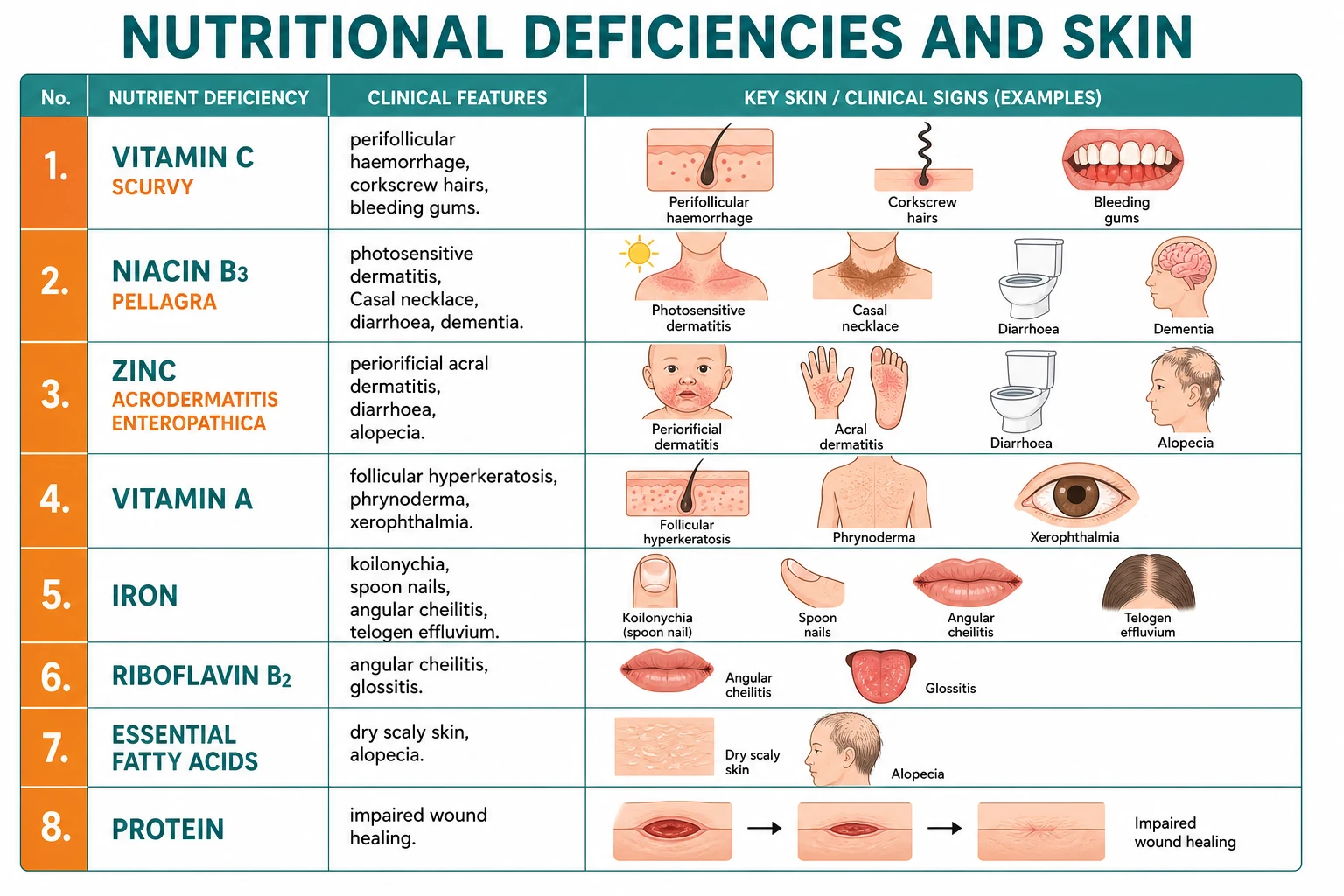
Nutritional Deficiencies — Cutaneous Signs
| Nutrient | Cutaneous sign | Defining structural or biochemical lesion |
|---|---|---|
| Vitamin A | Phrynoderma, xerophthalmia, Bitot spots, keratomalacia | Follicular hyperkeratosis; failure of retinal rhodopsin regeneration |
| Vitamin B1 (thiamine) | Glossitis, Wernicke encephalopathy, wet beriberi | Lactate accumulation from pyruvate dehydrogenase failure |
| Vitamin B2 (riboflavin) | Angular stomatitis, cheilosis, magenta tongue, photophobia | FAD depletion — atrophic epithelium of mouth and cornea |
| Vitamin B3 (niacin) | Pellagra — Casal necklace, gauntlet, 4 D's | NAD+ depletion in sun-exposed keratinocyte DNA repair |
| Vitamin B6 (pyridoxine) | Cheilosis, seborrhoeic dermatitis-like, drug-induced | PLP depletion — drug-induced (isoniazid, hydralazine) |
| Vitamin B7 (biotin) | Periorificial scaly dermatitis, alopecia, conjunctivitis | Five carboxylases inactive |
| Vitamin B9 (folate) | Glossitis, hyperpigmentation, megaloblastic anaemia | One-carbon transfer — failure of DNA synthesis |
| Vitamin B12 (cobalamin) | Patchy hyperpigmentation, Hunter glossitis, SCD | Methionine synthase failure |
| Vitamin C | Scurvy — perifollicular haemorrhage, corkscrew hairs, gum bleeding, poor wound healing | Prolyl and lysyl hydroxylases inactive — collagen disordered |
| Vitamin D | Often none in isolation; associates with AD/psoriasis severity; calcipotriol treatment of psoriasis | VDR-mediated keratinocyte dysregulation |
| Vitamin E | Xerosis, mild | Membrane lipid peroxidation |
| Vitamin K | Purpura, ecchymoses, neonatal bleeding, nodular purpura | Under-carboxylated clotting factors |
| Zinc | Acrodermatitis enteropathica — periorificial, acral, alopecia, diarrhoea | Numerous zinc-finger and metalloenzyme failures |
| Selenium | Pseudoalbinism, Keshan cardiomyopathy, brittle nails | Glutathione peroxidase failure — oxidative damage |
| EFA (omega-6, omega-3) | Scaly intertriginous dermatitis, alopecia, poor healing | Stratum corneum ceramide deficiency |
| Iron | Koilonychia, glossitis, telogen effluvium, Plummer-Vinson | Ribonucleotide reductase failure in matrix cells |
| Copper | Menkes kinky hair disease — pili torti, hypopigmentation, neurodegeneration | Lysyl oxidase and tyrosinase inactive |
| Protein | Kwashiorkor — flaky paint dermatosis, flag sign; marasmus — thin lax skin, monkey facies, lanugo | Albumin, keratin, pigment synthesis impairment |
Nutrition and Wound Healing
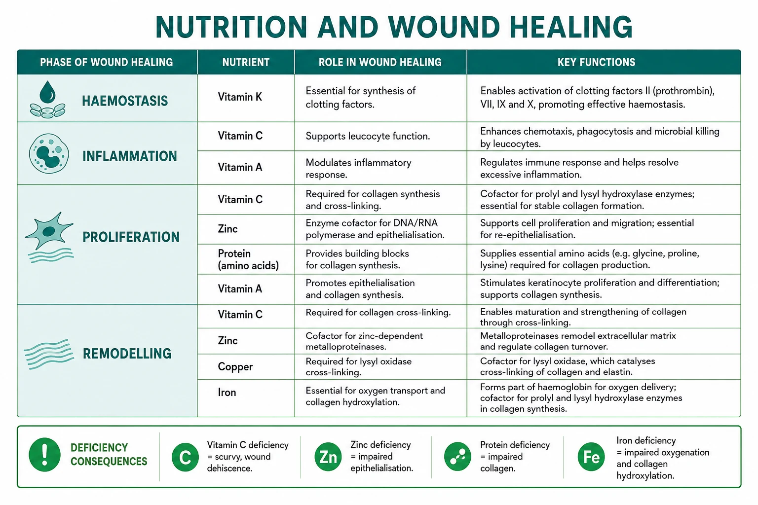
Key nutrients for wound healing:[3]
- Vitamin C: collagen synthesis (cofactor for prolyl and lysyl hydroxylase); deficiency = wound dehiscence (scurvy), dried old scars may break down.
- Zinc: enzyme cofactor (DNA and RNA polymerase, matrix metalloproteinases); epithelialisation, fibroblast migration; deficiency = impaired wound contraction and chronic leg ulcers.[5]
- Protein: amino acids (especially arginine, glutamine, methionine, cysteine) for collagen synthesis, fibroblast proliferation, immune function, and complement; deficiency = impaired healing, lymphopenia, infection risk.
- Iron: oxygen transport and delivery to wound bed; hydroxylation of proline and lysine in collagen; iron deficiency impairs oxygenation and collagen cross-linking.
- Vitamin A: epithelialisation, collagen synthesis, immune competence; especially important in chronic steroid users (retinoids reverse the antiproliferative effect of corticosteroids).
- Copper: lysyl oxidase crosslinks collagen and elastin fibres; deficiency produces brittle connective tissue.
- Vitamin K: gamma-carboxylation of clotting factors II, VII, IX and X; clotting and haemostasis.[16]
- Arginine: conditionally essential in wound healing — substrate for nitric oxide synthesis and protein synthesis; the trauma-specific diet supplemented with arginine has the best evidence base.
Nutrition in Common Skin Conditions
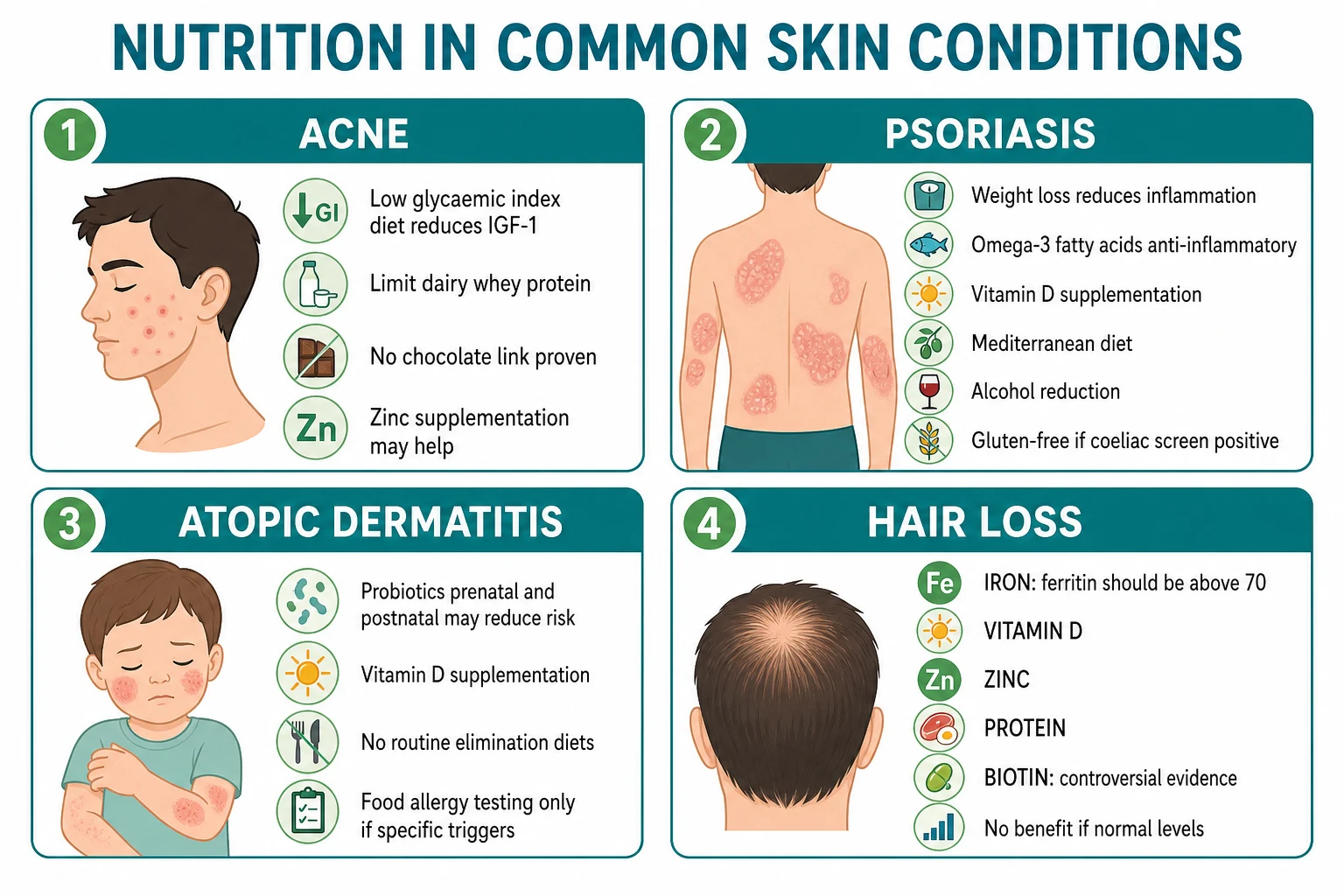
- Acne: low glycaemic index diet reduces IGF-1 → reduces sebum production and hyperkeratosis; limit dairy (especially skim milk and whey protein — both spike IGF-1, the lactoferrin fraction drives androgen receptor signalling); zinc supplementation (30 mg elemental zinc daily for 12 weeks) may help by anti-inflammatory suppression of TLR2 signalling; barberries (500 mg quercetin), omega-3, vitamin A (avoid high dose without monitoring), and probiotics (Lactobacillus rhamnosus) have positive signals but mixed evidence. The dietary intervention is an adjunct to standard retinoid/antibiotic therapy.[4]
- Psoriasis: weight loss (reduces systemic inflammation, decreases adipokine load, improves response to biologics); omega-3 fatty acids (anti-inflammatory, 2 to 4 g EPA plus DHA daily) reduce PASI in some trials; Mediterranean diet (fruit, vegetable, fish, olive oil) reduces incidence and severity; alcohol reduction; vitamin D supplementation and topical calcipotriol 50 ug per g daily; gluten-free diet only in patients with positive anti-tTG and biopsy-confirmed coeliac disease; vitamin B12/folate optimisation if methotrexate is planned.[4]
- Atopic dermatitis: probiotics (prenatal and postnatal Lactobacillus rhamnosus GG) may reduce incidence of AD in high-risk infants; vitamin D supplementation reduces severity in deficient patients; breastfeeding for at least 4 months may reduce AD risk; no routine elimination diets unless food allergy is proven — unguided exclusion may worsen the nutritional state.[3]
- Hair loss (telogen effluvium / female pattern hair loss): iron (ferritin target greater than 70 ug per L, often 100 ug per L in TE); zinc 15 to 30 mg elemental daily if low; vitamin D to keep 25-hydroxyvitamin D greater than 75 nmol/L; protein 60 to 80 g daily; biotin (controversial evidence; only benefits the rare patient with genuine biotin deficiency — supplementation without deficiency can disrupt biotin-streptavidin assays and falsely lower thyroid and troponin results).[3]
- Rosacea: trigger identification (alcohol, spicy food, hot drinks, heat, sunlight) — individualised; capsinoids and cinnamaldehyde are common dietary triggers; niacin skin flushing from niacin-containing foods or supplements can mimic rosacea; dietary modification is adjunctive to metronidazole or ivermectin topical and oral tetracycline.
- Hidradenitis suppurativa: weight loss (largest meta-analysis supports BMI reduction and dietary changes); Mediterranean diet; zinc gluconate 90 mg daily may reduce disease activity; vitamin D supplementation if deficient; avoid dairy and brewer's yeast in some cohorts (anti-TNF pathways).[4]
Refeeding — prevention and management
Refeeding syndrome — pre-refeeding checklist
The pre-refeeding checklist reflects the four pillars of safer refeeding:
- Prevent Wernicke encephalopathy by giving thiamine before any carbohydrate feed.
- Prevent hypophosphataemic cardiac and respiratory failure by replacing phosphate (intravenous potassium phosphate or sodium phosphate) before and during refeeding, and using a low-calorie starter feed (5 to 10 kcal/kg/day).
- Prevent vitamin A deficiency-related blindness and infection by single high-dose oral vitamin A (200,000 IU) in malnourished children.
- Prevent congestive cardiac failure from a sudden fluid and carbohydrate load on a cachectic myocardium. [1]
Special populations
[1]Exam Pearls
[1]Red Flags
Exam application bank (NEET-PG / INICET)
One-line answer
Micronutrient deficiency produces a characteristic cutaneous signature for almost every essential vitamin and trace element. This topic covers the nutrient-skin axis at the level of keratinocyte differentiation, sebocyte lipogenesis, melanocyte function, and wound biology; the named deficiency syndromes (scorbutic rosary, pellagra / Casal's necklace, acrodermatitis enteropathica, phrynoderma, kwashiorkor flaky-paint dermatosis, Menkes kinky hair disease, Keshan cardiomyopathy); the four prototypes of nutrient-driven skin disease — follicular, periorificial, photodistributed, and mucocutaneous; refeeding syndrome with thiamine-first resuscitation; and evidence-based nutritional therapy in acne, psoriasis, atopic dermatitis and hair loss.
Worked stems (answer without another resource)
Stem 1 — Classic presentation. Map symptoms to mechanism; name the first investigation and first treatment step with dose/route if drug therapy is standard. [1]
Stem 2 — Unstable / complicated. List red flags that force immediate resuscitation, theatre, ICU, antidote, or reperfusion — and what you do in the first 15 minutes. [1]
Stem 3 — Atypical group. Elderly, pregnancy, child, or immunocompromised: how presentation and thresholds change. [1]
Stem 4 — Differential trap. Name the three closest mimics and one discriminator for each. [1]
Stem 5 — Disposition. Who goes home with safety-netting, who is admitted, who needs HDU/ICU/theatre, and what follow-up is mandatory. [1]
Rapid viva checklist
- Definition + classification
- Pathophysiology chain
- Bedside signs / criteria
- Score with exact components (if any)
- Emergency bundle
- Definitive therapy with doses
- Complications of disease and of treatment
- Special populations
- Guideline/trial name if classic
- Three exam traps
Coverage self-check
If you cannot answer any stem above from this page alone, re-read the matching section — the page is intended to be self-sufficient for final-prof and NEET-PG/INICET questions on Nutrition and the skin.
References
- [1]Pullar JM, Carr AC, Vissers MCM. The Roles of Vitamin C in Skin Health Nutrients, 2017.PMID 28805671
- [2]Minich DM, Henning M, Darley C, et al. Is Melatonin the Next Vitamin D?: A Review of Emerging Science, Clinical Uses, Safety, and Dietary Supplements Nutrients, 2022.PMID 36235587
- [3]Seth I, Lim B, Cevik J, et al. Impact of nutrition on skin wound healing and aesthetic outcomes: A comprehensive narrative review JPRAS Open, 2024.PMID 38370002
- [4]Garbicz J, Całyniuk B, Górski M, et al. Nutritional Therapy in Persons Suffering from Psoriasis Nutrients, 2021.PMID 35010995
- [5]Ogawa Y, Kinoshita M, Shimada S, et al. Zinc and Skin Disorders Nutrients, 2018.PMID 29439479
- [6]Ndukwe C et al. Shining light on vitamin C deficiency and scurvy in Canada: A scoping review protocol of risk profiles, health outcomes, and interventions PLoS One, 2026.PMID 41801959
- [7]Ghosh SK. B vitamins in dermatology Clin Dermatol, 2026.PMID 41692080
- [8]Monshi B et al. Phrynoderma and acquired acrodermatitis enteropathica in breastfeeding women after bariatric surgery J Dtsch Dermatol Ges, 2015.PMID 26513075
- [9]Sina RE. Riboflavin Deficiency 2026.PMID 29262062
- [10]McNulty H et al. Causes and Clinical Sequelae of Riboflavin Deficiency Annu Rev Nutr, 2023.PMID 37603429
- [11]Elgharably N et al. Vitamin B group levels and supplementations in dermatology Dermatol Reports, 2023.PMID 37063401
- [12]Piraccini BM et al. Biotin: overview of the treatment of diseases of cutaneous appendages and of hyperseborrhea G Ital Dermatol Venereol, 2019.PMID 31638351
- [13]Mathur M et al. Skin Hyperpigmentation: An Under-Recognized Dermatological Clue to Vitamin B12 Deficiency Clin Case Rep, 2026.PMID 42052311
- [14]Hao S et al. Linear and nonlinear modeling of selenium biomarker dynamics in Keshan disease: Insights for precision prevention from rats and human populations Ecotoxicol Environ Saf, 2026.PMID 42296709
- [15]Kaler SG. Whole genome sequencing from dried blood spots for newborn screening of Menkes disease and 36 other actionable inherited neurometabolic disorders Mol Genet Metab, 2026.PMID 41687279
- [16]Ballal S et al. Unusual Manifestation of Vitamin K Deficiency, Nodular Purpura: A Case Series Indian Dermatol Online J, 2025.PMID 39850712
- [17]Walker J et al. Koilonychia: an update on pathophysiology, differential diagnosis and clinical relevance J Eur Acad Dermatol Venereol, 2016.PMID 27531645
- [18]Vijayasankar P et al. Flaky Paint Dermatosis in Kwashiorkor Am J Trop Med Hyg, 2021.PMID 34607303