Dermatology · Medicine
Paronychia
Also known as Paronychia · Acute paronychia · Chronic paronychia · Nail fold infection · Onychia / Perionychia · Herpetic whitlow · Felon (finger pulp abscess) · Green nail syndrome (Pseudomonas) · Periungual pyogenic granuloma (drug-induced) · Acrodermatitis continua of Hallopeau (pustular psoriasis of nail unit)
Paronychia is infection or inflammation of the nail fold (proximal and lateral) — the cuticle is the protective seal whose loss enables entry of organisms and irritants. ACUTE paronychia is dominantly Staphylococcus aureus, single-finger, fluctuant abscess, treated with warm saline soaks, incision-along-the-cuticular-edge and partial nail-plate avulsion if subungual, plus oral flucloxacillin 500 mg QDS 5-7 days for surrounding cellulitis; HERPETIC WHITLOW (HSV-1/2, vesicles, burning pain) is NEVER incised — aciclovir/valaciclovir; FELON is a closed-space pulp abscess that demands urgent I&D; GREEN NAIL SYNDROME (Pseudomonas / pyocyanin) needs acetic acid soaks and topical antibiotic drops. CHRONIC paronychia is multi-finger, boggy, with LOST CUTICLE and secondary nail dystrophy; it is fundamentally a WET-WORK-INDUCED IRRITANT CONTACT DERMATITIS with secondary Candida and bacterial colonisation, treated with strict wet-work avoidance (cotton-lined rubber gloves, SLS-free cleanser, emollient, barrier), topical tacrolimus 0.1% BD 6 weeks (steroid-sparing), and — if candidal culture positive or bacterial inflammatory component — oral fluconazole 50 mg OD 3-6 weeks or doxycycline 100 mg BD 6-12 weeks; refractory disease needs eponychial marsupialisation. Drug-induced paronychia from retinoids, EGFr / MEK / BTK inhibitors and antiretrovirals is an increasingly important iatrogenic group; the nail is regrown in 3-6 months (fingers) or 12-18 months (toes).
On this page & tools
Your progress
Saved locally on this device.
Exam tags
Red flags
Overview and Definition
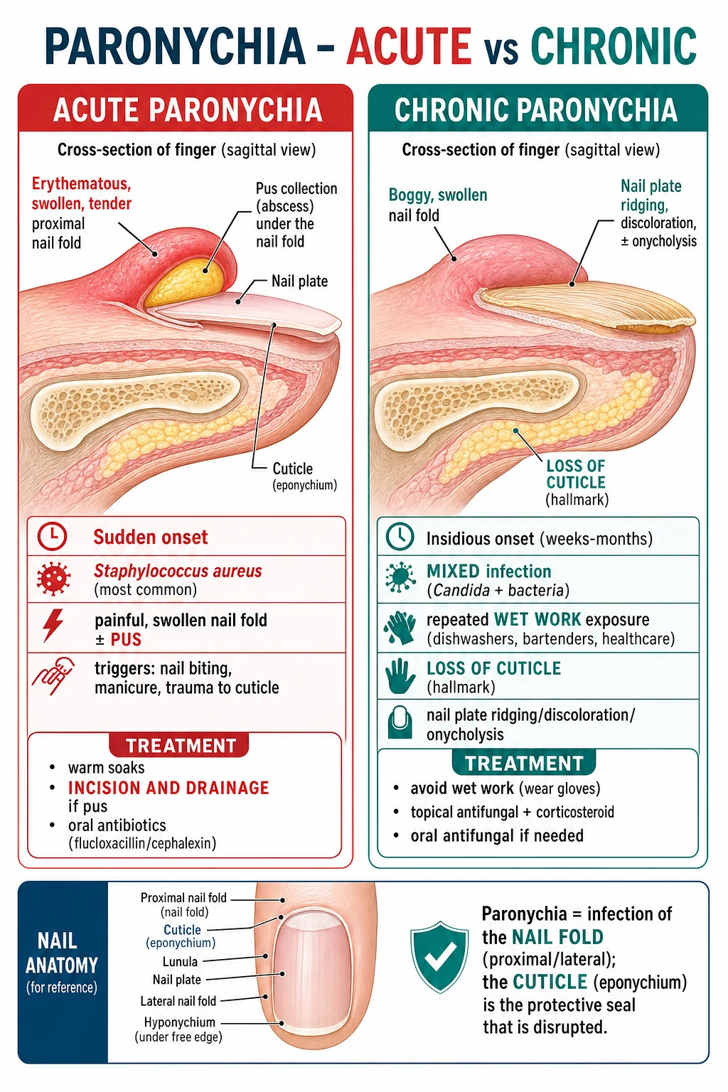
Paronychia is inflammation or infection of the nail fold — the skin that frames the nail plate on three sides (proximal and two lateral folds). The proximal nail fold (PNF) is the deepest and most clinically important: its free edge forms the cuticle (eponychium), a thin keratinised seal that runs from the dorsal surface of the PNF onto the dorsal surface of the nail plate for ~1 mm. The cuticle is the protective watertight barrier whose loss enables entry of resident flora (Staphylococcus aureus from anterior nares, Candida from gut and perineum), water, detergents and chemical irritants — and whose loss is the single most important pathophysiological event in both acute and chronic paronychia.[1][2][11]
Three anatomic landmarks complete the nail unit (and recur in examinations): the germinal matrix (proximal nail matrix, ~50% of the matrix, underlies the PNF and produces ~80% of the nail plate), the sterile matrix (nail bed, firmly adherent to the nail plate, longitudinally ridged, contributes the third remaining plate thickness), and the hyponychium (the distal seal under the free edge of the plate, onychocorneal band). The nail plate is fully keratinised dead keratin; it has no intrinsic blood supply, regenerates from the matrix, and grows at ~3 mm/month (fingernail) and ~1 mm/month (toenail).[11]
The condition can be classified along two orthogonal axes that examiners test: [1]
ACUTE paronychia
- Onset: hours to days (under 6 weeks).
- Site: ONE nail fold (proximal and / or one lateral — index, middle and thumb dominant from bite / pick exposure).
- Mechanism: breach of cuticle by trauma, hangnail, manicure, nail biting, avulsion of a torn cuticle, sustained wetness → portal of bacterial entry.
- Pathogen: Staphylococcus aureus (70-80%); Streptococcus pyogenes; anaerobes in nail-biters and clenched-fist injuries; Pseudomonas if green discoloration.
- Clinical: painful, erythematous, swollen nail fold ± yellow-green pus that LIFTS THE CUTICLE; ± fluctuation; ± subungual extension; rarely systemic.
- Course: settles in days to a week with soaks ± incision-and-drainage; rarely recurs.
CHRONIC paronychia
- Onset: weeks to months (>6 weeks).
- Site: MULTI-FINGER, usually dominant hand; can become bilateral in heavy wet-work exposure.
- Mechanism: chronic wet-work + chemical irritant (SLS detergents, vegetable juice, flour, salt) + occlusive glove use → chronic barrier disruption → irritant contact dermatitis; secondary Candida and bacterial colonisation;
- Pathogens: Candida albicans (coloniser), coagulase-negative staphylococci; the PRIMARY problem is the irritant dermatitis.
- Clinical: BOGGY, mildly tender, oedematous nail fold; LOST CUTICLE (hallmark); erythema ± scaling; secondary nail ridging, discoloration, onycholysis, Beau lines; recurrent acute flares.
- Course: takes MONTHS to resolve; recurs if wet-work resumed.
Classification [1]
Paronychia is classified by duration, pathogen, host, drug exposure and route of presentation — five axes the examiner tests: [1]
Paronychia — at-a-glance metric sheet
A — By duration
- Acute paronychia — symptoms ≤ 6 weeks; bacterial (Staph aureus dominant); single finger; painful; fluctuant; settles with soaks ± I&D.
- Chronic paronychia — symptoms > 6 weeks; multi-finger; boggy with LOST CUTICLE; irritant contact dermatitis with secondary Candida / bacterial colonisation; months-long course.
- Recurrent acute — either repeated bacterial paronychia in the same finger (think occult foreign body, ingrown nail, malignancy) or recurrent flares on a chronic-paronychia background (think continued wet-work exposure, drug-induced, EGFr inhibitor). [1]
B — By organism
- Bacterial (acute dominant): Staphylococcus aureus; Streptococcus pyogenes; Pseudomonas aeruginosa (green nail syndrome / chronic); anaerobic mouth flora (Eikenella corrodens, Fusobacterium, Prevotella, Peptostreptococcus) in clenched-fist / human-bite injuries; Klebsiella, Enterobacter, Serratia, Proteus in immunocompromised hosts.
- Fungal (chronic coloniser): Candida albicans > C. parapsilosis > C. tropicalis > C. glabrata; always treat as a coloniser — never monotherapy antifungal (fails without irritant removal).
- Viral: HSV-1 / HSV-2 — herpetic whitlow (different compartment, NO incision).
- Atypical: non-tuberculous mycobacteria (M. marinum in fish / aquarium handlers; M. chelonae in chronic steroid users); Bartonella henselae after cat scratch; deep fungi in severe neutropenia; Scedosporium, Fusarium in the profoundly immunocompromised. [1]
C — By host & route
- Primary occupational — wet-work (dishwashers, bartenders, butchers, bakers, domestic workers, healthcare workers, cleaners, fishermen, florists, swimmers).
- Secondary (drug-induced): retinoids (isotretinoin, acitretin, alitretinoin); EGFr antibodies (cetuximab, panitumumab); EGFr TKIs (gefitinib, erlotinib, osimertinib, dacomitinib, amivantamab); MEK inhibitors (trametinib, cobimetinib); BTK inhibitors (ibrutinib, acalabrutinib); antiretrovirals (indinavir, lamivudine); mTOR inhibitors (everolimus); taxanes (docetaxel); targeted therapies (vemurafenib, dabrafenib).
- Secondary (disease): eczema / atopic dermatitis; psoriasis (especially acrodermatitis continua of Hallopeau); diabetes mellitus; HIV; malnutrition; chronic kidney disease; Raynaud phenomenon; peripheral vascular disease. [1]
D — By complication / outcome
- Acute without abscess — settles with soaks.
- Acute with subungual extension — needs partial nail-plate avulsion.
- Chronic refractory — needs marsupialisation.
- Complicated — flexor tendon sheath infection (Kanavel), distal phalanx osteomyelitis, necrotising fasciitis, lymphangitis / bacteraemia. [1]
Epidemiology and Risk Factors
Global numbers
Acute paronychia is one of the commonest hand infections seen in primary care and emergency departments, accounting for ~30-35% of adult acute hand infections in large series (Fowler and Ilyas, 2013, urban US cohort).[17] It is seasonal (summer peak in temperate regions — barefoot trauma, gardening, sweaty hands), young-adult-skewed (peak incidence 20-40 years), and equally common in men and women except where occupational exposure drives a sex bias (chronic paronychia is female-skewed in butchers / beauticians / cleaners / canteen workers). Chronic paronychia affects ~1-3% of adults in occupational cohorts and dominates in the top two causes of work-related hand dermatitis after glove-related contact allergy.[1][2][7]
Modifiable risk factors
- Repetitive wet/dry cycles (the central mechanism of chronic paronychia).
- Wet-work occupations: dishwasher, bartender, butcher, baker, healthcare worker (especially nurses, dental hygienists, theatre scrub nurses), cleaner, fisherman, florist, hairdresser, beautician, swimmer.
- Occlusive glove use — thin nitrile gloves trap sweat and worsen maceration; cotton-lined rubber gloves are the protective standard.
- Manual labour with chemical irritants — vegetable juice (onion, garlic, citrus), flour, salt, sugar, fish juice, hydrocarbons, lubricants, photographic chemicals.
- Nail biting / onychophagia — disrupts cuticle, inoculates oral flora (anaerobes in clenched-fist / nail-fold bite).
- Hangnail / cuticle picking / manicure (especially cuticle pushing / cutting — REMOVES the cuticle barrier).
- Artificial nails / gel polish / acrylic — trap water, prevent cuticle regeneration, harbour Pseudomonas.
- Tinea manuum / pedis — interdigital portal; Trichophyton causes chronic paronychia in some series.
- Tight or constricting shoes / wet footwear.
- Smoking — vasoconstriction impairs cuticle healing.
- Poor glycaemic control — impaired neutrophil chemotaxis, faster progression to abscess / deeper infection. [1]
Non-modifiable risk factors
- Diabetes mellitus — recurrent / refractory chronic paronychia warrants HbA1c; up to 1 in 5 patients with refractory chronic paronychia have undiagnosed diabetes.[1][2]
- Atopic dermatitis / eczema / contact dermatitis — defective barrier.
- Peripheral vascular disease, Raynaud phenomenon — slow cuticle healing.
- HIV / AIDS — higher prevalence of atypical mycobacterial, fungal, and Bartonella paronychia; ART-related paronychia (indinavir / lamivudine).[15]
- Chemotherapy-induced neutropenia, transplant recipients, biologics — broader differential, atypical organisms, lower threshold for I&D and IV cover.
- Iron deficiency, malnutrition, hypoproteinaemia — impaired epithelial regeneration.[2]
Drug-induced paronychia — high-yield list
Drugs that cause paronychia — mnemonic CROPS
CROPS
EGFr antibodies (cetuximab, panitumumab) 10-20%, EGFr TKIs (osimertinib, gefitinib, erlotinib, dacomitinib) 30-50%, MEK inhibitors (trametinib, cobimetinib), mTOR inhibitors (everolimus), BRAF inhibitors (dabrafenib).
Isotretinoin / acitretin / alitretinoin — chronic dryness, pyogenic granuloma (periungual), fissuring; dose-dependent; resolves on withdrawal or dose reduction.
Indinavir (crystal paronychia and ingrown nail); lamivudine-induced periungual pyogenic granulomas (PLHIV); protease-inhibitor class effect.
Docetaxel (nail-fold changes, onycholysis); capecitabine (hand-foot syndrome with secondary nail-fold erythema); doxorubicin (rare).
BTK inhibitors (ibrutinib, acalabrutinib); JAK inhibitors; CDK4/6 inhibitors; CDK-7 inhibitors; PI3K inhibitors. Periungual pyogenic granuloma is the dominant lesion.
Regional deltas (epidemiology + clinician awareness)
- India / South Asia (ICMR context, NEET-PG): higher prevalence of barefoot walking, manual agricultural labour, repetitive harvest trauma, and chronic wet-work exposure in domestic workers and restaurant hands; empirical amoxicillin-clavulanate 625 mg TDS is often used first-line because of mixed oral-flora coverage; HIV-associated paronychia is more common in high-prevalence districts; fluconazole is cheap, widely available, and used in 150 mg weekly pulse rather than 50 mg OD continuous, when the patient cannot afford daily dosing.
- UK / Europe (NICE CKS): NICE CKS favours oral flucloxacillin 500 mg QDS 5-7 days; clarithromycin 250-500 mg BD 7 days for penicillin allergy; doxycycline / clindamycin / trimethoprim-sulfamethoxazole reserved for documented MRSA.
- US (IDSA 2014): purulent paronychia coverage includes empirical MRSA cover (doxycycline / TMP-SMX / clindamycin) where CA-MRSA prevalence > 20%.
- Australia / NZ (eTG): dicloxacillin 500 mg QDS is the empirical first-line (analogous to flucloxacillin); chronic paronychia is described explicitly as "wet-work-induced irritant contact dermatitis" with dry-work directive as the cornerstone.
- Beers Criteria: in the elderly, prolonged tetracyclines are listed as potentially inappropriate — prefer short courses of flucloxacillin or cephalexin in elderly paronychia. [1]
Pathophysiology
Nail unit anatomy as it relates to paronychia
The protective nail apparatus is built from four layered keratinised elements and three bacteriologic seals.[11] From surface to depth:
- Cuticle (eponychium) — thin keratinised cornified layer that runs from the dorsal surface of the proximal nail fold onto the dorsal surface of the nail plate for ~0.5-1.0 mm. Forms the proximal seal.
- Lateral nail folds × 2 — skin folds that grip the sides of the plate; their distal end seals against the nail at the lateral grooves. With chronic wet-work these seals are the first to soften.
- Hyponychium (onychocorneal band) — under the free edge of the plate; the distal seal. Loss of this seal produces distal subungual onychomycosis and is the gateway for fungal / Pseudomonas paronychia of the distal fold.
- Nail plate — fully keratinised dead keratin, no blood supply, no nerves. Regenerates from the matrix. [1]
The proximal nail fold has a dorsal and a ventral surface; the ventral surface of the PNF is the area that inflames acutely (acute paronychia) and thickens + loses its cuticle chronically (chronic paronychia). [1]
Step 1 — Loss of the cuticle (the gateway event)
The cuticle is the single most important barrier and the single most commonly breached barrier. Loss results from: [1]
- Mechanical: manicure (especially cuticle pushing / cutting), hangnail picking, nail biting, artificial-nail application, nail-plate avulsion, subungual foreign body.
- Chemical: detergents (sodium lauryl sulphate = SLS), solvents, vegetable juices (onion / garlic / citrus), flour (proteolytic enzymes), salt, sugar, photographic chemicals.
- Occlusive maceration: prolonged water contact (dishwasher, bartender, swimmer, fisherman); occlusion under gloves or bandages prevents the cuticle from re-keratinising.
- Inflammatory: acute dermatitis, atopic dermatitis, psoriasis, lichen planus.
- Drug: retinoids, EGFr / MEK inhibitors, antiretrovirals. [1]
Step 2 — Inoculation
Once the cuticle is breached, resident flora colonises the sub-cuticular space: [1]
Portal of entry → dominant organism (mnemonic pair-up)
Step 3 — Inflammation of the nail fold
In acute paronychia, neutrophil-rich pus accumulates under the lifted cuticle; the fold becomes tense, erythematous, fluctuant; the abscess either drains spontaneously (skin → pus tracks under the cuticle and onto the plate) or tracks under the proximal nail plate to form a subungual abscess. [1]
In chronic paronychia, repeated wet/dry cycles and irritant exposure cause a lymphohistiocytic + eczematous inflammation in the proximal nail fold; the cuticle retracts (lost cuticle — hallmark) and is replaced by a moist, erythematous, oedematous fold with poor keratinisation. Secondary Candida and bacterial colonisation is secondary, not causal; this is why monotherapy antifungal fails — the wet-work irritant must be removed.[7][13]
Step 4 — Nail-plate damage (chronic phase)
The chronic inflamed fold is a poor matrix for the emerging plate. Typical nail changes: [1]
- Beau lines — transverse ridges where the matrix briefly stopped growing.
- Median / longitudinal ridging — chronic matrix disturbance.
- Green-brown discoloration — Pseudomonas pyocyanin on onycholytic plate; or Candida brown staining.
- Distal onycholysis — plate separates from the bed (hyponychium breach). [1]
Drug-induced paronychia mechanism
- Retinoids (isotretinoin / acitretin / alitretinoin) cause dryness of the periungium and reduce keratinocyte proliferation → chronic fissuring, pyogenic granuloma formation, ingrown nails. Dose-dependent.
- EGFr inhibitors impair keratinocyte proliferation, neutrophil chemotaxis and barrier repair; a VEGF-driven angiogenesis in the periungium produces friable, easily-bleeding granulation tissue (periungual pyogenic granuloma).[15]
- MEK inhibitors cause brittle nails and onycholysis.
- Indinavir (antiretroviral) causes paronychia and ingrown toenails via retinoid-like dysregulation.
- Lamivudine has been described as causing periungual pyogenic granulomas in PLHIV (case series 2025).[15]
- BTK / JAK / CDK4/6 inhibitors — emerging class effects with paronychia and granuloma formation.
Herpetic whitlow pathophysiology
HSV-1 (oral shedders, dental / healthcare exposure) or HSV-2 (genital contact, autotransmission) enters via microtrauma → primary inoculation into the dermis of the pulp / fold → replication in epidermal keratinocytes → vesicular coalescence with Tzanck-positive multinucleate giant cells → ascendant lymphangitis → ganglionic latency in the dorsal root ganglion or trigeminal ganglion → reactivation in 30-50% of patients, often with prodromal burning/tingling.[14]
Clinical Presentation
Acute paronychia — diagnostic clinical pattern
- Onset: hours to days (often < 48 h from the precipitating event — hangnail, cuticle cut, nail-bite).
- Pain: throbbing; interferes with sleep and use of the hand.
- Sign: erythema, oedema, warmth of the proximal and / or one lateral nail fold; a yellow-green sub-cuticular collection that lifts the cuticle is pathognomonic.
- Fluctuance: the cardinal sign that pus is present and I&D is indicated.
- Extension: around the lunula, beneath the nail plate (subungual abscess); rarely into the pulp — then think FELON.
- Systemic: usually absent; fever / lymphangitis signals spreading cellulitis or early flexor tendon sheath infection.
- Distribution: commonly one finger — index, middle and thumb dominant (the "bite / pick fingers").
- Subacute on chronic background: if acute paronychia happens on a finger with chronic paronychia, a cure requires BOTH the I&D and the wet-work avoidance. [1]
Chronic paronychia — diagnostic clinical pattern
- Onset: weeks to months; often first noticed because the cuticle is GONE.
- Pain: dull, mild, episodic — much less than acute; some patients deny pain.
- Sign: BOGGY, mildly tender, oedematous proximal nail fold; LOST CUTICLE is the hallmark; erythema ± scaling; recurrent small flares of acute inflammation superimposed.
- Nail plate: ridging, transverse grooves / Beau lines, green-brown discoloration (if Pseudomonas or Candida), distal onycholysis; permanent nail changes if matrix damaged.
- Distribution: MULTI-FINGER, usually dominant hand; often symmetric in bilateral wet-work exposures.
- Predisposing background: occupation (dishwasher, bartender, cleaner), diabetes, eczema, recent drug start (retinoid, EGFr inhibitor, antiretroviral).
- Systemic: none. [1]
Atypical clinical patterns
[1]- Neonatal / congenital candidiasis — sterile paronychia with pustules on multiple digits + oral thrush + intertriginous rash; mother had vaginal candidiasis; settles with topical nystatin / miconazole.
- Immunocompromised — broader organism list (atypical mycobacteria, Bartonella, deep fungi); atypical presentation with rapid spread.
- Elderly diabetic — masked pain, rapid spread, risk of necrotising fasciitis and distal phalanx osteomyelitis.
- Drug-induced EGFr / MEK / BTK-inhibitor paronychia — periungual pyogenic granuloma (vascular, friable, bleeds on contact), bilateral, slow resolution.
- Bartonella paronychia — cat-scratch exposure; tender regional lymphadenitis; doxycycline 100 mg BD 6 weeks.
- Mycobacterium marinum paronychia — fish-tank / aquarium / fish-handling exposure; chronic indolent course; sporotrichoid nodules up the arm (lymphocutaneous spread); biopsy for AFB and culture; requires triple therapy (rifampicin + ethambutol ± clarithromycin) for 3-6 months.
- Acrodermatitis continua of Hallopeau — pustular psoriasis of the nail unit; sterile pustules; chronic erosive course; adjacent psoriatic plaques. [1]
Red-flag clinical features that mandate escalation
- Systemic toxicity — fever, rigors, tachycardia disproportionate to local signs, hypotension, confusion → sepsis pathway, IV antibiotics within 1 hour.
- Pain out of proportion to signs, dusky discolouration, crepitus, bullae → necrotising soft-tissue infection; immediate surgical debridement.
- Flexor tendon sheath infection (Kanavel signs) — fusiform swelling of the entire finger, finger held in flexion, tenderness along the tendon sheath, pain on passive extension of the distal interphalangeal joint. Hand surgery within hours; missing this causes permanent tendon necrosis and flexion contracture.
- Distal phalanx osteomyelitis — chronic paronychia + bone pain + systemic signs or persistent discharge; MRI / X-ray.
- Spreading cellulitis past the wrist → admit for IV antibiotics.
- Immunocompromised host + any paronychia → culture, IV cover, lower threshold for admission.
- Single-digit chronic dystrophy → biopsy to exclude squamous cell carcinoma / melanoma of the nail unit.
- Pigment extension onto the proximal nail fold (Hutchinson sign) → melanoma of the nail matrix until proven otherwise — biopsy the matrix, not just the plate. [1]
Differential Diagnosis
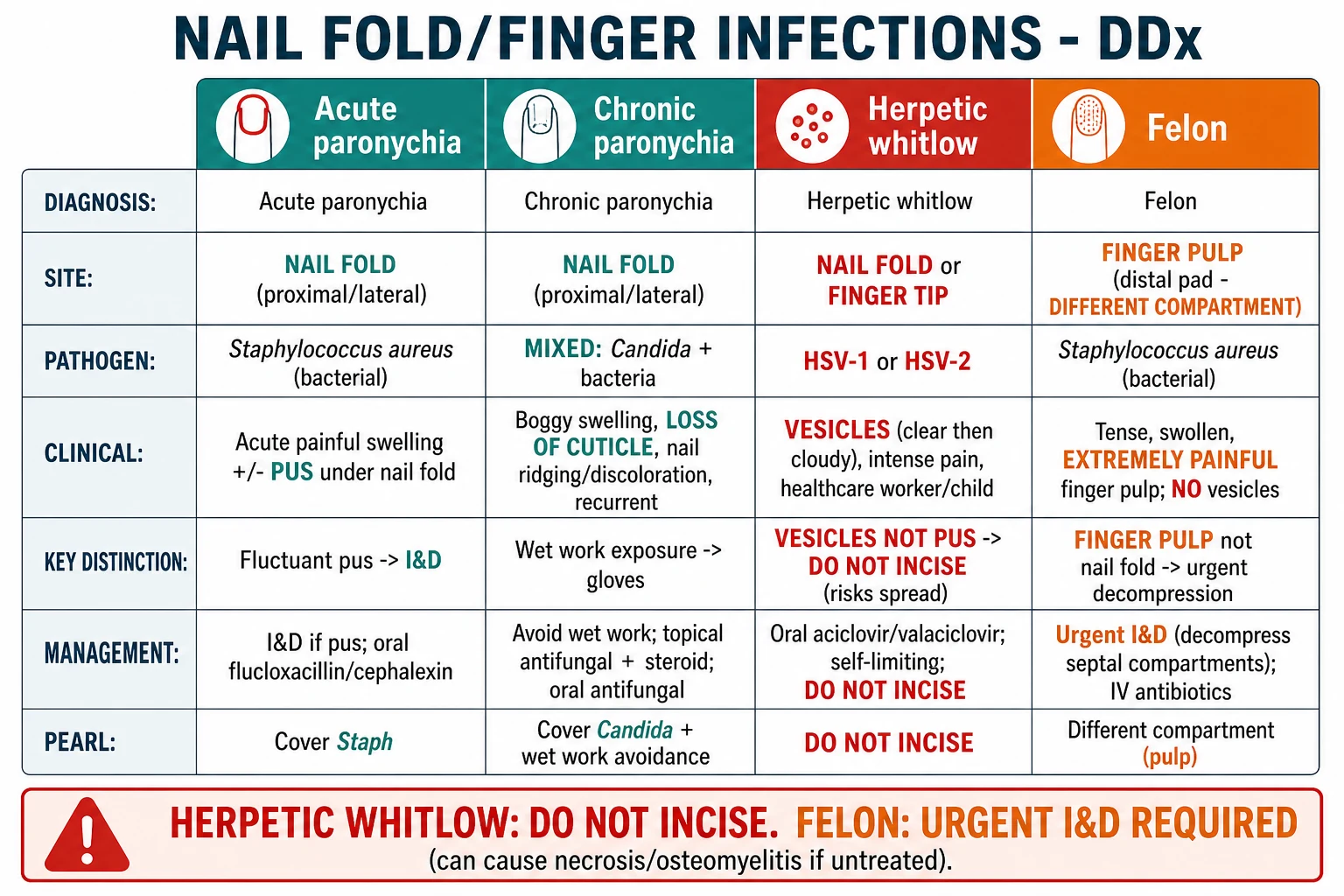
The diagnosis of paronychia is clinical. The differential is anatomic (which compartment is inflamed) plus morphologic (fluid vs. solid vs. papular) plus host context. [1]
| Diagnosis | Compartment | Onset | Fluid / morphology | Pathogen / cause | Key bedside sign | Action |
|---|---|---|---|---|---|---|
| ACUTE paronychia | Proximal nail fold | Hours-days | Yellow-green pus lifting cuticle | Staphylococcus aureus | Fluctuant; tender | Warm soaks → I&D along cuticular edge; oral flucloxacillin 500 mg QDS 5-7 d |
| Herpetic whitlow | Finger pad / fold / face | 2-7 days from inoculum | Clear then cloudy vesicles on erythematous base | HSV-1 / HSV-2 | Burning pain; lymphangitis | DO NOT INCISE; aciclovir 400 mg 5x/d 7 d; PCR confirm |
| Felon | Finger PULP (different compartment) | 7-10 d post-puncture | Pus under tension in pulp | Staph aureus / mixed | Pain more than paronychia; loss of pulp crease | Urgent I&D (lateral or volar pulp incision); IV antibiotics |
| Green-nail syndrome | Onycholytic nail plate (no fold abscess) | Weeks in wet-work | Green-black plate; onycholysis | Pseudomonas aeruginosa (pyocyanin) | Plate green; no cuticle pus | Acetic acid soaks; topical ciprofloxacin / ofloxacin drops |
| Periungual pyogenic granuloma | Periungual skin | Weeks on therapy | Friable red papule, bleeds on contact | Drug (EGFr / MEK / BTK inhibitors, retinoid, indinavir) | No pus; bleeds on touch | Dose modification; topical steroid + silver nitrate; topical timolol |
| Acrodermatitis continua of Hallopeau | Distal finger / nail unit | Chronic — months | Sterile pustules; erosive nail bed | Pustular psoriasis; sterile | Adjacent psoriatic plaques; sterile cultures | Topical clobetasol + calcipotriol; systemic methotrexate / biologics |
| Subungual exostosis | Subungual bone | Chronic — months | Hard nodule lifting plate; pain on pressure | Benign bony outgrowth (trauma) | X-ray diagnostic (bony projection) | Hand-surgery referral; excision |
| Squamous cell carcinoma of nail | Single digit; chronic | Months-years | Warty / bleeding subungual lesion | Malignancy; HPV-16 in some | Single-digit chronic dystrophy; bone erosion on X-ray | Biopsy — single-digit chronic disease is cancer until proven otherwise |
| Melanoma of nail matrix | Single digit (thumb / great toe dominant) | Months-years | Longitudinal melanonychia widening | Acral lentiginous melanoma | Hutchinson sign (pigment on proximal nail fold) | Matrix biopsy — DO NOT incise the plaque before biopsy |
| Nail-bed squamous-cell carcinoma mimicking paronychia | Single digit | Chronic | Wart-like, bleeds, discharging | HPV-16; chronic inflammation | Single-digit disease; bone erosion on X-ray | Biopsy before any further I&D or amputation |
Cannot-miss differentials (miss = significant morbidity)
Cannot-miss differentials — mnemonic HARD
HARD
Vesicles + burning pain + lymphangitis. DO NOT INCISE — viral encephalitis risk to operator; bacterial superinfection in patient. PCR-confirm; aciclovir / valaciclovir. Cover the lesion.
Hutchinson sign (pigment on proximal fold) is positive in up to 70% of subungual melanomas. Wide excisional biopsy of the matrix — do not incise the lesion before biopsy.
Pain out of proportion, dusky discolouration, crepitus, bullae, systemic toxicity. Immediate surgical debridement + broad-spectrum IV cover (flucloxacillin 2 g IV 6-hourly + clindamycin 600-900 mg IV 8-hourly ± vancomycin).
Kanavel signs (fusiform swelling, finger in flexion, sheath tenderness, pain on passive extension). Hand surgery within hours. Failure to decompress → permanent tendon necrosis and flexion contracture.
Clinical and Bedside Assessment
A complete bedside assessment has six steps — the examiner expects you to demonstrate all six. [1]
Step 1 — Inspection of the affected and contralateral fingers
- Look at all 20 nails — chronic paronychia is usually multi-finger.
- Look at the proximal and both lateral folds — and the distal hyponychium.
- Look at the pulp — to exclude concurrent felon.
- Look at the plate — for Beau lines (matrix damage), green-black discoloration (Pseudomonas), ridging, onycholysis.
- Look at the lunula — for pigmentation extension (Hutchinson sign). [1]
Step 2 — Palpation and bedside manoeuvres
- Fluctuance — pressing the fold with a gloved fingertip detects fluid.
- Tense pulp — distinguishes felon (pulp under tension) from paronychia (fold under tension).
- Lymphangitis — red streak ascending the forearm.
- Regional nodes — epitrochlear + axillary.
- Capillary refill — exclude arterial compromise (Buerger, thromboangiitis obliterans, vasculitis). [1]
Step 3 — Vascular and neurological screen
Capillary refill < 2 seconds; light touch / pin-prick of the distal pulp; peripheral pulses (radial, ulnar, brachial). Vasculopathy and neuropathy change prognosis. [1]
Step 4 — Identify the portal of entry
Hangnail / cuticle trauma, manicure or cuticle pushing within 7 days, nail bite, artificial nail, ingrown nail, tinea manuum (interdigital scale), puncture wound (splinter / thorn / fish bone / sewing needle), recent HSV exposure (dental visit, thumb-sucking child). [1]
Step 5 — Drug and disease history
Retinoid, EGFr / MEK / BTK inhibitor, antiretroviral, chemotherapy, biologics, recent surgery. Diabetes, eczema, psoriasis, HIV, Raynaud, peripheral vascular disease, smoking, malnutrition. [1]
Step 6 — Severity and systemic status
Temperature, pulse, BP, RR, mental state, NEWS2 / qSOFA — basic vital signs before any treatment decision. [1]
[1]Investigations
Acute paronychia is a clinical diagnosis. The role of investigations is to (i) confirm the organism when it matters for treatment, (ii) screen for underlying disease in chronic / recurrent / refractory disease, (iii) rule out herpetic whitlow when morphology is uncertain, and (iv) exclude osteomyelitis / necrotising infection in severe disease. [1]
Acute paronychia — when to investigate
Investigations — who, what and when
Investigations for HSV (herpetic whitlow — when in doubt)
- Vesicle PCR — gold standard; sensitivity 95%+; can type HSV-1 / HSV-2.
- Tzanck smear — multinucleate giant cells; fast (1 h) but low sensitivity; positive in HSV-1, HSV-2 and VZV; cannot distinguish.
- Direct immunofluorescence antigen detection (DFA) — type-specific, fast.
- Viral culture — historically the gold standard; sensitivity 50-70% and slow (3-7 days); now largely replaced by PCR.
- HSV serology (IgM / IgG) — useful in primary infection to confirm seroconversion; not useful in recurrences. [1]
Investigations for chronic paronychia
In chronic paronychia the investigations are largely about (a) confirming the role of Candida (culture is supportive, not diagnostic), (b) screening for predisposing disease, (c) excluding malignancy in single-digit disease. [1]
Adjunctive screening for recurrent / refractory / chronic disease
- HbA1c / fasting glucose — diabetes mellitus.
- HIV 4th-generation Ag/Ab — single exposure for high-prevalence populations.
- FBC + differential — neutropenia, lymphopenia, eosinophilia.
- Ferritin + iron studies — iron deficiency impairs neutrophil oxidative burst.
- Urea / creatinine / eGFR — renal dosing for flucloxacillin, cephalexin, tetracyclines.
- LFTs — baseline before fluconazole, itraconazole, doxycycline.
- TSH — hypothyroidism presents with brittle nails.
- Patch testing — when contact allergy is suspected (chromate, rubber, fragrance, preservative).
- Nail clippings for mycology — if tinea unguium concurrent. [1]
Imaging
- Plain X-ray of the distal phalanx if chronic paronychia + bone pain + systemic signs → osteomyelitis (cortical destruction, periosteal reaction).
- Ultrasound for occult abscess / foreign body.
- MRI if necrotising fasciitis suspected (deep fascial oedema, gas, non-enhancing muscle); reserved for severely ill patients.
- CT rarely needed; reserved for deep hand collections. [1]
Drug concentration monitoring
- Vancomycin trough at 15-20 mg/L (severe MRSA SSTI) — 25-30 mg/kg loading then 15-20 mg/kg 12-hourly with levels.
- Aminoglycoside extended-interval dosing (gentamicin, tobramycin) — once-daily 5-7 mg/kg with level-guided adjustment. [1]
Management — Resuscitation
Resuscitation is rarely needed in paronychia. Recognise the patients in whom it is: [1]
[1]For severe or necrotising disease: [1]
- ABCDE + oxygen to SpO2 94-98%.
- Two large-bore IV cannulae + fluid bolus 30 mL/kg balanced crystalloid if hypotensive or lactate ≥ 2.
- Bloods: FBC, U&E, CRP, lactate, blood cultures × 2, coagulation, group + save.
- Empirical IV antibiotics within 1 hour — flucloxacillin 2 g IV 6-hourly + clindamycin 600-900 mg IV 8-hourly (anti-toxin for streptococcal / staphylococcal superantigen toxins) ± vancomycin 25-30 mg/kg loading then 15-20 mg/kg 12-hourly (MRSA risk).[3][6]
- Urgent surgical consultation — for immediate debridement if necrotising fasciitis suspected; for Kanavel signs / flexor tendon sheath infection; for compartment syndrome of the digit.
- Tetanus cover — any breach + open wound needs tetanus assessment and a booster if last dose > 10 years or wound > 6 hours old / contaminated.
Management — Definitive and Stepwise
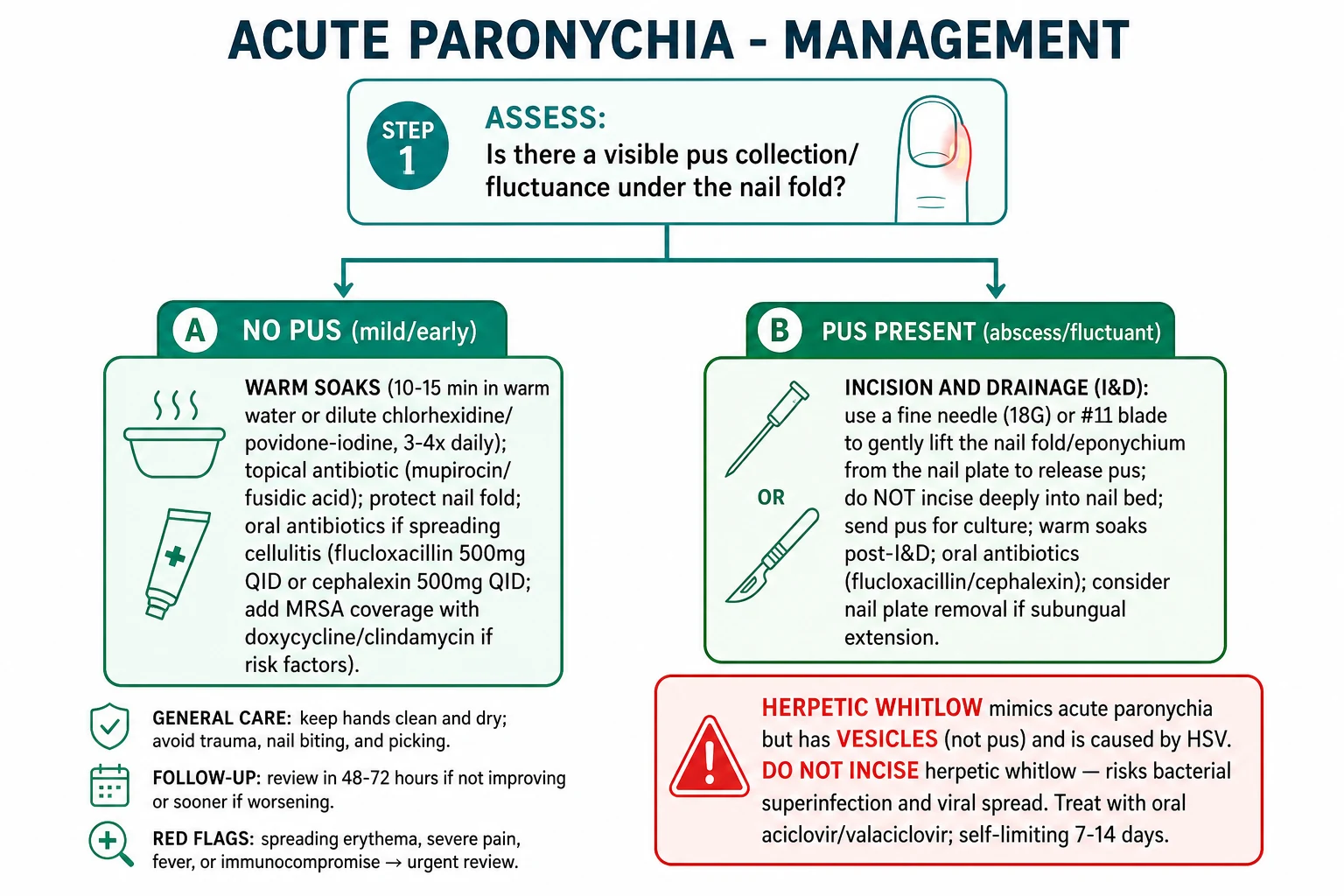
A — Acute paronychia — staged ladder
Acute paronychia — SOAKS to AVULSION ladder
SOAKS
Warm saline / dilute antiseptic soaks 10-15 min × 3-4 daily; topical antibiotic (mupirocin 2% / fusidic acid 2% / neomycin-bacitracin-polymyxin) TDS-QDS; rest the hand. Reassess at 48-72 h. Duration 5-7 days.
Fluctuant abscess: use a fine-gauge needle (18G) or a #11 blade tip to make a SMALL incision along the LONGITUDINAL AXIS of the cuticle side, lift the cuticle off the plate to release pus; DO NOT incise deeply into the nail bed (risk of matrix scar). Send pus for MCS.
Add oral flucloxacillin 500 mg QDS 5-7 d for surrounding cellulitis / systemic features / immunocompromise (UK NICE first-line; eTG equivalent); amoxicillin-clavulanate 625 mg TDS if mixed oral flora suspected (bite); clindamycin 300 mg QDS for documented MRSA.
Watch for Kanavel signs (fusiform swelling, finger in flexion, sheath tenderness, pain on passive extension). If positive, hand surgery within hours.
Subungual abscess or chronic nail-fold track: PARTIAL nail-plate AVULSION (≤25% of the plate) to expose the subungual abscess bed; drain and pack; reculture.
B — Acute paronychia — drug choice (drug + dose + route + timing + rationale)
First-line empirical cover for ACUTE paronychia
C — Chronic paronychia — staged ladder [1]
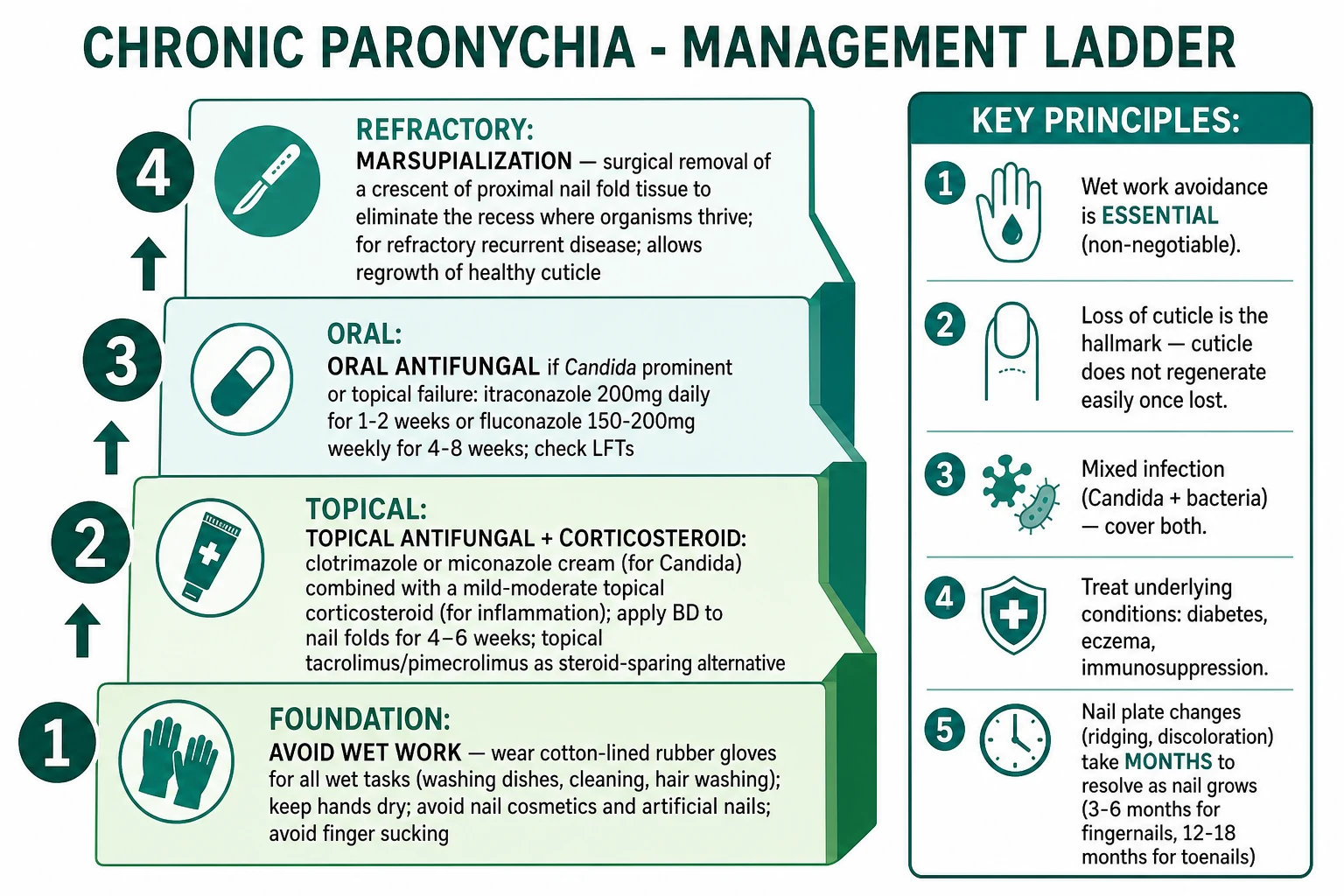
[1]Chronic paronychia — DRY Rx mnemonic
DRY
STOP wet-work exposure (the foundation). Cotton-lined rubber gloves for all wet tasks; SLS-free hand cleanser; emollient after every wash (white soft paraffin, urea-based); barrier cream (dimeticone); keep nails short and unpolished; avoid artificial nails.
Topical tacrolimus 0.1% ointment BD × 6 weeks (steroid-sparing first-line per Rigopoulos 2009 BRJ Dermatol RCT vs. betamethasone valerate 0.1% — equivalent efficacy, no atrophy); OR potent topical corticosteroid (clobetasol propionate 0.05% ointment BD 2-4 weeks). Apply a thin film to the affected fold and gently massage in.
Topical clotrimazole 1% / miconazole 2% cream BD 4-6 weeks; oral fluconazole 50 mg OD 3-6 weeks / 150 mg weekly 4-8 weeks (palmar-printer option); oral itraconazole 200 mg BD pulse week per month × 2; oral doxycycline 100 mg BD 6-12 weeks (anti-inflammatory + antistaphylococcal) OR minocycline 100 mg BD 6-12 weeks.
D — Chronic paronychia — drug choice (drug + dose + route + timing + rationale)
- Tacrolimus 0.1% ointment — BD application to the affected fold × 6 weeks; steroid-sparing first-line per Rigopoulos 2009 BR J Dermatol RCT vs betamethasone valerate 0.1% — equivalent efficacy in chronic paronychia, with no skin atrophy risk; safe on the proximal nail fold where atrophy from chronic potent steroids would expose the matrix. Counsel on transient burning sensation (3-10% of patients).[8][9]
- Clobetasol propionate 0.05% ointment — BD × 2-4 weeks; potent topical corticosteroid for short-course anti-inflammatory cover; limit duration to avoid fold atrophy and matrix damage.
- Clotrimazole 1% / miconazole 2% cream — BD × 4-6 weeks; topical antifungal if candidal culture positive.
- Fluconazole 50 mg OD PO × 3-6 weeks OR 150 mg weekly PO × 4-8 weeks — for culture-confirmed Candida in chronic paronychia; pulse weekly therapy is the standard low-cost option in low-/middle-income countries and the regimen most widely used in Indian practice.
- Itraconazole 200 mg BD PO × 1 week per month × 2 pulses — for refractory or extensive chronic candidal paronychia; check LFTs.
- Doxycycline 100 mg BD PO × 6-12 weeks — for bacterial / inflammatory component (anti-Staphylococcal + matrix metalloproteinase inhibition anti-inflammatory); effective in chronic paronychia even without MRSA coverage; avoid in pregnancy / under 12 y; sun protection.
- Minocycline 100 mg BD PO × 6-12 weeks — alternative to doxycycline; dizziness / vestibular side effects; hyperpigmentation at higher cumulative doses.
- Eponychial marsupialisation — surgical removal of a crescent of proximal nail fold tissue to eliminate the deep recess where organisms thrive; for refractory chronic paronychia after 3 months of conservative management.[7]
E — Subtype-specific drug choice
- Herpetic whitlow — oral aciclovir 400 mg 5× daily × 7 days OR oral valaciclovir 1 g BD × 7 days within 72 h of vesicle onset; cover the lesion; analgesia; self-limiting 2-3 weeks. Recurrence: chronic suppressive valaciclovir 500 mg BD for healthcare workers / dentists / hygienists.[14]
- Felon — IV flucloxacillin 1-2 g 6-hourly + urgent I&D via lateral or volar midline incision through the pulp; decompress septal compartments; drain pus; pack loosely or insert a small wick; remove at 24-48 h; hand therapy to prevent stiffness.[3][12]
- Green-nail syndrome — dilute acetic acid soaks (1:4 vinegar:water) BD × 4-6 weeks; topical ciprofloxacin 0.3% / ofloxacin 0.3% / gentamicin 0.3% drops BD × 4-6 weeks; trim onycholytic plate; partial nail avulsion if subungual extension; keep nail dry.[10]
- EGFr / MEK / BTK-inhibitor paronychia — dermatology–oncology liaison; dose interruption / reduction of the targeted agent; topical clobetasol BD × 2-4 weeks; chemical cautery of periungual pyogenic granulomas with silver nitrate; topical timolol 0.5% gel BD × 4-8 weeks; avoid systemic retinoids (worsen the granuloma); secondary staphylococcal infection → oral flucloxacillin 500 mg QDS 5-7 days.[15]
- Acrodermatitis continua of Hallopeau — topical clobetasol + calcipotriol combination; oral methotrexate 7.5-15 mg weekly; acitretin 0.3-0.5 mg/kg/d; ciclosporin 3-5 mg/kg/d; TNF-inhibitors (adalimumab 40 mg SC fortnightly); IL-23 / IL-17 inhibitors for refractory disease; bone-marrow-sparing tetracyclines for matrix protection.[18]
- Mycobacterium marinum paronychia — biopsy for AFB; specialist referral; triple therapy (rifampicin + ethambutol ± clarithromycin) for 3-6 months.
- Bartonella henselae paronychia — doxycycline 100 mg BD 6 weeks; check for regional lymphadenitis.
- Traumatic subungual haematoma with secondary paronychia — evacuate haematoma (paper-clip cautery through the plate); cover with antibiotic if paronychia settles.
F — Procedural technique in detail
Incision and drainage (I&D) of acute paronychia
- Anaesthesia — digital nerve block (plain lidocaine 1% without adrenaline; for the digit), or local infiltration over the fold.
- Aseptic prep — chlorhexidine or povidone-iodine.
- Incision — small longitudinal incision along the distal edge of the affected nail fold, parallel to the nail plate, just deep enough to release sub-cuticular pus. Use the sharp tip of a #11 blade (no. 15 or no. 11), OR an 18G needle alongside the plate. Do NOT incise deeply — the matrix is just beneath.
- Lift the cuticle from the plate — with the needle tip or the fine tip of a mosquito forceps — to release pus. Allow pus to drain.
- Irrigate with normal saline.
- Insert a small wick (gauze wisp) for 24 h if the cavity is large; otherwise leave open to drain.
- Send pus for MCS — bacterial culture ± Gram stain.
- Post-procedure — warm saline soaks TDS × 3-5 days; dressing with antibiotic ointment; oral flucloxacillin 500 mg QDS 5-7 days for surrounding cellulitis. [1]
Partial nail-plate avulsion
- Anaesthesia as above.
- Elevate the affected strip of plate by inserting a flat spatula or a Freer periosteal elevator under the proximal edge of the plate — between the plate and the nail bed.
- Cut the strip longitudinally with a nail splitter / scissors.
- Avulse the lateral strip with a haemostat — this exposes the subungual abscess bed.
- Irrigate and leave to drain.
- Phenolisation of the lateral nail-matrix horn is reserved for ingrown toenail, not paronychia. [1]
Eponychial marsupialisation (chronic paronychia — refractory)
- Digital block.
- Excise a crescent of proximal nail fold tissue from the dorsum of the finger, ~3-4 mm wide × 3-4 mm long, with the apex at the proximal edge of the cuticle.
- Undermine the fold proximally and remove the proximal portion of the underlying tissue to expose the germinal matrix.
- Hemostasis; close with simple sutures.
- The folded-back fold can no longer host the moist pocket. [1]
Felon I&D
- Digital block.
- Incision — lateral longitudinal incision along the ulnar side of the thumb or the radial side of the index / middle finger (to avoid the digital nerve and the flexor tendon sheath); OR a volar midline longitudinal incision through the pulp; incision should not cross the distal interphalangeal flexion crease.
- Decompress all septal compartments of the pulp using a mosquito haemostat.
- Irrigate and loosely pack with gauze for 24 h.
- IV antibiotics until clinical improvement; switch to oral to complete 7-10 days.
- Hand therapy within 48 h to prevent stiffness. [1]
G — Escalation triggers
- 48-72 h without improvement → reassess diagnosis (herpetic whitlow, felon, malignancy); broaden cover; MCS; image for abscess / osteomyelitis / necrotising infection; refer to plastic / hand surgery.
- Spreading cellulitis past the wrist → IV antibiotics + admission.
- Any necrotising fasciitis sign → immediate debridement + ICU.
- Immunosuppressed → lower threshold for IV, culture, admission.
- Chronic paronychia + single-digit disease → biopsy for malignancy. [1]
Specific Subtypes and Scenarios
A — Herpetic whitlow
- Organisms: HSV-1 (dental / oral shedders; healthcare workers; thumb-sucking children); HSV-2 (genital contact; autotransmission).
- Incubation: 2-21 days (commonly 5-7).
- Clinical: prodromal burning / tingling → tense vesicles (clear fluid first, then cloudy) on erythematous base → coalescence → crust over → resolution over 2-3 weeks. Tender epitrochlear / axillary nodes; lymphangitis ascending the arm; constitutional symptoms (fever, malaise, headache) in primary infection.
- Investigations: HSV PCR (gold) from vesicle fluid + roof; Tzanck smear (positive in 50-70%); DFA for type; viral culture (lower yield).
- Management: oral aciclovir 400 mg 5× daily 7 days OR oral valaciclovir 1 g BD 7 days, started within 72 h of vesicle onset; cover the lesion to prevent viral transmission; analgesia; DO NOT INCISE; recurrent episodes — chronic suppressive valaciclovir 500 mg BD.
- Differential moment: confirm with PCR if morphology uncertain; HSV-1 and HSV-2 immunoglobulin (IgM / IgG) helps to confirm primary seroconversion.
- Complication in operator: herpetic paronychia of the doctor / dentist from contact with the patient's vesicle fluid. Standard precautions (gloves) protect. [1]
B — Felon (finger pulp abscess)
- Pathogen: Staph aureus most common; mixed oral flora in puncture from a tooth / bite.
- Compartment: distal phalanx pulp — divided by fibrous septa into 15-20 closed compartments (septa anchor the dermis to the distal phalanx periosteum).
- Onset: 7-10 days after penetrating injury (thorn, splinter, fish bone, sewing needle, toothpick, clenched-fist / tooth).
- Clinical: tense, exquisitely tender finger pad; throbbing pain disturbing sleep; loss of pulp crease; surrounding erythema; lymphangitis and regional nodes; distinguish from paronychia by anatomy (pulp vs fold).
- Investigations: clinical + X-ray distal phalanx (foreign body, osteomyelitis); MCS from aspirate / pus.
- Management: urgent I&D within 24 hours by longitudinal lateral or volar midline incision; decompress all septal compartments; pack and remove at 24-48 h; IV flucloxacillin 1-2 g 6-hourly then oral to complete 7-10 days; hand therapy.
- Complications: distal phalanx osteomyelitis, flexor tendon sheath infection, septic arthritis of the DIP joint, flexion contracture if not mobilised early. [1]
C — Green-nail syndrome (Pseudomonas paronychia)
- Pathogen: Pseudomonas aeruginosa — produces pyocyanin (blue-green) + pyoverdine (yellow-green) + pyorubin (red-brown) pigments that stain the nail plate; biofilm growth in onycholytic plates.
- Risk factors: chronic wet-work exposure, onycholysis, artificial nails, occlusive footwear, hot-tub / spa exposure in chronic paronychia.
- Clinical: green-black discolouration of the nail plate, onycholysis, sometimes a foul smell; fold inflammation if chronic Pseudomonas paronychia.
- Management: trim onycholytic plate; dilute acetic acid soaks (1:4 vinegar:water) BD × 4-6 weeks; topical ciprofloxacin 0.3% / ofloxacin 0.3% / gentamicin 0.3% drops BD × 4-6 weeks; partial nail avulsion if subungual extension; oral ciprofloxacin reserved for refractory disease. [1]
D — Drug-induced paronychia (the modern epidemic)
- Retinoid-induced: isotretinoin / acitretin / alitretinoin — chronic dryness, pyogenic granuloma, fissuring; manage with emollient + topical steroid + dose reduction.
- EGFr-inhibitor-induced: periungual pyogenic granuloma (friable, bleeds on contact); bilateral; affects multiple fingers / toes; dose-dependent (cetuximab ~10-20%; osimertinib, dacomitinib 30-50%); manage with topical clobetasol + silver nitrate cautery + topical timolol 0.5%; dermatology–oncology liaison for dose modification; avoid systemic retinoids (paradoxical worsening).[15]
- MEK-inhibitor-induced: brittle nails, onycholysis; dose modification.
- BTK-inhibitor-induced: periungual pyogenic granulomas (ibrutinib / acalabrutinib); dose modification.
- Antiretroviral-induced: indinavir (crystal paronychia and ingrown nail); lamivudine-induced periungual pyogenic granulomas in PLHIV (case series 2025).[15]
- Taxane-induced: docetaxel — nail-fold changes, onycholysis.
E — Acrodermatitis continua of Hallopeau
- Pustular psoriasis of the nail unit; sterile pustules; chronic erosive course; adjacent psoriatic plaques; bone / matrix involvement in severe cases.
- Differentiate from bacterial paronychia by sterile cultures and adjacent psoriasis.
- Management: topical clobetasol + calcipotriol combination; systemic methotrexate 7.5-15 mg weekly OR acitretin 0.3-0.5 mg/kg/d OR ciclosporin 3-5 mg/kg/d; for refractory disease, TNF-inhibitors (adalimumab 40 mg SC fortnightly), IL-23 / IL-17 inhibitors.[18]
F — Atypical mycobacterial paronychia
- Mycobacterium marinum — fish-handler / aquarium / fishing exposure; chronic indolent course; sporotrichoid nodules up the arm (lymphocutaneous spread).
- Mycobacterium chelonae / fortuitum — chronic steroid users, transplant recipients, TNF-inhibitor patients.
- Diagnosis: biopsy for AFB and culture; specialist referral.
- Management: triple therapy (rifampicin + ethambutol ± clarithromycin) × 3-6 months; surgical excision for refractory lesions. [1]
G — Bartonella henselae paronychia
- Cat-scratch exposure; tender regional lymphadenitis (epitrochlear / axillary); doxycycline 100 mg BD × 6 weeks. [1]
H — Sporotrichosis paronychia
- Soil / thorn / gardening exposure; nodular lymphangitic spread up the arm (rose-garden gardener's disease).
- Itraconazole 200 mg BD × 3-6 months is the first-line systemic treatment. [1]
I — Pediatric paronychia
- Mostly Staph aureus from thumb-sucking / finger-sucking.
- Herpetic whitlow in HSV-1 oral shedders.
- Congenital candidiasis — multiple sterile pustules + oral thrush; topical nystatin.
- Weight-based dosing: flucloxacillin 62.5-125 mg QDS < 12 years, cephalexin 25-50 mg/kg/d divided QDS.
- Minocycline / doxycycline contraindicated < 12 years. [1]
J — Diabetic paronychia
- Tighter glycaemic control accelerates healing.
- Broader organism list; lower threshold for culture, IV cover, admission.
- Chronic paronychia + distal phalanx osteomyelitis is a particular concern; image with X-ray / MRI.
- Foot-care and footwear education in recurrent lower-limb paronychia (great-toe ingrown nail + chronic paronychia is common in diabetics with neuropathy). [1]
K — EGFr / MEK / BTK inhibitor patients
- Oncology liaison is essential — dose modification may be needed.
- Topical therapy + silver nitrate / topical timolol for the granuloma.
- Secondary bacterial infection is treated with oral flucloxacillin 500 mg QDS 5-7 days.
- Recurrence is the rule until the targeted agent is withdrawn or dose-adjusted. [1]
Complications and Pitfalls
Complications
- Local: nail-fold abscess extension around the lunula / subungual abscess; chronic nail dystrophy (ridging, Beau lines, onycholysis); permanent nail-plate loss after deep I&D; lateral fold hypertrophy; flexor tendon sheath infection (Kanavel signs); distal phalanx osteomyelitis; nail-unit squamous cell carcinoma misdiagnosed as chronic paronychia (years of mismanagement); melanonychia / nail-matrix melanoma misdiagnosed as chronic paronychia.
- Distant: lymphangitis / lymphadenitis; bacteraemia; metastatic infection (endocarditis, septic arthritis, osteomyelitis, septic pulmonary emboli); necrotising fasciitis (in elderly diabetics and immunocompromised, mortality up to 30%).
- Chronic / drug: chronic paronychia course is months-long; recurrence rate 30-50% if wet-work resumed; EGFr-induced disease recurs until the drug is dose-adjusted / withdrawn; herpetic whitlow can recur monthly in healthcare workers (suppressive valaciclovir needed). [1]
Pitfalls
[1]Prognosis and Disposition
Predictors of poor outcome
- Immunocompromise (HIV, transplant, chemotherapy, biologics).
- Diabetes mellitus with poor control.
- Peripheral vascular disease, Raynaud phenomenon.
- Sepsis at presentation (qSOFA ≥ 2).
- Delayed antibiotics / I&D.
- Misdiagnosis (felon called paronychia; herpetic whitlow incised; nail-unit malignancy treated as paronychia for years). [1]
Disposition
- Acute paronychia, no systemic features — outpatient; warm soaks + topical antibiotic + oral flucloxacillin 500 mg QDS 5-7 days; outpatient review at 48-72 h.
- Acute paronychia with abscess — same-day I&D in ED / minor theatre; oral antibiotics; pack / wick review at 24-48 h.
- Severe / necrotising paronychia / Kanavel / felon — inpatient IV antibiotics + theatre; hand-surgery referral.
- Chronic paronychia — outpatient; reset expectation of months-long recovery; nail regrowth 3-6 months (fingers) / 12-18 months (toes).
- EGFr / MEK / BTK-induced paronychia — managed with oncologist liaison; dose interruption / reduction may be needed; topical / chemical cautery; secondary infection treated as acute paronychia.
- Recurrent furunculosis / recurrent acute paronychia — screen for nasal S. aureus carriage, diabetes, HIV, iron deficiency; consider decolonisation (mupirocin 2% intranasal BD 5 days + chlorhexidine body wash 5 days) for index case and close contacts (Lin 2021 Cochrane; Ibler 2014 review). [1]
Recurrence
- Acute paronychia — 10-30% recurrence with predisposing factors (chronic paronychia background, ingrown nail, EGFr inhibitor, wet-work exposure).
- Chronic paronychia — 30-50% if wet-work exposure resumed before cuticle regenerates; recovery takes months; patience is part of the prescription.
- EGFr-inhibitor paronychia — recurs until the drug is withdrawn or dose-adjusted.
- Herpetic whitlow — recurs in 30-50%; chronic suppressive valaciclovir for healthcare workers.
- Felon — recurrence rate low after complete I&D + antibiotics. [1]
Safety-net advice for patients
- Return if erythema spreads past the wrist or up the forearm (lymphangitis).
- Return if fever > 38.3 °C, rigors, systemic illness.
- Return if pain is out of proportion to signs, or new bullae / dusky discolouration / crepitus.
- Return if the nail-fold abscess recurs within 2 weeks (consider occult foreign body, ingrown nail, malignancy).
- Return if no improvement at 48-72 h of oral antibiotics + soaks. [1]
Special Populations
Children
- Paronychia mostly Staph aureus from thumb-sucking / finger-sucking.
- Herpetic whitlow in HSV-1 oral shedders; congenital candidiasis with sterile pustules in neonates.
- Weight-based dosing: flucloxacillin 62.5-125 mg QDS < 12 years; cephalexin 25-50 mg/kg/d in divided QDS doses.
- Minocycline / doxycycline contraindicated < 12 years (tooth staining, enamel hypoplasia).
- Tetracycline and fluoroquinolones contraindicated in children. [1]
Pregnancy
- β-lactams (flucloxacillin, cephalexin, amoxicillin-clavulanate) — Category B — SAFE.
- Erythromycin / azithromycin — Category B — SAFE for penicillin-allergic alternative.
- Avoid doxycycline / tetracycline (tooth discolouration in 2nd / 3rd trimester), fluoroquinolones (cartilage toxicity), metronidazole in 1st trimester, retinoids (teratogenic), valaciclovir in 1st trimester (relative).
- I&D safe with lidocaine local (avoid bupivacaine in early pregnancy).
- Aciclovir safe in pregnancy (Category B; large registry data). [1]
Elderly
- Blunted fever and pain — lower threshold for blood cultures, admission, IV therapy.
- Comorbidity decompensation (heart failure, atrial fibrillation, falls).
- Peripheral vascular disease — slower healing.
- Avoid prolonged tetracyclines per Beers Criteria; prefer short courses of flucloxacillin / cephalexin.
- Avoid fluoroquinolones (tendonopathy, aortic dissection).
- Review renal function before any antibiotic; dose adjust accordingly. [1]
Immunocompromised
- Broader differential (Gram-negative bacilli, atypical mycobacteria, deep fungi, Bartonella, Nocardia, Scedosporium, Fusarium).
- Lower threshold for culture (bacterial + mycobacterial + fungal), biopsy, imaging (US / MRI), IV cover, admission.
- Neutropenic patients — add empirical anti-pseudomonal cover (piperacillin-tazobactam, meropenem).
- Transplant / biologic patients — review with transplant team before dose-adjusting immunosuppression.
- HIV — chronic candidal paronychia is common; Bartonella paronychia with cat-scratch exposure; ART-related paronychia (indinavir, lamivudine). [1]
Anticoagulated patients
- Paronychia is NOT a contraindication to therapeutic anticoagulation.
- I&D with continued anticoagulation; localised pressure + adrenaline infiltration controls bleeding.
- Avoid spinal / epidural blocks; warfarin / DOAC continued through the procedure.
- Topical haemostatic agents (silver nitrate, ferric subsulfate) control granuloma bleeding in EGFr-induced paronychia. [1]
Evidence, Guidelines and Regional Differences
UK NICE CKS — acute paronychia
- First-line: oral flucloxacillin 500 mg QDS 5-7 days; advise warm soaks.
- Penicillin allergy: clarithromycin 250-500 mg BD 7 days.
- Fluctuant abscess: refer to surgery for I&D; topical antiseptic (chlorhexidine, povidone-iodine) for soaks; oral antibiotics as above if cellulitis surrounding.
- MRSA cover: reserved for documented MRSA; doxycycline / clindamycin / TMP-SMX. [1]
US IDSA 2014 — purulent SSTI
- Purulent paronychia → cover MRSA in regions with CA-MRSA prevalence > 20%.
- Doxycycline / TMP-SMX / clindamycin are the empirical options.
- Severe (systemic toxicity, rapidly spreading): IV vancomycin ± piperacillin-tazobactam; surgical I&D as the cornerstone. [1]
Australia eTG — chronic paronychia
- Chronic paronychia is an irritant contact dermatitis; the cornerstone is wet-work avoidance.
- Topical tacrolimus 0.1% BD × 6 weeks as steroid-sparing first-line.
- Potent topical steroid (clobetasol) for short course.
- Antifungal if candidal culture positive.
- Marsupialisation for refractory disease. [1]
India (ICMR) — regional considerations
- Empirical amoxicillin-clavulanate 625 mg TDS 5-7 days for acute paronychia if mixed oral-floral inoculation suspected.
- Fluconazole weekly pulse 150 mg × 4-8 weeks is the standard low-cost option for chronic candidal paronychia.
- Higher prevalence of barefoot-walking + agricultural hand infections; recalcitrant chronic paronychia.
- PLHIV paronychia — broader differential including Bartonella, atypical mycobacteria, ART-related paronychia.
- Herpetic whitlow in dental professionals and rural practitioners — common due to under-utilised barriers. [1]
Rigopoulos 2009 / 2010 BR J Dermatol RCT — tacrolimus vs betamethasone
- Tacrolimus 0.1% ointment BD × 6 weeks vs betamethasone valerate 0.1% BD × 6 weeks in chronic paronychia — equivalent efficacy.
- Tacrolimus is steroid-sparing and not atrophogenic; preferred for chronic use on the proximal nail fold.[8][9]
Lin 2021 Cochrane (and Ibler 2014 review) — recurrent furunculosis / recurrent acute paronychia
- Bundle of care: decolonisation (intranasal mupirocin 5 days + chlorhexidine body wash 5 days); hygiene; treat close contacts; screen for diabetes, HIV, iron deficiency.[2]
Relhan 2014 Indian J Dermatol Venereol Leprol — management of chronic paronychia
- Comprehensive Indian guideline emphasising barrier restoration as the cornerstone; topical tacrolimus; oral antifungals for candidal culture-positive cases; marsupialisation for refractory disease.[7]
Iorizzo 2024 Hand Surg Rehabil — bacterial and viral infections of the nail unit
- Practical update on bacterial and viral paronychia with emphasis on the herpetic whitlow DO-NOT-INCISE rule.[5]
Iorizzo 2025 J Am Acad Dermatol — acrodermatitis continua of Hallopeau
- Modern clinical review and management algorithm for pustular psoriasis of the nail unit.[18]
Exam Pearls and High-Yield Minutiae
[1]Hutchinson sign mnemonic — CUT OIL PUS
CUT
Insidious multi-finger boggy nail fold with lost cuticle; wet-work avoidance is the foundation; not antifungal monotherapy.
Dry-work → topical tacrolimus → antifungal if candidal culture positive → doxycycline → marsupialisation. Recurrence 30-50% if wet-work resumed.
HSV PCR is gold standard; DO NOT incise; aciclovir / valaciclovir within 72 h of vesicle onset.
Single best answer (SBA) rehearsal questions (the examiner's mental map)
-
A 35-year-old dishwasher has 4 months of boggy, mildly tender proximal nail folds on both thumbs with loss of cuticle and nail ridging. What is the most appropriate first-line management? Answer: Stop wet work with cotton-lined rubber gloves; SLS-free cleanser; emollient; barrier cream; topical tacrolimus 0.1% BD 6 weeks. (Wet-work avoidance + topical steroid / tacrolimus is the foundation; antifungals are adjuncts, not primary.) [1]
-
A 28-year-old dentist develops vesicles on an erythematous, oedematous thumb pad with burning pain and ascending forearm lymphangitis. What is the definitive management? Answer: Oral aciclovir or valaciclovir; confirm with HSV PCR from vesicle fluid; DO NOT incise. (Herpetic whitlow — viral spread risk on incision; bacterial superinfection risk in patient.) [1]
-
A 40-year-old diabetic presents with a tense, exquisitely tender finger pad (the pulp, not the fold), throbbing pain disturbing sleep, 7 days after a splinter. What is the most appropriate next step? Answer: Urgent incision and drainage of the pulp (lateral or volar midline incision) + IV flucloxacillin. (Felon — closed-space infection of the finger pulp; risk of distal phalanx osteomyelitis.) [1]
-
A 50-year-old on cetuximab for metastatic colorectal cancer develops friable, easily bleeding periungual tissue on multiple fingers and toes. What is the diagnosis and management? Answer: EGFr-inhibitor-induced periungual pyogenic granuloma; topical clobetasol + silver nitrate cautery + topical timolol 0.5%; oncology liaison for dose modification. (Periungual pyogenic granulomas — VEGF-driven angiogenesis from EGFr inhibition.) [1]
-
A 24-year-old nail-biter has a 3-day painful, erythematous, swollen proximal nail fold of the index finger with a yellow-green collection that lifts the cuticle. What is the most appropriate next step? Answer: Incision along the longitudinal axis of the cuticular edge to release pus; warm saline soaks; oral flucloxacillin 500 mg QDS 5-7 days. (Acute bacterial paronychia; Staph aureus; incision and drainage ± oral antibiotics.) [1]
-
A 65-year-old with chronic paronychia has green-black discoloration of multiple nails with onycholysis. What organism and treatment? Answer: Pseudomonas aeruginosa (pyocyanin); dilute acetic acid soaks + topical ciprofloxacin / ofloxacin / gentamicin drops; trim onycholytic plate; partial nail avulsion if subungual extension. (Green-nail syndrome.) [1]
-
A 12-year-old thumb-sucker with HSV-1 oral lesions develops vesicles on the thumb pulp. What is the management? Answer: Confirm with HSV PCR; oral aciclovir 400 mg 5× daily × 7 days; cover the lesion; analgesia; DO NOT incise. (Herpetic whitlow in HSV-1 oral shedder.) [1]
-
A 30-year-old presents with chronic, recalcitrant paronychia of one finger only, unresponsive to wet-work avoidance + topical tacrolimus + antifungal for 6 months. What is the next step? Answer: Biopsy the affected nail unit to exclude squamous cell carcinoma / acral melanoma / acrodermatitis continua of Hallopeau. (Single-digit chronic dystrophy is cancer until biopsied.) [1]
-
A 60-year-old diabetic with a chronic paronychia + distal phalanx bone pain + persistent discharge + recent fevers. What is the next investigation? Answer: X-ray distal phalanx (cortical destruction, periosteal reaction) + MRI; bone biopsy / culture; IV antibiotics + hand-surgery referral. (Distal phalanx osteomyelitis.) [1]
-
A healthcare worker recurs monthly with herpetic whitlow. What is the chronic management? Answer: Oral valaciclovir 500 mg BD suppressive therapy; cover lesions during flares; standard precautions; hygiene; avoid finger-to-mouth in oral HSV-1 episodes. (Chronic suppressive therapy for healthcare workers with HSV.) [1]
Exam application bank (NEET-PG / INICET)
One-line answer
Paronychia is infection or inflammation of the nail fold (proximal and lateral) — the cuticle is the protective seal whose loss enables entry of organisms and irritants. ACUTE paronychia is dominantly Staphylococcus aureus, single-finger, fluctuant abscess, treated with warm saline soaks, incision-along-the-cuticular-edge and partial nail-plate avulsion if subungual, plus oral flucloxacillin 500 mg QDS 5-7 days for surrounding cellulitis; HERPETIC WHITLOW (HSV-1/2, vesicles, burning pain) is NEVER incised — aciclovir/valaciclovir; FELON is a closed-space pulp abscess that demands urgent I&D; GREEN NAIL SYNDROME (Pseudomonas / pyocyanin) needs acetic acid soaks and topical antibiotic drops. CHRONIC paronychia is multi-finger, boggy, with LOST CUTICLE and secondary nail dystrophy; it is fundamentally a WET-WORK-INDUCED IRRITANT CONTACT DERMATITIS with secondary Candida and bacterial coloni [1]
Worked stems (answer without another resource)
Stem 1 — Classic presentation. Map symptoms to mechanism; name the first investigation and first treatment step with dose/route if drug therapy is standard. [1]
Stem 2 — Unstable / complicated. List red flags that force immediate resuscitation, theatre, ICU, antidote, or reperfusion — and what you do in the first 15 minutes. [1]
Stem 3 — Atypical group. Elderly, pregnancy, child, or immunocompromised: how presentation and thresholds change. [1]
Stem 4 — Differential trap. Name the three closest mimics and one discriminator for each. [1]
Stem 5 — Disposition. Who goes home with safety-netting, who is admitted, who needs HDU/ICU/theatre, and what follow-up is mandatory. [1]
Rapid viva checklist
- Definition + classification
- Pathophysiology chain
- Bedside signs / criteria
- Score with exact components (if any)
- Emergency bundle
- Definitive therapy with doses
- Complications of disease and of treatment
- Special populations
- Guideline/trial name if classic
- Three exam traps
Coverage self-check
If you cannot answer any stem above from this page alone, re-read the matching section — the page is intended to be self-sufficient for final-prof and NEET-PG/INICET questions on Paronychia.
[1]References
- [1]Leggit JC. Acute and Chronic Paronychia Am Fam Physician, 2017.PMID 28671378
- [2]Lee DK, Lipner SR. Optimal diagnosis and management of common nail disorders Ann Med, 2022.PMID 35238267
- [3]Gottlieb M, Long B, Koyfman A. Management of Finger Felons and Paronychia: A Narrative Review J Emerg Med, 2025.PMID 40945390
- [4]Macneal P, Nypaver A, Chen GJ, et al. Paronychia Drainage 2026.PMID 32644572
- [5]Iorizzo M, Bellavista J, D'Ippolito PS, et al. Bacterial and viral infections of the nail unit: Tips for diagnosis and management Hand Surg Rehabil, 2024.PMID 36427761
- [6]Rerucha CM, Ewing JT, Oppenlander KE, et al. Acute Hand Infections Am Fam Physician, 2019.PMID 30763047
- [7]Relhan V, Bansal S, Vijay A, et al. Management of chronic paronychia Indian J Dermatol, 2014.PMID 24470654
- [8]Rigopoulos D, Gregoriou S, Belyayeva E, et al. Efficacy and safety of tacrolimus ointment 0.1% vs. betamethasone 17-valerate 0.1% in the treatment of chronic paronychia: an unblinded randomized study Br J Dermatol, 2009.PMID 19120329
- [9]Rao A, Chan HH, Yu J, et al. Efficacy and safety of tacrolimus ointment 0.1% vs. betamethasone 17-valerate 0.1% in the treatment of chronic paronychia: an unblinded randomized study Br J Dermatol, 2010.PMID 20353455
- [10]Zhang A, Wang H, Liu Y, et al. Pseudomonas aeruginosa and Burkholderia cepacia complex co-infection in green nail syndrome: a case report Front Med (Lausanne), 2026.PMID 41810233
- [11]de Berker D. Nail anatomy Clin Dermatol, 2013.PMID 24079579
- [12]Barger J, Kedia S, Goyal N. Fingertip Infections Hand Clin, 2020.PMID 32586457
- [13]Bahunuthula RK, Thappa DM, Laxmisha C, et al. Evaluation of role of Candida in patients with chronic paronychia Indian J Dermatol Venereol Leprol, 2015.PMID 26087081
- [14]Lieberman L, Castro T, Arbeit RD, et al. Case report: palmar herpetic whitlow and forearm lymphangitis in a 10-year-old female BMC Pediatr, 2019.PMID 31752766
- [15]Rapparini L, Cedirian S, Pileri A, et al. Management of periungual pyogenic granulomas due to EGFR inhibitors in real world J Eur Acad Dermatol Venereol, 2026.PMID 42307258
- [16]Gianni C, Morelli G, Riccardo T, et al. Treatment and prevention of paronychia using a new combination of topicals: report of 30 cases G Ital Dermatol Venereol, 2015.PMID 25854670
- [17]Fowler JR, Ilyas AM. Epidemiology of adult acute hand infections at an urban medical center J Hand Surg Am, 2013.PMID 23647640
- [18]Iorizzo M, Lipner SR, Piraccini BM, et al. Acrodermatitis continua of Hallopeau-clinical review and proposed management algorithm J Am Acad Dermatol, 2025.PMID 40480373