Dermatology · Medicine
Pemphigus foliaceus and paraneoplastic pemphigus
Also known as Pemphigus foliaceus (PF) · Fogo selvagem (endemic pemphigus foliaceus) · Pemphigus erythematosus (Senear-Usher) · Paraneoplastic autoimmune multiorgan syndrome (PNP/PAMS) · IgA pemphigus
Pemphigus foliaceus is a superficial autoimmune blistering disease from IgG anti-desmoglein 1 antibodies producing subcorneal/subgranular acantholysis, scaly crusted plaques, and (usually) no mucosal involvement; variants include endemic fogo selvagem and pemphigus erythematosus. Paraneoplastic pemphigus is a distinct, often fatal entity of multifocal refractory stomatitis and polymorphous rash with antibodies to desmogleins and plakin family proteins (envoplakin, periplakin) and an underlying neoplasm, classically lymphoproliferative. IgA pemphigus is a neutrophilic variant with IgA anti-desmocollin. Fellowship-level assessment demands mastery of the superficial split, DIF/ELISA patterns, drug-induced and endemic triggers, and the recognition and workup of paraneoplastic pemphigus.
On this page & tools
Your progress
Saved locally on this device.
Exam tags
Red flags
Overview
Pemphigus foliaceus (PF) is a superficial autoimmune blistering disease caused by IgG autoantibodies against desmoglein 1 (Dsg1), producing a subcorneal/subgranular acantholysis, scaly crusted plaques, and (usually) no mucosal involvement. Variants include endemic fogo selvagem (rural Brazil), pemphigus erythematosus (Senear-Usher), and drug-induced pemphigus foliaceus (penicillamine, captopril). Paraneoplastic pemphigus (PNP/PAMS) is a distinct, often fatal entity of multifocal refractory stomatitis and a polymorphous rash with antibodies to desmogleins and plakin family proteins, and an underlying neoplasm. IgA pemphigus is a neutrophilic variant with IgA anti-desmocollin antibodies.[9][10][6]
PF and PNP sit at opposite ends of the pemphigus spectrum: PF is the most superficial and the least severe variant (subcorneal split, no mucosa, mortality <5%); PNP is the most aggressive and the most lethal (suprabasal acantholysis + lichenoid interface, severe stomatitis, bronchiolitis obliterans, mortality 75 to 90%). The clinical skill is to distinguish them by split level on biopsy, by DIF pattern (PF upper epidermis only; PNP dual intercellular + basement membrane zone), and by autoantibody profile (PF anti-Dsg1 only; PNP anti-Dsg + anti-plakin).[9][5]
Pemphigus foliaceus & paraneoplastic pemphigus - high-yield numbers
Definition and classification
Pemphigus is a family of intraepidermal autoimmune blistering diseases in which IgG (or IgA) autoantibodies disrupt desmosomal adhesion between keratinocytes, producing acantholysis (loss of cell-cell adhesion) and an intraepidermal split. The split level within the epidermis, and the autoantibody target, define the variants.[9][10]
Pemphigus foliaceus (PF)
- Anti-desmoglein 1 (Dsg1) IgG
- Subcorneal/subgranular split - the most superficial
- Superficial, scaly, crusted plaques in seborrhoeic distribution
- Mucosa characteristically spared (Dsg3 compensation)
- Variants: sporadic, drug-induced (thiol), fogo selvagem, pemphigus erythematosus
Pemphigus vulgaris (PV)
- Anti-desmoglein 3 (Dsg3) ± Dsg1 IgG
- Suprabasal split with 'tombstone' basal cells
- Painful mucosal erosions (oral in 50-70%) + flaccid cutaneous bullae
- Nikolsky positive; more severe than PF
- Mucosal-dominant (Dsg3 alone) vs mucocutaneous (Dsg3 + Dsg1)
Paraneoplastic pemphigus (PNP/PAMS)
- Anti-Dsg1 + Dsg3 + plakin family (envoplakin, periplakin, desmoplakins, BP230, plectin)
- Suprabasal acantholysis + lichenoid/interface dermatitis
- Severe, painful, refractory stomatitis + polymorphous rash (lichenoid, targetoid, EM-like)
- Underlying neoplasm (NHL, CLL, Castleman, thymoma, sarcoma)
- Bronchiolitis obliterans is the killer - 75-90% mortality
IgA pemphigus
- IgA anti-desmocollin 1 (Dsc1) ± Dsc2/3
- Subcorneal pustules (SPD type) or intraepidermal neutrophilic pustules (IEN type)
- Annular/polycyclic plaques with peripheral pustules and central clearing
- Mucosa usually spared; dapsone-responsive
- Possible IgA gammopathy association
Epidemiology and risk factors
- Sporadic PF is uncommon (incidence ~0.5 to 1 per 100,000), affecting middle-aged adults (40 to 60 years) with a slight female predominance; seen worldwide but rare in childhood.[8]
- Endemic fogo selvagem occurs in rural Brazil and other parts of South America (Colombia, Bolivia, Peru, Tunisia), with a high prevalence in the Amazon and Central-West regions; children and young adults are affected, with a familial clustering and strong association with black fly (Simulium) and sandfly (Lutzomyia) exposure - an environmental epitope (LJM11 salivary antigen) is thought to trigger molecular mimicry with the EC1/EC2 domains of Dsg1.[1][2][3][11]
- Paraneoplastic pemphigus is rare (~200 to 500 cases reported in the literature) but carries high mortality (75 to 90%), typically in adults with an underlying neoplasm; men and women are equally affected.[5][13]
- IgA pemphigus is the rarest subtype, predominantly in adults >50 years.[7]
- HLA associations: PF and fogo selvagem are associated with HLA-DRB1*04 and HLA-DQB1*0302 in endemic regions; PV is associated with HLA-DRB1*04:02 and HLA-DRB1*14:01 in Ashkenazi Jewish, Mediterranean, and South Asian populations.[11]
- Drug triggers: thiol-containing drugs (penicillamine, captopril, tiopronin) carry the highest risk; non-thiol drugs (penicillins, cephalosporins, rifampicin, enalapril, NSAIDs) are less common triggers. Penicillamine-induced PF develops in up to 7% of RA patients on long-term therapy.[6]
- Autoimmune comorbidities: PF has a higher prevalence of other autoimmune diseases (myasthenia gravis, thyroid disease, type 1 diabetes, pernicious anaemia, rheumatoid arthritis).[8]
Pathophysiology and immunology
IgG autoantibodies target desmoglein 1 (Dsg1), a desmosomal cadherin expressed predominantly in the upper epidermis (granular and upper spinous layers). Antibody binding disrupts cell-cell adhesion in the superficial epidermis, producing a subcorneal or subgranular split with acantholytic keratinocytes. Because Dsg3 (targeted in pemphigus vulgaris) maintains basal-layer and mucosal adhesion, and oral mucosa expresses Dsg3 throughout its thickness, Dsg3 compensation prevents mucosal involvement in PF - the "compensation theory" of pemphigus, formalised by Mahoney and Amagai.[9][10]
[1]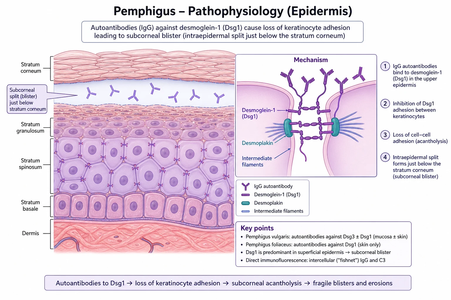
- IgG subclasses: in chronic/active disease, IgG4 is the dominant pathogenic subclass; IgG1 appears early in the disease. IgG4 has a low affinity for Fc receptors and does not fix complement well, but steric hindrance of desmosomal adhesion is the dominant mechanism.[9]
- Fogo selvagem: an environmental antigen from the saliva of the sandfly Lutzomyia longipalpis (LJM11) shares homology with the EC1/EC2 domains of Dsg1, triggering pathogenic autoantibodies in genetically susceptible individuals (HLA-DRB104 and HLA-DQB10302). This is the molecular mimicry model of endemic autoimmunity.[2][11]
- Drug-induced PF: thiol-containing drugs (penicillamine, captopril) disrupt desmosomal adhesion by biochemical (direct disruption of Dsg1) and immunological (autoantibody induction) mechanisms. Thiol drugs also activate plasminogen to plasmin, which directly cleaves Dsg1 and causes acantholysis independent of autoantibodies - hence autoantibody-negative drug-induced PF can occur.[6]
- Paraneoplastic pemphigus: the neoplasm triggers autoantibodies against both desmogleins (Dsg1 and Dsg3) and plakin-family proteins (envoplakin, periplakin, desmoplakins I/II, BP230, plectin) - the multi-antigen target profile distinguishes PNP from other pemphigus variants. Cell-mediated (CD8+ T-cell) cytotoxicity against plakin-expressing epithelia is thought to drive the lichenoid/interface dermatitis and the bronchiolitis obliterans.[5][13]
- IgA pemphigus: IgA autoantibodies target desmocollin 1 (subcorneal pustular dermatosis type) or desmocollin 1 to 3 (intraepidermal neutrophilic type), recruiting neutrophils into the epidermis via complement activation and Fc-alpha receptor signalling.[7]
Clinical features
Pemphigus foliaceus (sporadic)
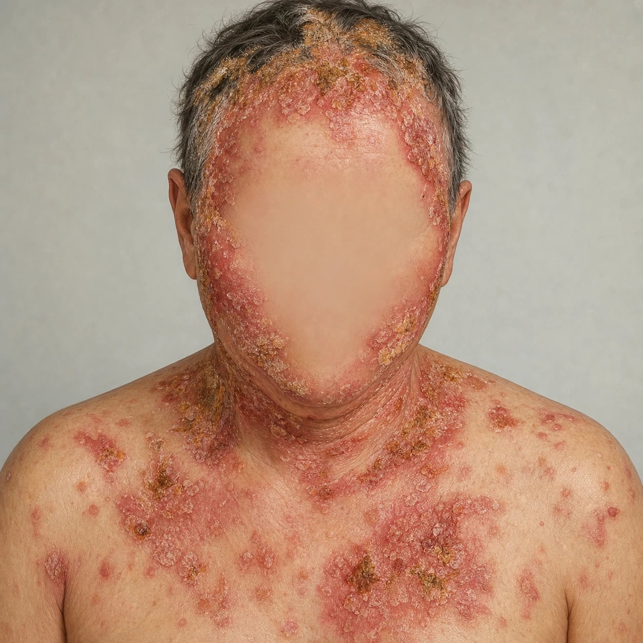
- Onset: insidious; small, flaccid, superficial blisters on apparently normal or mildly erythematous skin that rupture easily, leaving thin, scaly crusts over an erythematous base.
- Distribution: seborrhoeic / photo-exposed areas - scalp, face (especially malar), upper chest, back, and upper arms; may generalise to an exfoliative erythroderma (pemphigus foliaceus erythrodermicus) in severe cases.
- Nikolsky sign: positive (both sliding and direct pressure), as in all pemphigus variants.
- Mucosa: characteristically spared (key distinguishing feature from PV); if oral lesions are present, consider PV or PNP.
- Symptoms: mild burning or pruritus; the condition is generally less severe than PV, though extensive involvement can cause fluid/electrolyte disturbance similar to a burn.[6][9]
Variants
- Pemphigus erythematosus (Senear-Usher syndrome): a localised, photosensitive variant with butterfly (malar) facial distribution resembling lupus erythematosus, with scaly, crusted, hyperkeratotic plaques on the nose, cheeks, scalp, and chest; often associated with positive ANA and anti-Ro/SSA; responds well to hydroxychloroquine and sun protection.[6]
- Drug-induced PF: clinically and immunologically indistinguishable from sporadic PF; penicillamine (up to 7% of RA patients), captopril, and other thiol drugs (enalapril, rifampicin, cephalosporins); withdrawal of the offending drug may induce remission.[6]
- Pemphigus herpetiformis: a pruritic, annular, vesicular variant mimicking dermatitis herpetiformis; anti-Dsg1 ± anti-Dsg3; dapsone-responsive; the most steroid-sparing-responsive PF variant.[6]
Paraneoplastic pemphigus (PNP/PAMS)
- Hallmark: painful, multifocal, refractory stomatitis affecting the lips (with characteristic haemorrhagic crusting), buccal mucosa, tongue, oropharynx, and often extending to the oesophagus and conjunctiva; the stomatitis is usually the first and most prominent manifestation and is notoriously refractory to immunosuppression.[5][15]
- Cutaneous rash: polymorphous - may be bullous, lichenoid (violaceous plaques), erythema-multiforme-like (targetoid), morbilliform, or graft-versus-host-disease-like; palms, soles, and periungual areas may be involved; the lichenoid variant is the most common.
- Underlying neoplasm: classically non-Hodgkin lymphoma (NHL), chronic lymphocytic leukaemia (CLL), Castleman disease (especially in young Asian patients), Waldenström macroglobulinaemia, thymoma, and sarcomas (including retroperitoneal sarcoma); the neoplasm may precede, coincide with, or follow the mucocutaneous eruption by months to years.[5][6][13]
- Bronchiolitis obliterans: a life-threatening complication unique to PNP - progressive, irreversible small-airway obstruction; the leading cause of death; often unresponsive to immunosuppression. PFTs (FEV1, FVC, flow-volume loop with an obstructive pattern, decreased forced expiratory flow) should be performed at diagnosis and at 3-month intervals.[5][13]
PNP neoplasm associations
CASTLE
Hyaline-vascular type, especially in young Asian patients; PNP may be the presenting feature
Rarely associated; always consider if no lymphoproliferative source found
Retroperitoneal sarcoma, dendritic cell sarcoma, others
May precede or coincide with PNP; screen with chest CT
Non-Hodgkin B-cell lymphoma (most common); CLL, Waldenström macroglobulinaemia
Castleman + Lymphoma + Thymoma + Sarcoma account for >90%; the rest are exceptional
IgA pemphigus
- Two clinical subtypes: subcorneal pustular dermatosis (Sneddon-Wilkinson) type - annular/polycyclic plaques with peripheral pustules and central clearing on the trunk/intertriginous areas; and intraepidermal neutrophilic (IEN) type - sunflower-like configuration of pustules. Mucosal involvement is uncommon.[7]
- Associations: rare association with IgA gammopathy, HIV, and inflammatory bowel disease; consider screening in refractory cases.[7]
Histopathology
- PF: subcorneal or subgranular acantholysis - a superficial intraepidermal split just beneath the stratum corneum with detached, acantholytic keratinocytes in the blister cavity (may resemble a "row of tombstones" if basal layer remains attached); often mild or absent dermal infiltrate. Eosinophilic spongiosis may be an early clue.
- PNP: suprabasal acantholysis (as in PV) plus keratinocyte necrosis and a lichenoid/interface dermatitis with basal cell vacuolar change and dyskeratotic cells - the combination of acantholysis and lichenoid change is characteristic of PNP and the histological clue that should prompt the PNP workup.
- IgA pemphigus: intraepidermal neutrophilic pustules (subcorneal or mid-spinous) with minimal acantholysis; direct staining for IgA.[4]
Histology - row of tombstones
TOMBSTONES
Basal cells remain attached to BMZ like tombstones in the churchyard of an intraepidermal cemetery
Flaccid roof ruptures easily; only erosions and crusts visible clinically
Mucosa spared in PF (compensation); involved in PV/PNP
Subcorneal split in PF; suprabasal split in PV
Most superficial split in the pemphigus family
Acantholytic rounded keratinocytes on Tzanck smear
Characteristically spared in PF - if oral involvement, consider PV or PNP
Lateral pressure detaches epidermis
Early clue on biopsy
Variant of subcorneal split
Investigations
Direct immunofluorescence (DIF)
- PF: intercellular (fishnet) IgG deposition in the upper epidermis only - distinguishing it from PV where staining is throughout the full epidermal thickness (PV targets both Dsg1 and Dsg3). C3 may also be present.
- PNP: intercellular IgG/C3 plus complement deposition along the basement membrane zone (dual pattern: intercellular + BMZ) - highly characteristic.
- IgA pemphigus: intercellular IgA deposition in the superficial epidermis (subcorneal type) or throughout (IEN type).[4]
Serology - ELISA and immunoblot
- Anti-Dsg1 ELISA: positive in PF (the target antigen); anti-Dsg3 ELISA negative (distinguishes from PV). ELISA titres correlate with disease activity and can be used to titrate therapy.[4]
- PNP: positive for anti-Dsg1 AND anti-Dsg3, plus anti-envoplakin and anti-periplakin (confirmed by immunoblot or ELISA); rat bladder epithelium (RBE) IIF is the most sensitive single screening test for PNP (>75% sensitivity, >95% specificity) because rat bladder transitional epithelium expresses abundant plakin proteins, while monkey oesophagus IIF detects desmoglein antibodies but may miss plakin-only cases.[4][5][13]
- IgA pemphigus: anti-desmocollin 1 ELISA (research) or indirect immunofluorescence on IgA substrate.[7]
Additional workup
- Skin biopsy (shave or punch) for H&E and perilesional skin for DIF.
- For suspected PNP: whole-body CT/PET-CT to identify the underlying neoplasm, lymph node biopsy, serum protein electrophoresis and immunofixation (for IgA gammopathy and monoclonal proteins), flow cytometry (for CLL/NHL), and pulmonary function tests (PFTs) to screen for bronchiolitis obliterans.[5][13]
- For fogo selvagem: vector exposure history (sandfly, black fly), travel/residence in endemic Brazilian regions.[1][2]
Diagnostic-test sensitivity at a glance
Differential diagnosis
The differential is wide because the clinical appearance mimics eczema, infection, or other autoimmune blistering disease. The diagnostic triad (H&E + DIF + ELISA) resolves almost every case.[4]
- Seborrhoeic dermatitis: fine, greasy scales on scalp/face/chest without crusting or blistering; negative Nikolsky; DIF negative.
- Impetigo: honey-coloured crusts, positive bacterial culture (S. aureus / GAS); responds to antibiotics. Bullous impetigo is mediated by staphylococcal exfoliative toxins (ETA/ETB) that cleave Dsg1 - clinically indistinguishable from early PF, but DIF is negative in bullous impetigo (no autoantibody).
- Subcorneal pustular dermatosis (Sneddon-Wilkinson disease): resembles IgA pemphigus subcorneal type; DIF distinguishes (IgA pemphigus has intercellular IgA; Sneddon-Wilkinson is DIF-negative).
- Pemphigus vulgaris: mucosal involvement is the key discriminator; suprabasal split on histology; anti-Dsg3 positive.
- Bullous pemphigoid: tense bullae (not flaccid), elderly patients, subepidermal split, linear IgG/C3 along BMZ on DIF.
- Erythema multiforme / Stevens-Johnson syndrome: targetoid lesions, drug/infection trigger, full-thickness epidermal necrosis; for PNP, the lichenoid/targetoid rash may mimic EM/SJS, but the persistent severe stomatitis and the underlying neoplasm are clues.
- Acute graft-versus-host disease: post-HSCT timing distinguishes; PNP histology is similar (lichenoid interface) but HSCT history is absent.
- Lupus erythematosus: for pemphigus erythematosus (Senear-Usher); ANA, anti-dsDNA, and histopathology distinguish.[6][4]
Clinical and bedside assessment
PF cannot be diagnosed clinically, but the bedside examination localises the disease, gauges severity, and identifies complications. [1]
- Nikolsky sign: lateral pressure on perilesional skin detaches the epidermis - positive in all pemphigus variants, including PF; if negative, consider pemphigoid (subepidermal, not intraepidermal).
- Asboe-Hansen sign (bulla extension sign): pressure on top of an intact bulla extends it outward - characteristic of pemphigus and bullous pemphigoid.
- Full mucocutaneous examination is mandatory, especially in suspected PNP - oral cavity (lips, buccal mucosa, palate, gingiva, tongue, oropharynx), oesophageal (dysphagia, odynophagia), conjunctival (ophthalmology referral if involvement), genital (vulvar, penile), nasal (epistaxis, crusting), and anal (erosions, pain). The stomatitis of PNP characteristically has haemorrhagic lip crusting and persistent, painful erosions of the entire oral mucosa, often extending to involve the vermillion border.[15]
- Severity scoring: PDAI (Pemphigus Disease Area Index) for skin activity, skin damage, mucosal activity, and mucosal damage; ABSIS (Autoimmune Bullous Skin Disorder Intensity Score) as an alternative.
- PFTs and flow-volume loop are essential at every PNP visit - early inspiratory crackles, reduced FEV1/FVC, and the "notched" or "M-shaped" expiratory flow-volume loop signal bronchiolitis obliterans before the patient is symptomatic.
Management
The management strategy is dictated by disease severity (PF) and the underlying neoplasm (PNP). EADV 2020 S2K guidelines govern PF; EADV 2023 S2K guidelines govern PNP.[12][13]
Pemphigus foliaceus
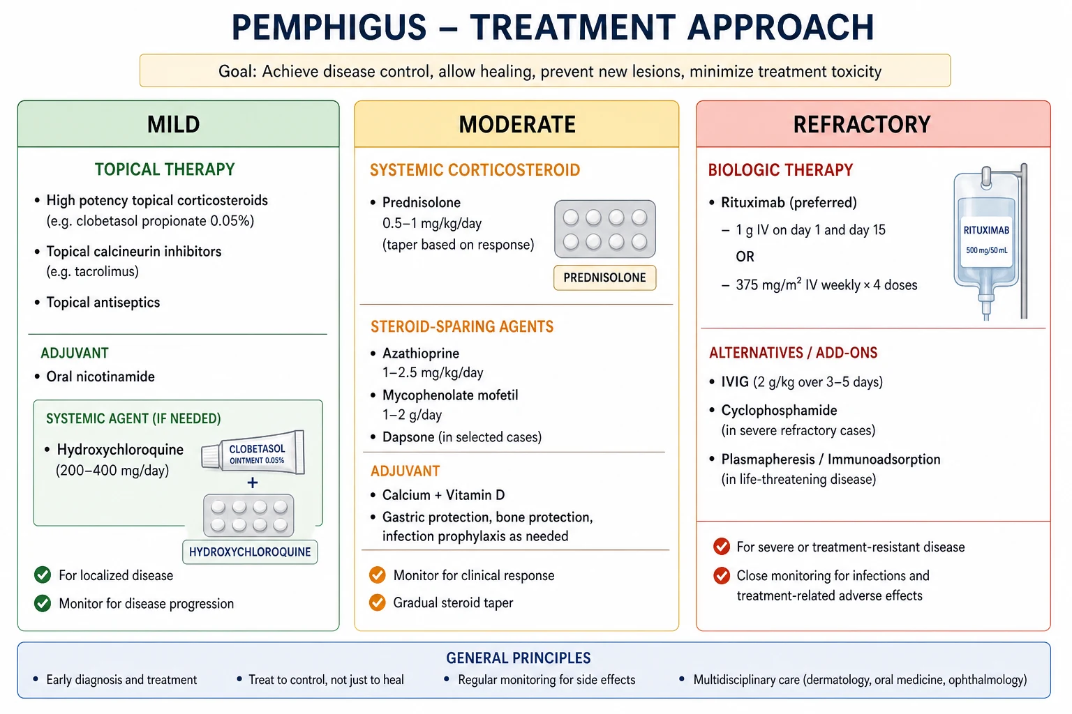
- Mild / localised disease: superpotent topical corticosteroids (clobetasol propionate 0.05%) to affected areas; hydroxychloroquine 200 to 400 mg daily (especially for pemphigus erythematosus and photosensitive variants); sun protection.[12]
- Moderate disease: oral prednisolone 0.5 to 1 mg/kg/day (lower doses than PV) in a tapering regimen; combine with steroid-sparing agents - hydroxychloroquine, nicotinamide (niacinamide) 500 mg TDS + tetracycline/doxycycline, or dapsone (especially if neutrophilic component).[6][12]
- Severe / refractory disease: rituximab (anti-CD20 monoclonal antibody) 1 g IV on day 1 and day 15 - highly effective, now first-line biologic for refractory pemphigus following the RITUX 3 trial (Werth et al., NEJM 2021, PMID 34097368); consider mycophenolate mofetil 2-3 g/day or azathioprine 2-2.5 mg/kg/day (after TPMT testing) as steroid-sparing agents.[9][10][12][14]
- Drug-induced PF: withdraw the offending drug (penicillamine, captopril); spontaneous improvement often follows within 4 to 12 weeks. Refractory cases are managed as sporadic PF.[6]
- Endemic fogo selvagem: WHO multidrug therapy regimens adapted from leprosy (prednisolone + azathioprine or MMF); rituximab for severe/refractory cases.[1]
Paraneoplastic pemphigus (EADV 2023 S2K)
- Treat the underlying neoplasm first: surgical resection (Castleman disease, thymoma) or chemotherapy / rituximab-based regimens (NHL, CLL) is the most effective approach - removing the antigen source can induce remission. Unicentric Castleman disease cured by surgery can produce full PNP remission.[5][13]
- Mucocutaneous disease: notoriously refractory to conventional immunosuppression; combinations of rituximab, cyclophosphamide, IVIG, and high-dose corticosteroids are used; rituximab shows the best evidence for disease control.
- Bronchiolitis obliterans: progressive and often fatal; azithromycin (immunomodulatory at sub-antibiotic doses), montelukast, and lung transplantation have been attempted with limited success. Early high-dose corticosteroids and avoidance of further lung injury (infection, aspiration) are the mainstays.[5][13]
- Supportive care: aggressive analgesia (lidocaine viscous mouthwash, opioid analgesics), nasogastric feeding for severe oral/oesophageal involvement, intensive wound care, fluid/electrolyte management, infection surveillance, and DVT prophylaxis.
IgA pemphigus
- Dapsone (50 to 150 mg daily) is first-line - check G6PD before initiation (screen all patients; haemolysis in G6PD deficiency can be fatal); monitor for haemolysis, methaemoglobinaemia, agranulocytosis, motor neuropathy, and the dapsone hypersensitivity syndrome (DRESS-like).[7]
- Alternatives: acitretin/isotretinoin, colchicine, nicotinamide + tetracycline, or rituximab for refractory cases.[7]
Drug dose table (PF / PNP / IgA pemphigus)
Prognosis
- PF: generally better prognosis than PV; mortality <5% with modern therapy; drug-induced PF has the best prognosis (remission on drug withdrawal); fogo selvagem has a chronic, relapsing course but responds to MDT. Most patients achieve complete remission off therapy (CRoff) within 2 to 5 years of rituximab-based regimens.[8][12]
- PNP: poor prognosis with 75 to 90% mortality at 5 years, largely from bronchiolitis obliterans, sepsis, or the underlying malignancy; early diagnosis and treatment of the neoplasm may improve outcomes. Patients with resectable unicentric Castleman disease or thymoma have the best outcomes; those with NHL/CLL fare worse.[5][13]
- IgA pemphigus: chronic but usually benign; responds well to dapsone; spontaneous remission may occur. Rarely progresses to IgA gammopathy or myeloma.[7]
Special populations
- Pregnancy: PF may flare in the 2nd to 3rd trimester; prednisolone and hydroxychloroquine are first-line; azathioprine is acceptable (after TPMT testing). Rituximab should be avoided (B-cell depletion in the neonate; neonatal pemphigus possible from transplacental IgG4). Dapsone is contraindicated (neonatal haemolysis, methaemoglobinaemia). Monitor anti-Dsg1 titres monthly.[12]
- Elderly: rituximab preferred over high-dose steroids when possible; infection surveillance; falls and bone health on steroids (vitamin D, calcium, bisphosphonate if prolonged steroids).
- Children: rare; rituximab used off-label; growth considerations with steroids; consider fogo selvagem in children from endemic Brazilian regions (children and young adults predominantly affected).[1]
- Immunosuppressed patients (HIV, transplant): rituximab acceptable but cautious; infection risk compounded; coordinate with HIV/transplant physician.
Complications and pitfalls
- Complications of PF: secondary bacterial infection (impetigo, cellulitis, sepsis), dehydration, electrolyte disturbance, weight loss, exfoliative erythroderma, progression to PV (rare, by epitope spreading).
- Complications of systemic corticosteroids: diabetes, hypertension, osteoporosis, adrenal suppression, Cushingoid features, mood disturbance, infection, avascular necrosis.
- Complications of rituximab: infusion reactions, hepatitis B reactivation (fatal if untreated), progressive multifocal leukoencephalopathy (PML, rare), hypogammaglobulinaemia, late-onset neutropenia, PJP pneumonia.
- Complications of dapsone: haemolysis in G6PD deficiency (screen first), methaemoglobinaemia, agranulocytosis, motor neuropathy, dapsone hypersensitivity syndrome.
- Complications of PNP: bronchiolitis obliterans (the most lethal, unique to PNP), severe oral pain interfering with eating, opportunistic infection from immunosuppression, sepsis, mortality from the underlying neoplasm.
- Diagnostic pitfalls: missing mucosal involvement in PV, missing PNP in a patient labelled "erythema multiforme", misdiagnosing PF as seborrhoeic dermatitis or impetigo, missing fogo selvagem in a returned traveller, not testing G6PD before dapsone.
- Management pitfalls: abrupt steroid taper (relapse), missing hepatitis B screening before rituximab (fatal reactivation), stopping immunosuppression during infection (paradoxical flare), missing bronchiolitis obliterans screening in PNP (PFTs every 3 months). [1]
Evidence, guidelines and controversies
- EADV 2020 S2K guidelines on PV and PF (Joly et al., JEADV 2020, PMID 32830877): rituximab plus short-course prednisone is the first-line option for moderate-to-severe PV; rituximab principles extend to refractory PF.[12]
- EADV 2023 S2K guidelines on PNP/PAMS (Antiga et al., JEADV 2023, PMID 36965110): the first disease-specific PNP guideline; treatment of the underlying neoplasm is the cornerstone; rituximab, IVIG, and cyclophosphamide combinations for mucocutaneous disease; bronchiolitis obliterans screening at diagnosis and every 3 months.[13]
- RITUX 3 trial (Werth et al., NEJM 2021, PMID 34097368): rituximab + prednisone superior to mycophenolate mofetil + prednisone for complete remission off therapy at 36 months in PV; principles extend to refractory PF and PNP.[14]
- British Association of Dermatologists (BAD) pemphigus guideline: concordant with EADV.
- IADVL (India) consensus: pemphigus variants (PV > PF) more common than in the West; resource-limited DIF/ELISA access; rituximab gaining traction.
- Brazilian and South-American guidelines: endemic fogo selvagem in the Amazon, Central-West and Goias states; vector control and WHO multidrug regimens adapted from leprosy.
- Open controversies: rituximab protocol (RA vs lymphoma), role of efgartigimod (anti-FcRn) and other emerging biologics, when to use cyclophosphamide vs rituximab in PNP, role of IVIG as monotherapy, screening for and treatment of bronchiolitis obliterans, role of dapsone vs rituximab first-line for PF, optimal duration of steroid-sparing therapy in fogo selvagem.
Exam pearls
[1]Red flags
Exam application bank (NEET-PG / INICET)
One-line answer
Pemphigus foliaceus is a superficial autoimmune blistering disease from IgG anti-desmoglein 1 antibodies producing subcorneal/subgranular acantholysis, scaly crusted plaques, and (usually) no mucosal involvement; variants include endemic fogo selvagem and pemphigus erythematosus. Paraneoplastic pemphigus is a distinct, often fatal entity of multifocal refractory stomatitis and polymorphous rash with antibodies to desmogleins and plakin family proteins (envoplakin, periplakin) and an underlying neoplasm, classically lymphoproliferative. IgA pemphigus is a neutrophilic variant with IgA anti-desmocollin. Fellowship-level assessment demands mastery of the superficial split, DIF/ELISA patterns, drug-induced and endemic triggers, and the recognition and workup of paraneoplastic pemphigus.
Worked stems (answer without another resource)
Stem 1 — Classic presentation. Map symptoms to mechanism; name the first investigation and first treatment step with dose/route if drug therapy is standard. [1]
Stem 2 — Unstable / complicated. List red flags that force immediate resuscitation, theatre, ICU, antidote, or reperfusion — and what you do in the first 15 minutes. [1]
Stem 3 — Atypical group. Elderly, pregnancy, child, or immunocompromised: how presentation and thresholds change. [1]
Stem 4 — Differential trap. Name the three closest mimics and one discriminator for each. [1]
Stem 5 — Disposition. Who goes home with safety-netting, who is admitted, who needs HDU/ICU/theatre, and what follow-up is mandatory. [1]
Rapid viva checklist
- Definition + classification
- Pathophysiology chain
- Bedside signs / criteria
- Score with exact components (if any)
- Emergency bundle
- Definitive therapy with doses
- Complications of disease and of treatment
- Special populations
- Guideline/trial name if classic
- Three exam traps
Coverage self-check
If you cannot answer any stem above from this page alone, re-read the matching section — the page is intended to be self-sufficient for final-prof and NEET-PG/INICET questions on Pemphigus foliaceus and paraneoplastic pemphigus.
Expanded exam teaching (depth pass)
Clinical reasoning
For Pemphigus foliaceus and paraneoplastic pemphigus, examiners test whether you can prioritise life threats, choose the right first test, and give specific therapy (agent, dose, route, timing). Generic phrases without numbers score poorly.
Mechanism → feature map
Build a short chain: cause → pathophysiologic intermediate → clinical feature → complication. Every major symptom in the classic vignette should sit on that chain.
Investigation strategy
- Bedside/first-line tests that change immediate management
- Confirmatory or staging tests
- What a normal result does not exclude
- When not to delay treatment for imaging (unstable patient)
Management ladder
- Resuscitation / ABC / sepsis or haemorrhage bundle as relevant
- Specific antidote / procedure / antimicrobial / reperfusion / surgery
- Supportive care and monitoring targets
- Definitive long-term therapy and secondary prevention
- Disposition and safety-net advice
Special populations
Always prepare one line each for children, pregnancy, elderly, renal/hepatic impairment, and immunocompromised patients when the topic allows.
Pitfalls that fail candidates
- Treating the number not the patient
- Missing pregnancy status when relevant
- Imaging before stabilisation
- Wrong empiric cover or wrong antidote timing
- Incomplete counselling on recurrence, adherence, or red-flag return
Pemphigus foliaceus is a superficial autoimmune blistering disease from IgG anti-desmoglein 1 antibodies producing subcorneal/subgranular acantholysis, scaly crusted plaques, and (usually) no mucosal involvement; variants include endemic fogo selvagem and pemphigus erythematosus. Paraneoplastic pemphigus is a distinct, often fatal entity of multifocal refractory stomatitis and polymorphous rash with antibodies to desmogleins and plakin family proteins (envoplakin, periplakin) and an underlying n [1]
[1]References
- [1]Hans-Filho G, Aoki V, Bittner NRH, et al. Fogo selvagem: endemic pemphigus foliaceus An Bras Dermatol, 2018.PMID 30156612
- [2]Aoki V, Rivitti EA, Diaz LA. Update on fogo selvagem, an endemic form of pemphigus foliaceus J Dermatol, 2015.PMID 25558948
- [3]Diaz LA, Sampaio SA, Rivitti EA, et al. Endemic pemphigus foliaceus (Fogo Selvagem): II. Current and historic epidemiologic studies J Invest Dermatol, 1989.PMID 2642512
- [4]van Beek N, Holtsche MM, Atefi I, et al. State-of-the-art diagnosis of autoimmune blistering diseases Front Immunol, 2024.PMID 38903493
- [5]Anderson HJ, Huang S, Lee JB. Paraneoplastic pemphigus/paraneoplastic autoimmune multiorgan syndrome: Part I. Clinical overview and pathophysiology J Am Acad Dermatol, 2024.PMID 37597771
- [6]Malik AM, Tupchong S, Huang S, et al. An Updated Review of Pemphigus Diseases Medicina (Kaunas), 2021.PMID 34684117
- [7]Kridin K, Patel PM, Jones VA, et al. IgA pemphigus: A systematic review J Am Acad Dermatol, 2020.PMID 31812619
- [8]Kridin K. Pemphigus group: overview, epidemiology, mortality, and comorbidities Immunol Res, 2018.PMID 29479654
- [9]Kasperkiewicz M, Ellebrecht CT, Takahashi H, et al. Pemphigus Nat Rev Dis Primers, 2017.PMID 28492232
- [10]Schmidt E, Kasperkiewicz M, Joly P. Pemphigus Lancet, 2019.PMID 31498102
- [11]Culton DA, Qian Y, Li N, et al. Advances in pemphigus and its endemic pemphigus foliaceus (Fogo Selvagem) phenotype: a paradigm of human autoimmunity J Autoimmun, 2008.PMID 18838249
- [12]Joly P, Horvath B, Patsatsi A, et al. Updated S2K guidelines on the management of pemphigus vulgaris and foliaceus initiated by the european academy of dermatology and venereology (EADV) J Eur Acad Dermatol Venereol, 2020.PMID 32830877
- [13]Antiga E, Bech R, Maglie R, et al. S2k guidelines on the management of paraneoplastic pemphigus/paraneoplastic autoimmune multiorgan syndrome initiated by the European Academy of Dermatology and Venereology (EADV) J Eur Acad Dermatol Venereol, 2023.PMID 36965110
- [14]Werth VP, Joly P, Mimouni D, et al. Rituximab versus Mycophenolate Mofetil in Patients with Pemphigus Vulgaris N Engl J Med, 2021.PMID 34097368
- [15]Rashid H, Lamberts A, Diercks GFH, et al. Oral Lesions in Autoimmune Bullous Diseases: An Overview of Clinical Characteristics and Diagnostic Algorithm Am J Clin Dermatol, 2019.PMID 31313078