Dermatology · Medicine
Pemphigus vulgaris
Also known as Pemphigus vulgaris · Pemphigus · PV
Pemphigus vulgaris is a potentially life-threatening autoimmune mucocutaneous blistering disease caused by pathogenic IgG4 autoantibodies against desmoglein 3 (Dsg3) ± desmoglein 1 (Dsg1), producing loss of keratinocyte adhesion (acantholysis) and flaccid blisters/erosions. Mucous membranes are frequently involved (oral, pharyngeal, oesophageal, conjunctival, genital). Diagnosis rests on the triad of histology (suprabasal acantholysis with tombstoning), direct immunofluorescence (intercellular IgG4/C3 in a chicken-wire pattern — pathognomonic), and serology (indirect immunofluorescence on monkey oesophagus plus anti-Dsg3/Dsg1 ELISA titres, which track disease activity). First-line management now combines systemic corticosteroids with rituximab (RITUX 3 trial, NEJM 2021) or a steroid-sparing agent (mycophenolate/azathioprine). Fellowship-level assessment demands mastery of the desmoglein compensation theory (explaining the mucosal vs mucocutaneous phenotype), the diagnostic triad with DIF, rituximab protocols and monitoring (hepatitis B reactivation, hypogammaglobulinaemia, PJP), paraneoplastic pemphigus (associated lymphoid malignancy/Castleman/thymoma), and the anti-Dsg ELISA as a disease-activity biomarker.
On this page & tools
Your progress
Saved locally on this device.
Exam tags
Red flags
Definition & Classification
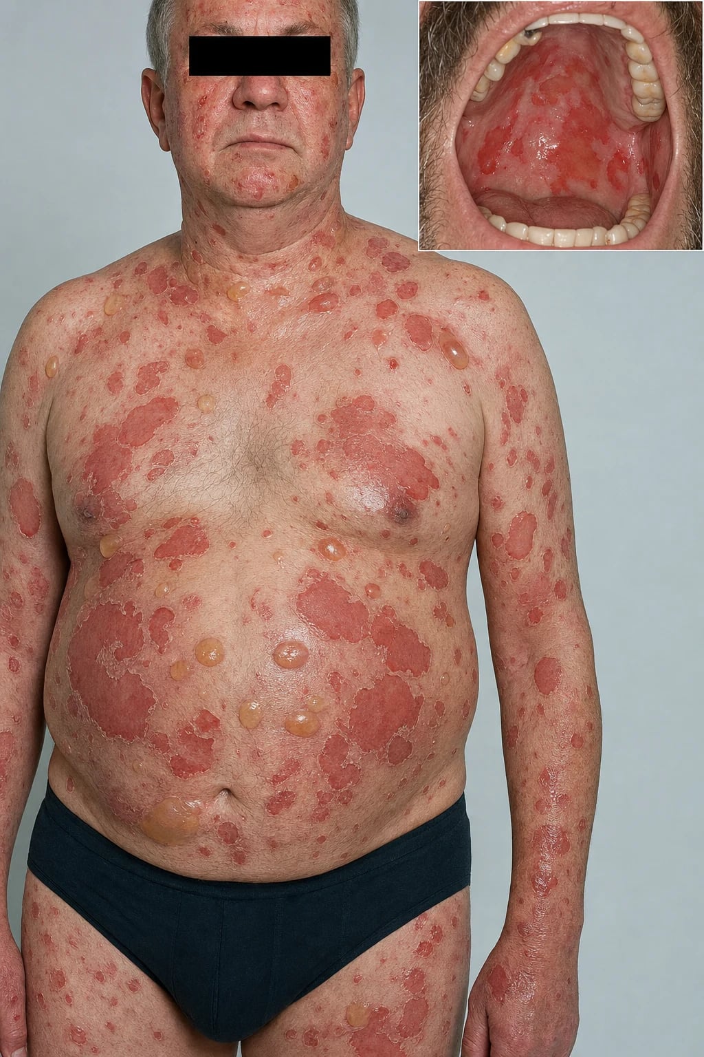
Pemphigus vulgaris (PV) is a potentially life-threatening autoimmune mucocutaneous blistering disease caused by pathogenic IgG4 autoantibodies against desmoglein 3 (Dsg3) ± desmoglein 1 (Dsg1), which disrupt keratinocyte adhesion and produce acantholysis (loss of intercellular adhesion) and flaccid blisters.[1][2]
Pemphigus subtypes (distinguished by antibody profile and phenotype):[2]
| Subtype | Antibody target | Phenotype |
|---|---|---|
| Pemphigus vulgaris — mucosal-dominant | Anti-Dsg3 only | Mucosal erosions ± minimal skin |
| Pemphigus vulgaris — mucocutaneous | Anti-Dsg3 + anti-Dsg1 | Skin AND mucosa |
| Pemphigus foliaceus | Anti-Dsg1 only | Superficial skin (corneocytes), spares mucosa |
| Paraneoplastic pemphigus (PNP) | Anti-envoplakin, anti-periplakin + others | Severe mucocutaneous + polymorphous eruption; associated with malignancy |
| IgA pemphigus | IgA anti-Dsg1/Dsg3 or anti-desmocollin | Pustular, often subcorneal |
Epidemiology
- Incidence: ~0.5-10 per million per year (varies by population); age of onset 40-60; equal sex distribution overall with slight female predominance in Mediterranean and Indian cohorts (F:M ~1.3-1.5:1).[2][8]
- Ethnic predisposition: higher prevalence in Ashkenazi Jewish (~16-32 per million per year), Mediterranean (Greek, Italian, Sephardic Jewish), Middle Eastern, Indian/South Asian and Japanese populations. Driven by HLA class II susceptibility alleles, in particular HLA-DRB1*04:02 (odds ratio ~10.8), DRB1*14:01, DRB1*14:04, DQB1*05:03 and DQB1*03:02, which restrict presentation of Dsg3 peptides to autoreactive CD4+ T cells.[2][3]
- Environmental triggers: drugs containing a thiol group (D-penicillamine, captopril, tiopronin) and other thiol-/thio-disrupting agents, checkpoint inhibitors (anti-PD-1/PD-L1 — pembrolizumab, nivolumab), and (less robustly) seasonal UV exposure, dental procedures and emotional stress.
- Associations: other autoimmune diseases (myasthenia gravis, autoimmune thyroid disease, type 1 diabetes, pernicious anaemia, rheumatoid arthritis, vitiligo, alopecia areata); thymoma (paraneoplastic link and overlap with myasthenia gravis).[8]
- Mortality trends: pre-corticosteroid mortality was ~75% within 1 year; with corticosteroid-sparing immunosuppression it fell to ~25-30%; with rituximab-era protocols (post-2018), disease-specific mortality is under 5% at 5 years in most contemporary cohorts, with infection (bacterial sepsis, PJP during combination immunosuppression) replacing disease activity as the leading cause of death.
Pemphigus Variants
The "pemphigus" family encompasses several clinicopathologically distinct entities that share IgG- or IgA-driven acantholysis but differ in antigen, phenotype, prognosis and treatment. Recognising the variant changes everything — for example, an isolated oral eruption in an older patient with lymphadenopathy must trigger a paraneoplastic work-up, and a verrucous flexural plaque in a young adult points to pemphigus vegetans rather than Hailey-Hailey disease.[2][10]
Pemphigus vegetans (Pveg)
Pemphigus vegetans is the chronic, hypertrophic variant of PV (~1-2% of all pemphigus). It carries the same anti-Dsg3 IgG4 (often with anti-Dsg1) antibody profile as mucosal-dominant PV but produces exuberant verrucous, vegetative plaques in flexural and intertriginous sites (axillae, groin, inframammary folds, perineum, scalp, face). Two clinical patterns are recognised: the Neumann type (begins as flaccid bullae that evolve into vegetative erosions) and the Hallopeau type (begins as pustules that evolve into verrucous plaques — historically the more benign). Oral involvement (cheek, tongue, lip) with cerebriform tongue (plicate tongue) is characteristic and may precede skin lesions by months. Histology shows suprabasal acantholysis plus prominent epidermal hyperplasia, hyperkeratosis and intraepithelial eosinophilic microabscesses. DIF is identical to PV (intercellular IgG4 chicken-wire). Treatment mirrors PV — systemic steroids plus rituximab — though vegetans can be more refractory to rituximab monotherapy and often needs adjunctive dapsone or immunosuppression. Prognosis is similar to classical PV once remission is induced.[2][3]
Paraneoplastic pemphigus (PNP) / paraneoplastic autoimmune multiorgan syndrome (PAMS)
PNP is a distinct, often lethal paraneoplastic autoimmune mucocutaneous disease triggered by an underlying neoplasm — most commonly non-Hodgkin B-cell lymphoma, Castleman disease, thymoma, follicular dendritic cell sarcoma and (rarely) CLL or solid tumours. The autoantibody profile is broader than PV: anti-plakin antibodies (envoplakin, periplakin, desmoplakin I/II, BP230, plectin) plus anti-Dsg3 and (less often) anti-Dsg1. The humoral and cellular immune response (CD8+ cytotoxic T cells, type II/III IFN signature) drives multi-organ autoimmunity — not just skin and mucosa but also bronchiolitis obliterans (BO), which is the leading cause of death. Clinically, PNP presents with severe, painful, often haemorrhagic stomatitis (the dominant clue; affects lips, tongue, buccal mucosa), a polymorphous cutaneous eruption (lichenoid, erythema multiforme-like, bullous, erosive), and pseudomembranous conjunctivitis with progressive scarring. DIF shows intercellular IgG AND linear BMZ deposition ("dual pattern") in 50-60% — this combination is highly suggestive. Serology by immunoblotting identifies anti-plakin antibodies; rat bladder IIF is the most specific substrate (intercellular staining of transitional epithelium). Treatment is treatment of the underlying malignancy first — resection of Castleman tumour or thymoma, chemotherapy for lymphoma — combined with high-dose steroids, rituximab, IVIG, and supportive care for BO (often needs lung transplantation). Mortality is high (~75-90% in BO-associated disease); early recognition is critical.[10][11]
IgA pemphigus
IgA pemphigus is a rare (under 5% of pemphigus cases), IgA-mediated neutrophilic/subcorneal acantholytic disease with two main subtypes: subcorneal pustular dermatosis (SPD) type (anti-desmocollin 1 IgA) and intraepidermal neutrophilic (IEN) type (anti-Dsg1 or anti-Dsg3 IgA). It presents with flaccid vesicopustules or annular pustular plaques with a characteristic "sunflower" or "flower-petal" configuration, often on the trunk, proximal limbs and intertriginous areas. Mucosa is usually spared. Histology shows subcorneal or intraepidermal pustules filled with neutrophils (and occasional eosinophils), with minimal acantholysis. DIF is the diagnostic clincher — intercellular IgA deposition (chicken-wire) in the upper epidermis or throughout; IgG is absent or weak. The disease course is chronic but relatively indolent; first-line is dapsone 50-150 mg/day (with G6PD screening), followed by retinoids, colchicine, low-dose steroids or mycophenolate. Rituximab has been used in refractory IgA pemphigus but the evidence base is limited to case series.[2][3]
Drug-induced pemphigus
Drug-induced PV is most commonly triggered by thiol-containing drugs that interact with desmoglein cysteines and disrupt desmosomal adhesion. The leading culprits are D-penicillamine (~7% of long-term users develop pemphigus — most often foliaceus phenotype), captopril and other ACE inhibitors with a sulfhydryl group, and tiopronin. Non-thiol triggers (less common) include rituximab-induced pemphigus (paradoxical), nivolumab/pembrolizumab (checkpoint-inhibitor pemphigus), penicillins, nifedipine, phenobarbital, and (rarely) interferon-α and piroxicam. Onset ranges from weeks to years after drug initiation. Clinically, drug-induced pemphigus more often resembles pemphigus foliaceus (superficial, scaly erosions) than PV, but mucosal and full PV phenotype is well described, especially with penicillamine. Management: withdraw the offending drug (resolves ~50% within weeks to months), then treat as for the idiopathic counterpart. Anti-Dsg antibodies may remain positive for months after resolution.[2][13]
VEGAN — pemphigus variants
Verrucous flexural plaques; anti-Dsg3 + IgG4; same DIF as PV; rituximab + steroids
Anti-plakin antibodies (envoplakin, periplakin, desmoplakin, BP230); Castleman, lymphoma, thymoma; bronchiolitis obliterans is lethal
IgA anti-desmocollin 1 (SPD) or anti-Dsg1/Dsg3 (IEN); vesicopustules sunflower pattern; dapsone first-line
D-penicillamine, captopril, tiopronin; often PF-like phenotype; withdraw drug + treat as idiopathic
CT chest/abdomen/pelvis + lymph node exam; serum protein electrophoresis; treat underlying malignancy first
Pemphigus vegetans (Pveg)
- Anti-Dsg3 ± Dsg1 (same as PV); flexural verrucous plaques; cerebriform tongue; suprabasal acantholysis + eosinophilic microabscesses
- DIF: intercellular IgG4 chicken-wire (identical to PV)
- Treatment: systemic steroids + rituximab (same as PV); can be more refractory; dapsone adjunct
Paraneoplastic pemphigus (PNP/PAMS)
- Anti-plakin family (envoplakin, periplakin) + Dsg3; associated with lymphoma, Castleman, thymoma
- Severe haemorrhagic stomatitis + polymorphous skin eruption + bronchiolitis obliterans (lethal)
- DIF: intercellular AND linear BMZ (dual pattern); rat bladder IIF positive; treat the tumour
IgA pemphigus
- IgA anti-desmocollin 1 (SPD type) or anti-Dsg1/Dsg3 (IEN type); vesicopustules sunflower pattern
- Subcorneal pustules with neutrophils; DIF intercellular IgA; minimal acantholysis
- Indolent course; first-line dapsone 50-150 mg/day (G6PD screen); refractory: retinoids, colchicine
Drug-induced PV
- Thiol drugs: D-penicillamine, captopril, tiopronin (also checkpoint inhibitors, rituximab rarely)
- Often PF-like phenotype; resolves in ~50% on drug withdrawal; otherwise treat as idiopathic
- Anti-Dsg antibodies can persist months after clinical resolution
Pathophysiology
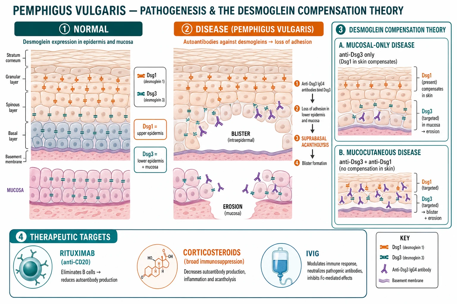
- Autoantibodies: predominantly IgG4 (pathogenic) and IgG1 against the extracellular cadherin domains of desmoglein 3 (Dsg3) and desmoglein 1 (Dsg1).[1][2]
- Acantholysis: antibody binding disrupts desmosome-mediated keratinocyte adhesion, causing cells to round up and detach — producing the characteristic intraepidermal blister.
- Desmoglein compensation theory (explains the clinical phenotype):[2]
- Dsg3 is concentrated in the lower epidermis and mucosal epithelium; Dsg1 is concentrated in the upper epidermis and skin surface.
- Mucosal-dominant PV = anti-Dsg3 only → Dsg1 in the upper skin compensates, so the skin is relatively spared.
- Mucocutaneous PV = anti-Dsg3 + anti-Dsg1 → no compensation in the skin → widespread cutaneous blistering.
- Paraneoplastic pemphigus involves additional antibodies (anti-envoplakin, anti-periplakin) and a broader, more severe phenotype driven by an underlying neoplasm.[10]
Quick numbers for the examiner
SUPER SUPERFICIAL — pemphigus vulgaris findings
Row of tombstones — basal cells attached to basement membrane but separated from upper epidermis
Bullae rupture easily leaving painful erosions with collarette of detached epidermis
Lateral pressure causes epidermal slippage; positive Asboe-Hansen sign
Oral erosions in 50-70% at presentation; may extend to pharynx, oesophagus, conjunctiva, genitalia
First-line with corticosteroids (RITUX 3 trial 2017); has transformed prognosis
Clinical Presentation [1]
- Skin: [1]
Quick numbers for the examiner
flaccid blisters on an erythematous or normal-appearing base that rupture easily, leaving painful erosions with a peripheral collarette of detached epidermis; the trunk, scalp, face and proximal limbs are common sites. Nikolsky sign positive (lateral pressure causes epidermal detachment); Asboe-Hansen sign (bulla extension with pressure) positive.[1][12]
- Mucous membranes are involved in ~50-70% of patients — often the presenting feature: oral erosions (buccal mucosa, palate, gingiva, tongue), pharyngeal, oesophageal (dysphagia, odynophagia — may need endoscopy), conjunctival, genital (vulvar, penile, anal), and nasal erosions.[9][12]
- Clinical assessment: calculate extent using the Pemphigus Disease Area Index (PDAI); examine all mucosal surfaces (oral, nasal, conjunctival, genital, rectal); test Nikolsky and Asboe-Hansen signs.
PV quick numbers
Differential Diagnosis [1]
| Mimic | Distinguishing features |
|---|---|
| Bullous pemphigoid | Tense bullae on urticarial base, older patients, subepidermal; DIF linear IgG/C3 at BMZ |
| Linear IgA bullous dermatosis | "String of pearls"; DIF linear IgA at BMZ |
| Mucous membrane pemphigoid (cicatricial pemphigoid) | Predominantly mucosal, scarring (conjunctival, oral); DIF linear at BMZ |
| Epidermolysis bullosa acquisita | Mechanobullous, DIF linear IgG at BMZ, anti-type VII collagen |
| Hailey-Hailey disease | Familial, intertriginous, "dilapidated brick wall" acantholysis; ATP2C1 mutation |
| Impetigo / bullous impetigo | Superficial, positive bacterial culture, no IgG on DIF |
| Herpes simplex / zoster | Viral cytopathic effect; DIF negative for IgG; PCR/culture |
| Paraneoplastic pemphigus | Severe polymorphous eruption + underlying malignancy; anti-envoplakin/periplakin |
| Grover disease | Itchy papulovesicles on trunk; histology (acantholysis, no IgG) |
Diagnosis — The Diagnostic Triad
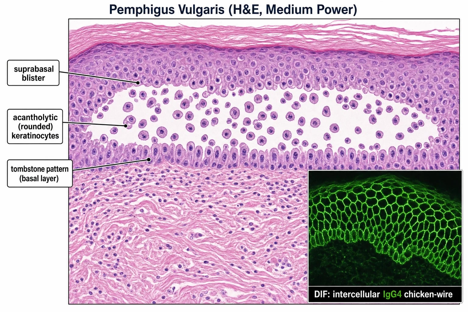
Diagnosis requires three investigations:[2][4]
- Histology (lesional biopsy): suprabasal acantholysis — a blister cavity just above the basal layer, with basal keratinocytes remaining attached to the basement membrane resembling a row of "tombstones", and rounded acantholytic keratinocytes floating in the blister cavity.
- Direct immunofluorescence (DIF) of perilesional skin — the pathognomonic finding: intercellular IgG4 (and C3) deposition throughout the epidermis in a "chicken-wire" pattern. This is the single most specific test.
- Serology:
- Indirect immunofluorescence (IIF) on monkey oesophagus substrate (high sensitivity for pemphigus).
- Anti-Dsg3 ELISA and anti-Dsg1 ELISA — positive in PV; titres correlate with disease activity and are used to monitor response to treatment.[3]
Management
First-line therapy
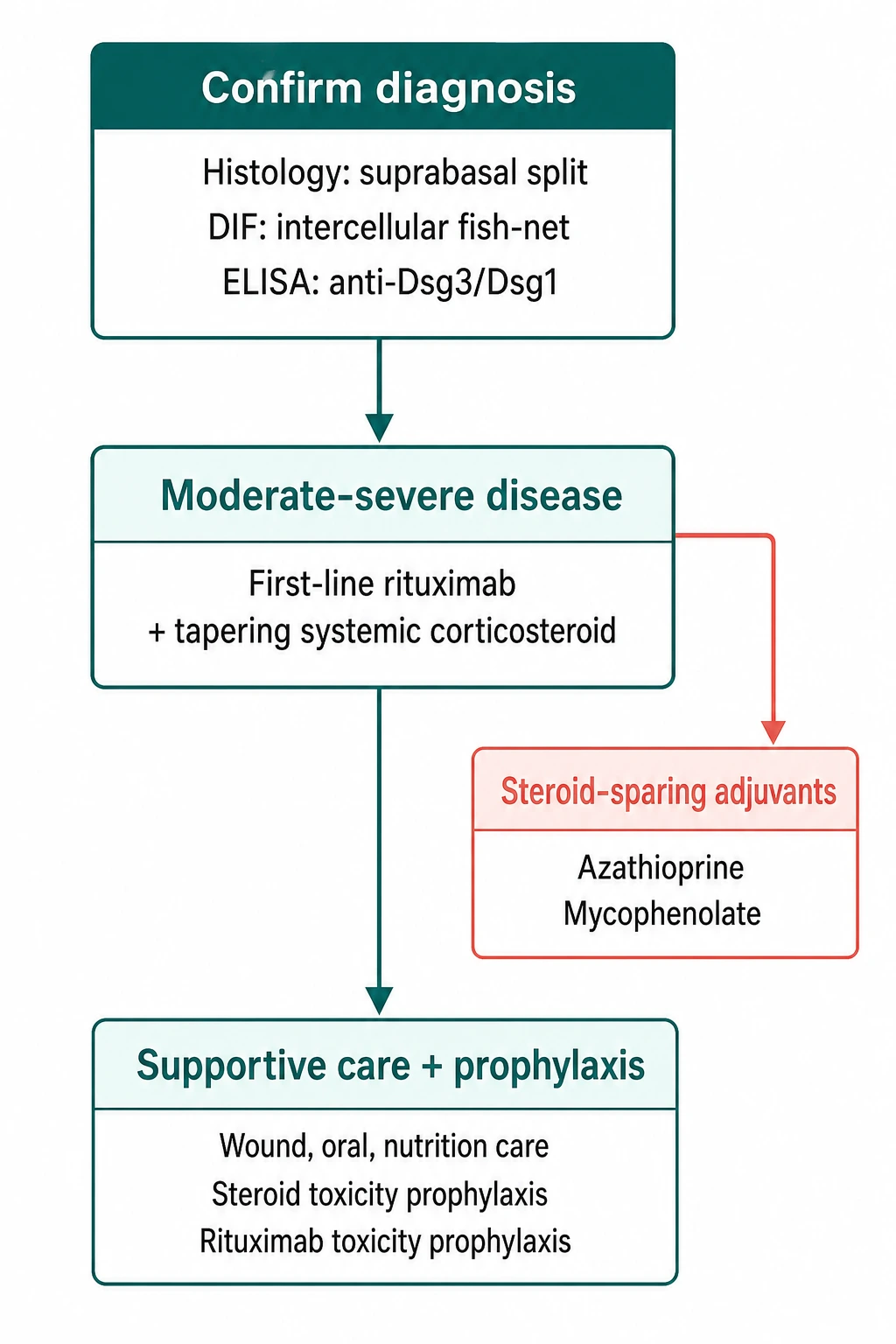
- Systemic corticosteroids: prednisolone 1-1.5 mg/kg/day (or equivalent) to induce remission; high initial doses, slow taper over months.[4]
- Steroid-sparing immunosuppressants: mycophenolate mofetil or azathioprine — used as adjuncts to reduce steroid exposure, though inferior to rituximab for inducing remission (RITUX 3).[5]
Rituximab — now first-line in many guidelines
- Rituximab (anti-CD20) depletes B cells and has transformed pemphigus outcomes. The RITUX 3 trial (NEJM 2021) showed rituximab (day 0 + day 15) was superior to mycophenolate mofetil for complete remission off therapy.[5]
- Dosing: either the lymphoma protocol (375 mg/m² weekly × 4 weeks) or the rheumatoid arthritis protocol (1 g on day 1 and day 15) — both effective in PV.[6]
- Monitoring (critical):[4]
- Hepatitis B screen before starting (rituximab causes reactivation — fatal if untreated); prophylaxis if positive.
- Serum immunoglobulins (hypogammaglobulinaemia with repeated cycles → infection risk).
- PJP prophylaxis (co-trimoxazole) especially with high-dose steroids.
- Vaccination (influenza, pneumococcal, COVID-19) before starting if possible.
- Repeat cycles may be needed for relapse; anti-Dsg3 ELISA titres guide retreatment decisions.
Refractory / escalation therapy
- Cyclophosphamide (IV pulse or oral) for refractory disease — effective but limited by toxicity (gonadal, bladder, malignancy risk).
- IVIG (2 g/kg/cycle) — rapid but temporary effect; useful as bridging or in acute severe disease.
- Immunoadsorption / plasmapheresis — removes pathogenic antibodies; rapid temporary effect.
- Emerging therapies: anti-neonatal Fc receptor (efgartigimod), BTK inhibitors, anti-BAFF (belimumab) — under investigation.[4]
Paraneoplastic pemphigus
- Suspect in new-onset pemphigus in an older patient with lymphadenopathy, organomegaly or weight loss.[10]
- Associated malignancies: lymphoid malignancies (CLL, NHL, Castleman disease), thymoma, sarcomas.
- Management: treat the underlying malignancy (the skin disease often parallels the tumour); rituximab + aggressive immunosuppression; prognosis driven by the malignancy.[10][11]
Supportive care
- Topical corticosteroids for localised or residual erosions.
- Pain management (oral anaesthetic gels, systemic analgesia).
- Nutritional support (soft/liquid diet if oral erosions severe; nasogastric if oesophageal involvement).
- Multidisciplinary team: dermatology, oral medicine, ophthalmology, gynaecology/urology, gastroenterology (oesophageal), ENT. [1]
Special Populations
- Pregnancy: pemphigus can flare; corticosteroids and azathioprine are relatively safe; rituximab is avoided (B-cell depletion in neonate); monitor anti-Dsg titres.
- Elderly / frail: rituximab preferred over high-dose steroids when possible; careful infection surveillance.
- Paraneoplastic pemphigus: as above — investigate for and treat underlying malignancy. [1]
Prognosis & Surveillance
- Before modern therapy: high mortality (infection, dehydration, sepsis).
- With rituximab: complete remission off therapy in the majority; anti-Dsg3 ELISA titres track disease activity and predict relapse.[5]
- Mortality now driven mainly by infection (especially pneumonia, sepsis) and treatment complications rather than the disease itself.[8]
- Surveillance: clinical assessment + anti-Dsg3/Dsg1 ELISA titres every 3-6 months during active disease, every 6-12 months in remission; monitor immunoglobulins if repeated rituximab cycles.
Evidence, Guidelines & Regional Differences
- EADV 2022 S2K guideline on pemphigus vulgaris and foliaceus — the principal European guideline, recommends rituximab as first-line alongside or instead of corticosteroids.[4]
- RITUX 3 trial (NEJM 2021): rituximab superior to mycophenolate for complete remission off therapy.[5]
- Japanese guidelines, British Association of Dermatologists — broadly concordant; rituximab increasingly first-line.
- Controversies: rituximab protocol choice (lymphoma vs RA); role of anti-FcRn (efgartigimod); when to use cyclophosphamide vs rituximab in refractory disease.
Prevention
- No primary prevention (autoimmune); secondary prevention = early diagnosis, treatment to remission, infection prevention (vaccination, PJP prophylaxis), and sun protection (UV may trigger flares).
- Family screening not routinely recommended (low penetrance). [1]
Exam Pearls
[1]Red Flags
Exam application bank (NEET-PG / INICET)
One-line answer
Pemphigus vulgaris is a potentially life-threatening autoimmune mucocutaneous blistering disease caused by pathogenic IgG4 autoantibodies against desmoglein 3 (Dsg3) ± desmoglein 1 (Dsg1), producing loss of keratinocyte adhesion (acantholysis) and flaccid blisters/erosions. Mucous membranes are frequently involved (oral, pharyngeal, oesophageal, conjunctival, genital). Diagnosis rests on the triad of histology (suprabasal acantholysis with tombstoning), direct immunofluorescence (intercellular IgG4/C3 in a chicken-wire pattern — pathognomonic), and serology (indirect immunofluorescence on monkey oesophagus plus anti-Dsg3/Dsg1 ELISA titres, which track disease activity). First-line management now combines systemic corticosteroids with rituximab (RITUX 3 trial, NEJM 2021) or a steroid-sparing agent (mycophenolate/azathioprine). Fellowship-level assessment demands mastery of the desmoglein
Worked stems (answer without another resource)
Stem 1 — Classic presentation. Map symptoms to mechanism; name the first investigation and first treatment step with dose/route if drug therapy is standard. [1]
Stem 2 — Unstable / complicated. List red flags that force immediate resuscitation, theatre, ICU, antidote, or reperfusion — and what you do in the first 15 minutes. [1]
Stem 3 — Atypical group. Elderly, pregnancy, child, or immunocompromised: how presentation and thresholds change. [1]
Stem 4 — Differential trap. Name the three closest mimics and one discriminator for each. [1]
Stem 5 — Disposition. Who goes home with safety-netting, who is admitted, who needs HDU/ICU/theatre, and what follow-up is mandatory. [1]
Rapid viva checklist
- Definition + classification
- Pathophysiology chain
- Bedside signs / criteria
- Score with exact components (if any)
- Emergency bundle
- Definitive therapy with doses
- Complications of disease and of treatment
- Special populations
- Guideline/trial name if classic
- Three exam traps
Coverage self-check
If you cannot answer any stem above from this page alone, re-read the matching section — the page is intended to be self-sufficient for final-prof and NEET-PG/INICET questions on Pemphigus vulgaris.
Expanded exam teaching (depth pass)
Clinical reasoning
For Pemphigus vulgaris, examiners test whether you can prioritise life threats, choose the right first test, and give specific therapy (agent, dose, route, timing). Generic phrases without numbers score poorly.
Mechanism → feature map
Build a short chain: cause → pathophysiologic intermediate → clinical feature → complication. Every major symptom in the classic vignette should sit on that chain.
Investigation strategy
- Bedside/first-line tests that change immediate management
- Confirmatory or staging tests
- What a normal result does not exclude
- When not to delay treatment for imaging (unstable patient)
Management ladder
- Resuscitation / ABC / sepsis or haemorrhage bundle as relevant
- Specific antidote / procedure / antimicrobial / reperfusion / surgery
- Supportive care and monitoring targets
- Definitive long-term therapy and secondary prevention
- Disposition and safety-net advice
Special populations
Always prepare one line each for children, pregnancy, elderly, renal/hepatic impairment, and immunocompromised patients when the topic allows.
Pitfalls that fail candidates
- Treating the number not the patient
- Missing pregnancy status when relevant
- Imaging before stabilisation
- Wrong empiric cover or wrong antidote timing
- Incomplete counselling on recurrence, adherence, or red-flag return
Pemphigus vulgaris is a potentially life-threatening autoimmune mucocutaneous blistering disease caused by pathogenic IgG4 autoantibodies against desmoglein 3 (Dsg3) ± desmoglein 1 (Dsg1), producing loss of keratinocyte adhesion (acantholysis) and flaccid blisters/erosions. Mucous membranes are frequently involved (oral, pharyngeal, oesophageal, conjunctival, genital). Diagnosis rests on the triad of histology (suprabasal acantholysis with tombstoning), direct immunofluorescence (intercellular I [1]
[1]Drug doses quick reference
RITUX 3 - pemphigus treatment trial key doses
RITUX 3 trial: 1 g IV day 1 + day 15 + prednisone 0.5-1 mg/kg; 89% complete remission at 3 yr
Check IgG levels before each rituximab dose; replace if IgG under 4 g/L with infections
PCP prophylaxis during first year of rituximab + steroids; especially if CD4 low
Skin care; aqueous cream; avoid soap; treat impetiginised erosions with flucloxacillin
Screen for TB, hepatitis B/C, HIV; pregnancy test; risk of HBV reactivation
Continue rituximab q6 months for 18-24 months; taper steroids and immunosuppressants
Comprehensive drug dosing reference
| Drug | Indication / line | Adult dose | Frequency / route | Key monitoring / toxicity |
|---|---|---|---|---|
| Prednisolone / prednisone | First-line induction (all PV) | 0.5-1 mg/kg/day (typical 40-80 mg/day) | Oral daily morning | Weight, BP, glucose, BMD, eyes; taper over 6-12 months |
| Methylprednisolone (pulse) | Severe/rapid control | 500-1000 mg/day × 3 days | IV daily | Glycaemia, electrolytes, arrhythmia, sepsis |
| Rituximab (RA protocol) | First-line per EADV 2020/RITUX 3 | 1 g IV day 1 + day 15; repeat 500 mg at month 12 + 18 if needed | IV infusion | HBV/HCV/HIV screen, IgG, CD19; PJP prophylaxis |
| Rituximab (lymphoma protocol) | Alternative | 375 mg/m² weekly × 4 weeks | IV infusion | As above |
| Mycophenolate mofetil (MMF) | Steroid-sparing adjunct | 30-40 mg/kg/day (~2-3 g/day) | Oral BD | FBC, LFTs; teratogenic |
| Mycophenolic acid (sodium) | Alternative to MMF | 720 mg BD (equivalent to MMF 1 g BD) | Oral BD | As above |
| Azathioprine | Steroid-sparing adjunct | 1-3 mg/kg/day (max 150 mg/day) — adjust by TPMT | Oral daily | FBC, LFTs; TPMT before initiation |
| Cyclophosphamide (IV pulse) | Refractory disease | 500-750 mg/m² every 3-4 weeks | IV | FBC, urinalysis (haemorrhagic cystitis), MESNA, fertility |
| Cyclophosphamide (oral) | Refractory | 1-2 mg/kg/day | Oral daily | Bladder cancer risk; cytology annually |
| Dapsone | Pveg adjunct, IgA pemphigus | 50-150 mg/day (start 50 mg/day) | Oral daily | G6PD screen first; FBC, LFTs; methaemoglobinaemia |
| IVIG | Refractory / steroid-sparing | 2 g/kg/month (divided over 2-5 days) | IV | Aseptic meningitis, thrombosis, renal (sucrose-free) |
| Methotrexate | Steroid-sparing (PV/Pveg) | 7.5-25 mg weekly + folic acid 5 mg/week | Oral/SC weekly | FBC, LFTs, CXR; avoid in pregnancy |
| Methylprednisolone + RTX combo | Severe / mucosal-dominant | RTX 1 g day 1+15 + methylpred 0.5-1 mg/kg | IV + oral | As for both |
| PJP prophylaxis (TMP-SMX) | During RTX + steroid | 480 mg daily (or 960 mg TIW) | Oral | FBC, K+, LFTs; sulpha allergy |
| HBV prophylaxis (entecavir/tenofovir) | HBsAg+ or anti-HBc+ | Entecavir 0.5 mg/day or TDF 300 mg/day | Oral daily | LFTs, HBV DNA |
| Hydroxychloroquine | Adjunct (PF, mild PV) | 200-400 mg/day (under 6.5 mg/kg IBW) | Oral daily | Eye review at baseline + 5 yr |
| Colchicine | IgA pemphigus adjunct | 0.5-1.5 mg/day | Oral BD-TDS | FBC, diarrhoea; avoid in renal failure |
| Acitretin / isotretinoin | Pveg / IgA pemphigus | Acitretin 0.5-1 mg/kg/day; isotretinoin 0.5-1 mg/kg/day | Oral daily | LFTs, lipids; teratogenic |
| Topical clobetasol propionate 0.05% | Localised erosions / residual disease | Thin layer BD | Topical | Skin atrophy with prolonged use |
| Efgartigimod (anti-FcRn) | Refractory PV (emerging) | 10 mg/kg weekly × 4 | IV/SC | Headache, injection-site reactions; phase III data pending |
| Dupilumab (anti-IL-4Rα) | Refractory PV (off-label) | 300 mg SC q2w after loading | SC | Conjunctivitis, eosinophilia |
References
- [1]Schmidt E, Kasperkiewicz M, Joly P. Pemphigus Lancet, 2019.PMID 31498102
- [2]Kasperkiewicz M, Ellebrecht CT, Takahashi H, et al. Pemphigus Nat Rev Dis Primers, 2017.PMID 28492232
- [3]Malik AM, Tupchong S, Huang S, et al. An Updated Review of Pemphigus Diseases Medicina (Kaunas), 2021.PMID 34684117
- [4]Joly P, Horvath B, Patsatsi A, et al. Updated S2K guidelines on the management of pemphigus vulgaris and foliaceus initiated by the european academy of dermatology and venereology (EADV) J Eur Acad Dermatol Venereol, 2020.PMID 32830877
- [5]Werth VP, Joly P, Mimouni D, et al. Rituximab versus Mycophenolate Mofetil in Patients with Pemphigus Vulgaris N Engl J Med, 2021.PMID 34097368
- [6]Hebert V, Joly P. Rituximab in pemphigus Immunotherapy, 2018.PMID 29064314
- [7]Kaegi C, Wuest B, Schreiner J, et al. Systematic Review of Safety and Efficacy of Rituximab in Treating Immune-Mediated Disorders Front Immunol, 2019.PMID 31555262
- [8]Kridin K. Pemphigus group: overview, epidemiology, mortality, and comorbidities Immunol Res, 2018.PMID 29479654
- [9]Alramadhan SA, Islam MN. Vesiculobullous Lesions of the Oral Cavity Oral Maxillofac Surg Clin North Am, 2023.PMID 37019505
- [10]Anderson HJ, Huang S, Lee JB. Paraneoplastic pemphigus/paraneoplastic autoimmune multiorgan syndrome: Part I. Clinical overview and pathophysiology J Am Acad Dermatol, 2024.PMID 37597771
- [11]Huang S, Anderson HJ, Lee JB. Paraneoplastic pemphigus/paraneoplastic autoimmune multiorgan syndrome: Part II. Diagnosis and management J Am Acad Dermatol, 2024.PMID 37714216
- [12]Ingold CJ, Sathe NC, Khan MAB. Pemphigus Vulgaris 2026.PMID 32809695
- [13]Holtsche MM, Boch K, Schmidt E. Autoimmune bullous dermatoses J Dtsch Dermatol Ges, 2023.PMID 37070500