Dermatology · Medicine
Periorificial dermatitis
Also known as Periorificial dermatitis · Perioral dermatitis · Perioral-periorificial dermatitis · Steroid rosacea · Face cream dermatitis · Light-sensitive seborrhoeid · Granulomatous perioral dermatitis (FACE) · TOPICAL STEROID DAMAGED FACE (TSDF) overlap
Periorificial (perioral) dermatitis is a common inflammatory facial eruption of grouped erythematous micropapules and papulopustules clustered around the orifices — perioral (most common), perinasal, and periocular — with the HALLMARK sparing of the vermilion border (the lips themselves are unaffected, separated from the eruption by a 2-5 mm rim of clinically normal skin). The dominant trigger is chronic topical corticosteroid use on the face (over-the-counter misuse is endemic in South Asia and produces the so-called TOPICAL STEROID DAMAGED FACE / TSDF picture); cessation causes a rebound flare lasting 2-4 weeks. Other precipitants include inhaled / nasal corticosteroids (asthma, allergic rhinitis — deposition around the mouth and nose), fluoridated toothpaste, occlusive cosmetics and heavy moisturisers, sodium-lauryl-sulphate cleansers, physical sunscreens, and — debatably — Demodex mite overgrowth and Candida colonisation. The granulomatous variant (Facial Afro-Caribbean Childhood Eruption — FACE) is a separate clinicopathological entity of prepubertal children, often with skin of colour, with non-caseating granulomas on biopsy, frequently without a corticosteroid trigger. Management is anchored on ZERO THERAPY (discontinue ALL topical products on the face — steroids, cosmetics, heavy moisturisers — and switch to a non-fluoride, SLS-free toothpaste), with topical anti-inflammatories (metronidazole 0.75% BD, azelaic acid 15% BD, pimecrolimus 1% or tacrolimus 0.03-0.1% BD) for mild-moderate disease and oral tetracyclines (doxycycline 100 mg BD or modified-release 40 mg daily; lymecycline 408 mg daily; minocycline 100 mg BD; 6-12 weeks) for moderate-severe or refractory disease. Oral macrolides (erythromycin, azithromycin) replace tetracyclines in children, pregnancy, and tetracycline-intolerance. Low-dose isotretinoin (0.1-0.3 mg/kg/day for 3-6 months) and emerging topical roflumilast are options for refractory disease. The single most important management rule is NEVER to prescribe a potent or moderate topical corticosteroid on the face for long-term use — it worsens periorificial dermatitis (and rosacea) and produces a rebound flare on withdrawal.
On this page & tools
Your progress
Saved locally on this device.
Exam tags
Red flags
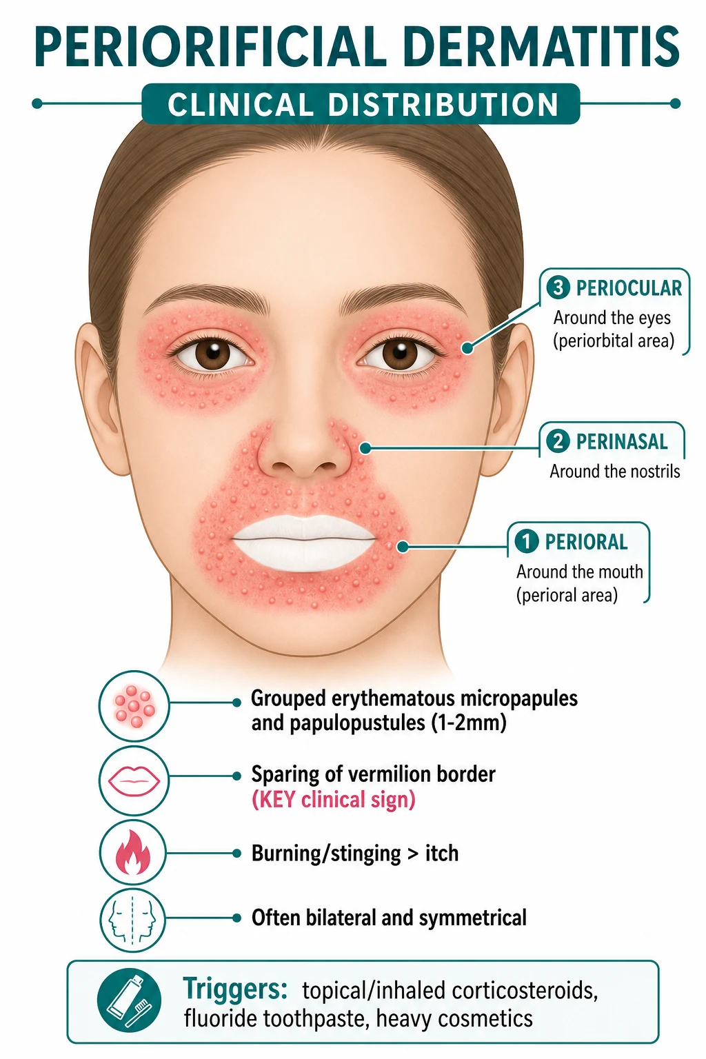
Definition and Classification
Periorificial (perioral) dermatitis is a common inflammatory facial dermatosis characterised by persistent or recurrent clusters of small (1-2 mm), monomorphic, erythematous micropapules and papulopustules distributed around the natural orifices of the face — most often the mouth (perioral), but also the nose (perinasal) and the eyes (periocular). The HALLMARK clinical sign is sparing of the vermilion border — a 2-5 mm rim of clinically normal skin separates the inflammatory papules from the lip margin itself, and the vermilion (the red, keratinised, mucosa-skin transition) is never involved.[1][3]
The terms perioral and periorificial are used interchangeably in clinical practice and in the literature, although purists reserve perioral for the strictly mouth-restricted disease and periorificial for the broader perioral + perinasal + periocular distribution. Most authorities — including the 2021 Searle review in J Cosmet Dermatol and the 2026 Acevedo-Fontanez synthesis in J Am Acad Dermatol — treat the two as a single clinicopathological entity and prefer periorificial dermatitis when perinasal or periocular involvement is present.[1][3]
The relationship to rosacea and acne is debated. Periorificial dermatitis overlaps with papulopustular rosacea morphologically (red papules, papulopustules, midface distribution) but is distinct in three ways: (1) no comedones (unlike acne); (2) no telangiectasia, no flushing, no ocular rosacea in the typical case (unlike rosacea); and (3) a clear iatrogenic precipitant (topical corticosteroid, fluoridated toothpaste) in most patients. The relationship to TOPICAL STEROID DAMAGED FACE (TSDF) is closer than to rosacea — TSDF is the broader iatrogenic syndrome that includes periorificial dermatitis as its most recognisable phenotype, but also features diffuse erythema, telangiectasia, hypertrichosis, atrophy, and hyper- or hypopigmentation across the face.[1][3][4]
The condition can be classified along five orthogonal axes that examiners test: [1]
Periorificial dermatitis — at-a-glance metric sheet
A — By distribution
- Perioral (most common, ~80% of adult cases) — papules clustered around the mouth, sparing the vermilion border.
- Perinasal (second most common) — papules around the alar grooves and nasal sill.
- Periocular / periorbital (less common, but tested) — papules around the lateral canthus and lower eyelid; must be distinguished from ocular rosacea, contact blepharitis, and demodicosis.
- Combined perioral + perinasal + periocular — when all three zones are involved, the disease is sometimes called periorificial dermatitis in the strict sense. [1]
B — By morphology
- Classic papulopustular — grouped erythematous micropapules and papulopustules on a background of erythema, often with fine scale.
- Granulomatous variant (Facial Afro-Caribbean Childhood Eruption — FACE) — monomorphic reddish-brown papules in prepubertal children, often with skin of colour; biopsy shows non-caseating granulomas.
- Lupus miliaris disseminatus faciei-like (LMDF-like) — discrete reddish-brown papules with central dell, may be perifollicular; biopsies show epithelioid granulomas with central necrosis. [1]
C — By trigger
- Steroid-induced (most common) — chronic topical corticosteroid misuse on the face (over-the-counter in South Asia, prescribed for facial eczema / pityriasis in adolescents, compounded "fairness" creams in South-East Asia).
- Inhaled / nasal corticosteroid-induced — deposition of aerosolised steroid at the mouth and nose; rinse mouth after each actuation.
- Cosmetic / moisturiser-induced — heavy occlusive moisturisers, foundation, sunscreen (chemical UV filters), SLS-containing cleansers.
- Fluoridated-toothpaste-associated — historically a much-debated trigger; methodologically weak evidence but consistently reported.
- Idiopathic — no obvious trigger, especially in children and in the granulomatous variant. [1]
D — By host
- Adult women (20-45 y) — the classic demographic; cosmetic, hormonal, and corticosteroid exposure.
- Children and adolescents — often granulomatous; may be related to inhaled corticosteroids, lip-licking, or fruit-juice contact.
- Patients with skin of colour — more prone to post-inflammatory hyperpigmentation and to the granulomatous variant.
- Pregnancy — tetracyclines and isotretinoin are contraindicated; topical metronidazole / azelaic acid first-line.
- Immunocompromised — consider demodicosis, candidiasis, and atypical infections (atypical mycobacteria, deep fungi). [1]
E — By severity (and management decision)
- Mild — scattered micropapules, no functional impact, responds to zero therapy + topical anti-inflammatory.
- Moderate — clustered papulopustules, cosmetic and psychosocial impact, may need oral tetracycline.
- Severe / refractory — extensive eruption, scarring dyspigmentation, treatment failure at 8-12 weeks; consider oral isotretinoin, biopsy to exclude alternative diagnoses. [1]
Periorificial dermatitis
- Perioral > perinasal > periocular; bilateral and symmetrical.
- Monomorphic 1-2 mm erythematous micropapules and papulopustules on a background of erythema and fine scale.
- SPARING of the vermilion border — the cardinal sign (2-5 mm normal-skin rim).
- Burning and stinging > itch (distinguishes from atopic / contact dermatitis).
- Topical corticosteroid is the classic iatrogenic trigger; cessation → rebound flare for 2-4 weeks.
- No comedones (distinguishes from acne); no telangiectasia / flushing (distinguishes from rosacea).
- Treatment: zero therapy → topical metronidazole / azelaic acid / calcineurin inhibitor → oral tetracycline 6-12 wk.
Papulopustular rosacea
- Central face (cheeks, nose, forehead, chin) — centrofacial distribution.
- Persistent centrofacial erythema with episodic flushing triggered by heat / alcohol / spicy food.
- Telangiectasia, papules, pustules; no comedones; may have ocular rosacea (50% — blepharitis, conjunctival injection, dry eye).
- Burning and stinging (similar to periorificial); stinging from topical agents is characteristic.
- Triggers: heat, alcohol, spicy food, sun, stress; not topical corticosteroids (rosacea can be worsened by them but is not caused by them).
- Same topical metronidazole / azelaic acid / ivermectin first-line; oral doxycycline 40 mg MR daily for moderate disease.
- Demodex density is increased in rosacea (especially the papulopustular subtype).
Epidemiology and Risk Factors
Periorificial dermatitis is a common condition in dermatology outpatient practice, accounting for an estimated 1-2% of all dermatology consultations in adult cohorts, with a striking female predominance (~9:1 female-to-male in most series), and a typical age range of 20-45 years in adults and 6 months to 18 years in the paediatric / adolescent population.[1][3][5]
Global patterns
The condition is reported worldwide, but the trigger mix differs sharply by region: [1]
- Western Europe / North America / Australasia — predominantly prescription topical corticosteroid (medium- or high-potency prescribed for facial eczema, seborrhoeic dermatitis, or contact dermatitis and used long-term) and cosmetic / moisturiser overuse ("over-skincare" with occlusive day / night creams, sheet masks, sunscreens, retinoid-containing anti-ageing products).
- South Asia (India, Pakistan, Bangladesh, Sri Lanka, Nepal) — over-the-counter topical corticosteroid misuse is endemic and produces the broader TOPICAL STEROID DAMAGED FACE (TSDF) phenotype. Combination creams containing clobetasol / mometasone / betamethasone with antifungal / antibacterial / depigmenting agents ("Skinlite", "Panderm", "Fourderm", "Betnovate-C", "Tenovate", "Dermikem") are sold OTC across India, Pakistan, Bangladesh and the Gulf. TSDF features periorificial dermatitis as its most recognisable component, but adds diffuse facial erythema, atrophy, telangiectasia, hypertrichosis, and hyper- or hypopigmentation. The Indian Association of Dermatologists (IADVL) has launched a sustained anti-OTC-topical-steroid campaign.
- Sub-Saharan Africa and the African diaspora — the granulomatous variant (FACE) is over-represented; prepubertal children, often with skin of colour.
- Latin America — fluoridated toothpaste and compounded fairness creams are additional triggers; melasma treatments containing unlabelled corticosteroids are a regional pitfall.
- East Asia (Korea, Japan, mainland China) — cosmetics and "K-beauty" multi-step skincare with multiple active ingredients can disrupt the barrier; high-potency whitening products occasionally contain unlabelled topical steroids. [1]
Recognised precipitants / triggers
The modern synthesis (Searle 2021; Acevedo-Fontanez 2026) groups the triggers into five categories that examiners test:[1][3]
Triggers of periorificial dermatitis — mnemonic
Why women are disproportionately affected
Several factors compound in adult women: [1]
- Higher prevalence of cosmetic, moisturiser, and sunscreen use — the modern multi-step skincare routine.
- Greater exposure to prescription topical corticosteroids — prescribed for facial "eczema" or "dermatitis" by non-dermatologists and then continued long-term.
- Hormonal modulation of skin immunity — the perimenstrual and perimenopausal skin is more reactive.
- Higher prevalence of adult acne and rosacea — and the (mis)use of topical corticosteroids for both. [1]
Risk factor — over-the-counter topical corticosteroid misuse in South Asia
The Indian / Pakistani / Bangladeshi TSDF epidemic is the single most important regional risk factor for periorificial dermatitis globally. The IADVL estimates that 20-30% of patients attending urban dermatology OPDs in India have a history of OTC topical steroid use, and that the periorificial distribution is the most common TSDF phenotype. The pattern is also seen in the South Asian diaspora (UK, US, Canada, Australia, Gulf) and is increasingly recognised in African and Latin American populations. Counsel patients and parents in plain language about the risks of OTC topical steroids on the face.[3][4]
Pathophysiology
Periorificial dermatitis is best understood as a chronic, relapsing, barrier-driven inflammatory dermatosis in which a permissive barrier dysfunction is triggered and perpetuated by external insults — most prominently the iatrogenic misuse of topical corticosteroids.[1][3]
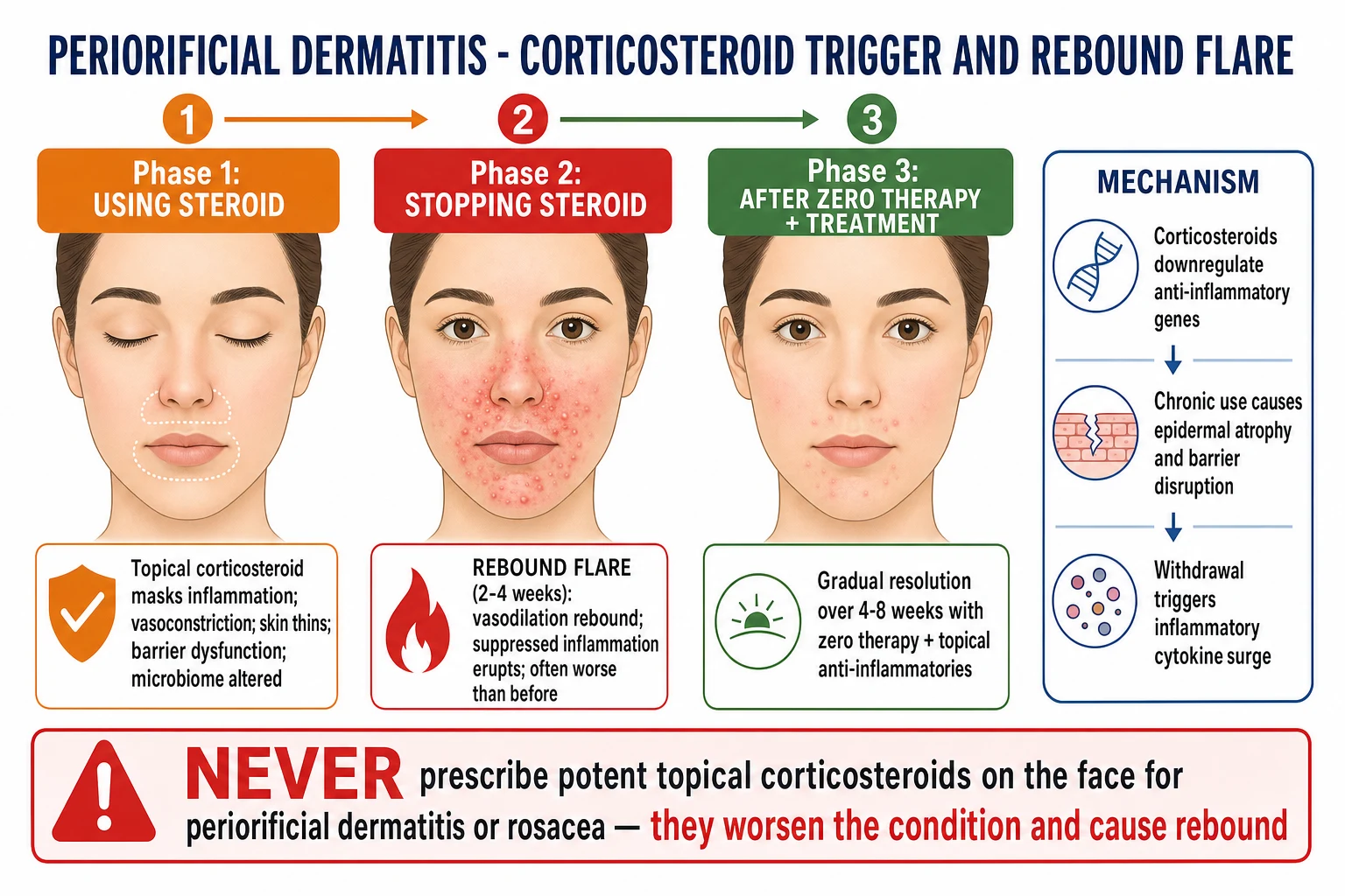
Step 1 — Skin barrier dysfunction (the permissive lesion)
The face — and the periorificial skin in particular — has a thinner stratum corneum, higher follicular density, and a more permeable barrier than the trunk or limbs. Loss of intercellular lipids (ceramides, cholesterol, free fatty acids) and filaggrin breakdown products compromises barrier integrity. The barrier dysfunction is the substrate on which the triggers act; alone it is usually insufficient to cause disease. [1]
Step 2 — Trigger exposure
The major triggers act through three mechanisms: [1]
- Direct barrier disruption — SLS cleansers, retinoids, AHAs / BHAs, physical exfoliants, chemical sunscreen penetration.
- Follicular occlusion with microbiome shift — heavy occlusive moisturisers, foundations, sheet masks, sunscreens in petrolatum base; Demodex mite overgrowth (their digestive enzymes and chitin exoskeletons are pro-inflammatory); Candida and Fusarium overgrowth (proposed but not confirmed).
- Vasoactive and immunomodulatory effects — topical corticosteroid vasoconstriction with rebound vasodilation on withdrawal; chronic barrier inflammation with aberrant innate-immune activation (TLR-2, cathelicidin LL-37, kallikrein-related peptidases). [1]
Step 3 — Topical corticosteroid — the dominant iatrogenic trigger
Topical corticosteroids (especially medium-, high-, and super-high-potency such as betamethasone valerate, mometasone furoate, clobetasol propionate, halobetasol) cause periorificial dermatitis through a stereotyped sequence:[4]
- Initial vasoconstriction and immunosuppression — the corticosteroid binds the cytoplasmic glucocorticoid receptor, translocates to the nucleus, and trans-represses NF-κB and AP-1; pro-inflammatory cytokine transcription is suppressed; the cutaneous vasculature constricts. The patient experiences a misleading early improvement (days 1-7).
- Epidermal and dermal atrophy — chronic use inhibits keratinocyte proliferation and collagen synthesis; the epidermis thins by 10-20% over weeks; the dermis shows reduced glycosaminoglycans and elastin.
- Barrier dysfunction — stratum corneum integrity falls, transepidermal water loss rises, ceramide content falls.
- Microbiome shift — Demodex density increases (the corticosteroid suppresses the cutaneous immune surveillance that normally keeps the mites in check); Staphylococcus and Candida overgrowth are reported.
- Rebound on withdrawal — when the corticosteroid is stopped, the suppressed inflammation erupts: rebound vasodilation, cytokine surge (IL-1α, IL-1β, TNF-α, IL-8, GM-CSF), recruitment of neutrophils and T cells. This is the rebound flare that peaks at 2-4 weeks and then slowly resolves over weeks to months. [1]
Step 4 — Why the vermilion border is spared
The vermilion border (the red margin of the lip) is anatomically and immunologically distinct from the surrounding perioral skin. It has no hair follicles, no sebaceous glands, a thinner stratum corneum, and a more mucosa-like epithelium. The absence of pilosebaceous units means the follicular occlusion and Demodex-driven inflammation that characterise the periorificial skin cannot develop. The vermilion also has a denser sensory innervation and a richer vascular plexus — but the vascular plexus is NOT susceptible to the corticosteroid-driven barrier and follicular changes that produce the eruption in the surrounding skin. This anatomic-physiologic asymmetry is the structural basis of the cardinal sign.[1][3]
Step 5 — Inhaled / nasal corticosteroid mechanism
Inhaled corticosteroids (fluticasone, budesonide, beclomethasone, mometasone) used for asthma or allergic rhinitis deposit on the oropharyngeal and perioral mucosa with each actuation. Large-particle aerosols and dry-powder inhalers deposit especially around the mouth and nose. Two mechanisms contribute: [1]
- Direct deposition — aerosolised steroid lands on the perioral skin and on the lip.
- Salivary redistribution — the steroid is dissolved in saliva and then redistributed to the perioral skin by tongue and lip movements (licking lips, licking the corners of the mouth). [1]
The clinical advice is to rinse the mouth with water and wash the face after each actuation; spacer devices reduce oropharyngeal deposition; switching to a non-steroid preventer (leukotriene-receptor antagonist, tiotropium, biologic) avoids the problem. [1]
Step 6 — The Demodex / Candida / Fusarium debate
The role of Demodex folliculorum and Demodex brevis (commensal face mites) is correlated but not proven causal. The mites are present in higher density on periorificial dermatitis skin than on normal facial skin; the standard skin-scraping method reports densities of 5-15 mites per cm² in periorificial dermatitis compared with 1-2 per cm² in controls. But density is also increased in rosacea and in healthy elderly skin, and eradicating the mites (with topical ivermectin or oral ivermectin) does not always cure the disease. The current synthesis is that Demodex is a co-factor and biomarker of barrier dysfunction rather than a true cause.[1][3]
Candida albicans is cultured from the perioral skin of up to 30% of periorificial dermatitis patients, and oral colonisation is common; the role of Candida is similarly debated and likely permissive (a coloniser of the moist, barrier-disrupted skin) rather than causal. Fusarium (a saprophytic mould) is reported in a small case series but is not a major factor. [1]
Step 7 — Innate immunity and the cathelicidin pathway
Rosacea and periorificial dermatitis share a common innate-immune signature: increased TLR-2 expression on keratinocytes, increased cathelicidin LL-37 production, and increased kallikrein-related peptidase 5 (KLK5) activity. The cathelicidin peptides are vasoactive and chemotactic; they are responsible for the erythema and the neutrophilic infiltrate. This is the pathway that tetracyclines target — doxycycline and minocycline inhibit KLK5 and cathelicidin processing, providing a mechanistic rationale for the empirical response to tetracyclines in both rosacea and periorificial dermatitis. [1]
Clinical Presentation
Periorificial dermatitis is a clinical diagnosis based on the triad of (1) morphology (grouped monomorphic micropapules and papulopustules), (2) distribution (perioral + perinasal + periocular with sparing of the vermilion border), and (3) history (a precipitating trigger, most often a topical corticosteroid).[1][3][5]
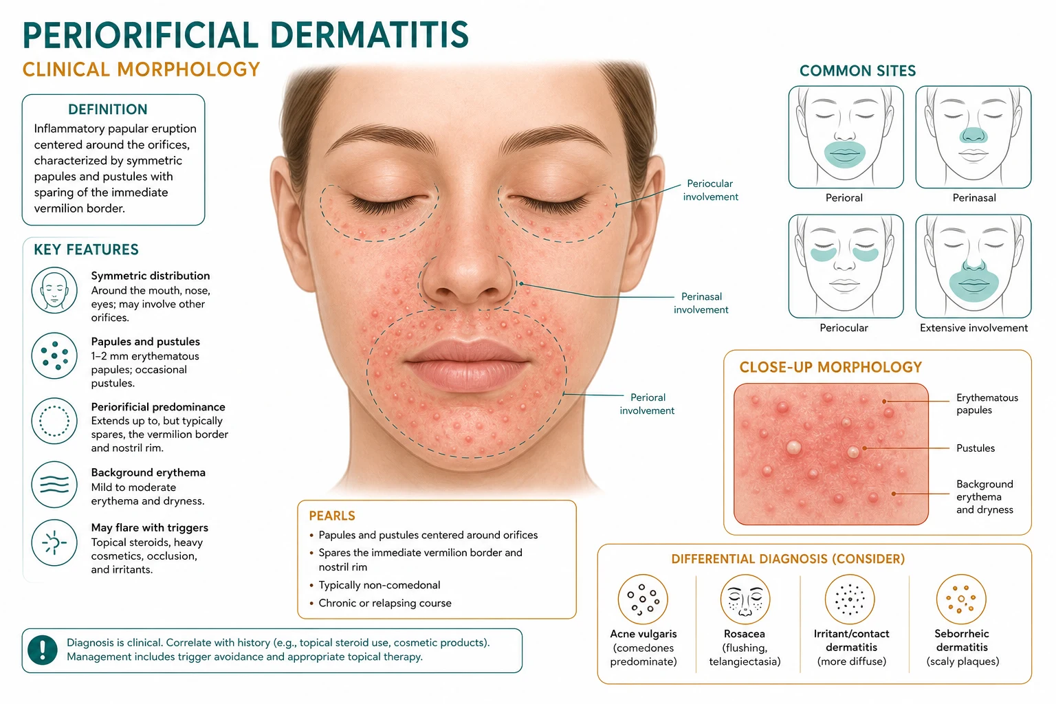
The diagnostic clinical pattern
- Onset: weeks to months; often insidious but recognised suddenly by the patient when the eruption is finally visible in the mirror or on a photograph.
- Morphology: grouped, monomorphic, 1-2 mm erythematous micropapules and papulopustules. The lesions are uniform (not polymorphic as in acne) and superficial (not deep-seated as in rosacea nodules).
- Background: ill-defined erythema, often with fine scale, sometimes with telangiectasia in chronic disease.
- Distribution: perioral (around the mouth) is the most common; perinasal (around the alar grooves and nasal sill) is the second; periocular (around the lateral canthus and lower lid) is the third. Bilateral and symmetrical.
- Cardinal sign — sparing of the vermilion border: a 2-5 mm rim of clinically normal skin separates the eruption from the lip margin. The vermilion (the red, keratinised, mucosa-skin transition) is never involved. This is the single most useful diagnostic sign.
- Sensation: burning and stinging are more prominent than itch. This distinguishes the disease from atopic and contact dermatitis, which itch more.
- Tempo: chronic and fluctuating; worse with continued corticosteroid use; rebounds within 2-4 weeks of corticosteroid withdrawal.
- Demographics: young to middle-aged adult women are the typical demographic (90%); also seen in children, adolescents, and the elderly. [1]
Clinical course and timeline
The natural history, if untreated, is chronic and relapsing over months to years. With appropriate therapy (zero therapy + topical anti-inflammatory + oral tetracycline for moderate-severe disease), the typical course is: [1]
- Week 0-2: cessation of corticosteroid; rebound flare begins.
- Week 2-4: rebound flare peaks (worst point); patient counselling is critical here.
- Week 4-8: gradual improvement begins; topical anti-inflammatories start to take effect.
- Week 8-12: most patients are clear or nearly clear.
- Week 12+: maintenance with bland emollient, sunscreen, trigger avoidance; oral tetracycline taper. [1]
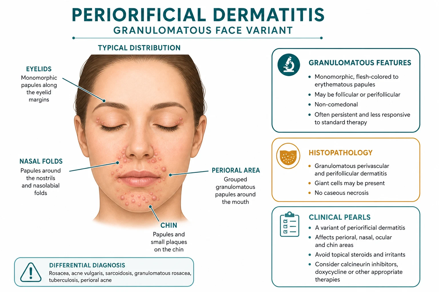
Granulomatous variant — Facial Afro-Caribbean Childhood Eruption (FACE)
The granulomatous variant (also called Facial Afro-Caribbean Childhood Eruption, FACE, or granulomatous perioral dermatitis) is a clinicopathological variant of periorificial dermatitis seen in prepubertal children, often with skin of colour (Afro-Caribbean, Hispanic, South Asian, sub-Saharan African ancestry).[3][5]
- Age: 6 months to 12 years; peak 3-7 years.
- Sex: slight male predominance (in contrast with the adult female predominance).
- Distribution: perioral, perinasal, periocular; often also involves the cheeks, chin, eyelids, and ears.
- Morphology: monomorphic, dome-shaped, reddish-brown to flesh-coloured papules 1-3 mm, often with a yellowish tinge on diascopy (the "apple-jelly" colour of granulomas).
- Symptoms: usually asymptomatic or mildly itchy; not burning.
- Trigger: often NO corticosteroid trigger is identified; the disease may arise in the context of a preceding irritant or contact dermatitis (lip-licking, fruit juice, SLS toothpaste), or it may be idiopathic.
- Histology: non-caseating granulomas in the superficial and mid-dermis, often perifollicular; mixed lymphohistiocytic infiltrate.
- Course: self-limiting over months to years (typically 1-3 years), but cosmetically distressing; treatment shortens the course.
- Treatment: topical metronidazole 0.75% BD, topical pimecrolimus 1% BD, oral erythromycin (30-50 mg/kg/day) or oral azithromycin (10 mg/kg three days a week) for 4-8 weeks. [1]
Periocular variant
- The periocular distribution is the least common in adults but the most diagnostically tricky.
- The lesions cluster around the lateral canthus and lower eyelid; the upper eyelid is often spared.
- Must be distinguished from ocular rosacea (with blepharitis, meibomian gland dysfunction, conjunctival injection), contact blepharitis (positive patch test, typically involves the upper lid), demodicosis (high Demodex density, cylindrical dandruff on the lash base), and atopic eyelid dermatitis (history of atopy, itch predominates, scaling and lichenification).
- Treatment: oral tetracycline + topical tacrolimus (preferred over topical steroid); refer to ophthalmology for ocular surface assessment. [1]
Paediatric variant (Chiriac 2025 update)
In infants and preschoolers, periorificial dermatitis presents with: [1]
- A milder, more papular pattern (less pustular than adults).
- A clear trigger in 60-70% of cases (fluoridated toothpaste, SLS-containing cleansers, fruit juice contact, lip-licking).
- A shorter, more treatment-responsive course (typically 4-8 weeks).
- Biopsy is more frequently required to distinguish from the granulomatous variant and from perioral sarcoidosis. [1]
Atypical presentations
- Unilateral periorificial dermatitis — reported after hemifacial topical corticosteroid use, after facial palsy (where corticosteroid is applied for presumed Bell's palsy), and after unilateral trigeminal neuralgia treatment. Recognise that the unilateral distribution reflects the unilateral application; treat the same as bilateral disease.
- Predominantly periocular — see above.
- Lupus miliaris disseminatus faciei (LMDF)-like — discrete, reddish-brown, centrofacial papules with central dell; histology shows epithelioid granulomas with central necrosis. Believed by some to be on the spectrum with granulomatous periorificial dermatitis; by others to be a separate entity (LMDF) within the granulomatous rosacea spectrum.
- Post-isotretinoin flare — periorificial dermatitis can flare on initiation of isotretinoin for acne, especially in patients with a subclinical rosacea / periorificial phenotype; manage expectantly with topical metronidazole and short-course oral tetracycline.
- Perioral sarcoidosis — lupus pernio of the perioral skin; papules and plaques; biopsy shows non-caseating granulomas; systemic features (lungs, eyes, joints) should be sought. [1]
Differential Diagnosis
The diagnosis of periorificial dermatitis is clinical, but several conditions enter the differential. The cardinal features that distinguish each are summarised below.[1][3][5]
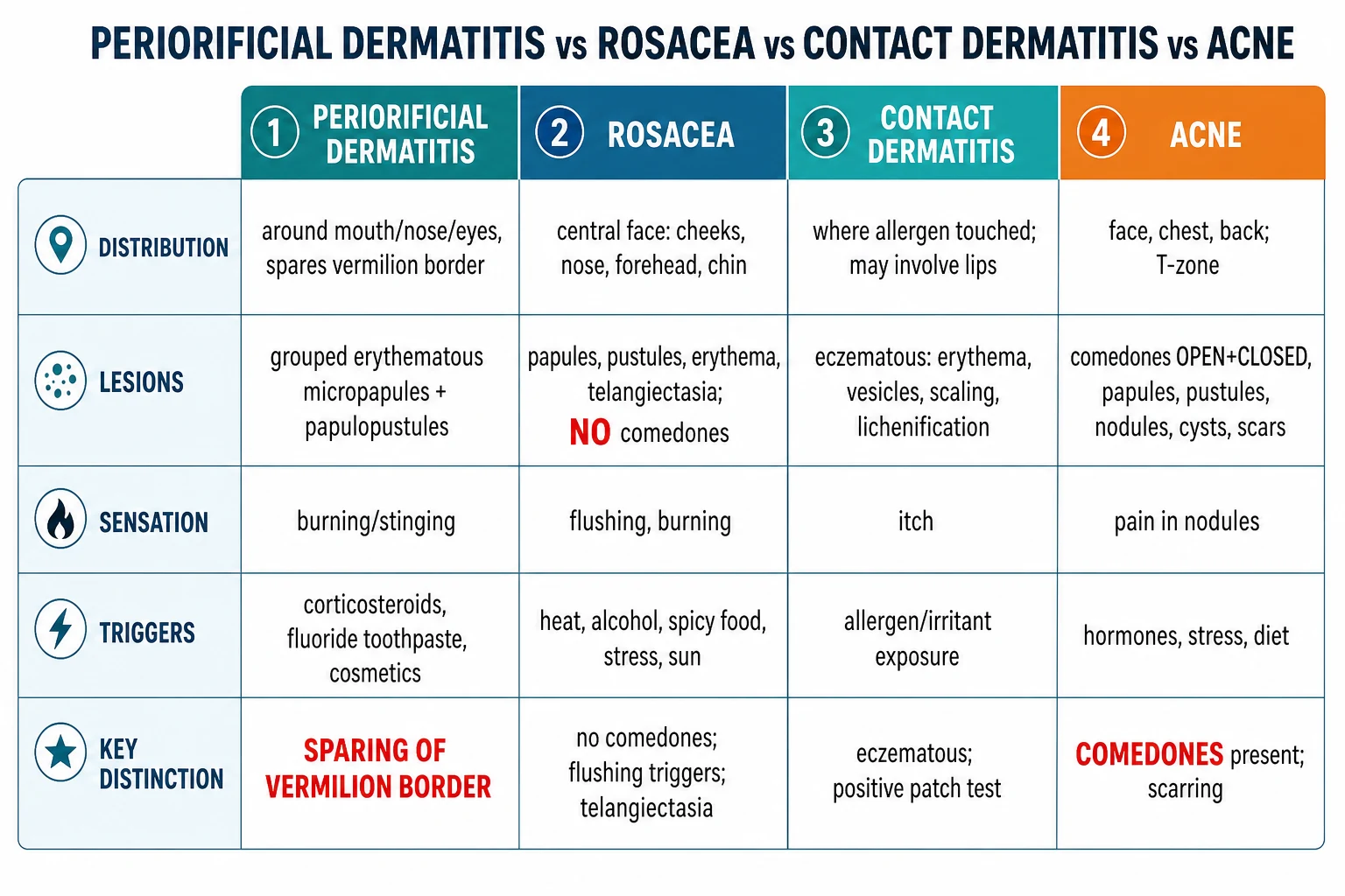
The differential at a glance
| Diagnosis | Distribution | Morphology | Sensation | Triggers | Cardinal distinguishing feature |
|---|---|---|---|---|---|
| Periorificial dermatitis | Perioral ± perinasal ± periocular; bilateral, symmetrical | Grouped monomorphic 1-2 mm micropapules + papulopustules on erythematous background | Burning and stinging > itch | Topical corticosteroid (most common), inhaled/nasal steroid, fluoride toothpaste, occlusive cosmetics | **SPARING OF THE VERMILION BORDER** (2-5 mm normal-skin rim) |
| Papulopustular rosacea | Central face (cheeks, nose, forehead, chin) | Persistent centrofacial erythema, papules, pustules, telangiectasia | Burning and stinging | Heat, alcohol, spicy food, sun, stress | Telangiectasia + flushing triggers; no vermilion-border sign |
| Allergic contact dermatitis | Where allergen contacted skin; perioral; may involve LIPS | Eczematous: erythema, vesicles, weeping, scaling, lichenification | Itch (PRURITUS) is dominant | Allergen exposure (toothpaste, lip balm, topical antibiotics, fragrance) | LIP INVOLVEMENT + positive patch test + itch predominates |
| Acne vulgaris | Face (T-zone, cheeks, jawline) + chest + back | COMEDONES (open and closed), papules, pustules, nodules, cysts, scars | Painful nodules/cysts; little itch | Androgens, diet, stress, occlusion | **COMEDONES** present; truncal involvement; scarring |
| Seborrhoeic dermatitis | Nasolabial folds, eyebrows, glabella, scalp, retroauricular | ERYTHEMA + YELLOW-GREASY SCALE; Pityrosporum-associated | Mild itch, burning | Cold weather, stress, Pityrosporum, HIV | SCALY YELLOW SCALE in nasolabial folds; scalp / ear involvement |
| Demodicosis | Face (forehead, cheeks, nose, perioral) | Rosacea-like papulopustules + cylindrical dandruff on lash base | Burning, itching, gritty eyes (ocular) | Immunosuppression, rosacea, elderly | HIGH DEMODEX DENSITY (≥5 mites/cm² on skin scraping); cylindrical dandruff on lashes |
| Lupus miliaris disseminatus faciei (LMDF) | Centrofacial; periorbital, perioral, nose, upper lip | Discrete reddish-brown papules, 1-3 mm, central dell; epithelioid granulomas with central necrosis on biopsy | Asymptomatic | Idiopathic; sometimes rosacea evolution | CENTRAL DELL on papule + biopsy with necrotic granulomas |
| Perioral sarcoidosis | Perioral, perinasal, periorbital, lupus pernio | Reddish-brown papules and plaques; apple-jelly on diascopy | Asymptomatic | Systemic sarcoidosis | NON-CASEATING GRANULOMAS + systemic features (lung, eye, joint, lymph) |
| Angular cheilitis (perlèche) | Corner of mouth (uni- or bilateral) | Fissuring, erythema, maceration, crusting | Pain, burning | Candida, Staph, denture, drooling, nutritional deficiency (B12, folate, iron) | ISOLATED TO MOUTH CORNERS; no perioral papules |
| Granuloma faciale | Face, often single plaque on cheek or nose | Reddish-brown to violaceous plaque; follicular openings exaggerated; chronic | Asymptomatic to mildly tender | Vasculitis of small vessels | SINGLE PLAQUE + leukocytoclastic vasculitis on biopsy |
Cannot-miss differentials
Three differentials are dangerous to miss: [1]
- Allergic contact dermatitis — the patient is often misdiagnosed as periorificial dermatitis, prescribed a topical corticosteroid, gets worse, and the cycle continues. Always ask about new toothpaste, lip balm, lipstick, foundation, sunscreen, fragrance, or topical antibiotics. Patch test if any doubt.
- Perioral sarcoidosis — a single biopsy may be the only way to distinguish; consider if the patient has systemic features (cough, dyspnoea, arthralgia, uveitis, lymphadenopathy) or if the disease is recalcitrant.
- Nail-unit SCC / perioral SCC — rare but tested; chronic single-site perioral lesion in an older patient that is not responding to appropriate therapy warrants biopsy. [1]
Diagnostic pointers (high-yield)
- Sparing of the vermilion border — present in periorificial dermatitis; absent in contact dermatitis, rosacea, and acne.
- Burning > itch — periorificial dermatitis and rosacea; atopic and contact dermatitis itch more.
- No comedones — periorificial dermatitis and rosacea; acne has comedones.
- No telangiectasia — periorificial dermatitis (typically); rosacea has telangiectasia.
- Perioral papules + inhaler use — inhaled-corticosteroid-induced periorificial dermatitis.
- Child with monomorphic centrofacial papules + skin of colour — granulomatous variant (FACE).
- Reddish-brown papules with central dell + centrofacial — LMDF.
- Single plaque on cheek / nose — granuloma faciale.
- Chronic single-site perioral lesion in older patient — perioral SCC until biopsied. [1]
Clinical and Bedside Assessment
The bedside assessment of suspected periorificial dermatitis has six steps that the examiner expects you to demonstrate. Each step has a yield — and each can be skipped only at the cost of missing a competing diagnosis.[1][3]
Step 1 — Focused history (the diagnostic yield is high)
- Duration and tempo — weeks to months; chronic and fluctuating; worse with continued corticosteroid; rebound within 2-4 weeks of corticosteroid withdrawal.
- Trigger inventory:
- Topical corticosteroids — what strength (mild hydrocortisone / moderate clobetasone butyrate / potent betamethasone / super-potent clobetasol propionate); for how long; OTC or prescribed; which area of the face; frequency.
- Inhaled / nasal corticosteroids — fluticasone, budesonide, beclomethasone, mometasone; mouth-rinsing habit.
- Toothpaste — fluoride? SLS? whitening? charcoal? herbal? "natural"?
- Cosmetics — foundation, concealer, primer, blush, bronzer, setting spray; how often applied; how often removed; with what cleanser.
- Sunscreen — chemical (oxybenzone, avobenzone) vs physical (zinc oxide, titanium dioxide); in what base (lotion, cream, stick, spray, gel).
- Skincare actives — retinoids (tretinoin, adapalene, retinol), AHAs (glycolic, lactic), BHAs (salicylic), vitamin C, niacinamide, peptides.
- Moisturisers — heavy occlusive (petrolatum, lanolin, mineral oil) vs light (hyaluronic acid, glycerin).
- Occupation — healthcare worker (hand hygiene + facial moisturiser), beautician (cosmetic exposure), food handler (citrus, garlic, flour), musician (wind instruments — lip contact), swimmer (chlorine), outdoor worker (sunscreen).
- Hobbies — running, hot yoga (flushing), spicy food, alcohol.
- Drugs and medical history — isotretinoin (paradoxical flare), EGFr / MEK / BTK inhibitors (pyogenic granuloma), hormone replacement / OCP, pregnancy.
- Family and social history — atopy, rosacea, perioral dermatitis in close contacts; smoking (nicotine patches can cause perioral dermatitis at the patch site).
- Prior treatments and response — what has been tried; what made it worse (most often a topical corticosteroid).
- Psychosocial impact — DLQI or similar; cosmetic and social impact; impact on work. [1]
Step 2 — Inspection of the affected area and all 20 nails (and the scalp)
- Affected area: grouped monomorphic micropapules and papulopustules, bilateral and symmetrical, on a background of erythema with fine scale.
- SPARING OF THE VERMILION BORDER — the cardinal sign. Confirm with the patient: "Are your lips involved?" The answer should be NO.
- Distribution zones: perioral, perinasal, periocular. Look at the glabella, chin, and cheeks for less common involvement.
- Background erythema and telangiectasia — telangiectasia suggests rosacea overlap; the disease is then often called rosacea + periorificial dermatitis and treated with a combined ladder.
- Atrophic features of TSDF — skin thinning (the skin is transparent, veins are visible), hypertrichosis, hyper- or hypopigmentation, striae (rare on the face but possible).
- All 20 nails — nail changes are uncommon in periorificial dermatitis, but onychomycosis and paronychia may coexist.
- Scalp and retroauricular — seborrhoeic dermatitis may coexist; look for scaly yellow scale.
- Eyes — blepharitis, conjunctival injection, meibomian gland dysfunction (ocular rosacea); refer to ophthalmology if present. [1]
Step 3 — Palpation and bedside manoeuvres
- Fluctuance — uncommon in periorificial dermatitis (the lesions are papulopustular, not abscess-forming); if fluctuance is present, consider secondary bacterial infection and swab.
- Diascopy — for the granulomatous variant (FACE), the apple-jelly colour of granulomas may be seen on pressure with a glass slide.
- Warmth and tenderness — present in acute flares; reduced in chronic quiescent disease.
- Cylindrical dandruff on lash base — Demodex cylindrical dandruff is a clue to demodicosis (and present in a minority of periorificial dermatitis patients).
- Tzanck smear / PCR — if vesicles are present, send HSV PCR to exclude herpetic involvement. [1]
Step 4 — Identify the portal of entry and the trigger
- The corticosteroid is the dominant iatrogenic trigger. Establish the strength, duration, area of application, and reason for original use. OTC use (South Asia, Gulf, Africa, Latin America) is common; do not be shy to ask.
- The toothpaste is the most common chemical trigger. Ask about brand, fluoride, whitening additives, and SLS.
- The cosmetics and skincare routine — ask the patient to bring in their products, or photograph them; many are unlabelled or compounded.
- Inhaled / nasal corticosteroid use — ask about asthma, allergic rhinitis, COPD, and spacer use. [1]
Step 5 — Drug, disease, and pregnancy history
- Retinoid, EGFr / MEK / BTK inhibitor, antiretrovirals, hormone replacement, OCP, pregnancy, lactation.
- Diabetes, HIV, immunosuppression, atopy, rosacea, seborrhoeic dermatitis. [1]
Step 6 — Severity and psychosocial impact
- Lesion count — mild (fewer than 20), moderate (20 to 50), severe (more than 50).
- Distribution — single zone vs multi-zone.
- Background erythema and scale — graded mild / moderate / severe.
- Psychosocial impact — DLQI; cosmetic impact; impact on social, work, and intimate life.
- Response to prior therapy — naïve vs refractory. [1]
Investigations
Periorificial dermatitis is a clinical diagnosis in the typical case. The role of investigations is to (1) exclude competing diagnoses when the morphology is atypical, (2) screen for underlying disease in refractory or recurrent disease, and (3) confirm the granulomatous variant when suspected.[1][3][5]
Investigations — who, what, and when
Histology of classic periorificial dermatitis
The histology of classic periorificial dermatitis is non-specific and resembles mild eczema with follicular accentuation: [1]
- Epidermis: focal spongiosis (intercellular oedema of the epidermis), focal parakeratosis, mild acanthosis.
- Follicular epithelium: mild follicular spongiosis and dilatation.
- Dermis: superficial perivascular and perifollicular lymphohistiocytic infiltrate with occasional neutrophils and plasma cells; oedema of the papillary dermis.
- No granulomas, no vasculitis, no microorganisms on special stains. [1]
The histology is helpful in distinguishing from: [1]
- Granulomatous rosacea / LMDF — epithelioid granulomas with central necrosis.
- Perioral sarcoidosis — tight, well-formed non-caseating granulomas with a "naked" appearance.
- Granuloma faciale — leukocytoclastic vasculitis with eosinophils and neutrophils.
- Pustular psoriasis / acrodermatitis continua — spongiform pustules of Kogoj in the epidermis. [1]
Histology of the granulomatous variant (FACE)
- Epidermis: usually normal.
- Dermis: non-caseating granulomas in the superficial and mid-dermis, often perifollicular; mixed lymphohistiocytic infiltrate with occasional eosinophils.
- Special stains (AFB, Fite, GMS, PAS) — negative (exclude mycobacteria and fungi). [1]
Patch testing
Patch testing is indicated when: [1]
- The lips are involved (suggests contact dermatitis from toothpaste, lip balm, lipstick, topical antibiotic).
- The morphology is eczematous rather than papulopustular.
- The patient has a history of multiple product changes.
- Standard therapy has failed. [1]
The standard series (European / North American baseline) plus a dental series and a cosmetic / fragrance series is the typical panel; specific allergens (cobalt, nickel, fragrance mix, balsam of Peru, propylene glycol, cocamidopropyl betaine) are reported in toothpaste and lip products. [1]
Dermoscopy (dermatoscopy)
Dermoscopy of periorificial dermatitis shows a non-specific pattern that can help distinguish from rosacea: [1]
- Pink-red background with structureless areas.
- Follicular openings preserved (rosacea shows follicular plugging).
- Linear and branching vessels in the background (rosacea shows polygonal vessels).
- White scales in fine flakes.
- No Demodex tails (Demodex tails are short whitish projections from follicular openings; present in demodicosis and rosacea, typically absent in periorificial dermatitis). [1]
KOH and fungal culture
KOH and fungal culture (CHROMagar Candida speciation) are indicated when: [1]
- The lesion is at the corner of the mouth (angular cheilitis) — Candida dominant.
- The patient is immunocompromised.
- The patient is a chronic oral or inhaled corticosteroid user.
- The morphology is pustular and chronic, with poor response to anti-bacterial therapy. [1]
Demodex quantification
Demodex quantification is indicated when: [1]
- The disease is refractory to standard therapy.
- The patient has rosacea-like features with high suspicion of demodicosis.
- The patient is immunocompromised (HIV, transplant, chemotherapy).
- Cylindrical dandruff is present on the lash base. [1]
Methods: standardised skin-surface biopsy (SSSB) with cyanoacrylate glue; skin scraping with a scalpel and immersion oil; modified standardized skin surface biopsy (1 cm² of stratum corneum lifted and examined under light microscopy). A density of ≥5 mites per cm² is suggestive of demodicosis. [1]
Management — Resuscitation
Periorificial dermatitis is not a dermatological emergency; resuscitation is rarely needed. The role of the first consultation is to: [1]
- Establish the diagnosis — confirm the morphology, the distribution, the sparing of the vermilion border, and the trigger.
- Discontinue the offending trigger — most importantly the topical corticosteroid.
- Counsel on the rebound flare — the patient must understand that the disease will worsen for 2-4 weeks before it improves; this is the single most common reason for treatment failure and loss to follow-up.
- Initiate the management ladder — see "Management — Definitive and Stepwise" below.
- Safety-net — when to return (worsening despite compliance; new symptoms; treatment failure at 8-12 weeks). [1]
Same-day specialist referral indications
- Diagnostic uncertainty (granulomatous variant vs sarcoidosis vs LMDF; rosacea vs periorificial dermatitis; cutaneous lymphoma).
- Severe flare with significant psychosocial impact.
- Pregnancy, lactation, or planned pregnancy.
- Child (especially prepubertal — consider granulomatous variant and biopsy).
- Treatment failure at 8-12 weeks of appropriate therapy.
- Recurrence on multiple attempts at trigger avoidance.
- Ocular involvement with vision changes (refer to ophthalmology urgently).
- Single chronic lesion in an older patient (rule out SCC). [1]
Psychological support
Periorificial dermatitis is cosmetically and psychologically distressing; patients are often misdiagnosed and given more topical corticosteroid, which perpetuates the cycle; and the rebound flare is psychologically devastating without prior warning. Validate the experience, acknowledge the cosmetic impact, and offer realistic timeframes for improvement. Consider DLQI monitoring and referral for psychological support if distress is significant. [1]
Management — Definitive and Stepwise
The management of periorificial dermatitis is anchored on three principles and a staged ladder that mirrors rosacea management. The principles are: [1]
- Stop the trigger. The corticosteroid (and the toothpaste, cosmetics, occlusive moisturisers, SLS cleansers) must be removed.
- Repair the barrier. Bland emollient, gentle cleanser, physical sunscreen.
- Calm the inflammation. Topical anti-inflammatory (metronidazole / azelaic acid / calcineurin inhibitor) for mild-moderate disease; oral tetracycline for moderate-severe. [1]
The staged ladder is presented in the figure and elaborated below.[1][2][3][5]
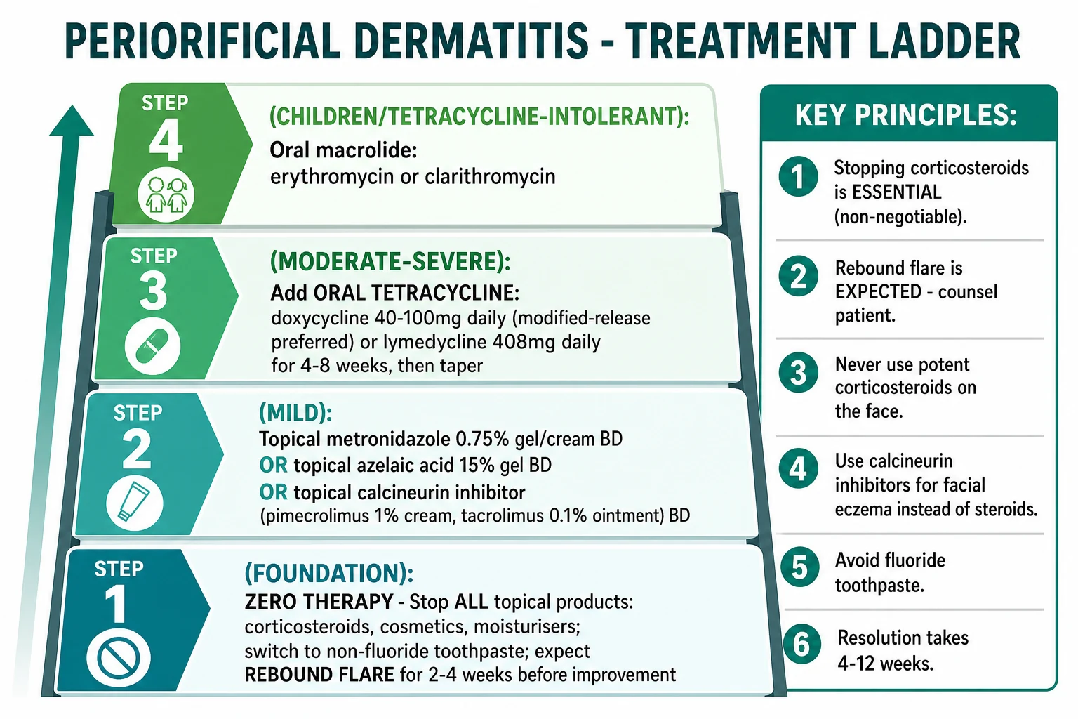
Step 1 — "Zero therapy" (the foundation)
- Discontinue ALL topical products on the face:
- Topical corticosteroids (non-negotiable — the dominant iatrogenic trigger).
- Cosmetics — foundation, concealer, primer, blush, bronzer, setting spray.
- Heavy moisturisers — petrolatum, lanolin, mineral oil, dimeticone in an occlusive base.
- Toners, exfoliants, and actives — retinoids, AHAs, BHAs, vitamin C, niacinamide, peptides.
- Sunscreens with chemical UV filters in an occlusive base.
- SLS-containing cleansers — switch to a non-foaming, SLS-free cleanser.
- Switch to a non-fluoride, SLS-free toothpaste — herbal or children's toothpaste (e.g., Aquafresh MildMint, Sensodyne Pronamel, Burt's Bees, or any toothpaste without sodium lauryl sulphate and without sodium fluoride).
- Expect a rebound flare for 2-4 weeks after stopping corticosteroids. The disease will temporarily worsen (more erythema, more papules) before it improves. Counsel the patient in plain language: "It will get worse for 2-4 weeks, then it will get better. The corticosteroid was masking the inflammation; the flare is the inflammation coming back. Persist with zero therapy." This single piece of counselling is the most common reason for treatment success vs failure.[4]
- Gentle cleansing with lukewarm water or a non-foaming, SLS-free cleanser (e.g., CeraVe hydrating cleanser, La Roche-Posay Toleriane, Aveeno Skin Relief).
- Sunscreen — physical (mineral) sunscreen with zinc oxide or titanium dioxide in a non-occlusive base. Avoid chemical UV filters in petrolatum.
- Bland emollient — a thin layer of white soft paraffin (Vaseline) or a fragrance-free, non-occlusive emollient (CeraVe moisturising cream, Vanicream, Eucerin Original) may be used sparingly for barrier support.
Step 2 — Topical anti-inflammatory (mild-moderate disease)
Introduce after 2 weeks of zero therapy (some authors introduce immediately; consensus favours waiting 2 weeks to allow the barrier to recover). [1]
- Topical metronidazole 0.75% gel, cream, or lotion — apply twice daily; anti-inflammatory and possibly anti-Demodex; safe in pregnancy (Category B); the most-prescribed topical agent in the literature. (Extrapolated from rosacea RCTs; no large perioral-specific RCT.)[1][2]
- Topical azelaic acid 15% gel or foam — apply twice daily; anti-inflammatory, anti-comedonal, and depigmenting; first-line in skin of colour for its dual role in reducing post-inflammatory hyperpigmentation; safe in pregnancy (Category B).[2]
- Topical calcineurin inhibitors — pimecrolimus 1% cream (Elidel) or tacrolimus 0.03% or 0.1% ointment (Protopic) — apply twice daily; steroid-sparing anti-inflammatory; first-line in children, granulomatous variant, and facial eczema comorbidity; transient burning or stinging is common. Avoid the 0.1% strength in children under 16; pimecrolimus is preferred in pregnancy. (Ollech 2020 — pimecrolimus is effective in paediatric periorificial dermatitis.)[2]
- Topical ivermectin 1% cream (Soolantra) — apply once daily; anti-inflammatory and anti-Demodex; first-line in rosacea; useful when rosacea overlap is suspected.
- Topical erythromycin 2% or clindamycin 1% — alternatives, especially in children and pregnancy; bacterial resistance is rising.
- Topical adapalene 0.1% or tretinoin 0.025% — controversial; can be irritating on a barrier-disrupted face; reserve for the maintenance phase after the acute flare has resolved.
Step 3 — Oral therapy (moderate-severe or refractory disease)
Indications: extensive eruption, failure of zero therapy + topical anti-inflammatory at 4-8 weeks, severe psychosocial impact, or rapid cosmetic and professional reasons for clearance. [1]
- Oral tetracyclines (the workhorse for moderate-severe disease):
- Doxycycline — 100 mg once or twice daily OR modified-release 40 mg once daily (sub-antimicrobial dose; preferred for tolerability and for avoiding antibiotic resistance).[2]
- Lymecycline — 408 mg once daily (one capsule); well-tolerated; commonly used in Europe.
- Minocycline — 100 mg once or twice daily; more vestibular side effects (vertigo) and rare DRESS / autoimmune hepatitis; reserve.
- Tetracycline — 250-500 mg twice daily; classical, less commonly used.
- Duration: 6-12 weeks. Continue for 4 weeks after clearance, then taper to topical maintenance.
- Oral macrolides (for children, pregnancy, lactation, tetracycline intolerance):
- Erythromycin — 250-500 mg twice daily in adults; 30-50 mg/kg/day in children.
- Azithromycin — 500 mg three times per week (Mon/Wed/Fri); pulse dosing; well-tolerated.
- Clarithromycin — 250-500 mg twice daily.
- Duration: 4-8 weeks.
- Oral metronidazole (alternative) — 200-400 mg twice daily for 4-6 weeks; rarely used due to taste aversion and potential for peripheral neuropathy with prolonged use.
Step 4 — Refractory disease (rare)
Reserved for patients who fail zero therapy + topical anti-inflammatory + oral tetracycline (6-12 weeks). [1]
- Low-dose isotretinoin — 0.1-0.3 mg/kg/day for 3-6 months; effective in refractory periorificial dermatitis (extrapolated from rosacea and from small case series; teratogenic; requires iPLEDGE / pregnancy prevention in women of childbearing age).[3]
- Topical roflumilast 0.3% cream (Zoryve) — phosphodiesterase-4 inhibitor; approved for plaque psoriasis and seborrhoeic dermatitis; emerging case-series evidence in periorificial dermatitis (Domingues 2026). Not yet first-line but promising.
- Combination therapy — topical metronidazole + oral tetracycline; topical pimecrolimus + oral tetracycline.
- Specialist referral — to a dermatology clinic with experience in refractory facial dermatoses.
Step 5 — Specific subtype management
- Granulomatous variant (FACE) — topical metronidazole 0.75% BD, topical pimecrolimus 1% BD, oral erythromycin 30-50 mg/kg/day (children) or azithromycin 10 mg/kg three times a week for 4-8 weeks. Self-limiting; treatment shortens the course.
- Inhaled-corticosteroid-induced — rinse mouth and wash face after each actuation; spacer device; consider non-steroid preventer (leukotriene antagonist, biologic).
- Pregnancy — topical metronidazole or azelaic acid first-line; pimecrolimus; oral macrolides if needed; tetracyclines and isotretinoin CONTRAINDICATED.
- Skin of colour — azelaic acid 15% (anti-pigment); physical sunscreen; early anti-inflammatory control to reduce post-inflammatory hyperpigmentation. [1]
Step 6 — Maintenance and trigger avoidance
- Bland emollient (white soft paraffin; non-occlusive moisturiser) — daily, twice a day.
- Physical sunscreen (zinc oxide / titanium dioxide) — daily, SPF 30-50, reapply every 2 hours.
- Trigger avoidance — non-fluoride toothpaste; SLS-free cleanser; minimal cosmetics; no facial topical corticosteroid (1% hydrocortisone only for short courses on the body, never on the face for more than 7 days).
- Taper oral therapy — continue 6-12 weeks; transition to topical maintenance; do not stop abruptly.
- Recurrence — repeat the same ladder; oral tetracycline can be restarted for a second course. [1]
Key principles (high-yield)
- Never use potent or moderate topical corticosteroids on the face — they worsen periorificial dermatitis (and rosacea) and produce rebound on withdrawal. For facial eczema, use topical calcineurin inhibitors instead.[4]
- Warn about the rebound flare — the disease will get worse for 2-4 weeks before it improves; this is expected and is the most common reason for treatment failure.
- Resolution takes 4-12 weeks with appropriate therapy; longer for the granulomatous variant in children.
- Avoid triggers permanently — non-fluoride toothpaste, no facial corticosteroid, minimal cosmetics, physical sunscreen.
Periorificial dermatitis initial management — mnemonic
Specific Subtypes and Scenarios
A — Granulomatous variant (Facial Afro-Caribbean Childhood Eruption, FACE)
- Demographics: prepubertal children (6 months to 12 years; peak 3-7 years); slight male predominance; often skin of colour.
- Morphology: monomorphic, dome-shaped, reddish-brown to flesh-coloured papules 1-3 mm, often with apple-jelly colour on diascopy.
- Distribution: perioral, perinasal, periocular, often cheeks, chin, eyelids, ears.
- Trigger: often NO corticosteroid trigger; may follow irritant or contact dermatitis; often idiopathic.
- Histology: non-caseating granulomas, often perifollicular.
- Course: self-limiting over months to years (typically 1-3 years); treatment shortens the course.
- Treatment: topical metronidazole 0.75% BD; topical pimecrolimus 1% BD; oral erythromycin (30-50 mg/kg/day) or azithromycin (10 mg/kg three times a week) for 4-8 weeks. Tetracyclines are contraindicated under 12 years (tooth discolouration). [1]
B — Inhaled / nasal corticosteroid-induced periorificial dermatitis
- Demographics: asthmatic / allergic-rhinitis / COPD patients using fluticasone, budesonide, beclomethasone, or mometasone.
- Distribution: perioral (lip-licking redistribution) and perinasal (deposition around the nares).
- Treatment: rinse mouth and wash face after each actuation; use a spacer device; consider non-steroid preventer (leukotriene antagonist, tiotropium, omalizumab, dupilumab, mepolizumab). Continue management ladder (zero therapy for the face + topical anti-inflammatory + oral tetracycline if needed). [1]
C — Pregnancy and lactation
- Topical metronidazole 0.75% — Category B; safe in pregnancy and lactation.
- Topical azelaic acid 15% — Category B; safe in pregnancy and lactation.
- Topical pimecrolimus 1% — Category C; use with caution; avoid in the first trimester.
- Oral erythromycin — Category B; safe (except the estolate form, which is contraindicated in pregnancy due to maternal cholestasis).
- Oral azithromycin — Category B; safe.
- Tetracyclines (doxycycline, minocycline, lymecycline) — CONTRAINDICATED in pregnancy (foetal tooth discolouration and bone growth retardation) and in children under 12 years.
- Isotretinoin — TERATOGENIC; absolute contraindication; two forms of contraception and monthly pregnancy tests required in women of childbearing age. [1]
D — Immunocompromised patients
- Demodicosis — consider if rosacea-like features with high Demodex density; treat with topical ivermectin 1% or oral ivermectin (200 µg/kg single dose, repeat in 1 week).
- Candidiasis — culture and KOH; treat with topical nystatin, miconazole, or oral fluconazole.
- Atypical mycobacteria / deep fungi — biopsy with AFB, Fite, GMS, PAS stains; mycobacterial and fungal culture; refer to infectious diseases.
- HIV-associated periorificial dermatitis — atypical morphology, more severe course, refractory to standard therapy; screen for and treat concurrent infections. [1]
E — Skin of colour (Fitzpatrick IV-VI)
- Post-inflammatory hyperpigmentation is a major concern; azelaic acid 15% is dual-action (anti-inflammatory + depigmenting) and is first-line.
- Granulomatous variant (FACE) is over-represented; biopsy to confirm; treat with topical metronidazole or pimecrolimus and oral macrolides.
- TSDF is more visible on darker skin — hypopigmentation, hyperpigmentation, and atrophy are more cosmetically impactful.
- Photoprotection — physical sunscreen; tinted mineral sunscreen for cosmetic camouflage. [1]
F — Periorificial dermatitis in the elderly
- Rosacea overlap is common; combined rosacea + periorificial dermatitis treated with combined ladder.
- Topical corticosteroid misuse history is often long (years to decades); psychological and education component is large.
- Tetracyclines are tolerated but watch for drug interactions (warfarin, statins, retinoids, methotrexate).
- Isotretinoin is an option for refractory disease. [1]
G — Post-isotretinoin flare
- Initiation of isotretinoin for acne can paradoxically flare periorificial dermatitis.
- Manage with topical metronidazole + short-course oral tetracycline during the first 2-3 months of isotretinoin.
- Continue isotretinoin for the underlying acne; the periorificial disease will improve as the barrier stabilises. [1]
Complications and Pitfalls
Disease-related complications
- Persistent erythema and telangiectasia — chronic disease can lead to telangiectasia; pulsed-dye laser or IPL for cosmetic improvement.
- Post-inflammatory hyperpigmentation — major concern in skin of colour; early anti-inflammatory control and azelaic acid reduce the pigmentary legacy.
- Post-inflammatory hypopigmentation — uncommon but reported after severe flares, especially in dark skin.
- Scarring — rare in periorificial dermatitis (unlike acne); when present, consider an alternative diagnosis.
- Psychosocial distress — DLQI is high; the cosmetic impact is severe and the rebound flare without prior warning is psychologically devastating.
- Secondary bacterial infection — S. aureus or Streptococcus; culture and antibiotics. [1]
Treatment-related pitfalls
- Corticosteroid rebound — the most common pitfall; abrupt cessation of long-term potent topical corticosteroid produces a severe rebound flare. Taper the corticosteroid (down-potency: super-potent → potent → moderate → mild → 1% hydrocortisone → stop) OR switch to a calcineurin inhibitor for 2-4 weeks before stopping the corticosteroid.
- Tetracycline photosensitivity — doxycycline (especially) causes phototoxic reactions; counsel on photoprotection and consider modified-release 40 mg daily (less phototoxic than 100 mg BD).
- Tetracycline teratogenicity — contraindicated in pregnancy and under 12 years; substitute macrolide.
- Tetracycline oesophagitis — take with a full glass of water; remain upright for 30 min.
- Vaginal candidiasis — common with tetracyclines; counsel and consider prophylactic antifungal.
- Antibiotic resistance — sub-antimicrobial-dose doxycycline (40 mg MR) avoids the resistance pressure; full-dose tetracyclines for short courses (6-12 weeks) are acceptable.
- Isotretinoin teratogenicity — absolute contraindication in pregnancy; iPLEDGE / pregnancy prevention programme.
- Calcineurin inhibitor malignancy warning — black-box warning for theoretical malignancy risk based on animal studies and rare case reports; the FDA has concluded the risk is unproven but requires a warning. [1]
Diagnostic pitfalls
- Mistaking periorificial dermatitis for eczema / contact dermatitis and prescribing more topical corticosteroid — the most common diagnostic error worldwide. The cycle is: eczema → topical steroid → apparent improvement → tolerance → rebound → "more eczema" → more topical steroid → periorificial dermatitis → topical steroid → worsening. The cardinal sign of sparing of the vermilion border and the history of topical corticosteroid are diagnostic.
- Mistaking periorificial dermatitis for rosacea and prescribing topical corticosteroid — the same cycle, the same trap. The presence of telangiectasia, flushing, and ocular rosacea is in favour of rosacea, but the periorificial distribution and the corticosteroid history favour periorificial dermatitis.
- Missing allergic contact dermatitis — lip involvement, itch, and a new product are clues. Patch test.
- Missing the granulomatous variant — the morphology and demographic are different; biopsy to confirm.
- Missing perioral sarcoidosis — single biopsy is diagnostic; consider if systemic features are present.
- Missing LMDF — central dell on the papule and epithelioid granulomas with central necrosis on biopsy are diagnostic. [1]
The "never do" list
- Never prescribe potent or moderate topical corticosteroid on the face for chronic use. The only acceptable facial topical corticosteroid is 1% hydrocortisone for ≤7-14 days, and only for a defined indication (acute contact dermatitis, acute eczema).
- Never incise a perioral pustule. The lesions are papulopustular, not abscess-forming; incision produces scarring and does not help.
- Never use topical antifungals as monotherapy for chronic periorificial dermatitis. Candida is a coloniser; antifungal monotherapy fails without barrier restoration and trigger removal.
- Never stop the oral tetracycline abruptly after 1-2 weeks. Continue for 6-12 weeks; taper to topical maintenance.
- Never forget to counsel on the rebound flare. The patient who is not warned will assume the treatment is failing and may restart the corticosteroid. [1]
Prognosis and Disposition
Natural history
Untreated, periorificial dermatitis is chronic and relapsing over months to years; spontaneous resolution is uncommon. The dominant reason for treatment success is the removal of the trigger (corticosteroid) and the resolution of the underlying barrier dysfunction; the dominant reason for treatment failure is the failure to remove the trigger or the lack of counselling on the rebound flare. [1]
With treatment
- Mild disease — 4-8 weeks to clearance with zero therapy + topical anti-inflammatory.
- Moderate disease — 6-12 weeks with zero therapy + topical anti-inflammatory + oral tetracycline.
- Severe / refractory disease — 12-16 weeks with combined therapy + low-dose isotretinoin for true refractory cases.
- Granulomatous variant (FACE) — 4-8 weeks of topical metronidazole + oral macrolide, but the disease may take 1-3 years to fully self-resolve. [1]
Recurrence
- 30-40% of patients have a recurrence if corticosteroids or other triggers are reused.
- Taper oral therapy to topical maintenance to reduce the recurrence risk.
- Long-term trigger avoidance (non-fluoride toothpaste, no facial corticosteroid, minimal cosmetics) is the cornerstone of secondary prevention. [1]
Disposition
- Outpatient management is the rule.
- GP + dermatologist for severe, refractory, or diagnostic-uncertain disease.
- Specialist referral (dermatology) for: severe disease, granulomatous variant, child, pregnancy, treatment failure at 8-12 weeks, suspected alternative diagnosis.
- Ophthalmology referral for ocular involvement (blepharitis, conjunctival injection, meibomian gland dysfunction).
- Infectious diseases referral for atypical mycobacterial or deep fungal infection in immunocompromised patients.
- Plastic / dermatologic surgery for pulsed-dye laser or IPL for persistent telangiectasia and erythema. [1]
Follow-up schedule
- Week 0 — initial consultation; trigger removal; counselling on rebound flare; topical anti-inflammatory or oral tetracycline prescribed.
- Week 2 (optional phone or in-person) — check on rebound flare, support compliance, adjust ladder.
- Week 4 — review; assess response; escalate if no improvement.
- Week 8 — review; assess response; consider biopsy if no improvement.
- Week 12 — review; maintenance plan; trigger-avoidance review.
- Week 24 — final review; safety-net advice; long-term plan. [1]
Safety-net advice for patients
- Avoid topical corticosteroids on the face permanently. 1% hydrocortisone only, maximum 7-14 days, only on defined indications (acute contact dermatitis, acute eczema).
- Switch to non-fluoride, SLS-free toothpaste.
- Use a non-foaming, SLS-free cleanser.
- Apply physical sunscreen (zinc oxide / titanium dioxide) daily, SPF 30-50, reapply every 2 hours.
- Use a bland, fragrance-free emollient.
- Rinse the mouth and face after each inhaled / nasal corticosteroid actuation.
- Return if the disease recurs despite compliance — repeat the ladder; consider biopsy if atypical. [1]
Special Populations
Children
- Demographics — childhood periorificial dermatitis is more common than traditionally reported; peak age 3-7 years; both sexes; both Fitzpatrick I-VI.
- Trigger — fluoridated toothpaste, SLS-containing cleansers, fruit juice contact, lip-licking, inhaled corticosteroids; less often topical corticosteroid misuse (more common in adults in South Asia).
- Granulomatous variant (FACE) — over-represented in children with skin of colour.
- Treatment — topical metronidazole 0.75% BD OR topical pimecrolimus 1% BD; oral erythromycin (30-50 mg/kg/day) or azithromycin (10 mg/kg three times a week) for moderate-severe disease. Tetracyclines are contraindicated under 12 years (tooth discolouration, bone growth retardation).
- Prognosis — self-limiting in many cases; treatment shortens the course. [1]
Pregnancy
- First-line — topical metronidazole 0.75% BD OR topical azelaic acid 15% BD.
- Second-line — topical pimecrolimus 1% BD; oral erythromycin (except estolate) or azithromycin.
- CONTRAINDICATED — tetracyclines (tooth discolouration, bone growth retardation); isotretinoin (teratogenic); topical retinoids (teratogenic).
- Pimecrolimus / tacrolimus — Category C; use with caution; avoid in first trimester if possible. [1]
Elderly
- Rosacea overlap is common.
- Topical corticosteroid misuse history often extends years to decades.
- Tetracyclines are tolerated but watch for drug interactions (warfarin, statins, retinoids, methotrexate).
- Isotretinoin is an option for refractory disease.
- Demodicosis is over-represented in the elderly (rosacea-like features with high Demodex density); consider topical ivermectin 1% or oral ivermectin. [1]
Immunocompromised
- HIV — atypical morphology, more severe course, refractory to standard therapy; screen for concurrent infections; biopsy if atypical.
- Transplant — high topical steroid exposure (used as part of immunosuppressive regimen for skin conditions); drug interactions with tetracyclines (e.g., calcineurin inhibitor levels).
- Chemotherapy / biologics — broader differential (atypical mycobacteria, deep fungi); biopsy if refractory. [1]
Skin of colour (Fitzpatrick IV-VI)
- Post-inflammatory hyperpigmentation — major concern; azelaic acid 15% is dual-action; physical sunscreen; early anti-inflammatory control.
- Granulomatous variant (FACE) is over-represented; biopsy to confirm; treat with topical metronidazole or pimecrolimus and oral macrolides.
- TSDF is more visible — hypopigmentation, hyperpigmentation, and atrophy are more cosmetically impactful. [1]
Patients using inhaled / nasal corticosteroids
- Asthma, allergic rhinitis, COPD — rinse mouth and wash face after each actuation; spacer device; consider non-steroid preventer (leukotriene antagonist, tiotropium, omalizumab, dupilumab, mepolizumab).
- Distinguish from perioral allergic contact dermatitis — the lip-licking redistribution can produce both perioral and perinasal periorificial dermatitis and perioral contact cheilitis. [1]
Evidence, Guidelines and Controversies
UK NICE CKS — perioral dermatitis
- First-line — discontinue topical corticosteroid; topical metronidazole 0.75% BD; review at 4 weeks.
- Second-line — topical pimecrolimus 1% BD; oral tetracycline (lymecycline 408 mg daily OR doxycycline 100 mg daily) for 6-12 weeks.
- Referral — refractory at 8-12 weeks; granulomatous variant; pregnancy; diagnostic uncertainty. [1]
US AAD / JAAD — Acevedo-Fontanez 2026 (the modern synthesis)
- First-line — zero therapy + topical anti-inflammatory (metronidazole or pimecrolimus) for 4-8 weeks.
- Second-line — oral tetracycline (doxycycline 100 mg BD or modified-release 40 mg daily) for 6-12 weeks.
- Refractory — low-dose isotretinoin; topical roflumilast (emerging).
- Special scenarios — granulomatous variant (FACE) and macrolide; pregnancy and topical metronidazole / azelaic acid; inhaled corticosteroid and mouth-rinsing.[3]
EADV / European guidelines (Ollech 2020; Tempark 2014)
- Topical pimecrolimus 1% is effective and safe in paediatric periorificial dermatitis (Ollech 2020 case series).
- Tetracyclines are first-line oral therapy for adults.
- Calcineurin inhibitors are steroid-sparing for chronic use and for facial eczema comorbidity.[5]
IADVL / Indian guidelines — TSDF perspective
- OTC topical corticosteroid misuse is the dominant iatrogenic trigger; the IADVL has launched a sustained anti-OTC-topical-steroid campaign.
- Counselling priority — identify the OTC product (often a combination cream such as "Skinlite", "Panderm", "Fourderm", "Betnovate-C", "Tenovate", "Dermikem") and discontinue.
- Treatment ladder mirrors the international standard; azelaic acid is favoured for skin of colour. [1]
ACD / Australian College of Dermatology
- Zero therapy + tetracycline is the cornerstone.
- Roflumilast 0.3% is emerging (Domingues 2026 case series). [1]
Gray 2022 — Cochrane / JEADV systematic review
The 2022 Gray systematic review in J Eur Acad Dermatol Venereol is the highest-quality evidence synthesis to date:[2]
- Search strategy — multiple databases; included RCTs, controlled clinical trials, and observational studies of pharmacological interventions for periorificial dermatitis in children and adults.
- Findings — the evidence base is low to very low quality, dominated by case series, retrospective cohort studies, and small open-label trials. No large, well-powered RCTs specifically for periorificial dermatitis exist. Most evidence is extrapolated from rosacea RCTs.
- Practical conclusions:
- Topical metronidazole 0.75% and topical pimecrolimus 1% are the most-studied topical agents and are effective.
- Oral tetracyclines (doxycycline, lymecycline, minocycline) are effective for moderate-severe disease.
- Topical ivermectin 1% and oral ivermectin are effective when Demodex is implicated.
- Low-dose isotretinoin is effective for refractory disease (limited evidence).
- The "zero therapy" foundation is supported by expert consensus; no RCT has tested it. [1]
The Demodex controversy
The role of Demodex mites in periorificial dermatitis is correlated but not proven causal. Density is higher on affected skin; eradicating the mites improves the disease in some patients. The current synthesis is that Demodex is a co-factor and biomarker of barrier dysfunction rather than a true cause. Topical ivermectin 1% is effective regardless of the underlying mechanism. [1]
The fluoride toothpaste controversy
The role of fluoridated toothpaste is historically reported but methodologically weak. The original observations in the 1970s-1980s were anecdotal case series; subsequent controlled studies have not consistently reproduced the association. The current consensus is that fluoridated toothpaste is not a primary cause but may be a perpetuator in some patients; a 4-week trial of non-fluoride toothpaste is reasonable in refractory disease. [1]
Regional and cultural differences
- South Asia (India, Pakistan, Bangladesh, Sri Lanka, Nepal) — TSDF endemic; OTC topical corticosteroid misuse; counselling priority.
- East Asia (Korea, Japan, mainland China) — multi-step skincare and "fairness" products with unlabelled steroids.
- Africa (sub-Saharan, North Africa) — TSDF via OTC; granulomatous variant (FACE) over-represented.
- Latin America — compounded fairness creams; melasma treatments with unlabelled steroids.
- Gulf (Saudi Arabia, UAE, Qatar, Kuwait, Bahrain, Oman) — South Asian diaspora; compounded fairness creams.
- North America / Western Europe / Australasia — prescribed topical corticosteroid misuse (chronic facial eczema); cosmetic / skincare barrier disruption. [1]
Exam Pearls and High-Yield Minutiae
[1]Mnemonics
Triggers of periorificial dermatitis — mnemonic
Initial management — mnemonic
Counsel patients — mnemonic
Single best answer (SBA) rehearsal questions (the examiner's mental map)
-
A 32-year-old woman presents with 3 months of grouped erythematous papules and papulopustules around the mouth, with sparing of the vermilion border. She has been applying betamethasone valerate 0.1% cream for "eczema" for 6 months. What is the most appropriate first step? Answer: Discontinue the topical corticosteroid ("zero therapy") and counsel on the expected rebound flare; introduce topical metronidazole 0.75% BD after 2 weeks; consider oral tetracycline if moderate-severe. [1]
-
A 6-year-old Afro-Caribbean boy has monomorphic reddish-brown papules around the mouth and eyes. A biopsy shows non-caseating granulomas. What is the diagnosis? Answer: Granulomatous perioral dermatitis (Facial Afro-Caribbean Childhood Eruption, FACE). Treat with topical metronidazole and oral erythromycin. [1]
-
A 28-year-old woman with asthma on inhaled fluticasone has perioral papules. What is the most likely cause? Answer: Inhaled corticosteroid deposition. Counsel on mouth-rinsing and spacer use; consider non-steroid preventer. [1]
-
A 35-year-old pregnant woman develops perioral papules. Which treatment is contraindicated? Answer: Tetracyclines (tooth discolouration, bone growth retardation) and isotretinoin (teratogenic). Use topical metronidazole or azelaic acid first-line. [1]
-
A 40-year-old man with perioral papules and a positive patch test to balsam of Peru. What is the most likely diagnosis? Answer: Allergic contact dermatitis (not periorificial dermatitis). Counsel on allergen avoidance. [1]
-
A 25-year-old woman with perioral papules and perioral comedones. What is the most likely diagnosis? Answer: Acne vulgaris (with perioral involvement). Note the comedones; periorificial dermatitis has NO comedones. [1]
-
A 50-year-old woman with perioral papules, telangiectasia, and flushing after alcohol. What is the most likely diagnosis? Answer: Papulopustular rosacea. Periorificial dermatitis typically lacks telangiectasia and flushing. [1]
-
A child with perioral papules and a skin scraping showing 12 Demodex mites per cm². What is the most likely diagnosis? Answer: Demodicosis (or rosacea overlap). Treat with topical ivermectin 1% or oral ivermectin. [1]
-
A 60-year-old man with a single chronic perioral plaque that has not responded to therapy. What is the next step? Answer: Biopsy to exclude squamous cell carcinoma. [1]
-
A patient with perioral papules is started on isotretinoin for acne and develops a flare. What is the most likely cause? Answer: Post-isotretinoin flare of underlying periorificial / rosacea phenotype. Manage with topical metronidazole + short-course oral tetracycline; continue isotretinoin. [1]
Red Flags
Exam application bank (NEET-PG / INICET)
One-line answer
Periorificial (perioral) dermatitis is a common inflammatory facial eruption of grouped erythematous micropapules and papulopustules clustered around the orifices — perioral (most common), perinasal, and periocular — with the HALLMARK sparing of the vermilion border (the lips themselves are unaffected, separated from the eruption by a 2-5 mm rim of clinically normal skin). The dominant trigger is chronic topical corticosteroid use on the face (over-the-counter misuse is endemic in South Asia and produces the so-called TOPICAL STEROID DAMAGED FACE / TSDF picture); cessation causes a rebound flare lasting 2-4 weeks. Other precipitants include inhaled / nasal corticosteroids (asthma, allergic rhinitis — deposition around the mouth and nose), fluoridated toothpaste, occlusive cosmetics and heavy moisturisers, sodium-lauryl-sulphate cleansers, physical sunscreens, and — debatably — Demodex
Worked stems (answer without another resource)
Stem 1 — Classic presentation. Map symptoms to mechanism; name the first investigation and first treatment step with dose/route if drug therapy is standard. [1]
Stem 2 — Unstable / complicated. List red flags that force immediate resuscitation, theatre, ICU, antidote, or reperfusion — and what you do in the first 15 minutes. [1]
Stem 3 — Atypical group. Elderly, pregnancy, child, or immunocompromised: how presentation and thresholds change. [1]
Stem 4 — Differential trap. Name the three closest mimics and one discriminator for each. [1]
Stem 5 — Disposition. Who goes home with safety-netting, who is admitted, who needs HDU/ICU/theatre, and what follow-up is mandatory. [1]
Rapid viva checklist
- Definition + classification
- Pathophysiology chain
- Bedside signs / criteria
- Score with exact components (if any)
- Emergency bundle
- Definitive therapy with doses
- Complications of disease and of treatment
- Special populations
- Guideline/trial name if classic
- Three exam traps
Coverage self-check
If you cannot answer any stem above from this page alone, re-read the matching section — the page is intended to be self-sufficient for final-prof and NEET-PG/INICET questions on Periorificial dermatitis.
[1]References
- [1]Searle T, Ali FR, Al-Niaimi F. Perioral dermatitis: Diagnosis, proposed etiologies, and management J Cosmet Dermatol, 2021.PMID 33751778
- [2]Gray NA, Tod B, Rohwer A, et al. Pharmacological interventions for periorificial (perioral) dermatitis in children and adults: a systematic review J Eur Acad Dermatol Venereol, 2022.PMID 34779023
- [3]Acevedo-Fontanez LA, Sánchez-Feliciano A, Ershadi S, et al. Periorificial dermatitis: Pathophysiology, diagnosis, and management J Am Acad Dermatol, 2026.PMID 41197738
- [4]Hengge UR, Ruzicka T, Schwartz RA, et al. Adverse effects of topical glucocorticosteroids J Am Acad Dermatol, 2006.PMID 16384751
- [5]Tempark T, Shwayder TA. Perioral dermatitis: a review of the condition with special attention to treatment options Am J Clin Dermatol, 2014.PMID 24623018