Dermatology · Medicine
Photosensitivity / Photodermatoses
Also known as Polymorphic light eruption (PLE) · Actinic prurigo · Chronic actinic dermatitis (CAD) · Solar urticaria · Hydroa vacciniforme · Phototoxic and photoallergic drug reactions
Photosensitivity disorders are cutaneous reactions to ultraviolet and visible radiation, spanning idiopathic photodermatoses (polymorphic light eruption, actinic prurigo, chronic actinic dermatitis, solar urticaria, hydroa vacciniforme), photoaggravated dermatoses (lupus, dermatomyositis), drug-induced phototoxic and photoallergic reactions, and genetic/metabolic disorders (xeroderma pigmentosum, porphyrias). Fellowship-level assessment demands mastery of the UV spectrum and skin photobiology, the clinical morphologies and action spectra, diagnostic phototesting (minimal erythema dose, monochromator phototesting, photopatch testing) and porphyrin studies, the culprit drug lists, sunscreen science (SPF, UVA protection, broad-spectrum, organic vs inorganic filters), and disorder-specific management from photoprotection and antimalarials through thalidomide for actinic prurigo and afamelanotide for erythropoietic protoporphyria.
On this page & tools
Your progress
Saved locally on this device.
Exam tags
Red flags
Overview
Photosensitivity disorders are a heterogeneous group of cutaneous reactions to ultraviolet (UV) and, less often, visible radiation. They are grouped into idiopathic (immune-mediated) photodermatoses, photoaggravated dermatoses (underlying disease worsened by light), drug- and chemical-induced phototoxic and photoallergic reactions, and genetic and metabolic photodermatoses (DNA-repair defects and porphyrias). Fellowship-level competence requires precise phenotyping, knowledge of action spectra and the responsible chromophores, structured phototesting and porphyrin biochemistry, the culprit-drug lists, sunscreen science, and disorder-specific management.[2][11]
UV spectrum and skin photobiology
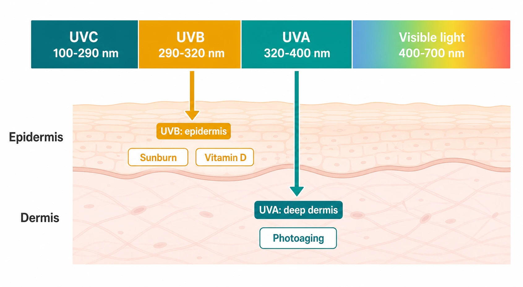
The solar UV radiation reaching the earth's surface is divided into:[2]
- UVA (320–400 nm) — penetrates deep into the dermis; largely responsible for photoaging and tanning, and for most drug- and disease-related photosensitivity reactions (UVA1 is 340–400 nm).
- UVB (290–320 nm) — largely absorbed in the epidermis; the principal cause of sunburn, most skin cancers, and vitamin D synthesis; the dominant action spectrum for many photodermatoses including polymorphic light eruption.
- UVC (100–290 nm) — filtered by the ozone layer and irrelevant to natural disease. [1]
Visible light (400–700 nm) provokes a minority of photodermatoses (notably chronic actinic dermatitis, solar urticaria, and some porphyrias) and is increasingly recognised for hyperpigmentation in skin of colour, addressed by tinted (iron-oxide) sunscreens.[23]
Quick numbers for the examiner
Idiopathic (immune-mediated) photodermatoses [1]
Polymorphic light eruption (PLE)
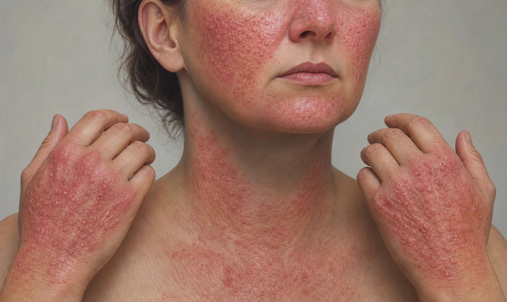
PLE is the commonest photodermatosis, affecting up to 10–20% of populations in temperate climates, with a female predominance and onset in young adulthood. It presents as itchy papules, papulovesicles, plaques, or erythema appearing hours to (more often) a day or two after sun exposure, on sun-exposed sites, and resolving without scarring over days. A hallmark is the "hardening" phenomenon — reduced reactivity as the summer progresses owing to acclimatisation. The action spectrum is usually UVA, sometimes UVB. Diagnosis is largely clinical; phototesting can reproduce lesions and exclude other photodermatoses. Management is graded photoprotection, prophylactic narrowband UVB or PUVA in early spring to induce hardening, antihistamines for itch, and short courses of topical or systemic corticosteroids for flares; severe refractory disease may respond to hydroxychloroquine or azathioprine.[3][4]
PLE - first-line management algorithm (BAD 2022 framework)
Photoprotection - first-line for all
Broad-spectrum SPF 30+ sunscreen with good UVA coverage (PPD 10+, critical wavelength over 370 nm), reapply every 2 hours; UPF 50+ clothing; wide-brimmed hat; avoid peak UV 10 am-4 pm; tinted (iron-oxide) sunscreens if visible-light contribution.
Symptomatic treatment of flares
Oral H1 antihistamines for itch; short course of potent topical corticosteroid (e.g. mometasone furoate 0.1 percent OD for 5 to 7 days) on lesions; rarely a short oral prednisolone taper (e.g. 20 mg for 5 days) for severe flares.
Prophylactic phototherapy - hardening
For patients with predictable annual flares, a 6 to 8-week course of narrowband UVB (or PUVA) starting 4 to 6 weeks before the expected sun exposure induces the 'hardening' phenomenon and prevents or attenuates seasonal flares.
Antimalarial therapy for refractory cases
Hydroxychloroquine 200 to 400 mg daily for 3 to 6 months during the spring and summer reduces flare frequency and severity in PLE that has not responded to photoprotection and hardening; monitor eyes at baseline and 5-yearly.
Immunomodulation for severe disease
Rarely needed; azathioprine 1 to 2 mg/kg/day, ciclosporin 3 to 5 mg/kg/day, or mycophenolate mofetil 1 to 2 g/day for refractory PLE under specialist supervision.
Actinic prurigo
A chronic, intensely pruritic, photo-distributed disorder with onset usually in childhood, strongly associated with HLA-DRB1*04:07, and commoner in Indigenous populations of the Americas. Lesions are excoriated papules, nodules, and lichenified plaques on sun-exposed sites (often the face and dorsal hands) that may extend to covered areas and to the lips (cheilitis) and conjunctiva. It persists year-round (no hardening) and into adult life. Thalidomide (50–100 mg at night, with strict pregnancy-prevention and neuropathy monitoring) is the most effective systemic agent; others include photoprotection, topical corticosteroids, calcineurin inhibitors, and in selected cases ciclosporin, mycophenolate, or omalizumab.[5][6]
Actinic prurigo - clinical severity spectrum
Generalised with mucosal disease
Excoriated papules, nodules, lichenified plaques extending to covered sites (buttocks, axillae) with haemorrhagic crusting, severe cheilitis and conjunctivitis; thalidomide 50 to 100 mg nightly is usually required, with specialist monitoring for neuropathy and teratogenicity.
Chronic actinic dermatitis (CAD)
A chronic, eczematous photo-distributed dermatitis of older men, often with a background of contact or atopic dermatitis, and characterised by an abnormal sensitivity to UVB, UVA, and frequently visible light. The action spectrum is broad. Clinically it resembles a persistent eczema of sun-exposed sites that may generalise to erythroderma, with histological features of chronic eczema and sometimes a CD8+ T-cell pseudolymphoma-like infiltrate. Investigations show a reduced minimal erythema dose (MED) to UVB/UVA and often abnormal responses to visible light on monochromator phototesting. Paradoxically, low-dose phototherapy can induce tolerance (desensitisation). Management combines strict photoprotection, topical corticosteroids and calcineurin inhibitors, azathioprine, ciclosporin, or mycophenolate for moderate-to-severe disease, and mycophenolate as a steroid-sparing option.[7][8]
Solar urticaria
An inducible (physical) urticaria in which UV or visible light triggers wheals within minutes of exposure, resolving within an hour. It can be debilitating and, rarely, systemic/anaphylactic on large-body-surface exposure. The action spectrum is variable (UVB, UVA, or visible light). It is distinguished from other photodermatoses by the rapid onset and fleeting nature. Management combines photoprotection, high-dose (up-dosed) second-generation H1-antihistamines, omalizumab in refractory cases, phototherapy-induced tolerance, and, in selected cases, ciclosporin or intravenous immunoglobulin.[9]
Hydroa vacciniforme
A rare, childhood-onset photodermatosis characterised by recurrent vesiculobullous lesions on sun-exposed sites that heal with varioliform (vacciniform) scarring. Most cases resolve by adolescence/young adulthood. Severe, persistent adult disease, especially with systemic features (fever, hepatosplenomegaly, lymphadenopathy), is associated with EBV-associated T/NK-cell lymphoproliferative disorder and warrants haematology evaluation and EBV monitoring. Photoprotection is the mainstay.[10]
Photoaggravated dermatoses
A number of dermatoses are exacerbated, but not caused, by sunlight: lupus erythematosus (especially subacute cutaneous lupus, characteristically sparing to the paronychia; photo-distributed annular/polycyclic or papulosquamous lesions), dermatomyositis (the shawl/V-sign and Gottron papules), rosacea, pemphigus (particularly pemphigus foliaceus), atopic eczema, seborrhoeic dermatitis, psoriasis (controversially), and pellagra (the Casal necklace). Recognition matters because the light-reactive dermatosis directs the work-up (autoantibodies, biopsy with immunofluorescence) and treatment (antimalarials for lupus; niacin for pellagra).[24][2]
Drug- and chemical-induced photosensitivity
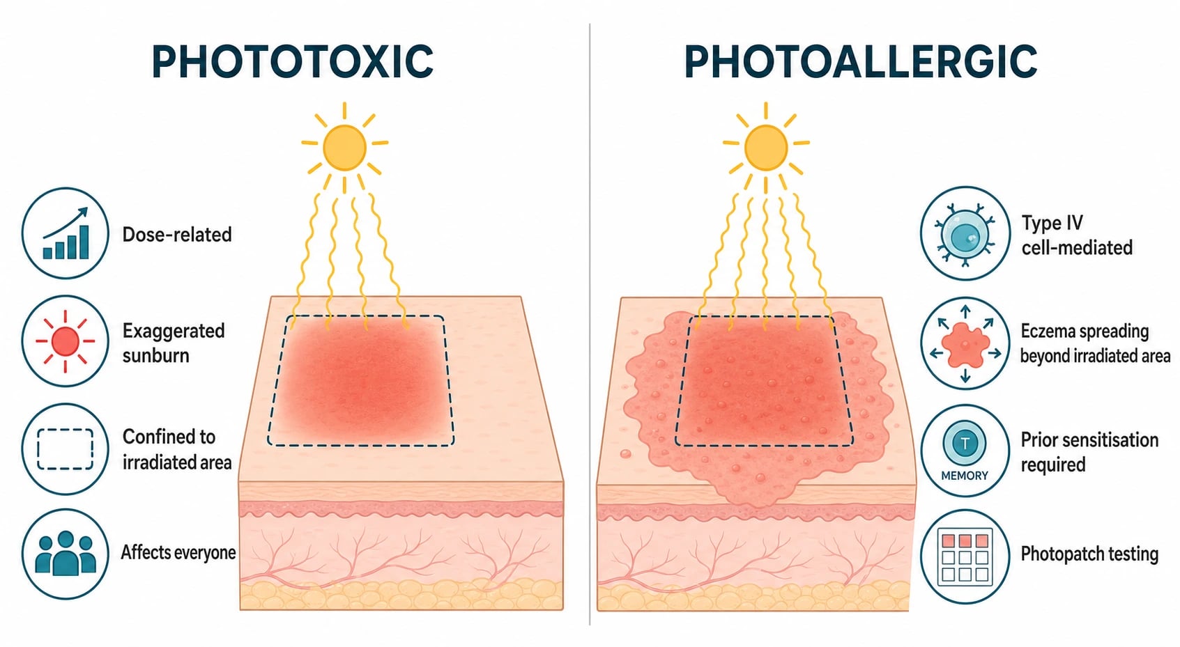
Drug photosensitivity is common and under-recognised, and is divided into phototoxic and photoallergic reactions:[11][12][13]
- Phototoxic reactions are dose-related, non-immunological, and resemble exaggerated sunburn (erythema, oedema, vesiculation, sometimes hyperpigmentation), confined to the irradiated area, occurring in anyone taking enough drug and receiving enough light. The drug (or its metabolite) absorbs UV — usually UVA — generating reactive oxygen species that damage cells.
- Photoallergic reactions are cell-mediated (type IV), require prior sensitisation, are eczematous and pruritic, may spread beyond the irradiated area, and persist at lower exposures once sensitised. Photopatch testing is diagnostic.[15]
Common culprit drugs and agents
High-yield drug classes:[11][12]
- Fluoroquinolones (especially ciprofloxacin, lomefloxacin) and tetracyclines (doxycycline, demeclocycline).
- NSAIDs (piroxicam, ketoprofen, celecoxib, naproxen).
- Sulphonamides and sulfonylureas.
- Phenothiazines (chlorpromazine, promethazine) and thiazide diuretics (hydrochlorothiazide).
- Amiodarone (slate-grey phototoxicity and hyperpigmentation).
- Furosemide, quinidine, dapsone, voriconazole (chronic phototoxicity, accelerated photoaging, and SCC risk on long-term use).
- Psoralens — phytophotodermatitis ("strimmer/limelight dermatitis") from plant contact (citrus, celery, parsnip, fig) plus sunlight, producing painful linear bullae with post-inflammatory hyperpigmentation.[14]
- St John's wort (hypericin).
Mnemonic for culprit photosensitising drug classes
High-yield photosensitising drug classes - mnemonic
CHATS-TAP-VD
Strong UVA-driven phototoxicity; lomefloxacin is the most photo-reactive
Photo-onycholysis with photo-distributed erythema
Slate-grey phototoxic hyperpigmentation in sun-exposed sites
Phototoxic onycholysis; lower with minocycline
Photoallergic eczema; cross-reaction between antibiotic and antidiabetic
Photoallergic contact dermatitis; persistent light reaction can last years
Increased SCC risk in transplant recipients
Plants (citrus, celery, parsnip, fig) plus UVA produce linear bullae
Chronic phototoxicity, accelerated photoaging, increased SCC risk
Photoallergy and dapsone hypersensitivity syndrome
Differential diagnosis of photo-distributed eruptions
A photo-distributed eruption has a wide differential, and the distribution of sparing is a key bedside clue. Sun-exposed sites affected, with sharp sparing of the submental triangle, behind the ears, under the chin, in the nasolabial folds, and beneath spectacle frames, watch straps, bra straps, or ring-covered fingers. Diseases that affect sun-exposed skin fall into several mechanistic groups:[2][11]
Idiopathic (immune-mediated) photodermatoses. Polymorphic light eruption, actinic prurigo, chronic actinic dermatitis, solar urticaria, hydroa vacciniforme, and juvenile spring eruption (a PLE variant affecting the boys' ear helices in spring). [1]
Photoaggravated dermatoses. Lupus erythematosus (systemic, subacute cutaneous, discoid), dermatomyositis (Gottron papules, heliotrope, shawl/V-sign), rosacea, atopic dermatitis (face and neck in summer), seborrhoeic dermatitis, psoriasis (in about 5-10 percent, controversial), pemphigus foliaceus, lichen planus actinicus (hyperpigmented annular plaques on sun-exposed limbs in patients with skin of colour), granuloma annulare, erythema multiforme, and pellagra (niacin deficiency with the Casal necklace and dermatitis-diarrhoea-dementia triad).[24]
Drug and chemical-induced photosensitivity. Phototoxic (tetracyclines, fluoroquinolones, NSAIDs, amiodarone, voriconazole, thiazides, phenothiazines), photoallergic (topical NSAIDs, fragrances, plant allergens), phytophotodermatitis (psoralen-containing plants), and chronic actinic photodermatosis from chemicals. [1]
Genetic and metabolic photodermatoses. Xeroderma pigmentosum, the cutaneous porphyrias (PCT, EPP, hepatoerythropoietic porphyria, congenital erythropoietic porphyria, variegate porphyria, hereditary coproporphyria), Bloom syndrome, Rothmund-Thomson syndrome, Cockayne syndrome, and Hartnup disease (pellagra-like photosensitive rash from impaired tryptophan absorption). [1]
Nutritional photosensitivity. Pellagra (niacin/B3 deficiency, classically from isoniazid, malabsorption, anorexia, or Hartnup disease), and B6 deficiency (sideroblastic anaemia photosensitivity), and kwashiorkor. [1]
Neoplastic and pre-neoplastic. Actinic keratoses, in-situ and invasive squamous cell carcinoma, basal cell carcinoma, lentigo maligna and melanoma, and Merkel cell carcinoma — distribution clues matter. [1]
Infectious. Photo-lupus can be confused with photosensitive viral exanthems; secondary syphilis can mimic PLE; cutaneous leishmaniasis on sun-exposed skin. [1]
The clinical morphology and latency from sun exposure are powerful discriminators: [1]
- Minutes (under 30 minutes) → solar urticaria (wheal-and-flare resolving in under an hour), erythropoietic protoporphyria (burning pain, no blister).
- Hours to 1-2 days → PLE (papules and plaques), phototoxic drug eruption (exaggerated sunburn), acute photodermatitis.
- Days to weeks → photoallergic drug eruption (eczema, may spread), chronic actinic dermatitis, subacute cutaneous lupus, dermatomyositis, lichen planus actinicus.
- Persistent with scarring → lupus (dyspigmentation, scarring alopecia), porphyrias (fragility, milia, hypertrichosis), chronic actinic dermatitis (lichenification), hydroa vacciniforme (varioliform scars), xeroderma pigmentosum (lentiginosis, poikiloderma, cancers). [1]
Genetic and metabolic photodermatoses
Xeroderma pigmentosum (XP)
An autosomal recessive defect in nucleotide excision repair producing extreme UV sensitivity, with onset in early childhood: severe sunburn on minimal exposure, freckle-like lentiginosis and poikiloderma on sun-exposed skin, and a more than 10,000-fold increased risk of skin cancers (basal cell, squamous cell, and melanoma) at a young age, with ocular and neurological involvement (sensorineural deafness, ataxia, cognitive decline) in some complementation groups. Management is absolute photoprotection, early cancer surveillance, and surgical excision of tumours; molecular diagnosis and genetic counselling are essential.[20]
Porphyrias
The porphyrias are metabolic defects of haem biosynthesis; the cutaneous porphyrias present with photo-induced skin fragility and blistering due to accumulation of phototoxic porphyrins that absorb UVA/visible light and generate reactive oxygen species.[19][25]
- Porphyria cutanea tarda (PCT) — the commonest porphyria, characterised by skin fragility, tense bullae, erosions, milia, and hypertrichosis on sun-exposed sites (dorsa of hands, forearms, face), with hyperpigmentation and sclerodermoid change. Trigger factors include alcohol, hepatitis C, oestrogens, iron overload, and haemochromatosis (HFE) mutations. Diagnosis is by a plasma/urine porphyrin profile (elevated uroporphyrin); management combines risk-factor modification (alcohol, oestrogen, HCV treatment), low-dose hydroxychloroquine, and therapeutic venesection.[16][17]
- Erythropoietic protoporphyria (EPP) — presents in early childhood with burning pain, erythema, and oedema within minutes of sun exposure, usually without blistering, with subsequent petechiae and waxy scarring. Accumulation of protoporphyrin (ferrochelatase deficiency) risks gallstones and, in a minority, cholestatic liver failure. Diagnosis is by elevated free erythrocyte protoporphyrin. Afamelanotide (an α-melanocyte-stimulating hormone analogue implant) increases melanin and photoprotection and is licensed for EPP; β-carotene and strict photoprotection are adjuncts.[18][19]
Drug doses in clinical practice
The disorders above have specific, evidence-based systemic therapies. The following dose cards give the starting and target adult doses, mechanism, adverse effects to monitor, and an exam-style tip for each:[5][17][18][26][27]
Hydroxychloroquine
First-line systemic agent for cutaneous lupus erythematosus (discoid, SCLE) and as adjunct in PLE; used in low dose in PCT
Dose
200 to 400 mg orally daily (max 6.5 mg/kg lean body weight per day to minimise retinal risk)
Thalidomide
Most effective systemic agent for moderate-to-severe actinic prurigo unresponsive to topical therapy
Dose
50 to 100 mg orally at night (some centres start 50 mg and titrate up to 200 mg); minimum effective dose maintained long-term
Afamelanotide (Scenesse)
First-in-class alpha-melanocyte-stimulating hormone analogue licensed for erythropoietic protoporphyria (EPP)
Dose
16 mg subcutaneous controlled-release implant every 2 months (inserted above the anterior supra-iliac crest under local anaesthetic); typically 3 to 4 implants per year
Beta-carotene
Adjunct photoprotective agent for EPP and other cutaneous porphyrias; historical first-line before afamelanotide
Dose
50 to 300 mg orally daily (adults); titrate to maintain serum carotene at 600 to 800 microgram/dL; paediatric dose 30 to 150 mg/day by weight
Nicotinamide (vitamin B3, amide form)
Skin-cancer chemoprevention in high-risk immunocompetent patients (more than 2 NMSC in 5 years); also Pellagra and Hartnup disease
Dose
500 mg orally twice daily (1 g/day); ONTRAC trial dose. Higher doses (1 to 3 g/day) have been used in Pellagra
Hydroxychloroquine (low-dose) for PCT
Low-dose regimen (100 to 200 mg twice weekly) mobilises hepatic porphyrin stores in PCT
Dose
100 mg orally twice weekly (low-dose PCT regimen); standard 200 to 400 mg daily for lupus
Xeroderma pigmentosum surveillance and prevention
Children with xeroderma pigmentosum require lifelong structured photoprotection and cancer surveillance, ideally delivered through a dedicated XP clinic. The regimen is more intensive than for any acquired photodermatosis and includes:[20]
XP - lifetime cancer-surveillance and prevention algorithm
Strict environmental control
UV-filtered home, school, and car windows (UVA passes through glass); avoid outdoor daylight between 10 am and 4 pm; turn off UV-emitting fluorescent lamps (or replace with UV-filtered LED); no sunlamps or tanning beds.
Cover-up clothing and physical sunscreens
UPF 50+ long-sleeved tops and trousers, wide-brimmed hats (over 7 cm brim), UV-blocking sunglasses, broad-spectrum SPF 50+ PA++++ mineral (zinc oxide, titanium dioxide) sunscreen reapplied every 2 hours and after swimming.
Skin examination every 3 to 6 months
Full-skin examination by a dermatologist familiar with XP; high-quality clinical and dermoscopic photographs of all nevi and freckles for change-detection; baseline and serial total-body photography from diagnosis.
Tissue-sparing excision of lesions
Treat actinic keratoses aggressively (topical 5-fluorouracil, imiquimod, ingenol mebutate, photodynamic therapy); excise suspicious lesions early with narrow margins to preserve skin; Mohs micrographic surgery where feasible.
Neurological and ophthalmic surveillance
Annual audiology (DeSanctis-Cacchione and Cockayne-like neurological phenotypes), ophthalmology (lid cancers, keratitis, exposure to UV), and developmental review; arrange genetic counselling for the family and offer prenatal/preimplantation diagnosis.
Special populations
Children and adolescents. PLE has its onset in young adulthood but actinic prurigo, hydroa vacciniforme, xeroderma pigmentosum, and erythropoietic protoporphyria all present in childhood. EPP in particular causes distress and avoidance of outdoor activity — schools must allow broad-brimmed hats, long sleeves, and access to shaded areas. Afamelanotide is licensed for adults but is used off-label in some paediatric EPP centres. XP demands a structured school environment with filtered lighting and constant photoprotection.[10][18][20]
Skin of colour. PLE, discoid lupus, lichen planus actinicus, and pellagra may be missed or under-diagnosed in patients with deeply pigmented skin because erythema is masked; hyperpigmentation is often the presenting sign. Visible light contributes to hyperpigmentation in skin of colour and is not blocked by standard UV sunscreens — tinted (iron-oxide) sunscreens are essential. Lupus photosensitivity in skin of colour is associated with greater disease damage and scarring alopecia.[2][23]
Pregnancy and lactation. Hydroxychloroquine is safe and should be continued in pregnancy for lupus — flare risk on stopping outweighs drug risk. Thalidomide is absolutely contraindicated and the pregnancy-prevention programme must be in place before prescribing. Topical corticosteroids and calcineurin inhibitors are preferred for skin disease flares; nbUVB phototherapy is acceptable as second-line. Voriconazole, isotretinoin, and tetracyclines are contraindicated.[5][6]
Organ transplant recipients. Chronic immunosuppression plus photosensitising drugs (azathioprine, ciclosporin, voriconazole) gives a 50 to 65-fold increased risk of cutaneous SCC. Aggressive photoprotection, regular skin surveillance, and consideration of switching from azathioprine to mTOR inhibitors (sirolimus, everolimus) reduces SCC risk in randomised trials.[11][12]
Investigations
A structured approach combines history (onset, latency, morphology, drug and chemical exposure, family history, seasonality), examination (distribution, scarring), and targeted testing:[2][15]
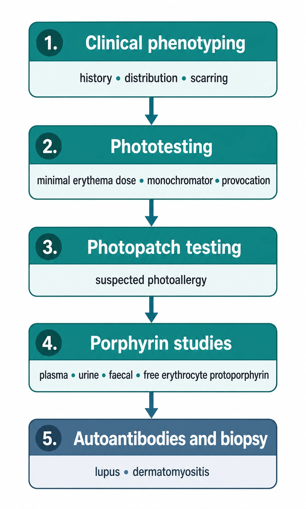
- Minimal erythema dose (MED) — the lowest UV dose producing perceptible erythema at 24 hours; a reduced MED to UVB/UVA indicates abnormal sensitivity.
- Monochromator phototesting — defines the action spectrum and abnormal responses across UVB, UVA, and visible light; essential for diagnosing chronic actinic dermatitis and solar urticaria.
- Provocation phototesting — repeated sub-erythemogenic UVA/UVB exposure to reproduce PLE or solar urticaria.
- Photopatch testing — suspected photoallergic drug/chemical reactions; allergens applied and irradiated with UVA.[15]
- Porphyrin studies — plasma, urine, and faecal porphyrins plus free erythrocyte protoporphyrin; cutaneous porphyrias show a characteristic pattern.
- Autoantibodies (ANA, Ro/SSA, La/SSB) and skin biopsy with immunofluorescence for suspected lupus and dermatomyositis.
Landmark trials and guidelines
The evidence base for photodermatology is dominated by small physiology studies supplemented by a few landmark trials and major consensus guidelines (BAD, EURO-PDT). Four anchor studies/guidelines define current practice:[26][27][28][29]
Langendonk phase 3 trials of afamelanotide in EPP
N Engl J Med (Langendonk JG et al.)
Two parallel multicentre randomised double-blind placebo-controlled phase 3 trials in the European Union and the United States (n = 168 EPP patients), 16 mg subcutaneous controlled-release implant every 2 months for up to 9 months
Key finding
Afamelanotide increased the duration of pain-free sun exposure (primary endpoint) by approximately 70 hours over 6 months compared with placebo, improved quality of life, and had an acceptable safety profile
Practice change
Established afamelanotide (Scenesse) as the first licensed disease-specific therapy for EPP; superseded beta-carotene as first-line in jurisdictions where it is licensed
ONTRAC trial - nicotinamide chemoprevention of NMSC
N Engl J Med (Chen AC et al.)
Phase 3 randomised double-blind placebo-controlled trial (n = 386 high-risk immunocompetent patients with at least two NMSCs in the previous 5 years); nicotinamide 500 mg orally twice daily for 12 months
Key finding
Nicotinamide reduced the rate of new non-melanoma skin cancers by 23 percent at 12 months compared with placebo, with significant reductions in actinic keratoses and an excellent safety profile
Practice change
Established oral nicotinamide 1 g/day as a chemopreventive option for high-risk immunocompetent NMSC patients; a follow-up ONTRANS trial in transplant recipients was less conclusive, so nicotinamide is not yet recommended in that population
EURO-PDT consensus on photodynamic therapy
J Eur Acad Dermatol Venereol (Morton CA et al.)
European consensus guideline on topical photodynamic therapy (PDT) for actinic keratosis, Bowen's disease, and basal cell carcinoma, developed by the European Society for Photodynamic Therapy in Dermatology (EURO-PDT) and the European Dermatology Forum
Key finding
PDT is a first-line field-directed treatment for actinic keratosis and field cancerization with excellent cosmetic outcomes; daylight PDT (when available) is as effective as conventional red-light PDT and substantially less painful
Practice change
Codified PDT as standard-of-care for multiple AK and field cancerization; supported the global shift from conventional to daylight PDT where climate permits
BAD/BPG guidelines for narrowband UVB phototherapy 2022
Br J Dermatol (Goulden V et al.)
British Association of Dermatologists (BAD) and British Photodermatology Group (BPG) evidence-based guideline for narrowband UVB phototherapy using GRADE methodology, covering PLE, solar urticaria, actinic prurigo, photoaggravated eczema, and hydroa vacciniforme
Key finding
NB-UVB is the preferred desensitisation regimen (hardening) for PLE and is effective for solar urticaria and actinic prurigo; PUVA is reserved for NB-UVB failures; treatment protocols, contraindications, and consent are standardised
Practice change
Standardised UK and international practice for nbUVB phototherapy; explicit recommendation of NB-UVB over PUVA for first-line hardening in PLE
Sunscreen science and photoprotection
Photoprotection is the foundation of management across all photodermatoses and comprises behavioural (sun avoidance, timing), physical (clothing, hats, sunglasses, window films), and chemical (sunscreens) measures.[2][21]
- SPF (sun protection factor) quantifies protection against UVB-induced erythema (the MED ratio with/without sunscreen); SPF is measured under standardised, high-application conditions that most users do not replicate, so real-world protection is lower than labelled.
- UVA protection is expressed by systems such as PPD (persistent pigment darkening) and the EU UVA circle (requiring UVA protection to be at least one-third of SPF). Broad-spectrum protection (UVB + UVA) is mandatory for photosensitivity patients.
- Organic (chemical) filters absorb UV (e.g., avobenzone, octocrylene, Tinosorb S/M, mexoryl); inorganic (physical) filters (zinc oxide, titanium dioxide) reflect and absorb and are preferred for sensitive and paediatric skin.
- Visible-light protection requires iron-oxide tinted sunscreens, important for hyperpigmentation in skin of colour and for visible-light photodermatoses (CAD, solar urticaria).[23]
- Sunscreen and vitamin D — routine sunscreen use does not cause vitamin D deficiency in the general population; high-risk photosensitivity patients may require oral vitamin D supplementation.[22]
Management by disorder
- Idiopathic photodermatoses — photoprotection, prophylactic phototherapy (hardening) for PLE, thalidomide for actinic prurigo, azathioprine/mycophenolate/ciclosporin for severe CAD, up-dosed antihistamines and omalizumab for solar urticaria.
- Drug photosensitivity — withdraw the culprit, substitute a non-photosensitising alternative, photoprotection, and topical corticosteroids for photoallergic reactions.
- Porphyria cutanea tarda — risk-factor modification, low-dose hydroxychloroquine, and venesection.[17]
- Erythropoietic protoporphyria — afamelanotide, β-carotene, strict photoprotection, and liver surveillance.[18]
- Xeroderma pigmentosum — absolute photoprotection and rigorous skin-cancer surveillance.
- Photoaggravated dermatoses — treat the underlying disease (antimalarials for lupus, niacin for pellagra).[24]
Clinical pearl
[1] [1]Photoprotection quick numbers
SLE-PROTECT - photoprotection essentials
Apply 30 min before exposure, reapply q2h
Lower lip is a common site for lupus cheilitis and SCC
UVA/UVB blocking; wraparound style; cataract prevention
Reflect UV; broad-spectrum; safe for sensitive skin
Or after swimming, sweating, towel drying
Seek shade, plan outdoor activities early/late
Critical for hyperpigmentation in skin of colour
UVA passes through standard window glass
Long sleeves, wide-brimmed hat, sunglasses
Adjunct to sunscreen; reduce oxidative damage
Red flags [1]
Exam application bank (NEET-PG / INICET)
One-line answer
Photosensitivity disorders are cutaneous reactions to ultraviolet and visible radiation, spanning idiopathic photodermatoses (polymorphic light eruption, actinic prurigo, chronic actinic dermatitis, solar urticaria, hydroa vacciniforme), photoaggravated dermatoses (lupus, dermatomyositis), drug-induced phototoxic and photoallergic reactions, and genetic/metabolic disorders (xeroderma pigmentosum, porphyrias). Fellowship-level assessment demands mastery of the UV spectrum and skin photobiology, the clinical morphologies and action spectra, diagnostic phototesting (minimal erythema dose, monochromator phototesting, photopatch testing) and porphyrin studies, the culprit drug lists, sunscreen science (SPF, UVA protection, broad-spectrum, organic vs inorganic filters), and disorder-specific management from photoprotection and antimalarials through thalidomide for actinic prurigo and afamelano
Worked stems (answer without another resource)
Stem 1 — Classic presentation. Map symptoms to mechanism; name the first investigation and first treatment step with dose/route if drug therapy is standard. [1]
Stem 2 — Unstable / complicated. List red flags that force immediate resuscitation, theatre, ICU, antidote, or reperfusion — and what you do in the first 15 minutes. [1]
Stem 3 — Atypical group. Elderly, pregnancy, child, or immunocompromised: how presentation and thresholds change. [1]
Stem 4 — Differential trap. Name the three closest mimics and one discriminator for each. [1]
Stem 5 — Disposition. Who goes home with safety-netting, who is admitted, who needs HDU/ICU/theatre, and what follow-up is mandatory. [1]
Rapid viva checklist
- Definition + classification
- Pathophysiology chain
- Bedside signs / criteria
- Score with exact components (if any)
- Emergency bundle
- Definitive therapy with doses
- Complications of disease and of treatment
- Special populations
- Guideline/trial name if classic
- Three exam traps
Coverage self-check
If you cannot answer any stem above from this page alone, re-read the matching section — the page is intended to be self-sufficient for final-prof and NEET-PG/INICET questions on Photosensitivity / Photodermatoses.
[1]References
- [1]Abdel Azim S, Bainvoll L, Vecerek N, et al. Sunscreens part 1: Mechanisms and efficacy J Am Acad Dermatol, 2025.PMID 38772426
- [2]Passeron T, Lim HW, Goh CL, et al. Photoprotection according to skin phototype and dermatoses: practical recommendations from an expert panel J Eur Acad Dermatol Venereol, 2021.PMID 33764577
- [3]Kadurina M, Kazandjieva J, Bocheva G. Immunopathogenesis and management of polymorphic light eruption Dermatol Ther, 2021.PMID 34676645
- [4]Guarrera M. Polymorphous Light Eruption Adv Exp Med Biol, 2017.PMID 29124691
- [5]Valbuena MC, Muvdi S, Lim HW. Actinic prurigo Dermatol Clin, 2014.PMID 24891055
- [6]Ross G, Foley P, Baker C. Actinic prurigo Photodermatol Photoimmunol Photomed, 2008.PMID 18811871
- [7]Paek SY, Lim HW. Chronic actinic dermatitis Dermatol Clin, 2014.PMID 24891057
- [8]Hawk JL. Chronic actinic dermatitis Photodermatol Photoimmunol Photomed, 2004.PMID 15533239
- [9]Goetze S, Elsner P. Solar urticaria J Dtsch Dermatol Ges, 2015.PMID 26612794
- [10]Saraswat N, Tripathy DM, Kumar S. Hydroa Vacciniforme JAMA Dermatol, 2023.PMID 37436742
- [11]Blakely KM, Drucker AM, Rosen CF. Drug-Induced Photosensitivity-An Update: Culprit Drugs, Prevention and Management Drug Saf, 2019.PMID 30888626
- [12]Montgomery S, Worswick S. Photosensitizing drug reactions Clin Dermatol, 2022.PMID 35190066
- [13]Honari G. Photoallergy Rev Environ Health, 2014.PMID 25274941
- [14]Ellis CR, Elston DM. Psoralen-Induced Phytophotodermatitis Dermatitis, 2021.PMID 33273237
- [15]Isaksson M, Bruze M. Photopatch testing Clin Dermatol, 1997.PMID 9255472
- [16]Bleasel NR, Varigos GA. Porphyria cutanea tarda Australas J Dermatol, 2000.PMID 11105361
- [17]Leaf RK, Dickey AK. Porphyria cutanea tarda: a unique iron-related disorder Hematology Am Soc Hematol Educ Program, 2024.PMID 39644053
- [18]Lecha M, Puy H, Deybach JC. Erythropoietic protoporphyria Orphanet J Rare Dis, 2009.PMID 19744342
- [19]Dickey AK, Leaf RK, Balwani M. Update on the Porphyrias Annu Rev Med, 2024.PMID 37540847
- [20]Leung AK, Barankin B, Lam JM, et al. Xeroderma pigmentosum: an updated review Drugs Context, 2022.PMID 35520754
- [21]Guan LL, Lim HW, Mohammad TF. Sunscreens and Photoaging: A Review of Current Literature Am J Clin Dermatol, 2021.PMID 34387824
- [22]Passeron T, Bouillon R, Callender V, et al. Sunscreen photoprotection and vitamin D status Br J Dermatol, 2019.PMID 31069788
- [23]Lyons AB, Trullas C, Kohli I, et al. Photoprotection beyond ultraviolet radiation: A review of tinted sunscreens J Am Acad Dermatol, 2021.PMID 32335182
- [24]Hołubiec P, Leończyk M, Staszewski F, et al. Pathophysiology and clinical management of pellagra - a review Folia Med Cracov, 2021.PMID 34882669
- [25]Muschalek W, Hermasch MA, Poblete-Gutiérrez P, et al. The Porphyrias J Dtsch Dermatol Ges, 2022.PMID 35304965
- [26]Langendonk JG, Balwani M, Anderson KE, et al. Afamelanotide for Erythropoietic Protoporphyria N Engl J Med, 2015.PMID 26132941
- [27]Chen AC, Martin AJ, Choy B, et al. A Phase 3 Randomized Trial of Nicotinamide for Skin-Cancer Chemoprevention N Engl J Med, 2015.PMID 26488693
- [28]Morton CA, Szeimies RM, Sidoroff A, Braathen LR. European guidelines for topical photodynamic therapy part 1: treatment delivery and current indications - actinic keratoses, Bowen's disease, basal cell carcinoma J Eur Acad Dermatol Venereol, 2013.PMID 23181594
- [29]Goulden V, Ling TC, Babakinejad M, et al. Covert poisoning with difenacoum: diagnosis and follow-up difficulties Clin Chem Lab Med, 2022.PMID 35621008