Dermatology · Medicine
Pityrosporum (Malassezia) and associated skin diseases
Also known as Malassezia-associated skin diseases · Pityriasis versicolor · Seborrhoeic dermatitis (Malassezia) · Malassezia folliculitis · Pityrosporum ovale/orbiculare
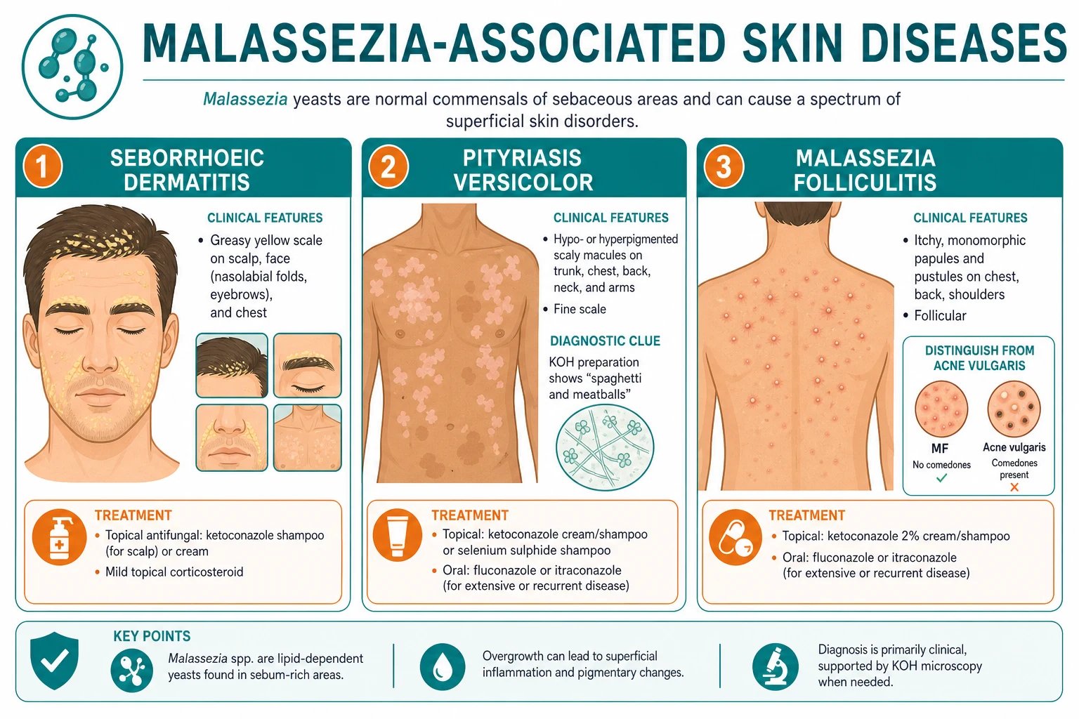
Malassezia species are lipophilic, lipid-dependent basidiomycetous yeasts of the normal cutaneous mycobiome that become pathogenic when heat, humidity, sebum, occlusion, or immunosuppression tip the host–commensal balance. They cause four clinical syndromes: (1) Pityriasis versicolor — hypo- or hyperpigmented, finely scaling macules on the trunk; KOH shows the diagnostic 'spaghetti and meatballs' and Wood's lamp a pale yellow-gold fluorescence; treat with topical ketoconazole 2% shampoo or selenium sulfide 2.5%, with oral itraconazole 200 mg daily for 7 days or fluconazole 300 mg weekly for 2 doses for extensive disease. (2) Malassezia folliculitis — itchy monomorphic follicular papules and pustules on the upper trunk with no comedones (the key distinction from acne); treat with topical ketoconazole or an oral azole. (3) Seborrhoeic dermatitis/dandruff — greasy yellow scale on scalp, face, and sternal chest (M. restricta, M. globosa; see separate topic). (4) Invasive/systemic Malassezia — catheter-related fungaemia in low-birth-weight neonates on lipid emulsions (M. furfur, M. pachydermatis); remove the line and treat with amphotericin B +/- flucytosine. Recurrence is common in humid climates; maintenance therapy is central.
On this page & tools
Your progress
Saved locally on this device.
Exam tags
Red flags
Overview & Definition
Malassezia (formerly Pityrosporum) is a genus of lipophilic, lipid-dependent, dimorphic basidiomycetous yeasts that colonise the superficial stratum corneum of warm-blooded vertebrates. In humans they form part of the normal cutaneous mycobiome, residing chiefly in sebum-rich areas — the scalp, face (especially the nasolabial folds and retroauricular skin), upper chest, back, and the keratinised plug of hair follicles. Colonisation begins in infancy and reaches peak density at puberty, when sebaceous gland activity rises. Most people carry Malassezia without consequence; disease emerges only when the balance between commensal and host is disturbed by heat and humidity, increased sebum, occlusion, sweating, immunosuppression, or broad-spectrum antibiotic or corticosteroid exposure.[1][14]
The genus gives rise to four recognisable clinical syndromes. The two archetypal cutaneous diseases are pityriasis versicolor (a superficial disorder of pigmentation) and Malassezia folliculitis (a follicular papulopustular eruption that mimics acne). Malassezia also plays a central pathogenic role in seborrhoeic dermatitis and dandruff (covered in a dedicated topic but discussed here because the same organism and the same topical agents are involved). The fourth syndrome — invasive or systemic Malassezia infection — is uncommon but clinically important, occurring almost exclusively in low-birth-weight neonates and immunocompromised adults with indwelling central venous catheters receiving lipid-containing parenteral nutrition, where the lipid emulsion provides the substrate the organism cannot synthesise for itself.[12][13]
A single unifying principle underpins the whole topic and explains almost every clinical feature: Malassezia cannot make its own fatty acids. Its genome lacks a functional fatty-acid synthase gene, so the yeast depends absolutely on exogenous lipids — supplied in the skin by sebaceous triglycerides and in the bloodstream by infused lipid emulsion. This lipid dependence is why Malassezia lives where sebum is plentiful, why occlusion and sweating precipitate folliculitis, and why a neonate on intralipid is the textbook host for invasive disease.[1]
Classification — the species and what they do
More than eighteen Malassezia species are now recognised by molecular methods, but a handful account for the vast majority of human disease. Different species predominate at different body sites and in different diseases, a fact examiners use to probe depth. The clinically important species, their distinguishing features, and their disease associations are summarised below.[1]
The remaining species — M. sympodialis (skin, seborrhoeic dermatitis, atopic dermatitis overlap), M. obtusa, M. slooffiae, M. dermatis, M. japonica, and others — are isolated less often and are examined rarely. The species-specific associations worth committing to memory are: pityriasis versicolor — M. globosa (and M. furfur); seborrhoeic dermatitis/dandruff — M. restricta and M. globosa; invasive neonatal disease — M. furfur and M. pachydermatis.[1][12]
Epidemiology & Risk Factors
Pityriasis versicolor is cosmopolitan but strikingly climate-dependent. In temperate regions it accounts for a small fraction of dermatology presentations, whereas in hot, humid tropical and subtropical climates it is one of the commonest skin disorders, with reported prevalence reaching 40 to 50 per cent of the population in some equatorial series. Disease peaks in adolescence and young adulthood, paralleling the sebum surge of puberty, and is uncommon before puberty and after the sixth decade. There is no true sex predilection, though hyperpigmented disease is more conspicuous and more often brought to medical attention in lighter skin, while hypopigmented disease is more visually prominent in darker skin.[4][5]
Malassezia folliculitis shares the same young-adult, sebum-rich demographic but is disproportionately seen in immunosuppressed patients — particularly those with advanced HIV infection (CD4 count under 200 to 300), solid-organ transplant recipients, and patients on systemic corticosteroids or prolonged broad-spectrum antibiotics. Occlusion (tight clothing, heavy emollients, wetsuits, occupational sweating), intense exercise with sweat, and humid environments are consistent precipitants.[6][7]
Seborrhoeic dermatitis affects roughly 3 to 5 per cent of immunocompetent adults but is dramatically over-represented in HIV/AIDS (where prevalence can reach 30 to 80 per cent and severity parallels immune decline) and in neurological disease, especially Parkinson disease, where it may be severe and refractory. The link to Parkinson disease is so consistent that sudden-onset or worsening seborrhoeic dermatitis in an older patient warrants a neurological assessment.[3][9][11]
Invasive Malassezia infection is rare and almost confined to the neonatal intensive care unit: low-birth-weight and preterm infants with central venous catheters receiving lipid-containing parenteral nutrition are the classic hosts. M. furfur causes most cases via the lipid-dependent bloodstream route, while M. pachydermatis — acquired from a hospital or pet reservoir — is notable as the species that can grow on ordinary media, sometimes leading to delayed recognition. Adult cases occur in immunocompromised patients with long-term central lines, especially on home parenteral nutrition.[12][13][14]
Pathophysiology
The pathophysiology of every Malassezia syndrome traces back to lipid dependence and the yeast-to-mycelium transition. On healthy skin the organism lives as a budding yeast (blastospore) in the stratum corneum, subsisting on sebum-derived triglycerides and saturated fatty acids. When the local environment shifts — heat, humidity, sweating, increased sebum, occlusion, or a change in skin barrier function — the organism converts to a mycelial (hyphal) form, penetrates the stratum corneum, and overgrows. This dimorphic switch is the histological signature of pityriasis versicolor, where both hyphae and budding yeast are visible together.[1][4]
In pityriasis versicolor the colour change is not caused by the yeast staining the skin but by Malassezia-derived metabolites acting on melanocytes. The organism secretes lipases that cleave sebum triglycerides into free fatty acids, and several of these (notably azelaic acid, produced by a Malassezia lipoxygenase) competitively inhibit tyrosinase, the rate-limiting enzyme of melanogenesis. Reduced melanin production in lesional skin produces the characteristic hypopigmentation, which is most conspicuous in darker skin types. Conversely, in lighter skin the dominant lesion is often hyperpigmented, driven by inflammatory mediators and increased epidermal turnover rather than pigment loss; irritation, scratching, and a heavier inflammatory burden favour hyperpigmentation. The same lesion can therefore look pale on one patient and brown on another — the basis of the name versicolor ("of changing colour").[4][5]
LIPID
Malassezia cannot synthesise its own fatty acids — the defining metabolic constraint
Azelaic acid (a Malassezia lipoxygenase product) blocks melanogenesis -> hypopigmentation in dark skin
Yeast overgrows within the hair follicle -> keratin plug + free fatty acids -> Malassezia folliculitis
Malassezia antigens + lipid metabolites drive the abnormal keratinocyte response of seborrhoeic dermatitis (M. restricta, M. globosa)
Intralipid in TPN supports bloodstream growth; biofilm forms on the catheter -> invasive disease in neonates
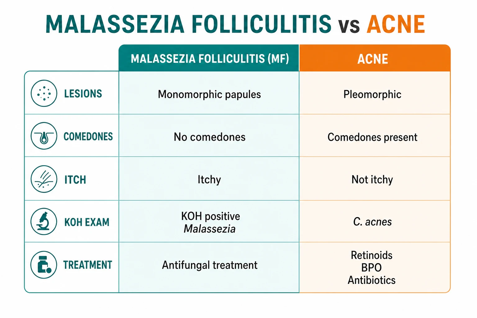
Malassezia folliculitis arises when the yeast proliferates within the hair follicle rather than on the surface. Accumulating organisms and their free-fatty-acid metabolites generate a follicular plug and provoke a neutrophilic inflammatory response, producing the characteristic monomorphic follicular papule or pustule. Crucially, the process does not generate comedones — the hallmark of acne vulgaris — because there is no keratinocyte cohesion abnormality at the infundibulum and no Cutibacterium acnes-driven microcomedone. The prominent itch reflects irritant and type I hypersensitivity mechanisms rather than the deeper, less itchy inflammation of acne.[6][7]
Seborrhoeic dermatitis and dandruff are not simple overgrowth but an abnormal host response to Malassezia antigens and lipids. M. restricta and M. globosa predominate on the scalp; their lipases release irritating free fatty acids (especially oleic acid), and their antigenic components (including malassezin) provoke keratinocyte activation, increased epidermal turnover (producing the visible scale), and a mixed non-immune and immune inflammatory infiltrate. The organism is necessary but not sufficient — its mere presence on healthy skin without inflammation shows that an individual susceptibility (barrier function, sebum composition, immune tone, neurological and HIV-related factors) determines who develops disease.[2][3][9]
Invasive disease is a story of substrate and access. When a lipid emulsion (such as 20% intralipid) is infused through a central venous catheter, the bloodstream and the catheter biofilm acquire the very substrate Malassezia cannot make. The organism — introduced from the patient's own skin (M. furfur) or from a zoonotic or environmental reservoir (M. pachydermatis, classically tracked to hospital staff pets or colonised incubators) — forms a biofilm on the catheter and seeds the bloodstream. Premature neonates are vulnerable because of immature immune defences, indwelling lines, frequent lipid exposure, and thin, easily colonised skin. The result is a catheter-related fungaemia with possible pneumonitis, thrombocytopenia, and persistent sepsis that resolves only when the substrate (lipid) and the conduit (catheter) are removed.[12][13][14]
Clinical Presentation
Pityriasis versicolor
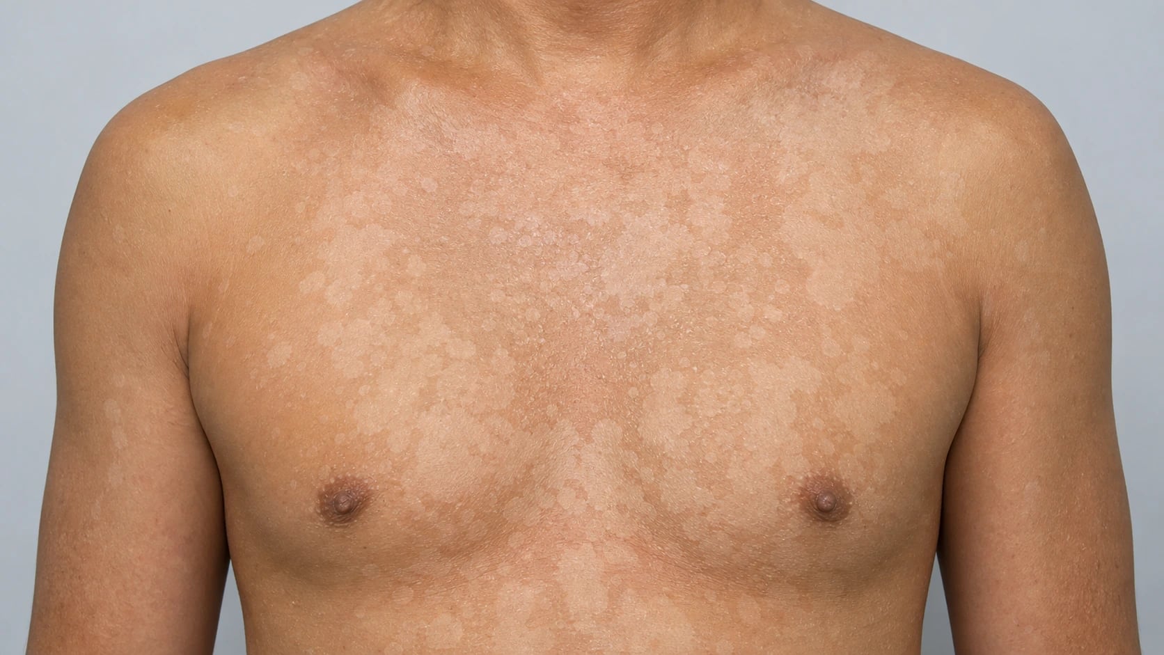
Pityriasis versicolor presents with multiple, discrete and then confluent, hypo- or hyperpigmented macules and thin patches covered in a fine, branny scale. The classical distribution is the upper trunk (chest, upper back, and shoulders), the neck, and the upper arms; the face is involved more often in children and in immunosuppressed patients. Individual lesions are round to oval, several millimetres to several centimetres across, and tend to coalesce over weeks into larger geographic patches. Pigmentation is the presenting complaint: in darker skin the lesions are typically hypopigmented (pale, sharply defined), while in lighter skin they are often hyperpigmented (fawn, salmon, or light brown), though either pattern can occur in any skin type. Symptoms are mild — slight itching is common, but many patients are entirely asymptomatic and present because of the cosmetic colour change.[4][5][10]
The scale is the key clinical clue and is easily missed. Stretching the skin or lightly scraping the surface accentuates a fine powdery scale (the scale sign, or Besnier's sign), which immediately distinguishes pityriasis versicolor from the completely smooth, scale-free depigmentation of vitiligo. In tropical climates hypopigmented disease is common enough that a truncal pale patch is the single most likely cause; the absence of scale (and thus the suspicion of vitiligo or leprosy) should prompt a scrape rather than a biopsy.[4]
Malassezia folliculitis
Malassezia (Pityrosporum) folliculitis presents as small (1 to 3 mm), monomorphic, erythematous, follicular papules and pustules on the upper trunk (chest, back, and shoulders) and upper arms, and less often on the face, neck, and buttocks. Two features distinguish it from acne vulgaris and should be sought deliberately: the eruption is monomorphic (all lesions at the same stage, no open or closed comedones) and strikingly itchy — the itch is often disproportionate to the visible lesion count and is the feature that brings patients back after "acne" treatment has failed. Lesions cluster in crops and may worsen with heat, sweating, occlusion, and exercise; they spare the comedone-bearing central face that dominates true acne.[6][7]
Malassezia folliculitis is over-represented in immunosuppressed patients — particularly those with HIV, transplant recipients, and patients on systemic steroids or prolonged antibiotics — and the diagnosis should be considered whenever an "acneiform" trunk eruption is monomorphic, itchy, comedone-free, and unresponsive to conventional acne therapy.[6]
Seborrhoeic dermatitis (the Malassezia role)
Seborrhoeic dermatitis and dandruff present with greasy, yellowish scale on the scalp (dandruff in its mildest form), the face (nasolabial folds, alae nasi, eyebrows, glabella, retroauricular folds), the external auditory meatus, and the sternal chest. Flexural variants occur in the axillae, groin, and submammary folds, and an infantile form (cradle cap) is common. Itching and erythema are mild to moderate. The role of Malassezia (M. restricta, M. globosa) is central to pathogenesis and underpins the use of topical antifungals; the condition is covered in detail in the seborrhoeic dermatitis topic.[2][8][9]
Invasive (systemic) Malassezia
Invasive disease is a diagnosis of the neonatal intensive care unit. The preterm, low-birth-weight infant with a central venous catheter on lipid-containing parenteral nutrition develops culture-negative or refractory sepsis, often with respiratory deterioration (pneumonitis), thrombocytopenia, and nonspecific signs of neonatal sepsis (apnoea, temperature instability, feeding intolerance). The link to the lipid infusion and the central line is the clue: deterioration during or soon after lipid administration, or persistence despite antibacterial therapy, should raise the diagnosis. Adult cases mirror this picture in immunocompromised patients with long-term central access, especially on home parenteral nutrition.[12][13]
Atypical presentations
Examiners test the edges of presentation. In HIV/AIDS, pityriasis versicolor, Malassezia folliculitis, and seborrhoeic dermatitis can all be severe, extensive, atypical in distribution (face, flexures), and refractory to standard topical therapy; their appearance or worsening may be the first clue to immune decline and mandates HIV testing. In darker skin, the hypopigmented variant of pityriasis versicolor is visually dramatic and generates disproportionate distress, driving presentation. In infants, seborrhoeic dermatitis (cradle cap) and rare neonatal invasive disease define the two ends of the age spectrum.[1][9]
Differential Diagnosis
The differential diagnosis of Malassezia disease splits along clinical lines: the pigmentary differential of pityriasis versicolor, the follicular differential of Malassezia folliculitis, and the seborrhoeic differential covered in the dedicated topic. Examiners reward candidates who can name the mimics and the single feature that separates each from Malassezia disease. [1]
The cardinal pigmentary mimic of hypopigmented pityriasis versicolor is vitiligo. Vitiligo produces chalk-white, completely depigmented (not merely hypopigmented) macules with no scale, no organisms on KOH, and bright blue-white accentuation under Wood's lamp — in stark contrast to the finely scaling, yellow-gold-fluorescent lesion of pityriasis versicolor. Pityriasis alba is a mild atopic dermatitis variant: ill-defined, slightly scaly, hypopigmented patches, usually on the face of children, self-limiting and associated with atopy. Post-inflammatory hypopigmentation follows any preceding inflammation; history is the clue. Leprosy produces hypoanaesthetic, hypopigmented lesions with thickened nerves and is excluded by sensation testing and slit-skin smear; in endemic regions it is the dangerous not-to-miss mimic. Secondary syphilis can produce a macular truncal rash but typically involves the palms and soles and is accompanied by systemic symptoms and serology.[4][5]
The hyperpigmented variant of pityriasis versicolor is confused with melasma (face, sun-related, women, pregnancy/OCP), post-inflammatory hyperpigmentation (history of preceding inflammation), confluent and reticulate papillomatosis (velopapillomatous brown papules coalescing centrally on the trunk, especially young women), and — in the flexures — erythrasma, distinguished instantly by its coral-red Wood's lamp fluorescence versus the pale yellow-gold of pityriasis versicolor.[4]
The follicular differential centres on the folliculitis versus acne vulgaris question, which is examined relentlessly because the two share a trunk distribution and a young-adult demographic but require entirely different treatment. Acne vulgaris has comedones (open and closed), is polymorphic (comedones, papules, pustules, nodules, cysts, scars), is not usually itchy, is driven by Cutibacterium acnes with androgen-driven sebum, and responds to retinoids, benzoyl peroxide, and antibiotics. Malassezia folliculitis has no comedones, is monomorphic, is prominently itchy, is KOH-positive for Malassezia, and responds only to antifungals — antibiotics do not help and may worsen it. Bacterial (staphylococcal) folliculitis tends to be larger, more painful, more superficially pustular, and culture-positive for Staphylococcus aureus. Eosinophilic folliculitis (Ofuji or the HIV-associated variant) is intensely itchy, urticarial, and histologically defined by eosinophilic folliculitis; it is the great mimic in advanced HIV and is treated very differently (antihistamines, topical corticosteroid, UVB, and ART optimisation). Miliaria (heat rash) follows intense heat/sweating with clear, superficial, non-follicular vesicopapules. Drug eruptions are suggested by a temporal drug link and a different morphology.[6][7]
Clinical & Bedside Assessment
Diagnosis of cutaneous Malassezia disease is made at the bedside by a focused skin examination and a small set of office tests; biopsy is rarely needed. Examine the distribution (seborrhoeic sites — scalp, face, chest, back, shoulders), the morphology (fine branny scale of pityriasis versicolor versus monomorphic follicular papules of Malassezia folliculitis versus greasy yellow scale of seborrhoeic dermatitis), and the pigment change. The scale sign (Besnier's sign) — visible powdery scale when the lesion is stretched or lightly scraped — is the single most useful bedside manoeuvre in pityriasis versicolor and immediately separates it from non-scaling vitiligo.[4]
The Wood's lamp (long-wave ultraviolet A, 365 nm) is an essential office tool here and earns its place by distinguishing three common conditions in seconds. In pityriasis versicolor, lesional scale fluoresces pale yellow-gold to pale green (coppery-orange) due to Malassezia-derived porphyrins. In erythrasma, the flexural scale fluoresces a vivid coral-red from Corynebacterium minutissimum porphyrins. In vitiligo, the depigmented patch shines bright blue-white with accentuation of contrast. A KOH preparation is then performed on scraped scale (for pityriasis versicolor) or on the roof of a pustule (for Malassezia folliculitis).[4][5]
Always take a focused history for immunosuppression in severe, extensive, or atypical cutaneous disease: HIV risk factors, transplant or immunosuppressive medication, systemic steroids, prolonged antibiotics, diabetes, and pregnancy. Severe or refractory seborrhoeic dermatitis, extensive or atypical pityriasis versicolor, and unexplained Malassezia folliculitis all warrant HIV testing. In the neonatal intensive care unit, the bedside assessment of invasive disease is clinical suspicion: any preterm infant with a central line on lipid TPN and refractory or culture-negative sepsis (with or without pneumonitis and thrombocytopenia) should trigger lipid-supplemented blood cultures.[12][13]
Investigations
Investigations are confirmatory, not diagnostic — the diagnosis is usually established clinically and at the bedside, with the laboratory used to resolve ambiguity or to support an unusual presentation. [1]
Potassium hydroxide (KOH) microscopy is the first-line test for pityriasis versicolor and Malassezia folliculitis. In pityriasis versicolor, scraped scale mounted in 10 to 20% KOH shows the pathognomonic 'spaghetti and meatballs' pattern: short, broad, stubby hyphae (the spaghetti) and round to oval budding yeast cells (the meatballs) of Malassezia together in the same field. The combination of hyphae and yeast reflects the dimorphic switch and is essentially diagnostic. In Malassezia folliculitis, the roof of a pustule similarly shows numerous budding yeast within follicular contents. By contrast, dermatophyte (tinea) KOH shows branching septate hyphae only (no spores), and Candida shows budding yeast and pseudohyphae.[4][5][10]
Wood's lamp examination shows the characteristic pale yellow-gold (coppery-orange) fluorescence of pityriasis versicolor in a darkened room. This is reasonably sensitive for pityriasis versicolor (the porphyrin fluorescence can be seen even in clinically subtle lesions), and its absence does not exclude the diagnosis after topical washing has removed surface porphyrins. The coral-red of erythrasma and the bright blue-white of vitiligo are the key contrast discrimina.[4]
Culture is not routinely required but is essential when the diagnosis is in doubt or invasive disease is suspected. Malassezia is fastidious: because it lacks fatty-acid synthase it will not grow on ordinary Sabouraud dextrose agar and requires a lipid supplement (Tween 80, oleic acid, or an olive oil overlay; or modified Dixon's or Leeming-Notman medium). The single exception is M. pachydermatis, the only non-lipid-dependent species, which grows on ordinary Sabouraud — a fact that explains its disproportionate role in recognised (rather than missed) neonatal invasive disease. Species identification uses molecular methods (sequencing of the large-subunit ribosomal RNA or internal transcribed spacer regions) and is a research or reference-laboratory exercise rather than a routine clinical tool.[1][12]
Skin biopsy (PAS or Gomori methenamine silver stain) is reserved for atypical or refractory cases. In pityriasis versicolor the histology shows a mildly acanthotic epidermis with hyphae and budding yeast in the stratum corneum (the tissue equivalent of 'spaghetti and meatballs') and a sparse superficial perivascular lymphocytic infiltrate; in dark skin there may be reduced epidermal melanin. In Malassezia folliculitis the histology shows a dilated hair follicle packed with yeast and a surrounding neutrophilic or mixed inflammatory infiltrate. Biopsy is most useful to exclude the dangerous mimics (cutaneous T-cell lymphoma, secondary syphilis) rather than to confirm Malassezia.[4][6]
Blood cultures are the key investigation in suspected invasive disease but are easily falsely negative if the wrong medium is used. Because the organism needs lipid, lipid-supplemented culture media (or subculture onto lipid-containing agar, with prolonged incubation) are required; standard bacterial and fungal blood culture bottles may fail to grow Malassezia, producing misleading "culture-negative sepsis". A high index of suspicion in the right clinical setting (preterm neonate, central line, lipid TPN) is therefore essential. Repeat cultures after catheter removal, and molecular identification where available, support the diagnosis and speciation.[12][13][14]
Management — Resuscitation & Escalation Context
Cutaneous Malassezia disease is an outpatient condition and does not require resuscitation. The resuscitation dimension of this topic belongs to invasive disease: the neonate with catheter-related Malassezia fungaemia is managed as neonatal sepsis — obtain cultures (using lipid-supplemented media), support the airway, breathing, and circulation, correct metabolic and haematological derangements, and remove the source (catheter and lipid emulsion) — alongside systemic antifungal therapy. The decisive intervention is almost always catheter removal, without which antifungal therapy frequently fails.[12][13]
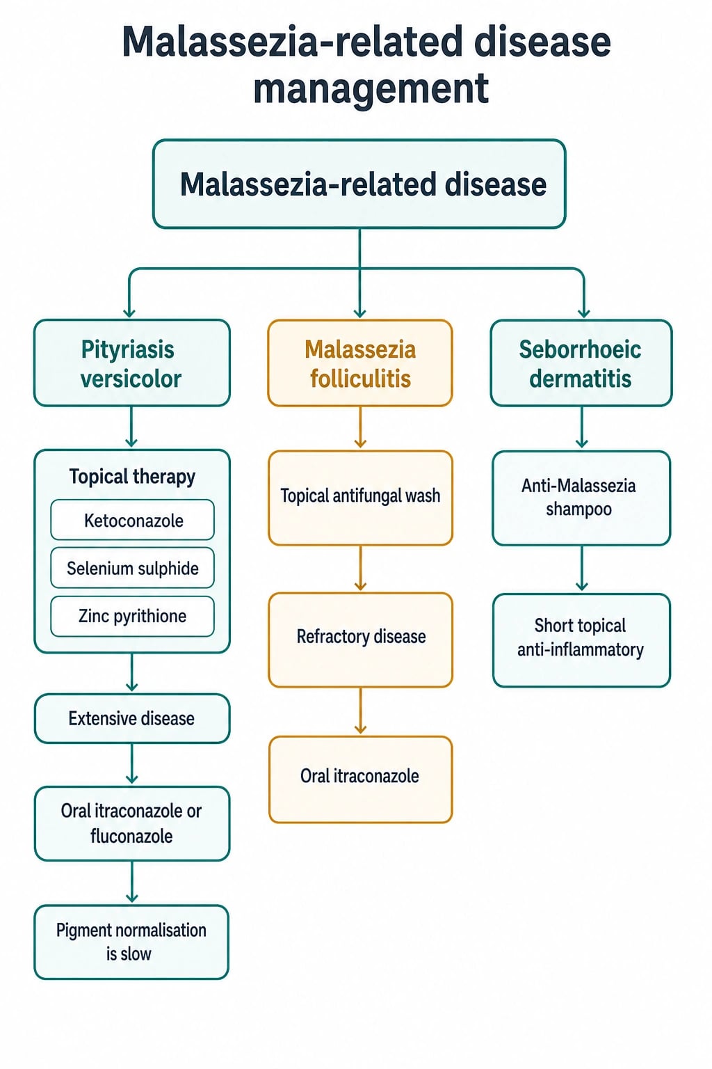
The escalation framework for cutaneous disease is straightforward: topical therapy first for limited disease; oral azole therapy added for extensive, recurrent, immunosuppression-associated, or treatment-refractory disease; and maintenance topical therapy to prevent the high recurrence rate in humid climates.[4][6]
Management — Definitive & Stepwise
Pityriasis versicolor — topical therapy
First-line treatment is topical, and the workhorse is the 2% ketoconazole shampoo, applied to all affected areas, left on for 3 to 5 minutes (or as a single overnight application), and rinsed off. A short intensive course — daily for 3 to 5 days, or a single overnight application repeated weekly — is highly effective at clearing the organism. Equivalent alternatives include selenium sulfide 2.5% lotion or shampoo (applied for 10 minutes daily or overnight), ciclopirox olamine gel or shampoo, clotrimazole 1% or miconazole 2% cream, and terbinafine 1% cream (noting that the allylamine terbinafine is less effective against yeasts than against dermatophytes and is therefore not the preferred topical azole).[4][5]
Ketoconazole 2% shampoo
Dose
Apply to all affected areas, leave 3 to 5 minutes, then rinse. Daily for 3 to 5 days, OR a single overnight application repeated weekly for 2 to 4 weeks.
Pityriasis versicolor — oral therapy
Oral azoles are reserved for extensive, recurrent, immunosuppression-associated, or refractory disease, or where topical therapy is impractical. Two regimens dominate practice: itraconazole 200 mg orally once daily for 7 days (or 100 mg daily for 2 weeks), and fluconazole 300 mg as a single weekly dose repeated after one week (extended to weekly for up to 4 weeks in resistant cases). Both are fungicidal against Malassezia in sebum and are effective for pityriasis versicolor and Malassezia folliculitis alike. Terbinafine is effective for dermatophytes but not reliably active against Malassezia and is not recommended. Pre-treatment and on-therapy monitoring of liver function is prudent for oral azoles, and potential drug interactions (itraconazole and CYP3A4 substrates; fluconazole and CYP2C9/2C19 substrates) must be reviewed before prescribing.[4][5]
Itraconazole
Dose
200 mg once daily for 7 days (alternative: 100 mg once daily for 2 weeks)
Fluconazole
Dose
300 mg as a single weekly dose, repeated after one week (extend to weekly for up to 4 weeks in resistant cases)
A vital counselling point
Patients must be told that the colour change persists long after the organism is killed. Repigmentation of hypopigmented pityriasis versicolor depends on melanocytes resuming melanin production and can take weeks to months, during which the skin looks unchanged even though it is microbiologically cured. Reassurance and daily broad-spectrum sunscreen (sun exposure darkens surrounding skin and accentuates the contrast) are central to management; without this counselling, patients return repeatedly convinced the treatment has failed.[4][10]
Malassezia folliculitis — therapy
Malassezia folliculitis is treated with topical ketoconazole 2% shampoo washed over the affected areas daily for 1 to 2 weeks, followed by maintenance application 1 to 2 times weekly; topical ketoconazole, ciclopirox, or selenium sulfide washes are alternatives. For extensive, recurrent, or immunosuppression-associated disease, add an oral azole — itraconazole 200 mg once daily for 7 to 14 days, or fluconazole — using the same principles and cautions as for pityriasis versicolor. Conventional acne therapy (retinoids, benzoyl peroxide, oral antibiotics) does not treat Malassezia folliculitis and may worsen it; the most common reason for failure to respond is misdiagnosis as acne and treatment with antibiotics.[6][7]
Seborrhoeic dermatitis — therapy (Malassezia-directed)
The Malassezia-directed arm of seborrhoeic dermatitis treatment is topical ketoconazole 2% shampoo to the scalp and body, used twice weekly; this is combined with a mild topical corticosteroid (e.g., hydrocortisone 1% or a low-potency agent) or a topical calcineurin inhibitor (tacrolimus/pimecrolimus, preferred for facial use to avoid steroid atrophy) for short courses to settle inflammation. Intermittent maintenance ketoconazole shampoo (once or twice weekly) reduces relapse. Full management, including paediatric cradle cap and refractory disease, is covered in the seborrhoeic dermatitis topic.[2][8][9]
Invasive (systemic) Malassezia — therapy
Invasive disease demands three simultaneous steps: (1) remove the central venous catheter (the source of the biofilm and persistent fungaemia — the single most important intervention, without which relapse is expected); (2) stop or modify the lipid emulsion (the growth substrate); and (3) systemic antifungal therapy — amphotericin B (deoxycholate or lipid formulation) is first-line, frequently combined with flucytosine for synergy; fluconazole is an alternative or step-down agent guided by susceptibility. Therapy is prolonged (weeks), and resolution is judged clinically and by repeat (lipid-supplemented) blood cultures. Removal of the catheter and the lipid source is often curative even before antifungal susceptibility is known.[12][13][14]
Invasive Malassezia bundle
Maintenance therapy and recurrence prevention
Because relapse is the rule rather than the exception in humid climates, maintenance therapy is part of treatment, not an afterthought. A practical and effective regimen is ketoconazole 2% (or selenium sulfide) shampoo applied to the trunk once weekly as a maintenance wash after the initial intensive course. Patients should be counselled that this is long-term, that pigment change resolves slowly, and that daily sunscreen minimises the cosmetic contrast. Prophylactic oral itraconazole (e.g., 200 mg on two consecutive days each month) has been used in highly recurrent cases but is rarely necessary and carries the usual azole cautions.[1][4][10]
Pityriasis versicolor — expected course after starting treatment
Specific Subtypes & Scenarios
Pityriasis versicolor — pigmentary variants
Pityriasis versicolor presents in hypopigmented (commonest in dark skin), hyperpigmented (commonest in light skin), and rare erythematous/atrophic variants. The pigmentary variant does not change management but is the chief driver of presentation and of patient anxiety; the counselling point about delayed repigmentation applies in all variants.[4]
Malassezia folliculitis in HIV
In advanced HIV, Malassezia folliculitis can be extensive, refractory to topical therapy, and clinically indistinguishable from eosinophilic folliculitis (Ofuji-type). The two are separated histologically (eosinophilic folliculitis shows eosinophilic spongiosis and a follicular eosinophilic infiltrate) and respond to different therapy: Malassezia folliculitis to antifungals, eosinophilic folliculitis to antihistamines, topical corticosteroid, UVB, and antiretroviral therapy (ART) optimisation. ART-induced immune recovery may itself precipitate a folliculitis that must be distinguished from both.[6][7]
Neonatal invasive disease
The preterm neonate on lipid-containing parenteral nutrition with a central line is the textbook invasive host. M. furfur predominates (acquired from the infant's own skin, with the lipid emulsion as substrate), while M. pachydermatis is notable as a zoonotic, hospital-reservoir species that grows on ordinary media — sometimes traced to staff pets or to contaminated incubators or healthcare workers' hands. Management is catheter removal, lipid withdrawal, and amphotericin B +/- flucytosine; outcome is generally good with source control, and mortality relates more to prematurity and comorbidity than to the yeast itself.[12][13]
Less common associations
Malassezia has been implicated, with varying evidence, in a range of other conditions including onychomycosis (especially proximal/subungual disease in immunosuppressed patients), dacryocystitis, and some acneiform and papulopustular eruptions of the face; these are uncommon, controversial, and examined rarely, but a candidate should know that Malassezia is not confined to pityriasis versicolor and folliculitis.[1]
Complications & Pitfalls
The complications of Malassezia disease are predominantly cosmetic and psychosocial, but two diagnostic pitfalls are genuinely dangerous. The most common complication is post-inflammatory hypopigmentation or hyperpigmentation, which persists for months after microbiological cure and is the chief source of patient distress and of repeated, unnecessary re-treatment. Recurrence is the other common problem: in humid and tropical climates, relapse rates of 60 to 80 per cent within twelve months are typical without maintenance therapy, making weekly topical maintenance a routine part of management rather than an optional extra.[4][5]
Exam application bank (NEET-PG / INICET)
One-line answer
Malassezia species are lipophilic, lipid-dependent basidiomycetous yeasts of the normal cutaneous mycobiome that become pathogenic when heat, humidity, sebum, occlusion, or immunosuppression tip the host–commensal balance. They cause four clinical syndromes: (1) Pityriasis versicolor — hypo- or hyperpigmented, finely scaling macules on the trunk; KOH shows the diagnostic 'spaghetti and meatballs' and Wood's lamp a pale yellow-gold fluorescence; treat with topical ketoconazole 2% shampoo or selenium sulfide 2.5%, with oral itraconazole 200 mg daily for 7 days or fluconazole 300 mg weekly for 2 doses for extensive disease. (2) Malassezia folliculitis — itchy monomorphic follicular papules and pustules on the upper trunk with no comedones (the key distinction from acne); treat with topical ketoconazole or an oral azole. (3) Seborrhoeic dermatitis/dandruff — greasy yellow scale on scalp, f [1]
Worked stems (answer without another resource)
Stem 1 — Classic presentation. Map symptoms to mechanism; name the first investigation and first treatment step with dose/route if drug therapy is standard. [1]
Stem 2 — Unstable / complicated. List red flags that force immediate resuscitation, theatre, ICU, antidote, or reperfusion — and what you do in the first 15 minutes. [1]
Stem 3 — Atypical group. Elderly, pregnancy, child, or immunocompromised: how presentation and thresholds change. [1]
Stem 4 — Differential trap. Name the three closest mimics and one discriminator for each. [1]
Stem 5 — Disposition. Who goes home with safety-netting, who is admitted, who needs HDU/ICU/theatre, and what follow-up is mandatory. [1]
Rapid viva checklist
- Definition + classification
- Pathophysiology chain
- Bedside signs / criteria
- Score with exact components (if any)
- Emergency bundle
- Definitive therapy with doses
- Complications of disease and of treatment
- Special populations
- Guideline/trial name if classic
- Three exam traps
Coverage self-check
If you cannot answer any stem above from this page alone, re-read the matching section — the page is intended to be self-sufficient for final-prof and NEET-PG/INICET questions on Pityrosporum (Malassezia) and associated skin diseases.
Prognosis & Disposition
Cutaneous Malassezia disease is benign and readily treatable. Most cases of pityriasis versicolor clear with a single short intensive topical course, and Malassezia folliculitis responds within 1 to 2 weeks of appropriate antifungal therapy. The cosmetic pigment change resolves slowly over weeks to months, and the long-term outcome is excellent. The principal challenge is recurrence, which is common in humid climates and in immunosuppressed patients; weekly maintenance ketoconazole shampoo substantially reduces relapse and should be offered routinely to patients in high-risk settings.[4][10]
Invasive Malassezia infection resolves with catheter removal and antifungal therapy, and the prognosis is generally favourable when source control is achieved promptly. Mortality relates more to the patient's prematurity, underlying immunosuppression, and comorbidity than to the yeast itself, but delayed recognition (particularly the missed use of lipid-supplemented culture media) prolongs illness and exposes the infant to unnecessary antibacterial therapy. Disposition for cutaneous disease is outpatient dermatology or primary care; invasive disease is managed in the neonatal or intensive care unit.[12][13]
Special Populations
Neonates. Invasive disease is the neonatal concern: any preterm, low-birth-weight infant with a central venous catheter on lipid-containing parenteral nutrition and unexplained sepsis, pneumonitis, or thrombocytopenia should be evaluated for invasive Malassezia. Use lipid-supplemented blood culture media, suspect the diagnosis early, and treat with catheter removal and amphotericin B +/- flucytosine. Cutaneous disease in neonates is rare but infantile seborrhoeic dermatitis (cradle cap) is common and benign.[12][13]
HIV / immunosuppressed. Cutaneous Malassezia disease (especially seborrhoeic dermatitis, pityriasis versicolor, and Malassezia folliculitis) is more severe, extensive, atypical in distribution, and refractory; recurrence is frequent; and oral therapy is more often required. Eosinophilic folliculitis enters the differential in advanced HIV and is managed differently. Severe or new-onset extensive disease warrants HIV testing, and management must be coordinated with the HIV physician because of azole–antiretroviral drug interactions (itraconazole and CYP3A4-metabolised agents).[1][9]
Pregnancy. Topical azoles (ketoconazole 2% shampoo, clotrimazole) and selenium sulfide 2.5% are regarded as safe for localised pityriasis versicolor and Malassezia folliculitis in pregnancy. Oral azoles should be avoided in pregnancy (teratogenic potential, particularly in the first trimester) and used only when the benefit clearly outweighs the risk; oral terbinafine is not reliably active against Malassezia and is best avoided. Coordinate with the obstetric team for extensive disease.[4]
Tropical and humid climates. The high baseline prevalence and recurrence rate mean that maintenance therapy and sun protection counselling are part of routine care, not optional. Patients should understand that recurrence is expected and that weekly topical maintenance, not repeated courses of oral therapy, is the sustainable strategy.[4][10]
Evidence, Guidelines & Regional Differences
There is no single global guideline for Malassezia disease; practice is informed by comprehensive reviews and the guidance of national dermatology societies. The two pivotal reference reviews are the Saunte, Gaitanis and Hay (2020) overview of Malassezia-associated skin diseases, diagnostics, and treatment, and the Leung et al. (2022) updated review of tinea (pityriasis) versicolor, which together establish the diagnostic and therapeutic framework used worldwide. The Łabędź et al. (2023) narrative review and the Andersen et al. (2025) clinical update consolidate the same approach for pityriasis versicolor.[1][4][5][10]
For Malassezia folliculitis, the Rubenstein and Malerich (2014) and Henning, Jemec and Saunte (2020) reviews establish the diagnostic criteria (monomorphic follicular papules and pustules, no comedones, prominent pruritus, KOH positivity, response to antifungal therapy) and the first-line topical azole, oral azole escalation paradigm. For seborrhoeic dermatitis, the Borda (2015, 2019), Clark (2015), and Dall'Oglio (2022) reviews underpin the ketoconazole-shampoo-plus-mild-steroid backbone.[2][3][8][9][11]
Saunte, Gaitanis, Hay 2020 — the modern reference review
Comprehensive review of Malassezia biology, species, diagnostics, and treatment across all clinical syndromes
Key finding
Consolidates the lipid-dependence paradigm, species-disease associations (M. globosa for PV; M. restricta/globosa for SD), the KOH 'spaghetti and meatballs' and Wood's lamp pale-yellow fluorescence diagnostics, and the topical-ketoconazole-first / oral-azole-for-extensive-disease treatment framework used worldwide.
Practice change
The single best starting reference for Malassezia disease; informs every current guideline and review.
Leung et al. 2022 — tinea versicolor updated review
Updated narrative review of pityriasis versicolor epidemiology, diagnosis, and management
Key finding
Confirms ketoconazole 2% shampoo as first-line topical, with oral itraconazole 200 mg daily for 7 days or fluconazole 300 mg weekly for 2 doses for extensive/recurrent disease; emphasises that pigment change resolves slowly over months after microbiological cure and that recurrence is common in humid climates.
Practice change
Establishes the current dose regimens and the central counselling point about delayed repigmentation.
For invasive disease, the Tashiro, Takazono and Izumikawa (2023) review and the Huang et al. (2020) case series and literature review of systemic M. pachydermatis infection in infants, together with the classic Marcon and Powell (1992) review of human infections due to Malassezia spp., establish catheter removal as the decisive intervention and amphotericin B (+/- flucytosine) as first-line systemic therapy, with the caveat that lipid-supplemented culture media are required for diagnosis.[12][13][14]
Practice is broadly concordant worldwide for cutaneous disease: topical azole first, oral azole for extensive/recurrent/immunosuppressed disease, and maintenance topical therapy for relapse prevention. Regional variation is greatest in (a) the first-choice azole vehicle (ketoconazole 2% shampoo is universal; ciclopirox, clotrimazole, miconazole, and econazole are alternatives where ketoconazole is unavailable or locally avoided), (b) the preferred oral azole (itraconazole is widely used in Europe and Asia; fluconazole is favoured in some North American centres for its convenience and interaction profile), and (c) the first-line antifungal for invasive disease (amphotericin B universally first; fluconazole step-down varies by susceptibility and local formulary).[1][4]
Australasian practice (Australasian College of Dermatologists) mirrors the global consensus, with ketoconazole 2% or selenium sulfide 2.5% first-line and oral azoles for extensive disease; the high prevalence in tropical northern Australia makes maintenance therapy and sun-protection counselling a routine part of care.[1]
Exam Pearls
SPAGHETTI
Short broad hyphae (spaghetti) + budding yeast (meatballs) on KOH — the pathognomonic PV finding
Wood's lamp finding in PV (vs coral-red erythrasma; bright blue-white vitiligo)
Malassezia lipoxygenase product -> hypopigmentation in dark skin; persists months after cure
M. globosa is the predominant species in pityriasis versicolor
60-80% recur within 12 months without weekly ketoconazole maintenance
Itraconazole 200 mg OD x 7 d; fluconazole 300 mg weekly x 2; terbinafine NOT reliable
Classic distribution of PV — seborrhoeic areas
MF has NO comedones, is itchy, KOH positive; antibiotics do not help and may worsen it
Neonate on lipid TPN; remove line + amphotericin B; M. pachydermatis is the only non-lipid-dependent species
Quick self-test — what causes the colour change in pityriasis versicolor?
Malassezia-derived azelaic acid (from a Malassezia lipoxygenase acting on sebum lipids) inhibits tyrosinase, reducing melanin production and producing hypopigmentation (most conspicuous in dark skin). In lighter skin, inflammatory mediators and increased epidermal turnover dominate, producing hyperpigmentation. The colour change is metabolic, not staining — and it persists for months after the organism is killed because melanocytes must resume melanin synthesis.[4]
Quick self-test — three itchy follicular papules on the chest that are NOT acne. Why?
Malassezia folliculitis: monomorphic, itchy, follicular papules and pustules with no comedones and KOH-positive budding yeast in follicular contents, driven by Malassezia overgrowth within the follicle. Treated with topical ketoconazole 2% (oral itraconazole/fluconazole if extensive) — NOT antibiotics. The key exam discriminator from acne is the absence of comedones and the prominent itch.[6][7]
Key Takeaways
Malassezia is a lipid-dependent commensal turned pathogen. Master four syndromes — pityriasis versicolor, Malassezia folliculitis, seborrhoeic dermatitis, and invasive neonatal line infection — and one unifying principle (no fatty-acid synthase). At the bedside, the scale sign, KOH 'spaghetti and meatballs', and Wood's lamp pale yellow-gold fluorescence confirm pityriasis versicolor; the absence of comedones and prominent itch flag Malassezia folliculitis. Treat topically first (ketoconazole 2% or selenium sulfide 2.5%), escalate to an oral azole (itraconazole 200 mg daily x 7 days or fluconazole 300 mg weekly x 2) for extensive disease, and counsel that pigment change resolves over months and that weekly maintenance is the answer to the high recurrence rate in humid climates. Invasive disease in the preterm neonate on lipid TPN is managed by removing the catheter, stopping the lipid, and giving amphotericin B +/- flucytosine — and is missed unless lipid-supplemented culture media are used.[1][4][6][12]
References
- [1]Saunte DML, Gaitanis G, Hay RJ. Malassezia-Associated Skin Diseases, the Use of Diagnostics and Treatment Front Cell Infect Microbiol, 2020.PMID 32266163
- [2]Borda LJ, Perper M, Keri JE. Treatment of seborrheic dermatitis: a comprehensive review J Dermatolog Treat, 2019.PMID 29737895
- [3]Borda LJ, Wikramanayake TC. Seborrheic Dermatitis and Dandruff: A Comprehensive Review J Clin Investig Dermatol, 2015.PMID 27148560
- [4]Leung AK, Barankin B, Lam JM, et al. Tinea versicolor: an updated review Drugs Context, 2022.PMID 36452877
- [5]Łabędź N, Navarrete-Dechent C, Kubisiak-Rzepczyk H, et al. Pityriasis Versicolor-A Narrative Review on the Diagnosis and Management Life (Basel), 2023.PMID 37895478
- [6]Rubenstein RM, Malerich SA. Malassezia (pityrosporum) folliculitis J Clin Aesthet Dermatol, 2014.PMID 24688625
- [7]Henning MA, Jemec GB, Saunte DM. [Malassezia folliculitis] Ugeskr Laeger, 2020.PMID 33215579
- [8]Clark GW, Pope SM, Jaboori KA. Diagnosis and treatment of seborrheic dermatitis Am Fam Physician, 2015.PMID 25822272
- [9]Dall'Oglio F, Nasca MR, Gerbino C, et al. An Overview of the Diagnosis and Management of Seborrheic Dermatitis Clin Cosmet Investig Dermatol, 2022.PMID 35967915
- [10]Andersen PL, Andersen SML, Henning MAS, et al. [Pityriasis versicolor] Ugeskr Laeger, 2025.PMID 40171908
- [11]Woolhiser E, Keime N, Patel A, et al. Nutrition, Obesity, and Seborrheic Dermatitis: Systematic Review JMIR Dermatol, 2024.PMID 39102684
- [12]Tashiro M, Takazono T, Izumikawa K. Invasive Malassezia Infections Med Mycol J, 2023.PMID 38030275
- [13]Huang CY, Peng CC, Hsu CH, et al. Systemic Infection Caused by Malassezia pachydermatis in Infants: Case Series and Review of the Literature Pediatr Infect Dis J, 2020.PMID 32118859
- [14]Marcon MJ, Powell DA. Human infections due to Malassezia spp Clin Microbiol Rev, 1992.PMID 1576583