Dermatology · Medicine
Porphyria cutanea tarda (PCT)
Also known as Porphyria cutanea tarda (PCT) · Chronic hepatic porphyria · Uroporphyrinogen decarboxylase deficiency · PCT · Hepatoerythropoietic porphyria (homozygous form) · Symptomatic porphyria
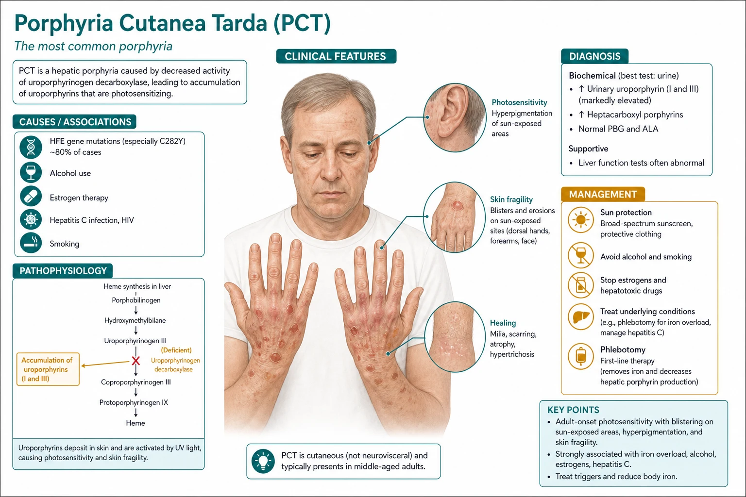
Porphyria cutanea tarda (PCT) is the most common type of porphyria, caused by reduced activity of uroporphyrinogen decarboxylase (UROD) in the liver, producing photosensitive blistering lesions on sun-exposed skin (dorsal hands, face, forearms), skin fragility, hypertrichosis, hyperpigmentation, and milia. Risk factors include hepatitis C virus (HCV), alcohol excess, iron overload (HFE mutations C282Y/H63D), oestrogens, HIV, and renal dialysis. The hallmark biochemical finding is elevated urinary uroporphyrin and heptacarboxyl porphyrin (which fluoresces pink-red under Wood's lamp) and a plasma fluorescence emission peak at 619-620 nm. Treatment: therapeutic phlebotomy (target ferritin ~ 25 ng/mL) or low-dose hydroxychloroquine (100 mg orally twice weekly); eradicate HCV with DAAs; stop alcohol and oestrogens; lifelong photoprotection.
On this page & tools
Your progress
Saved locally on this device.
Exam tags
Red flags
Overview & Definition
Porphyria cutanea tarda (PCT) is the most common porphyria worldwide, accounting for roughly 80–90 percent of all uroporphyrinogen decarboxylase-related disease and a dominant cause of acquired photosensitivity in adults. The disease is a disorder of the hepatic haem biosynthesis pathway in which activity of the fifth enzyme, uroporphyrinogen decarboxylase (UROD; EC 4.1.1.37), falls below a critical threshold (typically under 20 percent of normal hepatic activity). The result is a predictable accumulation pattern — uroporphyrin I, uroporphyrin III, and heptacarboxyl porphyrin build up in hepatocytes and overflow into plasma and urine, where the porphyrins settle in the upper dermis and the epidermis; when UV-A reaches the skin, the porphyrins become a localised generator of singlet oxygen and reactive oxygen species, producing the clinical phenotype of phototoxic blistering on sun-exposed skin.[1][2][3]
PCT is, by definition, a non-acute cutaneous porphyria. There are no abdominal crises, no peripheral neuropathy, no hyponatraemia, no seizures, and no psychotic episodes, the syndromic hallmarks of the acute hepatic porphyrias (acute intermittent porphyria, variegate porphyria, hereditary coproporphyria, ALA-dehydratase deficiency). A patient who walks in with skin fragility and tense bullae on the knuckles and who then goes on to develop abdominal pain and tachycardia has not 'flared' PCT — they have a second, superimposed acute porphyria that must be investigated separately.[1][2]
In the WHO and European Porphyria Network (EPNET) classification, PCT sits in the chronic cutaneous hepatic porphyria group alongside rarer disorders (plumboporphyria / ALAD deficiency is the acute correlate). Distinguishing PCT from the acute porphyrias is one of the most regularly tested clinical decisions in dermatology and clinical biochemistry finals.[4]
Classification
PCT is subclassified into four clinicopathological patterns. The examiner expect you to know the proportions and the genetic distinction at least to family-counselling depth. [1]
- Type I — Sporadic (acquired, ~80 percent of cases): UROD activity reduced in the liver only; no germline UROD mutation; clinical expression requires an acquired precipitant (HCV, alcohol, iron, oestrogens, HIV, drugs, dialysis).
- Type II — Familial (autosomal dominant, ~20 percent): germline heterozygous mutation in UROD at chromosome 1p34; reduced activity in all nucleated cells, including erythrocytes; penetrance < 10 percent unless a cofactor (iron, HCV, alcohol) is added — most carriers never develop PCT.
- Type III — Familial but liver-only deficiency (rare): kindred history of PCT but erythrocyte UROD is normal; behaves clinically like Type I.
- Homozygous Type II — Hepatoerythropoietic porphyria (HEP): autosomal recessive; severe photosensitivity from infancy; residual UROD activity below 10 percent; clinically resembles congenital erythropoietic porphyria (CEP, Gunther disease). [1]
The key biochemical discriminator in the laboratory is erythrocyte UROD activity: reduced → Type II (familial); normal → Type I or Type III. In routine adult practice, subtyping rarely changes first-line management (phlebotomy or low-dose hydroxychloroquine still works for all three), but a positive family history should prompt genetic counselling for the proband's siblings and offspring.[1][3]
Epidemiology & Risk Factors [1]
PCT is the most common porphyria, with an estimated prevalence of 1 in 5,000 to 1 in 25,000 in adult European populations, and a male-to-female ratio of approximately 2 : 1 to 5 : 1 in pre-menopausal cohorts, narrowing (and reversing) after the menopause when endogenous oestrogens fall. The disease is rarely seen in childhood; an infant or child with bullae and photosensitivity is far more likely to have erythropoietic protoporphyria (EPP), congenital erythropoietic porphyria (CEP), or hepatoerythropoietic porphyria (HEP) than adult-pattern PCT.[1][2]
The dominant acquired risk factor is chronic hepatitis C virus (HCV) infection. PCT series from Southern Europe, Japan, and Latin America report anti-HCV prevalence of 50 to 80 percent in PCT patients, compared with 10 to 20 percent in matched controls. The next largest independent risk factors are alcohol excess (> 60 g/day in most cohorts), iron overload (hereditary haemochromatosis; HFE C282Y or H63D mutations — heterozygous C282Y detected in 30–50 percent of PCT patients compared with ~10 percent of the Northern European background population), oestrogens (oral contraceptive pill, hormone replacement therapy), and HIV infection. The roles of hepatitis B virus, chronic renal failure / dialysis, and halogenated hydrocarbons (poly chlorinated biphenyls — PCB, hexachlorobenzene — historically relevant in industrial toxicology) are smaller but recognised.[1][3][4]
Mortality in PCT is dominated by chronic liver disease and hepatocellular carcinoma (HCC), with a 5-year cumulative HCC incidence of approximately 5 percent overall and substantially higher in patients with established cirrhosis at baseline. Cutaneous disease rarely kills; what kills the patient is the liver.[3][4]
Pathophysiology
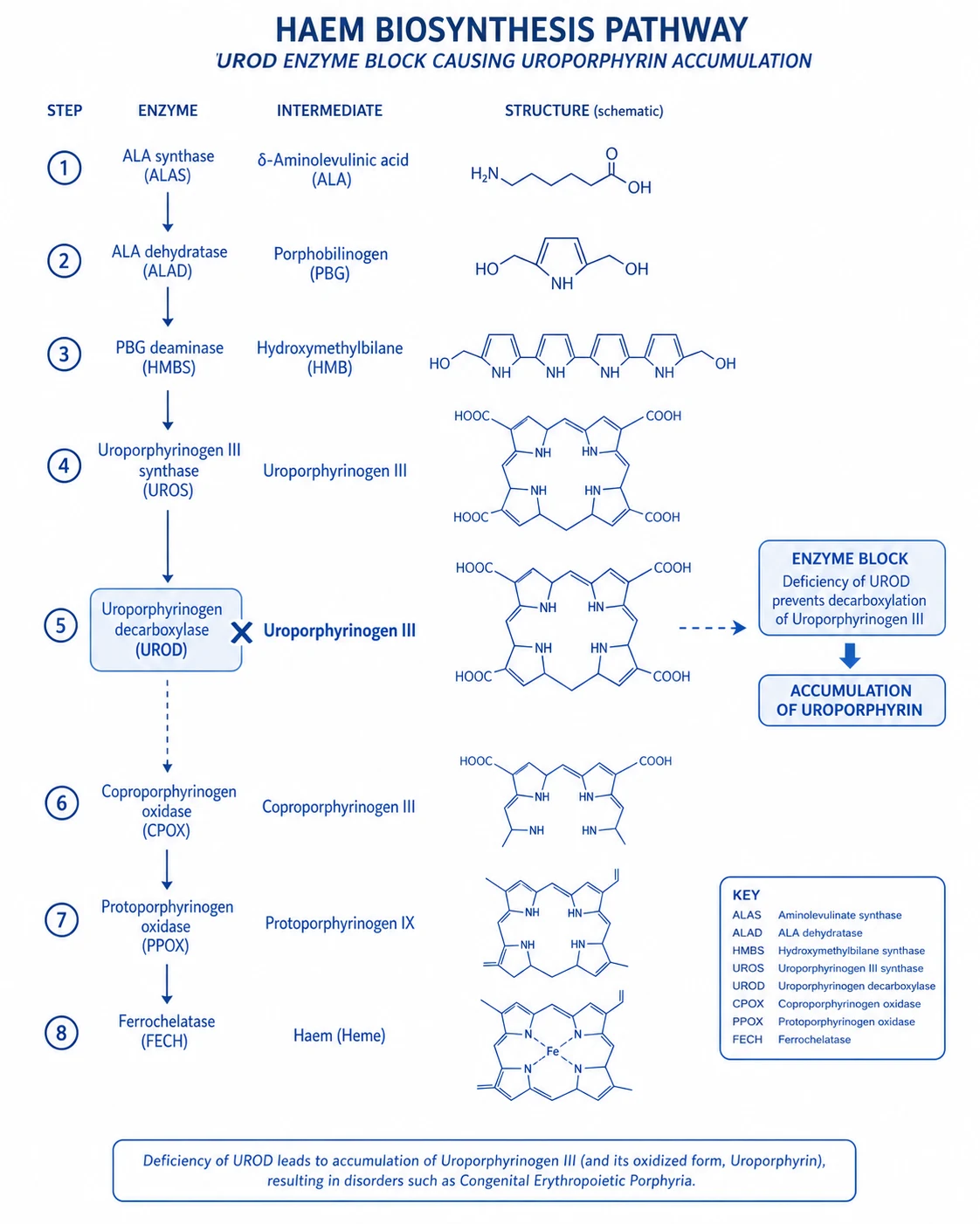
The haem biosynthesis pathway is an eight-step cascade that begins in the mitochondrion with the rate-limiting δ-aminolaevulinic acid synthase (ALAS2) reaction and finishes with ferrochelatase (FECH) inserting ferrous iron into protoporphyrin IX to form haem. The complete ordered sequence is: [1]
- Glycine + succinyl-CoA → δ-aminolaevulinic acid (ALA) — ALAS (1 in liver, 2 in erythroid).
- 2 ALA → porphobilinogen (PBG) — ALAD (δ-aminolaevulinic acid dehydratase, also called ALAD/PDS).
- 4 PBG → hydroxymethylbilane — PBGD / uroporphyrinogen I synthase / HMBS.
- Hydroxymethylbilane → uroporphyrinogen III — uroporphyrinogen III synthase (UROS).
- Uroporphyrinogen III → coproporphyrinogen III — uroporphyrinogen decarboxylase (UROD) — the deficient enzyme in PCT.
- Coproporphyrinogen III → protoporphyrinogen IX — coproporphyrinogen oxidase (CPOX).
- Protoporphyrinogen IX → protoporphyrin IX — protoporphyrinogen oxidase (PPOX).
- Protoporphyrin IX + Fe²⁺ → haem — ferrochelatase (FECH). [1]
In PCT, when hepatic UROD activity falls to less than ~ 20 percent of normal, the substrate uroporphyrinogen III accumulates and oxidises in the hepatocyte cytosol to uroporphyrin (cannot cross back easily once oxidised) and heptacarboxylic porphyrin. These enter plasma, bind albumin and lipoproteins, and reach skin where the dermis localises them near the upper capillaries and the basement membrane zone.[3][4]
The iron connection is the second pillar of PCT pathophysiology. Hepatic iron overload, whether from HFE mutations, dietary iron, multiple transfusions, or alcohol-driven increased gut absorption, drives a Fenton-style catalytic cycle (Fe²⁺ → Fe³⁺ with H₂O₂ → hydroxyl radical ·OH) that oxidises UROD substrate to a porphyrinogen-derived porphomethene. This porphomethene is itself a competitive inhibitor of UROD, and the resultant liver injury is augmented by iron-induced oxidative damage to hepatocyte membranes. The genetic disease is therefore 'silent' until environmental cofactors make the partial enzymatic deficiency clinically apparent — explaining why depletion of iron (by phlebotomy or low-dose hydroxychloroquine / chloroquine) reverses UROD inhibition and cures the syndrome, even though the UROD gene itself is unaffected in Type I.[3][1]
The cutaneous phototoxicity is the third pillar. In the skin, accumulated uroporphyrin and heptacarboxyl porphyrin absorb photons in the Soret band (approximately 400–410 nm, UV-A1) with a smaller secondary Q-band around 500–650 nm. The excited porphyrin transfers energy to molecular oxygen producing singlet oxygen (¹O₂) and other reactive oxygen species (superoxide, hydrogen peroxide, hydroxyl radical). These damage the lamina lucida of the dermal–epidermal junction, with activation of complement, mast-cell degranulation, and protease-mediated loss of keratinocyte adhesion. The histological corollary is a subepidermal blister with 'festooning' of dermal papillae, PAS-positive hyaline material around upper dermal vessels, and 'caterpillar bodies' (eosinophilic basement-membrane fragments in the blister roof) — all of which are useful bedside and laboratory correlates. Direct immunofluorescence (DIF) is negative in PCT, which is the key feature distinguishing it from the autoimmune subepidermal blistering diseases (bullous pemphigoid, EBA, linear IgA disease, mucous membrane pemphigoid).[1][2][3]
The contrast with the acute hepatic porphyrias is informative. Acute intermittent porphyria (AIP) has ALA + PBG accumulation (HMB synthase deficiency) — and these water-soluble monomers are neurotoxic; the resulting neurovisceral attack (abdominal pain, neuropathy, hyponatraemia, seizures, psychosis) is the dominant phenotype. In PCT the accumulated intermediates are uroporphyrin and heptacarboxyl porphyrin (not ALA/PBG), and they cause skin, not nerve, disease.[1][2]
Clinical Presentation
PCT is characteristically insidious: a middle-aged adult (often male, alcohol-using, HCV-positive) presents with months or years of skin fragility and recurrent blisters on the dorsal hands, more pronounced in spring and summer. The classic morphologies are: [1]
- Skin fragility and erosions: minor trauma to the dorsal hands (towel-drying, buttoning, gardening, washing dishes) shears off the epidermis — erosions are typically painless and heal with crusting.
- Tense vesicles and bullae: fluid-filled blisters 0.5–3 cm across on the dorsal hands (knuckles, dorsum), extensor forearms, V of the neck, face, and pinna of the ear; heal with milia and pigmentary change.
- Milia — small 1–2 mm subepidermal keratin cysts, often the most persistent feature.
- Hyperpigmentation — diffuse brown discolouration in sun-exposed areas; often mottled or reticulate.
- Hypertrichosis — fine dark terminal hairs on the temples, lateral cheeks, upper lip, and forearms; in women this is often the presenting complaint and is a high-yield examiner sign.
- Sclerodermoid (morphea-like) plaques — waxy, indurated, sclero-atrophic plaques on sun-exposed skin of long-standing PCT (rare but pathognomonic); histology shows dermal fibrosis with no scleroderma-pattern antibodies.
- Photo-onycholysis — separation of the nail plate from the nail bed; occasionally seen. [1]
Atypical and special populations that examiners deliberately test: [1]
- Post-menopausal women: endogenous oestrogens fall; PCT begins or recurs in this age band; prior OCP exposure may have masked earlier disease.
- Oestrogen therapy: OCP, HRT, and high-dose oestrogen therapy (e.g., prostate cancer treatment in men) induce PCT within months of starting.
- HIV co-infected adults: PCT is over-represented in HIV cohorts; consider aggressive screening and DAA treatment if HCV-coinfected.
- Chronic haemodialysis patients: 'dialysis porphyria' includes both pseudo-PCT (aluminium overload, plasma porphyrins normal) and true PCT (renal-excretion porphyrins accumulate even with normal UROD); check both possibilities.
- Children with bullae + photosensitivity: classic adult PCT is exceptional; think HEP, CEP, or EPP — and refer to a porphyria centre.
- Sclerodermoid PCT: a long-standing scarred subtype often misdiagnosed as morphea; histology with PAS and direct immunofluorescence (negative) confirms it.
- Darker skin phenotypes: hyperpigmentation may be less obvious, but milia, blistering, and hypertrichosis remain reliable signs;Wood's lamp of urine is the bedside discriminator.
- Pregnancy: PCT may flare from the oestrogen surge; hydroxychloroquine is contraindicated; phlebotomy is safe and pregnancy-compatible. [1]
The constitutional symptoms of PCT are notably absent or mild: fevers, weight loss, abdominal pain, vomiting, and joint disease should redirect the diagnostic workup. A PCT patient who develops any of these is more likely to have decompensated liver disease from cirrhosis, hepatocellular carcinoma, or coexistent viral hepatitis — and the workup must include liver imaging and a hepatic encephalopathy screen.[1][2][4]
Differential Diagnosis
PCT has a wide differential: bullous, photosensitive, and blistering disorders overlap. The 'must-not-miss' contributors are pseudo-PCT (because treatment is different), the acute porphyrias (because presentation is different), and the autoimmune subepidermal blistering diseases (because treatment is profoundly immunosuppressive and the wrong choice makes the patient worse). [1]
A complete differential of PCT must include: [1]
- Porphyria cutanea tarda (PCT) — typical tetrad (fragility, vesicles/bullae, milia, hypertrichosis), uroporphyrin / heptacarboxyl porphyrin elevated, plasma peak 619–620 nm.
- Pseudo-PCT — identical clinical picture but porphyrins NORMAL; drug- or dialysis-induced (NSAIDs, tetracyclines, amiodarone, voriconazole, retinoids, frusemide, PUVA).
- Variegate porphyria (VP) — cutaneous lesions PLUS acute neurovisceral attacks; plasma peak 626 nm; PPOX deficiency.
- Erythropoietic protoporphyria (EPP) — childhood onset; immediate PAINFUL burning on sun exposure (no delayed blistering); ferrochelatase deficiency; free erythrocyte protoporphyrin raised.
- Congenital erythropoietic porphyria (CEP, Gunther disease) — mutilating scarring from infancy; severe photosensitivity; UROS deficiency; haemolytic anaemia with erythrodontia.
- Bullous pemphigoid — elderly, tense subepidermal bullae on urticarial base; linear IgG/C3 at BMZ; BP180/BP230 antibodies.
- Epidermolysis bullosa acquisita (EBA) — adult trauma-induced bullae on extensor surfaces; salt-split dermal IgG; type VII collagen antibodies.
- Linear IgA bullous dermatosis — annular 'crown of jewels' vesicles; linear IgA at BMZ DIF.
- Dermatitis herpetiformis — pruritic vesicles on extensor surfaces; granular IgA in dermal papillae; gluten-sensitive enteropathy.
- Pemphigus vulgaris / foliaceus — flaccid intra-epidermal bullae, POSITIVE Nikolsky, mucosal involvement; IgG against desmogleins.
- Polymorphous light eruption (PLE) — pruritic papules/vesicles sparing dorsal hands; NORMAL porphyrins; phototest reproduces.
- Hydroa vacciniforme — childhood vesico-crusty on sun-exposed face / ears with vacciniform scarring; resolves by adulthood.
- Lupus erythematosus (cutaneous, subacute) — annular or papulosquamous photo-distributed lesions; ANA positive; histology with interface dermatitis.
- Solar urticaria — wheals within minutes of sun exposure; urticaria rather than vesicles. [1]
The discriminator with the highest exam value is the plasma fluorescence emission scan: 619–620 nm → PCT; 626 nm → VP; 634 nm → EPP. The discriminator with the most clinical value is the Wood's lamp fluorescence of urine (pink-red → PCT) — a finding that takes 30 seconds and is reliably present (in freshly voided, unpreserved urine) when urinary porphyrins are massively elevated.[1][2][5]
Clinical & Bedside Assessment
The bedside assessment has six components, and each one drives a piece of the differential or the management ladder. [1]
- Skin examination, daylight, with the patient gowned. Confirm bullae, erosions, milia, hyperpigmentation, and hypertrichosis in their characteristic distribution. Photograph the worst lesions (with consent) to compare against follow-up; PCT often has a 6–12 month clinical lag after biochemical remission.
- Wood's lamp (UV-A 365 nm) of freshly voided urine. Pink-red fluorescence in PCT is highly suggestive; absence argues strongly against PCT but does not exclude pseudo-PCT, where porphyrins are normal but the clinical picture looks identical. The bedside test is supplemented by formal HPLC separation of the porphyrins.
- Focused abdominal examination. Hepatomegaly, splenomegaly, caput medusae, spider naevi, palmar erythema, gynaecomastia, ascites, and peripheral oedema suggest chronic liver disease — relevant to the prognosis and the management ladder (anaesthesia for phlebotomy, anaesthesia and analgesia choice, HCC surveillance).
- Focused cardiovascular assessment. Pulse, blood pressure lying and standing, ankle-brachial pressure index in the elderly. PCT phlebotomy is safe only when the patient is haemodynamically stable; anaemia, severe cardiac disease, or postural hypotension mandates the low-dose hydroxychloroquine route instead.
- Mucosal and Nikolsky examination. PCT does not produce mucosal lesions. A cotton-tipped applicator shear test confirms a NEGATIVE Nikolsky sign. The discovery of flaccid bullae, positive Nikolsky, or oral erosions re-opens the differential to pemphigus.
- Focused drug, alcohol, and oestrogen history. Quantify alcohol intake (units/week), prescribed oestrogens (OCP, HRT, tibolone, oestrogen patches), over-the-counter NSAIDs (specifically naproxen, used heavily in osteoarthritis and sport), iron or vitamin-mineral supplements, HIV pre-exposure prophylaxis (some regimens interact with the liver). Use AUDIT-C; a positive screen prompts referral for alcohol support before disease-modifying therapy. [1]
The clinical signs that are most often missed on first assessment are hypertrophic scarring of the helix of the ear, sclerodermoid plaques on the V of the neck, and discreet symmetrical hypertrichosis over the temples — all of which point convincingly at PCT when found together.[1][2][4]
Investigations
The investigation ladder has three rings: diagnostic biochemistry to confirm PCT, mandatory associated tests to find a precipitant, and supporting investigations to stage the liver and monitor for HCC. [1]
First ring — Diagnostic porphyrin biochemistry
- 24-hour urinary porphyrin profile (HPLC separation) — uroporphyrin and heptacarboxyl porphyrin (types I and III) markedly raised (often > 10× the upper limit of normal); ALA and PBG are NORMAL; sample protected from light from collection to analysis.
- Plasma fluorescence emission spectroscopy — emission peak at ~ 619–620 nm confirms PCT; 626 nm is variegate porphyria; 634 nm is erythropoietic protoporphyria or congenital erythropoietic porphyria.
- Faecal porphyrin profile — isocoproporphyrin raised (highly characteristic of PCT); heptacarboxylic porphyrin raised.
- Erythrocyte UROD activity — reduced in Type II (familial); normal in Type I and Type III. [1]
Second ring — Mandatory precipitant and comorbidity screen (every patient)
- Hepatitis C — anti-HCV antibody + (if positive) HCV RNA (PCR); HCV genotype before DAA therapy.
- Hepatitis B — HBsAg, anti-HBc, anti-HBs.
- HIV — fourth-generation HIV Ag/Ab combo.
- Iron studies — serum ferritin, transferrin saturation, total iron binding capacity; raised ferritin + raised transferrin saturation support iron overload.
- HFE genotype — C282Y and H63D mutations; homozygous C282Y confirms hereditary haemochromatosis — the most aggressive precipitant.
- Liver function tests — ALT, AST, ALP, GGT, bilirubin, INR, albumin.
- Kidney function — urea, creatinine, eGFR; PCT symptoms worsen in renal failure because porphyrin excretion is impaired. [1]
Third ring — Liver staging and HCC surveillance
- Liver ultrasound with elastography (or FibroScan) at diagnosis to assess fibrosis and to look for focal lesions.
- Alpha-fetoprotein + hepatic ultrasound 6-monthly if cirrhosis is established; 12-monthly if no cirrhosis.
- Skin biopsy for histology and direct immunofluorescence — subepidermal blister with festooning, PAS-positive perivascular material, caterpillar bodies in the blister roof; DIF is NEGATIVE in PCT, distinguishing it from EBA, BP, and linear IgA disease.
- Genetic testing (UROD sequencing) — not routine, but offered when family screening is contemplated (Type II / familial disease).
- Pre-phlebotomy workup — full blood count, ferritin, TSAT, weight, blood pressure, baseline ECG in the elderly; defer phlebotomy if Hb < 12 g/dL. [1]
The investigation that most often settles the diagnosis when it is uncertain is the plasma fluorescence emission scan — it is fast, non-invasive, and discriminates the four common porphyrias in a single step.[1][2][4]
Management — Resuscitation
PCT is not a resuscitation-level diagnosis. The patient is rarely unwell at presentation; the exceptions are co-existing decompensated cirrhosis, severe alcohol withdrawal, or an opportunistic HIV infection that mandates inpatient stabilisation before disease-modifying therapy. [1]
In the meantime, photoprotection must be instituted at the first contact, even before biochemistry returns: [1]
- Opaque broad-spectrum SPF 50+ with zinc oxide 20 %+ or titanium dioxide 20 %+ physical blockers; chemical absorbers alone are insufficient because the Soret band sits in UV-A.
- UPF 50+ clothing, broad-brimmed hat, gloves while driving.
- Photoprotective window film on car and home windows, especially in regions with summer UV-A.
- Indoor activities between 10 a.m. and 4 p.m. where possible. [1]
Identify and withdraw all precipitants immediately: alcohol, oestrogens, iron supplements, NSAIDs (particularly naproxen, oxaprozin, celecoxib, diclofenac), tetracyclines, amiodarone, voriconazole, retinoids (isotretinoin and acitretin), frusemide, barbiturates, sulphonamides, rifampicin — and check all herbal remedies for pyrrolizidine alkaloids and other porphyrinogens.[1][5]
Management — Definitive & Stepwise
The definitive ladder has six steps. The expected duration of disease-modifying therapy is 6–12 months, and biochemical remission typically precedes clinical remission by 3–6 months. [1]
PCT Definitive Management Ladder
Step 1 — Stop precipitants
Step 2 — Treat the precipitating comorbidity
Step 3a — Therapeutic phlebotomy (first-line)
Step 3b — Low-dose hydroxychloroquine (alternative first-line)
Step 4 — Photoprotection and adjuncts
Step 5 — Surveillance and response monitoring
Step 6 — Escalation and specialist referral
Treatment-specific notes (high-yield examiner territory)
- Phlebotomy end-points: ferritin ~ 25 ng/mL; urinary uroporphyrin normalisation within 3–6 months; skin fragility reverts by 6 months; hypertrichosis and milia may persist longer; hyperpigmentation fades slowly over 12–24 months. Re-bleed if ferritin rebounds above ~ 50 ng/mL.
- Hydroxychloroquine end-points: the same ferritin and porphyrin end-points; full-dose antimalarial regimens are forbidden because they precipitate a transient porphyrin release and fulminant hepatocyte necrosis — the trap the examiner wants to test is the candidate who writes '200 mg orally daily' rather than '100 mg orally twice weekly'.
- Combined therapy is reasonable in severe or refractory PCT, particularly in those with elevated ferritin and heavy HCV burden in whom monotherapy with either modality is slow.
- Retreatment of relapses is acceptable, especially after resumption of alcohol or oestrogens; patient education about the triggers remains the cornerstone of long-term disease control.
- Drugs to NEVER use in PCT (full antimalarial doses; standard-dose retinoids): hydroxychloroquine 200–400 mg/day; chloroquine 250 mg/day; isotretinoin 0.5–1 mg/kg/day; deferasirox concurrent loading.[1][2][3][5]
Hydroxychloroquine (low-dose)
Dose
100 mg
Therapeutic phlebotomy
Dose
5–10 mL/kg per session (typically 450 mL of whole blood)
Specific Subtypes & Scenarios
- Type I — Sporadic (acquired). ~ 80 percent of cases. UROD activity reduced in liver only; erythrocyte UROD normal. Always look for the second insult: HCV (anti-HCV), iron overload (ferritin + TSAT + HFE), alcohol, oestrogens, HIV. Excellent response to phlebotomy + trigger withdrawal.
- Type II — Familial. ~ 20 percent. Heterozygous UROD mutation at 1p34; AD; reduced UROD in all tissues; erythrocyte UROD is the laboratory marker (half-normal). Penetrance < 10 percent unless an environmental cofactor is present; offer UROD sequencing and family screening if positive.
- Type III — Familial with normal erythrocyte UROD. Rare; behaves clinically as Type I; UROD variant may not be detectable without functional studies.
- Hepatoerythropoietic porphyria (HEP, homozygous Type II). Severe photosensitivity beginning in infancy; residual UROD activity below 10 percent; defends the differential with CEP; severe mutilating scarring; refer urgently to a specialist porphyria centre.
- Pseudo-PCT (drug- or dialysis-induced). Identical skin picture, NORMAL porphyrins. Causes: NSAIDs (naproxen most common; oxaprozin, celecoxib, diclofenac), tetracyclines (chlortetracycline, minocycline, doxycycline), amiodarone, voriconazole (long-term antifungals), retinoids (isotretinoin, acitretin), frusemide (high-dose), PUVA photochemotherapy, chronic haemodialysis (aluminium overload), and rarely tanning-bed overuse. Management: withdraw the trigger — phlebotomy and low-dose HCQ are NOT effective because there is no porphyrin accumulation.
- Alcohol- and HCV-driven PCT. The dominant adult pattern, especially in Southern European, Japanese, and Brazilian cohorts; typically men 40–60 years, heavy alcohol (> 80 g/day), elevated GGT, hyperferritinaemia, anti-HCV positive. Treat with abstinence + phlebotomy (or low-dose HCQ); eradicate HCV with DAAs.
- Oestrogen-induced PCT. Typically women on OCP/HRT (or men on oestrogen for prostate cancer); often reversible on stopping; switch to progesterone-only contraception; if hepatoerythropoietic progression, refer for genetic counselling.
- Dialysis-associated PCT. Mixed pathogenesis: impaired renal porphyrin clearance + aluminium overload + common precipitants (iron sucrose infusions, ESAs); chloroquine tolerated; erythrocytapheresis an option where blood-letting is unsafe. [1]
PCT TYPES — UROD classification at a glance
SPOT
Liver only, ~80 percent; no germline mutation; needs cofactor.
Familial AD; reduced UROD in all tissues; low penetrance unless cofactor.
Familial clustering with normal erythrocyte UROD — behaves as sporadic.
HEP — autosomal recessive severe infantile photosensitivity; UROD activity under 10 percent.
Complications & Pitfalls
- Hepatocellular carcinoma — risk up to 20–30-fold raised in PCT with cirrhosis; up to 5 percent develop HCC within 5 years of diagnosis; mandate lifelong USS + AFP (6-monthly in cirrhosis; 12-monthly otherwise).
- Liver cirrhosis and portal hypertension — chronic iron overload + alcohol + HCV drive progression; HCC surveillance must extend for life in established cirrhosis.
- Severe cutaneous morbidity — milia, scarring, hyperpigmentation, sclerodermoid PCT (morphea-like induration); may persist after biochemical remission and require cosmetic dermatology referral.
- Secondary infection of eroded skin — impetiginisation, cellulitis; sample and treat with flucloxacillin / non-penicillin alternative; antiseptic washes.
- Psychosocial morbidity — disfigurement from facial hypertrichosis and scarring contributes to GAD-7 / PCL-5 screen positivity; provide psychiatric support and onward referral.
- Drug toxicity — standard antimalarial doses (HCQ 200–400 mg/day) trigger fulminant PCT hepatitis from acute porphyrin release; never use these doses.
- Phlebotomy complications — anaemia, iron deficiency, syncope, hypovolaemia in cardiac patients, venous access failure; restart or switch modality at the first sign.
- Iron overload complications — hypogonadism, hypothyroidism, diabetes, arthropathy, cardiomyopathy; screen for end-organ iron damage in severe HFE phenotype.
- Treatment-emergent hepatic flare with full-dose HCQ — document in the patient's drug chart, allergy list, and clinic letters; refer explicitly to 'low-dose only' to prevent inadvertent re-prescribing.
- Avoid iron-containing vitamin and herbal preparations and PRN NSAIDs — both trigger recurrence.[1][2][3]
Prognosis & Disposition
- Biochemical remission typically within 3–6 months of phlebotomy (target ferritin ~ 25 ng/mL) or low-dose HCQ; clinical remission follows over 6–12 months.
- Relapse in approximately 10–20 percent after stopping treatment, especially if triggers resume (alcohol, oestrogens, HCV reactivation, iron overload).
- Sustained remission after HCV eradication with DAAs is essentially permanent unless a new precipitant is added.
- Skin disease does not shorten survival directly; mortality is dictated by chronic liver disease and hepatocellular carcinoma.
- Discharge criteria: ferritin and urinary porphyrins within normal range for at least 12 months; sun-tolerance restored; no active liver disease; no active trigger. Annual dermatology and hepatology review thereafter; HCC surveillance for life in patients with cirrhosis.
- Long-term follow-up: 6-monthly LFTs and urinary porphyrin in the first 2 years, 12-monthly thereafter; HCC surveillance (USS + AFP) 6-monthly in cirrhosis, 12-monthly otherwise.[1][2][3]
Special Populations
- Paediatric. Classic adult PCT is exceptionally rare; consider HEP, CEP, EPP. Phlebotomy volume adjusted to weight (5 mL/kg, max 250 mL). Hydroxychloroquine 3–4 mg/kg/day twice weekly may be used; specialist referral mandatory.
- Pregnancy. PCT may flare from the oestrogen surge + iron demand. Therapeutic phlebotomy is safe and effective. Hydroxychloroquine is CONTRAINDICATED in pregnancy — manage conservatively with photoprotection and obstetric monitoring; defer disease-modifying therapy until postpartum if possible. Coordinate with obstetric medicine. Monitor haemoglobin closely; iron deficiency anaemia is a relative contraindication to phlebotomy, so HCQ is preferred where safe (post-partum, breastfeeding compatible).
- Elderly. High prevalence of HCV and comorbidities; phlebotomy tolerated but pace to tolerance (every 2–3 weeks); baseline cardiac evaluation; avoid diuretics, iron supplementation, and indiscriminate NSAIDs for osteoarthritis; engage with falls-risk team for syncope prevention.
- HIV co-infected. High PCT prevalence in HIV cohorts. Use DAA for HCV. Maintain ART; monitor for ART-related hepatotoxicity. Iron studies and HFE in all HIV-positive patients with PCT.
- Chronic haemodialysis. Minimise venepuncture trauma; aluminium overload is thought contributory; deferasirox (off-label) and erythrocytapheresis are options where blood-letting is impossible; chloroquine tolerated; hepatitis screening mandatory; coordinate with nephrology.
- Immunosuppressed / post-transplant. PCT may emerge as drug-induced (calcineurin inhibitors, azathioprine) or secondary to HCV reactivation; coordinate with transplant team regarding immunosuppression adjustment and DAA therapy.
- Women of reproductive age. Oestrogens (OCP, HRT) are a reversible trigger; switch to progesterone-only methods (mini-pill, depot medroxyprogesterone, copper IUD); clarify perimenopausal HRT risks and benefits; avoid tibolone.
- Anticoagulated patients. Phlebotomy still first-line if INR is stable; avoid intramuscular iron and intramuscular vitamin injections; liaise with haematology for coagulopathy.
- Alcohol dependence. Address alcohol as the dominant modifiable driver of mortality; motivational interviewing; consider inpatient alcohol detoxification before phlebotomy if PT/INR deranged; naltrexone or acamprosate in eligible patients; link to community support services.
- First-degree relatives of familial Type II PCT. Offer UROD sequencing and genetic counselling; screen adult carriers annually with iron studies and counsel on avoiding cofactor insults (alcohol, oestrogens, iron, HCV).[1][2][3][4]
Evidence, Guidelines & Regional Differences
- Singal & Anderson 2019 (Mol Genet Metab) — comprehensive recent update; supports phlebotomy as first-line and low-dose HCQ as second-line; reaffirms urine/plasma/faecal porphyrin triad for diagnosis.[1]
- Bissell, Anderson & Bonkovsky 2024 (Liver Int) — contemporary clinical management update incorporating the DAA era; documents post-SVR PCT remission rates and the role of ferritin-targeting.[2]
- Phillips & Kushner 2024 (ASH Education Program) — frames PCT as an iron-related disorder, with iron as the linchpin that unifies disease and therapy.[3]
- Egger et al. 2010 (Best Pract Res Clin Gastroenterol) — bridges hepatology and dermatology; supports a co-managed clinic (hepatology + dermatology) for all PCT patients.[4]
- BAD / EPNET / American Porphyria Foundation consensus — phlebotomy is grade A first-line when ferritin > 50 ng/mL and there is no contraindication; low-dose HCQ grade A first-line when phlebotomy is contraindicated; surveillance USS + AFP for cirrhotic patients; DAA therapy for HCV-positive PCT once the patient is clinically stable.[1][2]
- Regional deltas:
Australasian Porphyria Registry (Melbourne) supplies ongoing diagnostic and treatment guidance; alcohol and iron overload remain dominant drivers; phlebotomy first-line in nearly all cases; Aboriginal and Torres Strait Islander populations have higher-than-background HCV prevalence in some regions.
- Controversies. Phlebotomy vs HCQ head-to-head — no adequately powered RCT comparing modalities directly, but multiple cohort series show similar remission rates (~ 70–90 percent at 12 months). Ferritin target — most groups aim for ~ 25 ng/mL, others prefer a higher ~ 50 ng/mL ceiling to avoid anaemia; both are acceptable and should be titrated to tolerance. Beta-carotene is supported by small trials for visible-light tolerance but not all guidelines endorse it. Deferasirox and other iron chelators are off-label but used where phlebotomy is impossible (renal failure, cardiac disease, severe anaemia). Vitamins B6 and lutein are adjuncts with modest supportive evidence; not disease-modifying.[1][2][5]
Exam Pearls
[1]Photosensitive + painful/burning differential
PAIN
Bullae + skin fragility + milia; no pain.
Pruritic papulovesicular; no bullae; spares dorsal hands.
Photoallergic contact dermatitis; topical NSAID, sunscreen component.
Burning pain on UV exposure; childhood onset; little blistering.
Self-test — plasma fluorescence peaks by porphyria
Plasma emission peak is the single highest-yield discriminator in cutaneous porphyria biochemistry. Match the peaks to the diseases: [1]
- 619–620 nm → Porphyria cutanea tarda (PCT)
- 626 nm → Variegate porphyria (VP)
- 634 nm → Erythropoietic protoporphyria (EPP) or congenital erythropoietic porphyria (CEP) [1]
The MBBS and FRCDerm examiner will expect you to identify these peaks without prompting.
Red flags
Exam application bank (NEET-PG / INICET)
One-line answer
Porphyria cutanea tarda (PCT) is the most common type of porphyria, caused by reduced activity of uroporphyrinogen decarboxylase (UROD) in the liver, producing photosensitive blistering lesions on sun-exposed skin (dorsal hands, face, forearms), skin fragility, hypertrichosis, hyperpigmentation, and milia. Risk factors include hepatitis C virus (HCV), alcohol excess, iron overload (HFE mutations C282Y/H63D), oestrogens, HIV, and renal dialysis. The hallmark biochemical finding is elevated urinary uroporphyrin and heptacarboxyl porphyrin (which fluoresces pink-red under Wood's lamp) and a plasma fluorescence emission peak at 619-620 nm. Treatment: therapeutic phlebotomy (target ferritin ~ 25 ng/mL) or low-dose hydroxychloroquine (100 mg orally twice weekly); eradicate HCV with DAAs; stop alcohol and oestrogens; lifelong photoprotection. [1]
Worked stems (answer without another resource)
Stem 1 — Classic presentation. Map symptoms to mechanism; name the first investigation and first treatment step with dose/route if drug therapy is standard. [1]
Stem 2 — Unstable / complicated. List red flags that force immediate resuscitation, theatre, ICU, antidote, or reperfusion — and what you do in the first 15 minutes. [1]
Stem 3 — Atypical group. Elderly, pregnancy, child, or immunocompromised: how presentation and thresholds change. [1]
Stem 4 — Differential trap. Name the three closest mimics and one discriminator for each. [1]
Stem 5 — Disposition. Who goes home with safety-netting, who is admitted, who needs HDU/ICU/theatre, and what follow-up is mandatory. [1]
Rapid viva checklist
- Definition + classification
- Pathophysiology chain
- Bedside signs / criteria
- Score with exact components (if any)
- Emergency bundle
- Definitive therapy with doses
- Complications of disease and of treatment
- Special populations
- Guideline/trial name if classic
- Three exam traps
Coverage self-check
If you cannot answer any stem above from this page alone, re-read the matching section — the page is intended to be self-sufficient for final-prof and NEET-PG/INICET questions on Porphyria cutanea tarda (PCT).
[1]References
- [1]Singal AK, Anderson KE. Porphyria cutanea tarda: Recent update Mol Genet Metab, 2019.PMID 30683557
- [2]Bissell DM, Anderson KE, Bonkovsky HL. The clinical management of porphyria cutanea tarda: An update Liver Int, 2024.PMID 38813949
- [3]Phillips JD, Kushner JP. Porphyria cutanea tarda: a unique iron-related disorder Hematology Am Soc Hematol Educ Program, 2024.PMID 39644053
- [4]Egger NG, Goeger DE, Payne DA, et al. Porphyria cutanea tarda--when skin meets liver Best Pract Res Clin Gastroenterol, 2010.PMID 20955974
- [5]Badiu C, Dicker M, Pfeifer W, et al. Drug-induced cutaneous porphyria Clin Dermatol, 1993.PMID 7907270