Dermatology · Medicine
Port-wine stain
Also known as Port-wine stain (PWS) · Naevus flammeus · Capillary malformation
Port-wine stain (PWS; naevus flammeus) is a congenital capillary malformation — a vascular malformation (NOT a tumour) of ectatic capillaries and post-capillary venules in the superficial dermis, present at birth, growing proportionally with the child, and never involuting. The lesion is caused by a somatic mosaic GNAQ R183Q activating mutation (the same mutation found in Sturge-Weber syndrome). It presents as a unilateral pink-to-purple macule/patch following a trigeminal dermatome (V1/V2/V3) on the face; it darkens and thickens with age. V1 distribution mandates screening for Sturge-Weber syndrome (leptomeningeal angiomatosis + glaucoma + seizures). Pulsed dye laser 585/595 nm is the gold standard treatment, most effective when started in early infancy.
On this page & tools
Your progress
Saved locally on this device.
Exam tags
Red flags
A port-wine stain (PWS) is one of the first vascular lesions a dermatologist, paediatrician, or plastic surgeon encounters in the neonatal examination. Its clinical importance is disproportionate to its apparent simplicity: the lesion itself is benign, but its distribution — particularly on the forehead and upper eyelid in the ophthalmic division of the trigeminal nerve — unlocks a chain of decisions that determines whether a child receives timely screening for glaucoma, leptomeningeal angiomatosis, and lifelong developmental surveillance. A candidate who can articulate the distinction between a malformation and a tumour, name the GNAQ mutation, recite the syndromic triads, and defend the evidence for early pulsed dye laser treatment will answer any question an examiner sets on this topic.[2][3]
Definition and Classification
Port-wine stain (PWS; naevus flammeus) is a congenital capillary malformation consisting of ectatic (dilated) but structurally normal capillaries and post-capillary venules in the superficial dermis. It is a developmental vascular anomaly — a malformation, not a neoplasm — and this single word defines its biology, its natural history, and its treatment. A malformation is present at birth, grows commensurately with the child, and never regresses; a tumour proliferates beyond the growth of the child and then (in the case of infantile haemangioma) involutes.[2][3]
Within the ISSVA classification (International Society for the Study of Vascular Anomalies), PWS sits in the category of simple vascular malformations under the capillary malformation subgroup. This framework separates all vascular anomalies into two superfamilies — vascular tumours (which proliferate) and vascular malformations (which are developmental errors present at birth) — and then by flow characteristics (slow-flow vs fast-flow). PWS is a slow-flow capillary malformation.[3]
Port-wine stain
Salmon patch (stork bite)
Infantile haemangioma
The common naevus flammeus neonatorum — the salmon patch, stork bite, or angel's kiss — is a clinically and biologically distinct entity that resolves spontaneously in the vast majority of infants by 12-24 months. Midline nuchal lesions (stork bite) may persist into adulthood as a cosmetic non-issue, but they do not darken, do not thicken, and carry no syndromic implication. The PWS, in contrast, is lateralised (or at least clearly off-midline on the face), follows a dermatomal pattern, and will darken and thicken inexorably if untreated. Confusing the two is the cardinal neonatal diagnostic error.[3]
Epidemiology and Risk Factors
PWS occurs in approximately 0.3 per cent of live births (range 0.1-0.5 per cent across studies). There is no sex predilection — males and females are equally affected — and no racial or ethnic predilection, though darker skin phototypes alter the clinical appearance and the response to laser treatment.[3]
PWS is congenital but not inherited. Because the causative GNAQ mutation is somatic and mosaic (confined to the affected tissue, not present in germ cells), the risk to siblings is not elevated above baseline, and affected individuals do not transmit the lesion to their offspring. This is a critical counselling point: parents of a newborn with a PWS are often anxious about recurrence risk and need explicit reassurance.[6]
The most important epidemiological figure for the exam concerns the V1 distribution: approximately 8-15 per cent of infants with a PWS involving the forehead or upper eyelid (V1 dermatome, particularly the ophthalmic distribution over the upper eyelid) have Sturge-Weber syndrome. The risk is highest when the PWS involves the upper eyelid skin itself, and it rises further with bilateral V1 involvement. A PWS confined to V2 or V3 carries essentially no SWS risk, though isolated exceptions exist.[1][4]
Pathophysiology
The molecular basis of PWS was established in 2013, when Shirley and colleagues demonstrated a somatic activating mutation in GNAQ (c.548G then A; p.R183Q) in the affected tissue of patients with both non-syndromic PWS and Sturge-Weber syndrome. The same mutation is found in leptomeningeal angiomatosis tissue, establishing a shared genetic basis for the cutaneous and neurological manifestations. The mutation is absent from the blood and unaffected skin, confirming its mosaic (post-zygotic) nature.[6]
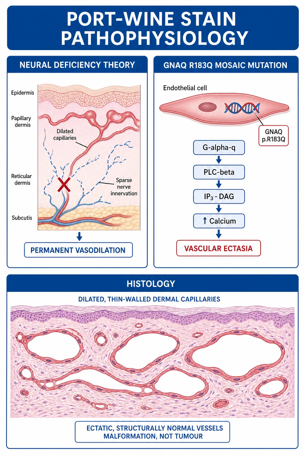
GNAQ encodes the Gαq subunit of a heterotrimeric G protein. The R183Q substitution impairs the intrinsic GTPase activity of Gαq, locking it in its active (GTP-bound) state. This produces constitutive signalling through the downstream effector cascade: Gαq activates phospholipase C-beta (PLCβ), which hydrolyses PIP2 to produce IP3 and DAG, leading to release of intracellular calcium and activation of protein kinase C. The net effect on the affected endothelial cells is a persistent pro-ectatic state — the vessels progressively dilate but do not proliferate, which is the histological hallmark distinguishing a malformation from a tumour.[4][6]
A second, complementary theory — the neural deficiency hypothesis — predates the molecular discovery and explains the dermatomal distribution. Histological studies demonstrate a reduction in perivascular sympathetic nerve density in PWS tissue compared with normal skin. In normal skin, tonic sympathetic vasoconstriction maintains capillary and venule tone; when this neural input is deficient, the vessels dilate passively and permanently. This theory elegantly explains why PWS follows a sensory dermatomal pattern (the affected vasculature corresponds to a developmentally deficient neural crest-derived sensory nerve territory) and why the lesion does not regress (the neural deficit is structural and permanent). Both theories are now understood to be facets of the same mosaic developmental error: GNAQ may act in both endothelial and neural crest-derived lineages during embryogenesis.[3][4]
The histopathology is consistent across all theories: the superficial dermis contains abnormally large numbers of ectatic, thin-walled capillaries and post-capillary venules with structurally normal endothelium and no proliferation. The channels are 10-150 micrometres in diameter and occupy the papillary and upper reticular dermis. There is no mitotic activity, no endothelial tufting, and no GLUT1 expression (distinguishing PWS from infantile haemangioma). With age, the vessels become progressively more ectatic, and the overlying epidermis may thicken and develop cobblestone nodularity — the histological correlate of the clinical darkening and thickening seen in untreated adult PWS.[3]
Clinical Presentation
A port-wine stain is visible at birth as a flat, sharply demarcated, pink-to-red macule or patch. The initial colour ranges from pale pink (in fair-skinned infants) to deep red. The lesion is non-blanching or only partially blanching on diascopy — pressure with a glass slide empties the ectatic vessels partially but they refill immediately, distinguishing PWS from the more readily blanching salmon patch.[3]
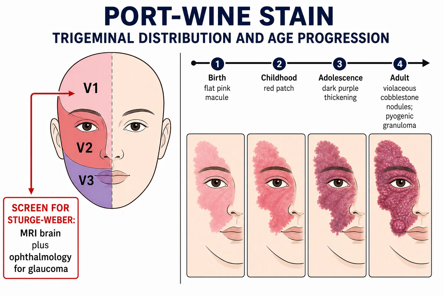
The distribution is the most important clinical feature. On the face, PWS follows a unilateral, dermatomal pattern respecting the sensory divisions of the trigeminal nerve:[3]
- V1 (ophthalmic): forehead, upper eyelid, and sometimes the scalp above the lesion. This is the highest-risk distribution for Sturge-Weber syndrome.
- V2 (maxillary): cheek, lower eyelid, and upper lip.
- V3 (mandibular): lower lip, chin, and jawline. [1]
Lesions are typically unilateral but can be bilateral (in approximately 10 per cent), and bilateral V1 involvement carries the highest SWS risk. Cross-midline extension is uncommon but recognised. Beyond the face, PWS may appear on the neck, trunk, or limbs, where it follows a broad geographic rather than strictly dermatomal pattern. Mucosal involvement (gingiva, buccal mucosa, conjunctiva) can occur ipsilateral to a facial lesion.[2][3]
The natural colour progression is characteristic and diagnostically useful when comparing PWS to fading salmon patches: [1]
In adulthood, an untreated PWS develops cobblestone nodularity — discrete vascular papules and nodules superimposed on the macular background. Pyogenic granuloma-like friable vascular nodules may arise within the lesion and cause recurrent bleeding. Underlying soft-tissue and bony hypertrophy is common, producing facial asymmetry, lip enlargement (macrocheilia), and gingival hyperplasia ipsilateral to the lesion. On a limb, the corresponding change is limb overgrowth with length discrepancy.[2][3]
Atypical presentations
- Segmental/large geographic PWS on the trunk or limb without facial involvement — screen for Klippel-Trenaunay if the limb is overgrown.
- Bilateral facial PWS — higher SWS risk; both hemispheres may be affected.
- Median/central forehead PWS — the area most strongly associated with SWS is the forehead and upper eyelid; even a small lesion here requires screening.
- Adult presenting with new nodularity/bleeding in a lifelong flat PWS — consider pyogenic granuloma (common, benign) but biopsy to exclude angiosarcoma (very rare). [1]
Differential Diagnosis
The differential diagnosis of a congenital vascular stain is one of the highest-yield exam questions. Each mimic has a single distinguishing feature that the examiner expects the candidate to articulate. [1]
Salmon patch / stork bite
Infantile haemangioma
Telangiectasia (e.g. HHT)
AV malformation
Segmental NF1 CAL macule
Naevus anaemicus
The single most important comparison is PWS versus salmon patch versus infantile haemangioma — the three-way neonatal/infantile vascular lesion decision. The PWS is present at birth and never involutes; the salmon patch is present at birth but resolves; the haemangioma is absent at birth, proliferates, and then involutes. This temporal distinction resolves the majority of diagnostic uncertainty without any investigation.[3]
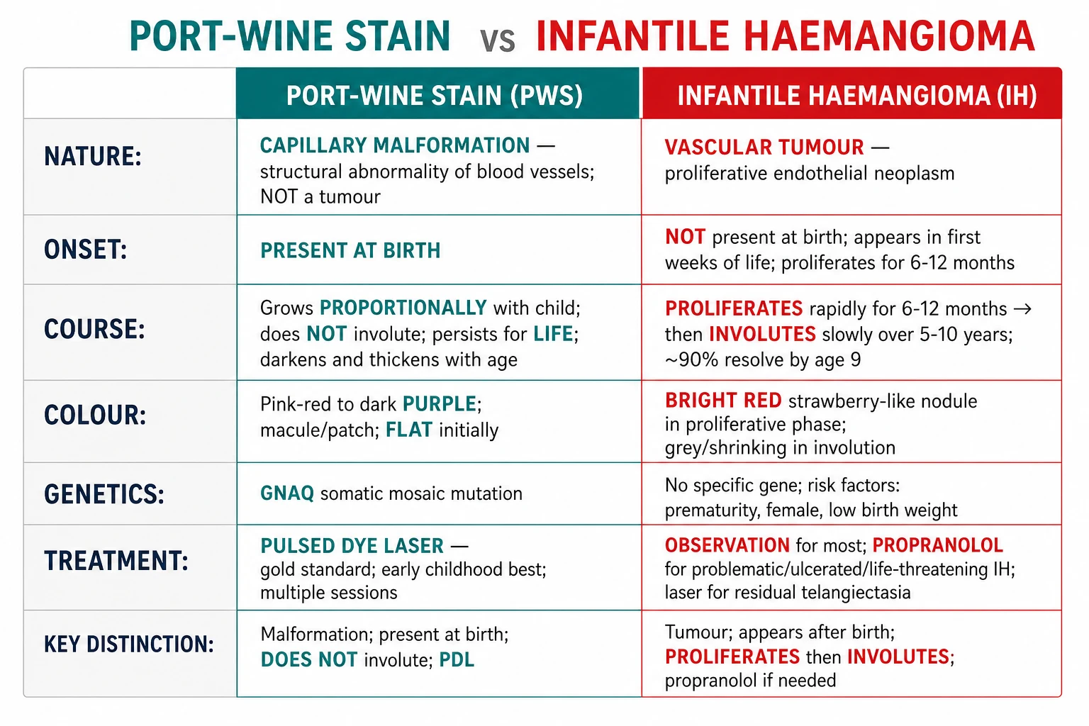
A candidate faced with a red vascular lesion on a baby's face at the viva table must first answer one question: was it present at birth? If yes, the lesion is a PWS or a salmon patch; if no, it is an infantile haemangioma. The second question — does it blanch and fade with pressure? — separates the readily blanching salmon patch from the fixed-colour PWS. These two bedside questions resolve more diagnostic dilemmas than any imaging study, and the examiner will reward a candidate who demonstrates this structured reasoning rather than reaching for investigations. The salmon patch (naevus simplex) is the most common vascular birthmark, present in 30-40 per cent of newborns; it sits in the midline (glabella, eyelids, forehead, nape of neck) and fades within 1-2 years, whereas the PWS is lateralised, follows a dermatome, and persists for life. [1]
Histopathology
Biopsy is rarely necessary — PWS is a clinical diagnosis — but the histology is examinable and underpins the laser treatment rationale. The diagnostic findings are:[3]
- Numerous ectatic, thin-walled capillaries and venules (10-150 micrometres diameter) in the papillary and superficial reticular dermis.
- Normal endothelium with no atypia, no mitoses, no tufting or proliferation.
- GLUT1-negative (the critical immunostain distinguishing PWS from infantile haemangioma, which is GLUT1-positive).
- In thickened adult lesions: additional deeper ectatic channels, increased connective tissue, and occasional capillary microproliferations (pyogenic granuloma-like foci). [1]
The progressive change in vessel architecture with age explains the clinical evolution. In infancy, the ectatic vessels are confined to the superficial papillary dermis (within 0.5 mm of the surface) and are uniformly small-calibre, which is why the lesion appears flat and responds well to the 585/595 nm PDL. Over years, some vessels dilate further and extend into the deeper reticular dermis (up to 2-3 mm), while new ectatic channels form — the histological basis of the clinical thickening, nodularity, and cobblestone change seen in untreated adult PWS, and the reason adult lesions respond less well to PDL alone (the deeper channels are beyond the laser penetration depth and require longer-wavelength devices such as Nd:YAG 1064 nm). The overlying epidermis may show acanthosis and elongation of rete ridges in established lesions, and the connective tissue stroma between the vessels becomes more dense and fibrotic with age.[3]
Other immunohistochemical markers that may be mentioned: the ectatic vessels are positive for CD31 and CD34 (endothelial markers) but negative for Wilms tumour 1 (WT1), Glut1, and Lewis Y antigen — the combination of GLUT1-negative, WT1-negative endothelium in a lesion of ectatic capillaries is diagnostic of a malformation and excludes infantile haemangioma (GLUT1-positive, WT1-positive, Lewis Y-positive). The lymphatic marker D2-40 (podoplanin) is negative, distinguishing the capillary malformation from a lymphatic malformation. [1]
The depth of the ectatic vessels in the dermis (predominantly superficial, less than 1 mm deep in infants) is the reason pulsed dye laser is effective — the 585/595 nm wavelength penetrates to precisely this depth and is absorbed by oxyhaemoglobin in these channels. [1]
Clinical and Bedside Assessment
The focused assessment of a suspected PWS has three goals: confirm the diagnosis clinically, identify the trigeminal distribution (especially V1), and screen for syndromic associations. [1]
Bedside manoeuvres:[3]
- Diascopy (press a glass slide or lens over the lesion): a PWS partially or completely blanches as the ectatic vessels empty; an AVM shows pulsation under the slide. A salmon patch blanches readily; a fixed telangiectatic mat does not.
- Wood's lamp examination helps distinguish PWS from café-au-lait macules (which brighten under Wood's light) and from ash-leaf macules of tuberous sclerosis (which become more conspicuous).
- Palpation for warmth, thrill, or bruit excludes an arteriovenous malformation or fistula. [1]
Neuro-cutaneous assessment (infant with V1 PWS):[1][4]
- Measure the lesion, photograph it for baseline documentation, and mark whether it crosses the midline.
- Examine for facial asymmetry, lip or gingival hypertrophy, and limb length discrepancy (Klippel-Trenaunay).
- Perform a focused neurological examination for asymmetry of tone, reflexes, or visual attention (early evidence of SWS hemiparesis or visual field deficit).
- Measure head circumference (SWS can cause developmental delay). [1]
Ophthalmic assessment (mandatory for V1 PWS):[1]
- Tonometry for intraocular pressure (congenital glaucoma).
- Anterior segment and fundus examination (choroidal haemangioma in SWS causes a tomato-ketchup fundus).
- Assessment for buphthalmos (enlarged eye) — a late sign of untreated congenital glaucoma. [1]
Limb assessment (for limb/trunk PWS):[7]
- Measure limb circumference and length bilaterally at fixed landmarks; document baseline for longitudinal comparison.
- Examine for varicose veins, lymphoedema, and venous flares (Klippel-Trenaunay). [1]
Investigations
PWS is a clinical diagnosis. No laboratory test, biopsy, or imaging study is required to confirm a typical lesion in a well-appearing neonate. Investigations are directed entirely at excluding or characterising the syndromic associations.[3]
When is imaging required?
Facial PWS in V1 distribution (forehead or upper eyelid): MRI brain with and without contrast to screen for leptomeningeal angiomatosis (Sturge-Weber). Perform in the neonatal period or early infancy.
If MRI shows leptomeningeal enhancement or choroidal haemangioma: refer to neurology and ophthalmology; establish seizure surveillance and developmental monitoring.
Limb/trunk PWS with overgrowth: Doppler ultrasound and/or MRI of the limb to characterise the venous, lymphatic, and arteriovenous components (Klippel-Trenaunay workup).
PWS with a palpable thrill or bruit: urgent vascular imaging (MRA or CTA) to exclude an arteriovenous malformation or fistula.
MRI brain findings in Sturge-Weber syndrome:[1][5]
- Leptomeningeal angiomatosis: pial angioma over the cerebral hemisphere ipsilateral to the facial PWS, best seen as leptomeningeal enhancement on post-contrast T1 sequences.
- Cortical calcifications: the classic tram-track or railroad-track sign — parallel curvilinear calcifications following the cortical gyri, best seen on CT or gradient-echo MRI.
- Choroidal haemangioma: diffuse choroidal thickening (the tomato-ketchup fundus).
- Accelerated myelination or cerebral atrophy ipsilateral to the angioma in advanced cases.
- Enlarged deep medullary and subependymal veins as collateral drainage. [1]
Note: MRI may be normal in the neonatal period even in infants who later develop SWS; some centres repeat MRI at 6-12 months if the initial scan is normal but clinical suspicion persists. CT is more sensitive for the tram-track calcifications but involves radiation and is reserved for older children.[5]
Molecular testing
GNAQ somatic mutation testing is now available through specialised laboratories but is not required for routine clinical management. It is useful in research contexts, in atypical cases (e.g., port-wine macrocheilia with recurrent nodules), and in confirming the diagnosis when clinical and imaging findings are ambiguous. The test requires affected tissue (biopsy or surgical specimen), as the mutation is mosaic and absent from blood.[6]
Ophthalmic surveillance
Every infant with a PWS involving the forehead or upper eyelid (V1) requires urgent ophthalmology referral — ideally within the first few weeks of life — for tonometry and fundoscopy. Glaucoma in SWS may be congenital (present at birth, causing buphthalmos) or develop later (most cases present within the first year). Surveillance tonometry is repeated at intervals dictated by the ophthalmologist, typically every 3-6 months in the first year and then annually if normal.[1]
Management — Topical and Physical Modalities
The management of PWS is divided into treatment of the cutaneous lesion itself (laser and adjuncts) and treatment of the syndromic associations (glaucoma, seizures, limb overgrowth). The cutaneous treatment is pulsed dye laser (PDL), which is the gold standard and first-line for all PWS regardless of location.[2][3]
Pulsed dye laser (PDL) — gold standard
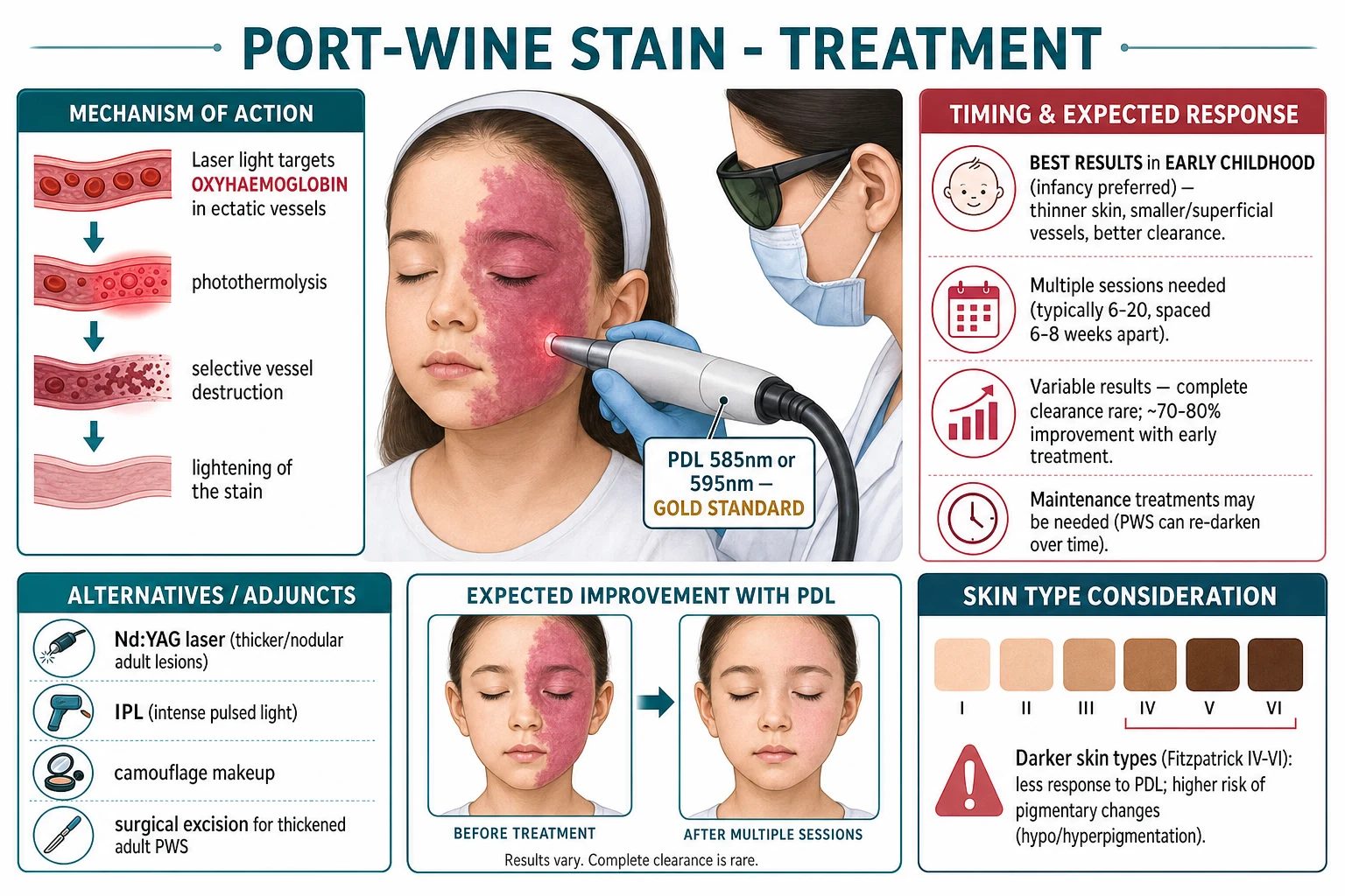
The 585 nm or 595 nm flashlamp-pumped pulsed dye laser is the gold-standard treatment for PWS. The mechanism is selective photothermolysis: the wavelength is preferentially absorbed by oxyhaemoglobin in the ectatic dermal vessels; the pulse duration (0.45-1.5 ms) is matched to the thermal relaxation time of the target vessels, confining thermal injury to the vessel and sparing the surrounding epidermis and dermis. The result is intravascular coagulation, vessel destruction, and progressive lightening of the stain over successive sessions.[3][8]
Key treatment parameters and evidence:[3][8]
- Timing: treatment should begin as early as possible, ideally in early infancy. The skin is thinner, the ectatic vessels are smaller and more superficial, and there is less competing epidermal melanin. Infants treated before 6 months of age achieve better clearance per session and require fewer total sessions.
- Number of sessions: typically 6-12 treatments are required, spaced 6-8 weeks apart. Some patients need 15-20 sessions for maximal clearance.
- Efficacy: approximately 70-90 per cent lightening is achieved with early, consistent treatment; complete clearance is rare (less than 10 per cent achieve near-total blanching). The response varies with anatomical site (central face responds less well than the periorbital or temporal regions), lesion size, and skin type.
- Maintenance: the PWS can re-darken over years after initial treatment; periodic maintenance sessions may be needed.
- Darker skin types (Fitzpatrick IV-VI): less clearance per session (competing epidermal melanin absorbs the laser energy) and a higher risk of post-inflammatory hyperpigmentation or hypopigmentation. Dynamic cooling devices (cryogen spray) protect the epidermis and allow higher fluences, improving results in all skin types.[9]
Technical parameters the candidate should be able to quote at viva: the 585 nm wavelength (original PDL) corresponds to the oxyhaemoglobin absorption peak and penetrates approximately 0.5-1.0 mm into the dermis; the 595 nm wavelength (modern PDL) provides slightly deeper penetration (up to 1.2 mm) with marginally less selective absorption but better coverage of deeper ectatic channels. Typical fluences are 5-10 J/cm squared with a spot size of 5-10 mm and a pulse duration of 0.45-1.5 ms. The epidermis is protected by a dynamic cooling device (DCD) that sprays cryogen (tetrafluoroethane) onto the skin surface milliseconds before the laser pulse, allowing higher fluences to be delivered to the target vessels without epidermal thermal injury. Purpura (the immediate endpoint, lasting 1-2 weeks) indicates vessel destruction. Subpurpuric fluences are sometimes used to reduce downtime but achieve less clearance per session and require more total treatments. Treatment is typically delivered every 6-8 weeks; the interval allows re-epithelialisation and resolution of purpura between sessions.[8][9]
Factors predicting better PDL response include younger age at treatment start, smaller lesion size, a more superficial (macular) lesion, location on the lateral face/neck/periorbital region (the central face, particularly the V2 maxillary distribution, responds least well), and lighter skin phototype. Thickened, nodular adult PWS and lesions on the central cheek or lip are the most refractory. Despite optimal PDL treatment, 10-20 per cent of PWS show minimal response, and complete clearance is achieved in fewer than 10 per cent — candidates should counsel parents honestly about the expected outcome (significant lightening, not disappearance).[3][8]
Why does early infancy yield better laser results?
The skin is thinner, the target vessels are smaller and more superficial (closer to the surface and within the PDL penetration depth of approximately 1 mm), there is less competing epidermal melanin, and the lesion has had less time to develop deep ectatic channels. Infants also heal faster and have less cumulative psychosocial burden.
Alternatives and adjunctive devices
For PWS that respond incompletely to PDL, or for specific anatomical situations, several alternative and adjunctive devices are used: [1]
- Long-pulsed Nd:YAG laser (1064 nm): penetrates more deeply than PDL; used for thickened, nodular, or cobblestone adult PWS where the ectatic channels extend into the deep dermis. Higher risk of scarring; requires expertise.[2]
- Alexandrite laser (755 nm): deeper penetration than PDL; used as a second-line for hypertrophic or PDL-resistant PWS.
- Intense pulsed light (IPL): broadband light source; may be used as an alternative or adjunct for macular PWS, particularly in adult patients with treatment-resistant areas. Less selective than PDL; more variable results.[8]
- Photodynamic therapy (PDT) with intravenous verteporfin (a photosensitiser activated by 689 nm light): targets the photosensitiser-loaded vascular endothelium. Emerging evidence, particularly from Chinese centres, suggests high clearance rates for refractory PWS; not yet standard of care outside specialist centres.[8]
- Topical timolol 0.5% gel: case reports and small series suggest modest lightening of superficial macular PWS, particularly in infants and in Sturge-Weber-associated birthmarks. The mechanism is beta-adrenergic-mediated vasoconstriction of the ectatic vessels. Evidence is preliminary; timolol is not a substitute for PDL but may be a useful adjunct in infants too young for laser or in treatment-resistant patches.[10]
When is topical timolol 0.5% gel used in PWS?
Timolol is an emerging adjunct, not a first-line treatment. It is considered for: (1) superficial macular PWS in neonates/infants before laser is technically feasible, (2) small treatment-resistant patches as an adjunct between PDL sessions, and (3) Sturge-Weber-associated facial PWS where systemic beta-blockade is undesirable. Evidence comes from small case series; large randomised trials are lacking.
Camouflage and psychosocial support
- Camouflage make-up (e.g. specialized skin-coloured cosmetic creams) is offered to all patients who decline laser, are awaiting treatment, or have treatment-resistant areas. Cosmetic camouflage clinics provide colour-matched preparations and instruction in application technique. This is an important option for patients with extensive lesions or Fitzpatrick VI skin where laser results are poor.
- Psychological support: facial PWS causes significant visible difference, and patients (and parents) benefit from access to counselling, patient support organisations (e.g. the Sturge-Weber Foundation, Changing Faces, the Birthmark Support Group), and school liaison. [1]
Management — Procedural and Surgical
Surgery has a limited but important role in PWS management. It is reserved for treatment-resistant or complicated adult PWS where the lesion has progressed to thickened, nodular, or cobblestone change that is refractory to laser:[2]
- CO2 laser resurfacing or surgical excision with primary closure or skin grafting for focal thickened nodular segments, particularly on the cheek, lip (macrocheilia reduction), or forehead.
- Gingivectomy or mucosal reduction for gingival hypertrophy.
- Orthognathic or bony contouring procedures for maxillary or mandibular overgrowth.
- Excision of pyogenic granulomas that bleed recurrently. [1]
Surgery does not treat the underlying malformation and new vessels may develop in the grafted or adjacent skin over time. It is therefore reserved for specific problem areas, not for whole-lesion treatment. [1]
Management of Syndromic Associations
Sturge-Weber syndrome — management
The management of SWS is multidisciplinary, involving dermatology, neurology, ophthalmology, and developmental paediatrics.[1][4][5]
- Glaucoma: topical beta-blockers (e.g. timolol 0.25% eye drops), carbonic anhydrase inhibitors, and prostaglandin analogues are first-line. Refractory cases require surgical trabeculotomy, goniotomy, or tube shunt. The goal is to prevent optic nerve damage and blindness; untreated congenital glaucoma causes buphthalmos and irreversible visual loss. Lifelong ophthalmic surveillance is mandatory.
- Seizures: antiseizure medications are tailored to the seizure type. Focal seizures are most common; levetiracetam, oxcarbazepine, and carbamazepine are frequently used. Infantile spasms may require vigabatrin or ACTH. Refractory epilepsy is an indication for epilepsy surgery (hemispherectomy or focal resection) in carefully selected patients — the earlier the seizure control, the better the developmental outcome.
- Stroke-like episodes and neurological decline: low-dose aspirin (3-5 mg/kg/day) is used at some centres for stroke-like episodes or progressive neurological decline, though the evidence base is limited and the practice is controversial. Physiotherapy and occupational therapy for hemiparesis.
- Developmental surveillance: regular assessment of cognitive, motor, and language development; early intervention services.
- Cutaneous PWS: standard PDL treatment as for non-syndromic PWS. [1]
Klippel-Trenaunay syndrome — management
Management is predominantly conservative and symptom-directed:[7]
- Compression therapy (custom-fitted graduated compression stockings or garments) is the cornerstone — it reduces limb swelling, pain, and the risk of venous ulceration and thrombosis.
- Limb length discrepancy: orthopaedic assessment; epiphysiodesis may be needed for significant discrepancy (greater than 2 cm) near skeletal maturity.
- Venous intervention: sclerotherapy or endovenous laser ablation of incompetent lateral varicosities in selected patients; embolisation of significant arteriovenous components if present.
- Lymphatic management: manual lymphatic drainage and compression for lymphoedema.
- DVT prophylaxis: KTS carries an increased risk of deep vein thrombosis and pulmonary embolism; prophylactic anticoagulation is considered in high-risk situations (surgery, prolonged immobility).
- Cutaneous PWS: standard PDL treatment for the capillary component. [1]
Associated Syndromes
Sturge-Weber syndrome (SWS)
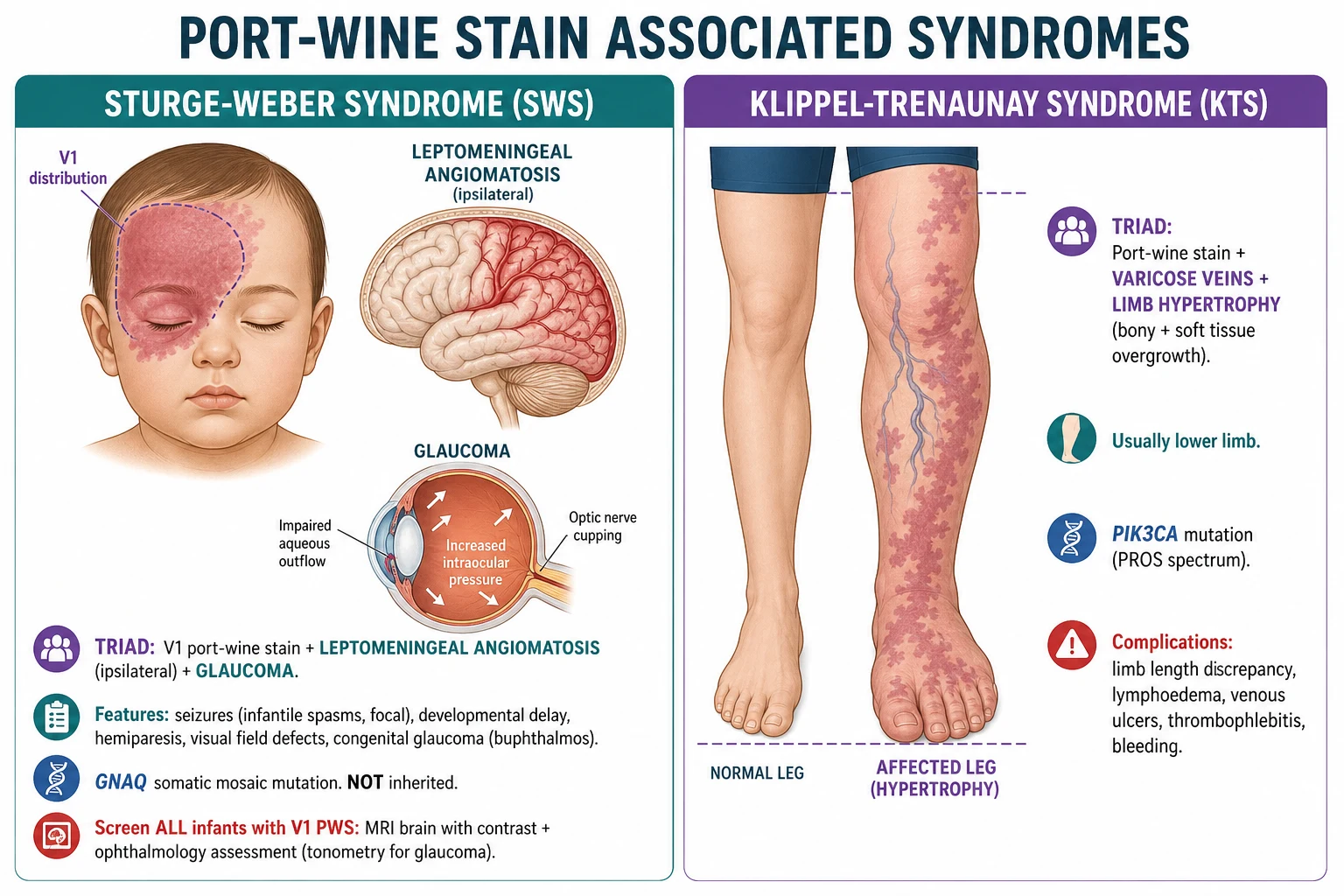
Sturge-Weber syndrome is a congenital, non-inherited neuro-oculo-cutaneous disorder caused by the same somatic mosaic GNAQ R183Q mutation that causes isolated PWS. The mutation occurs in the ectoderm during the first trimester, affecting tissues of common embryological origin: the facial skin (PWS in V1), the leptomeninges (pial angiomatosis), and the eye (glaucoma, choroidal haemangioma).[4][6]
The classic triad (all three present in the complete form, but partial forms are common):[1]
- Facial PWS in the V1 distribution (forehead, upper eyelid).
- Leptomeningeal angiomatosis (pial angioma over the ipsilateral cerebral hemisphere).
- Glaucoma (ipsilateral; may be congenital with buphthalmos, or later-onset). [1]
Neurological features: seizures (the presenting feature in 75-90 per cent, typically onset in the first year; focal, generalised, or infantile spasms), hemiparesis contralateral to the cerebral lesion, visual field defects (homonymous hemianopia), developmental delay, and learning difficulties. Stroke-like episodes with transient hemiparesis or visual loss occur and may be exacerbated by seizures. The seizures and neurological decline are thought to result from chronic venous congestion and ischaemia in the cortex underlying the pial angioma (the abnormal leptomeningeal vessels impair cortical venous drainage, causing venous hypertension, ischaemia, and progressive cortical calcification).[5]
The pathophysiology of the SWS neurological injury is worth understanding in detail because it explains the clinical features and guides management. The pial angioma that characterises leptomeningeal angiomatosis is a thin layer of ectatic leptomeningeal vessels overlying the cerebral cortex. These abnormal vessels are inefficient at draining venous blood from the underlying cortex, and they impair the development of normal cortical veins. The result is chronic venous hypertension in the cortex, leading to progressive cortical ischaemia, neuronal loss, and gliosis. The ischaemic cortex eventually calcifies (the tram-track sign on imaging) and atrophies. Seizures are both a consequence of the cortical injury and an accelerant of further injury (seizure-related metabolic demand exceeds the already-impaired perfusion). This is why early and aggressive seizure control is neuroprotective in SWS. Stroke-like episodes (acute transient hemiparesis or visual field deficit, sometimes with exacerbation of seizures) are thought to represent episodes of acute cortical ischaemia or venous infarction superimposed on the chronic perfusion deficit.[4][5]
The ophthalmic component of SWS — glaucoma — results from increased episcleral venous pressure caused by the episcleral haemangioma (the same vascular malformation on the surface of the eye), which impairs aqueous outflow through Schlemm's canal. The glaucoma may be congenital (present at birth, with buphthalmos, corneal oedema, and Haab striae) or develop in childhood or later. Choroidal haemangioma (a diffuse, flat vascular malformation of the choroid) is present in approximately 40-50 per cent of SWS patients and causes a characteristic tomato-ketchup fundus; it may cause retinal detachment, visual field constriction, or amblyopia, and is treated with low-dose verteporfin PDT or radiotherapy in symptomatic cases.[1][5]
Risk stratification: the risk of SWS in a neonate with a facial PWS is:[1][4]
- 8-15 per cent if the PWS involves the forehead or upper eyelid (V1).
- Rises to approximately 25 per cent if the PWS involves a quarter or more of the upper eyelid.
- Approximately 0 per cent if the PWS is confined to V2 or V3 without V1 involvement (rare exceptions reported).
- Higher in bilateral V1 involvement. [1]
Klippel-Trenaunay syndrome (KTS)
KTS is a complex combined vascular malformation (capillary-venous-lymphatic) with soft-tissue and bony overgrowth of an extremity. It is caused by a somatic mosaic mutation in PIK3CA (part of the PIK3CA-related overgrowth spectrum, PROS). The classic triad:[7]
- Capillary malformation (PWS) on the affected limb (usually a large geographic stain).
- Varicose veins (often lateral, persistent embryonic veins; may be extensive).
- Limb hypertrophy (soft-tissue and bony overgrowth with length discrepancy). [1]
Most commonly affects a lower limb; upper limb involvement is less common. Complications include lymphoedema, recurrent cellulitis, venous ulcers, thrombophlebitis, deep vein thrombosis, pulmonary embolism (significant risk), bleeding from superficial varicosities, and Kasabach-Merritt phenomenon (rare; more associated with kaposiform haemangioendothelioma than KTS).[7]
Proteus syndrome
A rare, sporadic, severe asymmetric overgrowth syndrome caused by a somatic mosaic activating mutation in AKT1. Features include asymmetric overgrowth of bones and soft tissues, connective tissue naevi (cerebriform connective tissue naevus of the sole is pathognomonic), epidermal naevi, vascular malformations (including capillary stains), lipomatosis, and a predisposition to thrombosis and certain tumours. The capillary component may resemble PWS but is accompanied by the striking asymmetric overgrowth that defines the syndrome. The diagnosis is clinical, supported by AKT1 mutation testing of affected tissue. [1]
PHACE syndrome
PHACE is a neurocutaneous syndrome classically associated with segmental infantile haemangioma (not PWS) of the face, but the distinction is important in the differential of a large facial vascular lesion in an infant. The acronym:[11]
- Posterior fossa brain malformations (Dandy-Walker complex).
- Haemangioma (segmental, facial; larger than 5 cm).
- Arterial anomalies (cerebral, aortic, or visceral).
- Cardiac defects (coarctation of the aorta, other congenital heart disease).
- Eye anomalies. [1]
PHACE syndrome is distinguished from SWS by the lesion being a proliferating haemangioma (not a flat congenital PWS) and by the arterial and cardiac anomalies. Any infant with a large segmental facial haemangioma (greater than 5 cm) requires screening for PHACE (MRI/MRA brain and neck, echocardiogram, ophthalmology).[11]
Cobb syndrome (spinal arteriovenous metameric syndrome)
A rare association of a spinal arteriovenous malformation with a cutaneous vascular stain (capillary, venous, or arteriovenous) in the same metameric dermatome overlying the spine. The cutaneous lesion may resemble a PWS but is accompanied by neurological signs from the spinal AVM (myelopathy, weakness, sensory deficit, sphincter disturbance). A vascular stain in a midline or paraspinal location with neurological symptoms requires spinal MRI/MRA to exclude Cobb syndrome. [1]
PWS SYNDROMES
Complications and Pitfalls
Cutaneous complications
- Progressive darkening and thickening: without treatment, the lesion darkens from pink to purple, develops cobblestone nodularity, and becomes progressively more difficult to treat with laser. This is the strongest argument for early PDL.
- Pyogenic granulomas: friable vascular nodules arising within the PWS, prone to recurrent bleeding. Treatment is excision, curettage, cautery, or pulsed dye laser.
- Soft-tissue and bony hypertrophy: facial asymmetry, macrocheilia, gingival hypertrophy, maxillary/mandibular overgrowth; limb length discrepancy with gait disturbance.
- Bleeding: from pyogenic granulomas, from trauma to thickened nodular PWS, or (in KTS) from varicosities. [1]
Systemic/syndromic complications
- Sturge-Weber: seizures, stroke-like episodes, hemiparesis, glaucoma/blindness, developmental delay, learning difficulties.[5]
- Klippel-Trenaunay: deep vein thrombosis, pulmonary embolism, lymphoedema, recurrent cellulitis, venous ulcers, chronic pain.[7]
Diagnostic pitfalls
- Assuming a salmon patch will persist — reassuring a parent that a V1 PWS is just a stork bite and will fade is the most dangerous error. A true PWS in V1 requires SWS screening; a salmon patch does not.
- Failing to screen a V1 PWS for Sturge-Weber syndrome — every infant with a PWS on the forehead or upper eyelid needs MRI brain and ophthalmology, regardless of how small the lesion appears.
- Missing congenital glaucoma — an infant with a V1 PWS and a hazy cornea, watering eye, photophobia, or buphthalmos has congenital glaucoma until proven otherwise; urgent ophthalmology is required.
- Confusing PWS with infantile haemangioma — leading to wrong counselling (telling parents it will involute when it will not) and wrong treatment (propranolol instead of laser).
- Not measuring a limb with a PWS — a limb PWS may be the first sign of Klippel-Trenaunay; serial limb measurements and venous assessment are needed. [1]
Prognosis and Disposition
The natural history of an untreated PWS is permanent, progressive darkening and thickening throughout life. The lesion is present at birth, grows proportionally with the child, and never regresses. In infancy it is a flat pink macule; by adulthood, without treatment, it is a thickened, violaceous, nodular plaque with soft-tissue hypertrophy and a risk of pyogenic granulomas and bleeding. The psychosocial burden of an untreated facial PWS is substantial.[2][3]
With pulsed dye laser treatment, the prognosis is substantially improved. Approximately 70-90 per cent of treated PWS achieve meaningful lightening, with the best results in infants treated early. Complete clearance is achieved in fewer than 10 per cent. The lesion may re-darken over years, requiring maintenance sessions. Early treatment (before 1 year of age, ideally in infancy) gives the best outcomes because the skin is thinner, the vessels are more superficial, the lesion is less established, and the child avoids the psychosocial burden of a visible untreated facial lesion during the formative years.[3][8]
Sturge-Weber neurological prognosis is highly variable and correlates with:[5]
- Age of seizure onset: earlier onset (before 6 months) predicts worse outcome.
- Seizure control: refractory epilepsy correlates with progressive cognitive decline.
- Extent of leptomeningeal involvement: bilateral involvement and large hemispheric angiomas predict worse outcome.
- Frequency of stroke-like episodes: recurrent episodes accelerate decline. [1]
With good seizure control and early intervention, many children with SWS attend mainstream school, though learning difficulties and hemiparesis are common. Without seizure control, progressive encephalopathy and severe cognitive impairment may develop. [1]
Klippel-Trenaunay prognosis is generally good with compression therapy and careful management of venous and lymphatic complications, though the limb overgrowth is permanent and thromboembolic risk persists throughout life.[7]
Disposition: [1]
- Neonate with V1 PWS: urgent ophthalmology (within 1-2 weeks) + MRI brain; dermatology for PDL planning.
- Neonate with V2/V3 facial PWS: dermatology for PDL; ophthalmology if any lesion touches V1 territory.
- Infant with limb PWS + overgrowth: vascular anomaly service, Doppler ultrasound, limb measurement, compression therapy.
- Adult with untreated PWS: dermatology for laser assessment; surgery for thickened nodular components. [1]
Special Populations
Neonates and infants
The neonate with a PWS is the primary clinical scenario. Every neonate with a facial PWS involving the forehead or upper eyelid must be referred urgently for ophthalmology (glaucoma) and neuroimaging (leptomeningeal angiomatosis). PDL treatment should begin as early as feasible — many centres start at 4-6 weeks of age, once the diagnosis and SWS status are established. Treatment under general anaesthesia or with topical anaesthesia and feeding is standard; neonates tolerate PDL well.[3]
Pregnancy
PWS itself is not affected by pregnancy. However, a thickened nodular PWS may become more congested during pregnancy due to increased blood volume and venous pressure, and pyogenic granulomas of pregnancy may arise within a pre-existing PWS. Klippel-Trenaunay syndrome in pregnancy carries a heightened thromboembolic risk requiring multidisciplinary planning and thromboprophylaxis. [1]
Darker skin phototypes (Fitzpatrick IV-VI)
PDL is less effective in darker skin types because epidermal melanin competes with oxyhaemoglobin for the 585/595 nm light, reducing the energy delivered to the target vessels and increasing the risk of post-inflammatory pigmentary change. Dynamic cooling devices (cryogen spray pre-cooling of the epidermis) are essential. Higher fluences and more sessions are needed. Patients should be counselled about the lower expected clearance and the risk of pigmentary alteration. Nd:YAG (1064 nm) may be preferred for deeper vessels in darker skin.[9]
Adults presenting with new nodularity
An adult with a lifelong flat PWS that develops new nodularity or bleeding almost always has a benign pyogenic granuloma or progressive cobblestone change. However, angiosarcoma arising within a longstanding PWS has been reported (very rare). Any rapidly growing, atypical, or treatment-resistant nodule within a PWS should be biopsied to exclude malignancy. [1]
Follow-up and Monitoring
The follow-up strategy is tailored to the clinical scenario and the presence or absence of syndromic features.[1][3]
Isolated (non-syndromic) PWS: dermatology review every 6-8 weeks during active PDL treatment to assess response and plan the next session. Once the treatment course is complete (typically 6-12 sessions), annual review is reasonable to monitor for re-darkening and to plan maintenance sessions. Between visits, the patient is counselled on sun protection (ultraviolet exposure darkens the lesion and increases post-laser pigmentary risk) and is advised to report any new nodularity, thickening, or bleeding promptly. [1]
Sturge-Weber syndrome (V1 PWS with confirmed leptomeningeal involvement): multidisciplinary follow-up involving neurology (seizure surveillance every 3-6 months, more frequent if seizures are poorly controlled), ophthalmology (tonometry every 3-6 months in the first year, then at least annually), and developmental paediatrics (periodic cognitive, motor, and language assessment). Repeat MRI brain is performed if there is clinical deterioration (new or worsening seizures, developmental regression, or stroke-like episodes). The Sturge-Weber Foundation provides family-facing resources and connects families with specialist centres.[4][5]
Klippel-Trenaunay syndrome: vascular anomaly service follow-up with annual limb measurement (circumference and length), Doppler ultrasound surveillance for deep vein thrombosis in symptomatic episodes, compression garment review, and orthopaedic assessment for progressive limb length discrepancy approaching skeletal maturity. Patients are counselled on DVT symptoms and prophylaxis for high-risk situations (surgery, long-haul travel, pregnancy).[7]
Patient and family education: parents of a child with a PWS should be given clear written information about the natural history (the lesion will not resolve spontaneously), the treatment plan and expected outcome (significant lightening but not total clearance), the rationale for early laser, the syndromic screening that has been done and the symptoms that should prompt urgent review (seizures, eye watering or cloudiness, limb swelling or pain), and the contact details for patient support organisations. A named coordinator (often a specialist nurse) improves the family experience and ensures the multidisciplinary appointments are scheduled efficiently. [1]
Evidence, Guidelines, and Regional Differences
Landmark evidence
The 2013 discovery by Shirley and colleagues of the GNAQ R183Q somatic mosaic mutation as the cause of both PWS and Sturge-Weber syndrome was transformative — it established the shared molecular basis, explained the dermatomal distribution (mosaic post-zygotic mutation), confirmed the non-inherited nature, and opened the door to potential future targeted therapies.[6]
Pulsed dye laser has been the gold standard since the 1990s. The evidence base supports: [1]
- Early treatment: multiple cohort studies show better clearance in infants treated before 1 year (and particularly before 6 months) compared with older children and adults, attributed to thinner skin, more superficial vessels, and less competing melanin.
- Adjuncts: a 2021 review confirmed that several adjunctive strategies (imiquimod, rapamycin, topical timolol, anti-angiogenic agents) have been studied as PDL adjuncts, with timolol showing the most promising preliminary signal but without robust randomised trial data.[8]
- PDT with verteporfin: predominantly from Chinese centres, reports high clearance rates (70-90 per cent) in refractory PWS; large multicentre randomised trials outside China are lacking.[8]
Regional differences
SWS screening protocols vary by centre and country. The consensus across US, UK, European, and ANZ centres is that all infants with a PWS involving the forehead or upper eyelid (V1) should have an MRI brain with contrast and ophthalmology assessment. The timing of MRI (neonatal vs deferred) and the interval for repeat imaging if normal vary. The Sturge-Weber Foundation and international consensus statements guide practice.
In Australia and New Zealand, PDL is available in public paediatric dermatology centres (e.g. major children's hospitals) and in private practice. Public funding and wait times for paediatric PDL vary by state. SWS screening follows the international consensus.
Controversies
- Low-dose aspirin in SWS: some centres use low-dose aspirin (3-5 mg/kg/day) prophylactically in infants with SWS to reduce stroke-like episodes and neurological decline, based on the hypothesis that microthrombosis contributes to cortical injury. Evidence is limited to retrospective series; a randomised trial is lacking, and the practice is not universal.
- Timing of PDL in infancy: while early treatment is consensus, the optimal starting age (neonatal period vs 2-3 months vs 6 months) and whether general anaesthesia is justified for treatment in very young infants are debated. Most centres start in the first few months.
- PDT vs PDL: in China, PDT with verteporfin is widely used and reported to outperform PDL for refractory PWS, but the technique and evidence base have not been widely adopted in Western centres. [1]
Exam Pearls
Red Flags
Exam application bank (NEET-PG / INICET)
One-line answer
Port-wine stain (PWS; naevus flammeus) is a congenital capillary malformation — a vascular malformation (NOT a tumour) of ectatic capillaries and post-capillary venules in the superficial dermis, present at birth, growing proportionally with the child, and never involuting. The lesion is caused by a somatic mosaic GNAQ R183Q activating mutation (the same mutation found in Sturge-Weber syndrome). It presents as a unilateral pink-to-purple macule/patch following a trigeminal dermatome (V1/V2/V3) on the face; it darkens and thickens with age. V1 distribution mandates screening for Sturge-Weber syndrome (leptomeningeal angiomatosis + glaucoma + seizures). Pulsed dye laser 585/595 nm is the gold standard treatment, most effective when started in early infancy.
Worked stems (answer without another resource)
Stem 1 — Classic presentation. Map symptoms to mechanism; name the first investigation and first treatment step with dose/route if drug therapy is standard. [1]
Stem 2 — Unstable / complicated. List red flags that force immediate resuscitation, theatre, ICU, antidote, or reperfusion — and what you do in the first 15 minutes. [1]
Stem 3 — Atypical group. Elderly, pregnancy, child, or immunocompromised: how presentation and thresholds change. [1]
Stem 4 — Differential trap. Name the three closest mimics and one discriminator for each. [1]
Stem 5 — Disposition. Who goes home with safety-netting, who is admitted, who needs HDU/ICU/theatre, and what follow-up is mandatory. [1]
Rapid viva checklist
- Definition + classification
- Pathophysiology chain
- Bedside signs / criteria
- Score with exact components (if any)
- Emergency bundle
- Definitive therapy with doses
- Complications of disease and of treatment
- Special populations
- Guideline/trial name if classic
- Three exam traps
Coverage self-check
If you cannot answer any stem above from this page alone, re-read the matching section — the page is intended to be self-sufficient for final-prof and NEET-PG/INICET questions on Port-wine stain.
References
- [1]Higueros E, Roe E, Granell E, et al. Sturge-Weber Syndrome: A Review Actas Dermosifiliogr, 2017.PMID 28126187
- [2]Urban MJ, Williams EF 3rd. Vascular Lesions Facial Plast Surg Clin North Am, 2024.PMID 37981409
- [3]Escobar K, Pandher K, Jahnke MN. Capillary Malformations Dermatol Clin, 2022.PMID 36243429
- [4]Sánchez-Espino LF, Ivars M, Antoñanzas J, et al. Sturge-Weber Syndrome: A Review of Pathophysiology, Genetics, Clinical Features, and Current Management Approache Appl Clin Genet, 2023.PMID 37124240
- [5]Yeom S, Comi AM. Updates on Sturge-Weber Syndrome Stroke, 2022.PMID 36263782
- [6]Shirley MD, Tang H, Gallione CJ, et al. Sturge-Weber syndrome and port-wine stains caused by somatic mutation in GNAQ N Engl J Med, 2013.PMID 23656586
- [7]John PR. Klippel-Trenaunay Syndrome Tech Vasc Interv Radiol, 2019.PMID 31864529
- [8]Wang B, Mei X, Wang Y. Adjuncts to pulsed dye laser for treatment of port wine stains: a literature review J Cosmet Laser Ther, 2021.PMID 35422188
- [9]Eckembrecher FJ, Eckembrecher DG, Camacho I. A review of treatment of port-wine stains with pulsed dye laser in fitzpatrick skin type IV-VI Arch Dermatol Res, 2023.PMID 37253863
- [10]Saffren B, Yassin SH, Guo S. Treatment of Port Wine Birthmarks in Sturge-Weber Syndrome Using Topical Timolol J Pediatr Ophthalmol Strabismus, 2020.PMID 33625518
- [11]Garzon MC, Epstein LG, Heyer GL, et al. PHACE Syndrome: Consensus-Derived Diagnosis and Care Recommendations J Pediatr, 2016.PMID 27659028