Dermatology · Medicine
Post-inflammatory hyperpigmentation
Also known as Post-inflammatory hyperpigmentation (PIH) · Postinflammatory hyperpigmentation · PIH
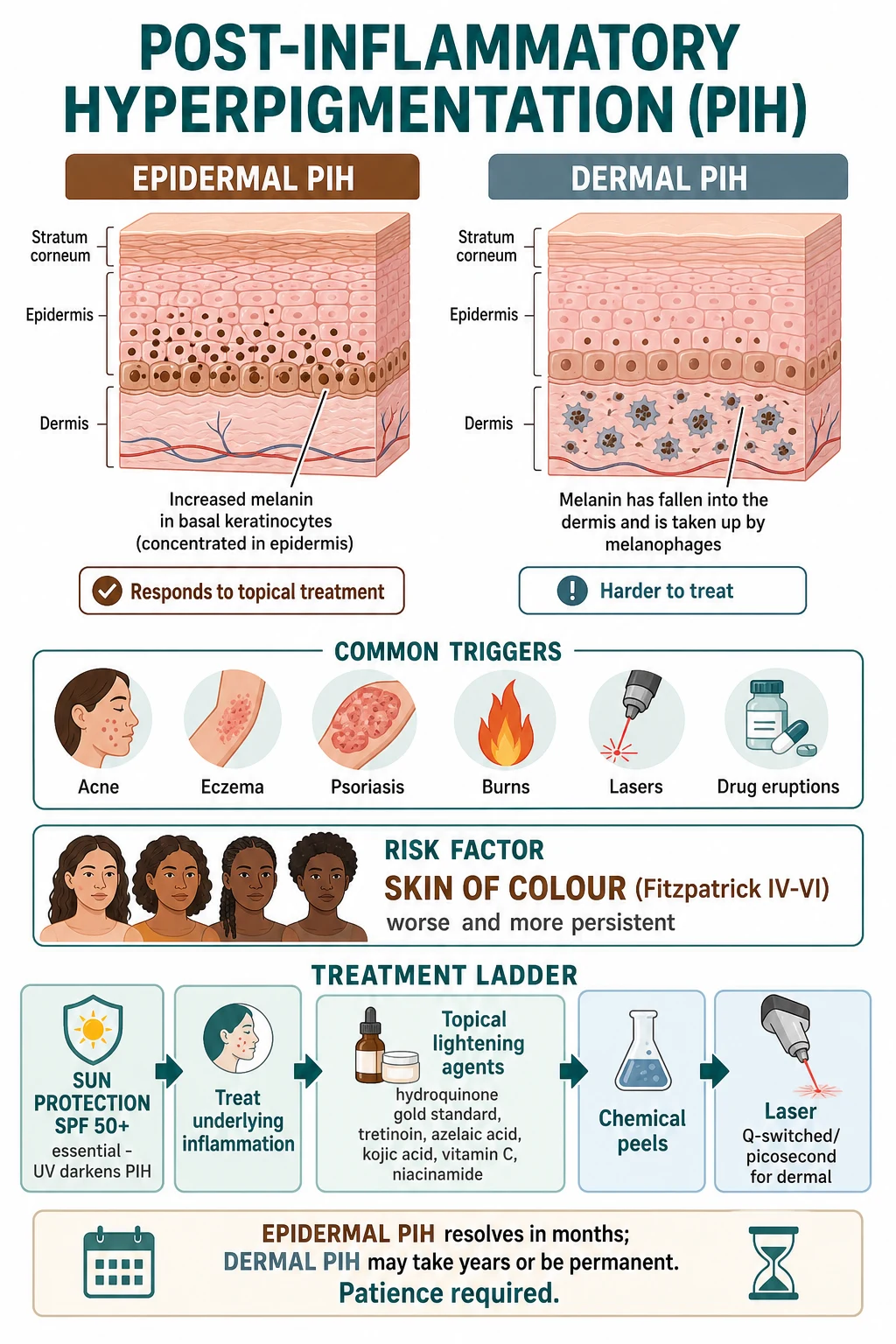
Post-inflammatory hyperpigmentation (PIH) = acquired excess melanin deposited in the skin as a sequela of cutaneous inflammation or injury (acne, eczema, psoriasis, insect bites, burns, chemical peels, lasers, cryotherapy, drug eruptions). It is one of the most common pigmentary disorders worldwide and is markedly more frequent, more conspicuous and more persistent in skin of colour (Fitzpatrick IV-VI). Three anatomic types exist: epidermal PIH (melanin retained in basal and suprabasal keratinocytes; light-brown to tan; brightens under Wood's lamp; responds to topical therapy; resolves over months), dermal PIH (melanin incontinence into the upper dermis with uptake by melanophages; grey-blue to slate; does NOT brighten under Wood's lamp; topicals penetrate poorly; persists for years and may be permanent), and mixed. Erythema from the vasodilatory phase of inflammation resolves in days to weeks but pigment persists because melanin turnover is slow. Management ladder: (1) rigorous photoprotection SPF 50+ plus visible-light (tinted iron-oxide) protection; (2) treat the underlying inflammation; (3) topical lightening agents — hydroquinone 2-4% nightly is the gold-standard tyrosinase inhibitor, with tretinoin, azelaic acid, kojic acid, niacinamide, vitamin C and the combination Kligman's formula (hydroquinone + tretinoin + corticosteroid); (4) chemical peels (glycolic, salicylic, Jessner's, mandelic) for epidermal PIH; (5) lasers — Q-switched ruby/alexandrite/Nd:YAG and picosecond — for dermal or refractory PIH. Oral tranexamic acid is an emerging systemic option. Caution: prolonged unsupervised hydroquinone causes exogenous ochronosis; peels and lasers can paradoxically worsen PIH in skin of colour.
On this page & tools
Your progress
Saved locally on this device.
Exam tags
Red flags
Overview & Definition
Post-inflammatory hyperpigmentation (PIH) is the acquired deposition of excess melanin (and, in some contexts, haemosiderin) within the skin that appears as a direct sequela of cutaneous inflammation or injury. The pigment follows — and is confined to — the distribution of the preceding inflammatory event, whether an acne papule, an eczematous patch, a thermal burn, a chemical peel, a laser injury, a fixed drug eruption or an insect bite. Unlike a primary pigmentary disorder such as melasma or vitiligo, PIH is reactive: remove the trigger and no new pigment forms, although what is already present takes months to years to clear.[2][5][6]
PIH is one of the most common reasons for dermatology consultation in populations of African, Hispanic, South Asian, East Asian and Middle Eastern descent, and it carries a disproportionate psychosocial burden because it most often appears on the face. The condition is biologically benign — it is not premalignant, it does not scar, and it does not endanger the patient — but its cosmetic and psychological impact can be severe, particularly in adolescents and young adults whose acne is the trigger. The clinical skill lies not in diagnosing PIH (which is usually obvious from the history) but in (a) determining how deep the pigment sits, because depth dictates prognosis and therapy; (b) excluding the mimics that are not PIH at all; and (c) counselling realistic expectations, because even optimal therapy clears epidermal pigment only over months and dermal pigment only over years.[1][3]
Classification — Epidermal, Dermal and Mixed
The single most useful classification of PIH is by the anatomical depth at which the excess melanin resides, because depth predicts colour, Wood's lamp behaviour, response to topical therapy and prognosis. Three patterns are recognised:[2][6]
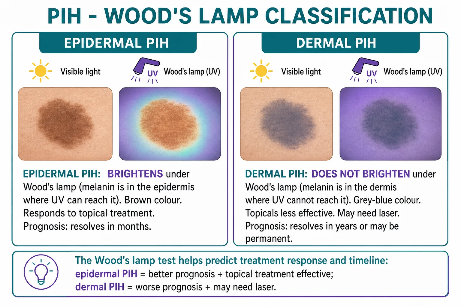
- Light-brown to tan to dark-brown macules
- Melanin in basal and suprabasal keratinocytes
- Wood's lamp: BRIGHTENS (enhances contrast)
- Topical therapy: GOOD response
- Prognosis: resolves over 3 to 12 months
- Grey-blue, slate or ashy-brown macules
- Melanin in upper-dermis melanophages (melanin incontinence)
- Wood's lamp: NO CHANGE (pigment too deep)
- Topical therapy: POOR response (topicals penetrate poorly)
- Prognosis: 6 to 24 months, sometimes permanent
- Brown with a greyish hue
- Both epidermal melanin and dermal melanophages
- Wood's lamp: partial / variable enhancement
- Topical therapy: partial response
- Prognosis: intermediate; often needs combination therapy or laser
The mechanistic basis of this distinction is the integrity of the dermo-epidermal junction. When inflammation is superficial and the basement membrane remains intact, the excess melanin produced by hyper-stimulated melanocytes is transferred to neighbouring keratinocytes and remains within the epidermis — producing the brown, topically-responsive, epidermal pattern. When inflammation is deep or severe enough to disrupt the basement membrane, melanin and melanosomes "fall through" (the phenomenon of melanin incontinence) into the papillary dermis, where they are phagocytosed by macrophages that become long-lived melanophages. Because there is no active mechanism to remove melanin from the dermis, dermal pigment turns over extraordinarily slowly and may persist indefinitely. This is the central reason dermal PIH is so refractory.[5][6]
Epidemiology & Risk Factors
PIH is universal across all skin types in principle, but its prevalence, severity, visibility and persistence are heavily skewed toward darker phototypes. [1]
Risk factors cluster into three groups:[1][4][5]
- Constitutive pigmentation (the dominant factor). Fitzpatrick IV-VI skin has larger, more numerous, more rapidly responding melanocytes that generate more melanin per unit stimulus, making PIH both more likely after any given insult and more clinically apparent against the darker background. This single biological fact explains why the entire management of PIH is calibrated to skin of colour.
- Depth and severity of the triggering inflammation. Lichenoid, blistering or deeply infiltrative processes (lichen planus, Stevens-Johnson syndrome/TEN, deep acne nodules, severe atopic dermatitis, deep chemical or thermal burns) disrupt the basement membrane and tend to produce dermal PIH, whereas superficial inflammation (mild acne, superficial eczema) usually produces epidermal PIH.
- External aggravators. Ultraviolet and visible-light exposure darken existing pigment; picking, squeezing and excoriation re-injure skin and renew the pigmentation cycle; harsh cosmetics, irritant contact dermatitis and over-aggressive procedures (peels, lasers, dermabrasion, cryotherapy) themselves become iatrogenic causes; and certain drugs — notably the fixed drug eruption culprits and minocycline, amiodarone, chloroquine, clofazimine and heavy metals — produce pigmentation that overlaps with and may be mislabelled as PIH. [1]
Pathophysiology
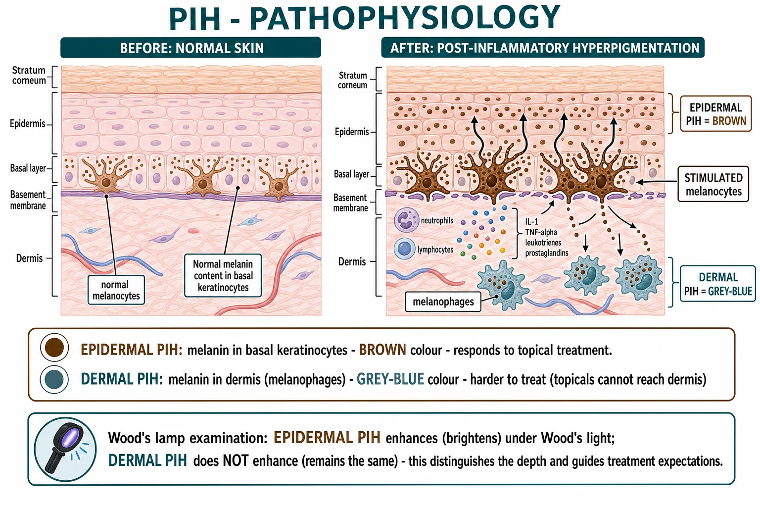
PIH is fundamentally a disorder of disregulated melanogenesis, and the cascade runs as follows.[5][6]
The inflammatory mediator cascade
When the skin is injured — by acne, eczema, a burn, a drug, a virus or a laser — keratinocytes, mast cells, neutrophils and resident immune cells release a cocktail of pro-pigmentary mediators. The principal actors are: [1]
- Interleukin-1 (IL-1 alpha and beta), tumour necrosis factor-alpha (TNF-alpha) and interleukin-6 (IL-6) — cytokines that upregulate melanocyte tyrosinase activity and melanocyte-proliferation signals.
- Leukotrienes (LTC4, LTD4, LTE4) — arachidonic-acid products with a powerful direct stimulatory effect on melanocyte proliferation and melanin synthesis; they are among the most experimentally reproducible inducers of pigmentation.
- Prostaglandins (PGE2, PGF2-alpha) and thromboxanes — eicosanoids that increase both melanogenesis and dendricity of the melanocyte, enhancing melanosome transfer.
- Endothelin-1 (ET-1) and stem cell factor (SCF) / c-Kit signalling — keratinocyte-derived peptides that drive melanocyte activation, survival and migration, and that are upregulated after UV exposure and inflammation.
- Alpha-melanocyte-stimulating hormone (alpha-MSH), adrenocorticotropic hormone (ACTH) and prostaglandin E2 — melanocortin-axis signals that bind the melanocortin-1 receptor (MC1R) on the melanocyte and switch on the cAMP-CREB-MITF pathway that culminates in tyrosinase, TYRP1 and DCT transcription — the three enzymes of the melanin-synthesis pathway. [1]
The net effect is a melanocyte that synthesises more melanin, extends longer dendrites, packages melanosomes more rapidly, and transfers them in greater numbers to the ~36 keratinocytes in its column. Clinically, this manifests as the hyperpigmented macule that appears within days to weeks of the inflammatory peak. [1]
Epidermal versus dermal fate of the pigment
The depth of the original inflammation decides where the excess melanin ends up. If the basement membrane zone is preserved, melanin is retained within basal and suprabasal keratinocytes and is carried outward with the normal epidermal turnover (roughly 28 to 40 days), which is why epidermal PIH fades over months as pigmented keratinocytes are shed. If the basement membrane is disrupted — by deep inflammation, blistering, lichenoid interface dermatitis, or destructive procedures — melanosomes and free melanin spill into the papillary dermis and are engulfed by macrophages. These melanophages are long-lived, resident cells with no efficient clearance pathway, which is why dermal PIH persists for years and may be permanent. The optical grey-blue colour of dermal PIH is a Tyndall effect: longer wavelengths of incident light are absorbed by the overlying tissue and shorter (blue) wavelengths are scattered back to the eye, exactly the same physics that makes superficial veins look blue.[5][6]
Why erythema resolves but pigment persists
A common teaching point and a frequent patient question: why does the redness of the original acne or burn fade in days to weeks while the brown mark lasts for months? The two phenomena are mechanistically distinct. Erythema reflects vasodilation of the superficial plexus, which resolves within days to a few weeks once the inflammatory mediators clear and vascular tone is restored. Pigment, by contrast, reflects melanin synthesis and deposition, a much slower process whose clearance depends either on epidermal turnover (months) or on melanophage depletion (years). The two trajectories decouple, and this decoupling is the biological reason patients must be counselled that "the redness going away does not mean the pigmentation is going away." [1]
The role of ultraviolet and visible light
Both UVB (290 to 320 nm) and, more importantly, UVA (320 to 400 nm) drive melanogenesis directly by damaging keratinocyte DNA (p53-mediated alpha-MSH release) and by upregulating the mediator cascade described above. In darker phototypes, visible light — particularly high-energy visible (HEV) blue light around 415 nm — is an additional and under-appreciated driver of pigmentation, acting through the opsin-3 (OPN3) receptor on melanocytes. This is the rationale for tinted sunscreens containing iron oxide, which absorb visible light, in skin of colour: a non-tinted broad-spectrum sunscreen blocks UV but permits HEV-induced pigmentation to continue.[1][11]
Histopathology
On routine haematoxylin-and-eosin staining, epidermal PIH shows increased melanin within basal and suprabasal keratinocytes with no increase in melanocyte number; dermal PIH shows melanin incontinence with melanophages in the papillary and upper reticular dermis. A Fontana-Masson silver stain highlights the melanin, and a S100, SOX10 or Melan-A stain confirms that the dendritic cells in the basal layer are melanocytes (not increased in number) rather than a proliferative melanocytic lesion. The histology is confirmatory rather than diagnostic — PIH is a clinical diagnosis, and biopsy is reserved for atypical or non-resolving cases. [1]
Clinical Presentation
PIH presents as macules (and occasionally slightly elevated plaques or papules) of altered pigmentation that occupy exactly the footprint of a preceding inflammatory or injurious event.[4][6]
- Colour. The colour is the single most informative clinical sign. Light-brown, tan, fawn or dark-brown macules indicate epidermal melanin. Grey, slate, blue-grey or ashy macules indicate dermal melanin. A brown macule with a discernible grey hue suggests a mixed pattern. The colour should be read in good natural or daylight light, as fluorescent lighting distorts the brown-grey distinction.
- Distribution. PIH follows the distribution of its trigger. Acne-induced PIH therefore clusters on the cheeks, jawline, forehead, upper back and chest; atopic-dermatitis PIH favours the flexures (antecubital fossae, popliteal fossae, neck, eyelids); insect-bite PIH is on exposed limbs; fixed-drug-eruption PIH is on the lips, genitalia, hands and feet; procedural PIH is on the treated site.
- Sites most often seen in the clinic. The face dominates the consultation because facial PIH is the most cosmetically distressing, but PIH is common on the neck, upper trunk, dorsal hands and proximal limbs.
- Morphology variants. Linear PIH follows excoriation and scratching tracks (Koebner isomorphic phenomenon); guttate PIH follows varicella, acne or folliculitis; confluent plaques follow eczema or friction; reticulate PIH follows lichenoid disorders; and perifollicular PIH follows folliculitis or acne.
- Temporal evolution. Pigment appears within days to weeks of the inflammatory peak. It is darkest at the height of the inflammatory reaction and then, in epidermal PIH, fades gradually over months; in dermal PIH it may persist unchanged for years.
- Symptoms. PIH itself is asymptomatic. Itch, pain or tenderness indicate that the underlying inflammatory process is still active and has not yet burned out — an important clue that treating the trigger is still the priority. [1]
Atypical presentations
Examiners probe the corners. The following atypical presentations are testable:[4]
- PIH in lichenoid disorders. Lichen planus produces a characteristic, deeply grey-blue, long-lasting dermal PIH on the flexor wrists, lumbar region and oral mucosa; the pigment may persist long after the violaceous papules have cleared.
- PIH after Stevens-Johnson syndrome or toxic epidermal necrolysis. Survivors develop extensive, dark, often reticulate PIH over the sites of epidermal detachment that can take a year or more to clear and may be permanent.
- PIH in infants and children with atopic dermatitis. Often diffuse, ill-defined, grey-brown, and persistent; distinguished from true post-inflammatory hypopigmentation by Wood's lamp (hyperpigmentation brightens, hypopigmentation does not).
- Photo-exacerbated PIH. Facial PIH that darkens strikingly through summer and lightens through winter signals ongoing UV/HEV-driven melanogenesis and mandates intensified photoprotection. [1]
A 24-year-old woman with Fitzpatrick V skin has acne papules on both cheeks that, once healed, leave dark-brown marks that have not faded at six months. The marks brighten clearly under Wood's lamp. What type of PIH is this, and what is the prognosis?
This is epidermal PIH: the brown colour and the Wood's-lamp enhancement confirm melanin confined to the basal keratinocytes. The prognosis is good — epidermal PIH responds to a combination of rigorous photoprotection, treatment of the active acne, and a topical lightening agent (hydroquinone 2 to 4%, azelaic acid 15 to 20%, or a tretinoin-based regimen), with clearance expected over the next 3 to 12 months.
Differential Diagnosis
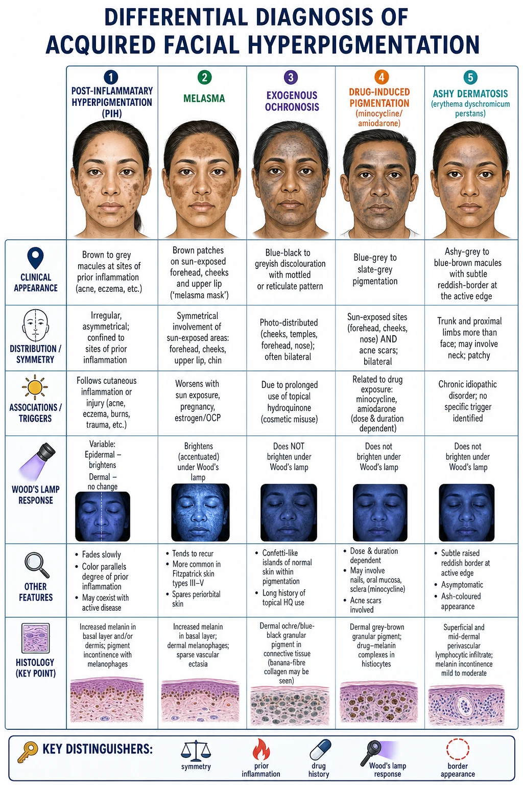
The differential diagnosis of acquired hyperpigmentation is broad, and several mimics are more important to exclude than PIH itself because they imply systemic disease, drug toxicity or a different (and sometimes irreversible) pigmentary disorder.[6][11][12]
| Differential | Distinguishing feature from PIH |
|---|---|
| Melasma | Symmetrical, sharply demarcated brown patches on the sun-exposed forehead, cheeks and upper lip (the "melasma mask"); no preceding inflammation; worsens with sun exposure, pregnancy and oral contraceptives; Wood's lamp may brighten (epidermal melasma) — the history of pregnancy/OCP and symmetry are the keys. |
| Exogenous ochronosis | Blue-black or grey-brown pigmentation with confetti-like islands of normal skin on photo-exposed areas in a patient with a history of prolonged, often unsupervised hydroquinone use; Wood's lamp does NOT brighten; biopsy shows ochre-coloured banana-shaped fibres in the dermis. |
| Drug-induced pigmentation | Minocycline (type I blue-black on acne scars, type II blue-grey on sun-exposed skin, type III diffuse muddy-brown on shins), amiodarone (slate-grey photo-distributed), chloroquine/hydroxychloroquine (blue-grey), clofazimine (reddish-brown), heavy metals (gold, silver — argyria is slate-grey); a careful drug history is diagnostic. |
| Ashy dermatosis (erythema dyschromicum perstans) | Asymptomatic ashy grey-brown macules with a subtle raised erythematous border at the active edge, mainly on the trunk and proximal limbs rather than the face; chronic; Wood's lamp negative (dermal). |
| Addison's disease | Generalised hyperpigmentation with accentuation in sun-exposed areas, flexures, palmar creases, oral mucosa, recent scars and gingival margins; accompanied by fatigue, weight loss, postural hypotension, salt-craving, hyponatraemia and hyperkalaemia — measure morning cortisol and ACTH. |
| Haemochromatosis | Diffuse bronze or grey-brown "bronze diabetes" pigmentation with hepatomegaly, diabetes, cardiac disease, gonadal failure; measure serum ferritin and transferrin saturation. |
| Melanoacanthoma | A benign solitary dark brown to black papule or plaque that grows rapidly; a deep pigmented variant of seborrhoeic keratosis; distinguished from PIH by being a single raised lesion rather than a macule at a healed inflammatory site. |
| Acanthosis nigricans | Velvety, thickened, hyperpigmented plaques in the flexures (axillae, neck, groin); mechanistically epidermal proliferation driven by insulin/IGF-1, not inflammation; usually indicates insulin resistance. |
| Linear epidermal naevus | Congenital or early-childhood onset, blaschkoid linear distribution, papillomatous surface — a hamartomatous malformation, not reactive. |
The cardinal question on history is "Was there an inflammatory or injurious event at this exact site before the pigmentation appeared?" A "yes" supports PIH. A "no", together with symmetry, mucosal involvement, drug exposure, or systemic symptoms, points to an alternative diagnosis that needs work-up.[6][11]
Clinical & Bedside Assessment
The diagnosis of PIH is clinical and made at the bedside; no blood test is required for the typical case. The structured assessment has four elements.[2][6]
- Confirm the trigger. Take a careful history of the preceding lesion: acne, eczema, burn, insect bite, procedure (peel, laser, cryotherapy, microneedling), infection (herpes zoster, impetigo, tinea), or drug (fixed drug eruption). The pigmentation should be confined to the footprint of the trigger.
- Establish the depth. Examine the colour in good light: brown = epidermal; grey-blue = dermal. Perform a Wood's lamp (long-wave UVA, 365 nm) examination in a darkened room — epidermal PIH brightens (enhances contrast), dermal PIH does not change. Note that Wood's lamp sensitivity falls in very dark (Fitzpatrick VI) skin because constitutive epidermal melanin absorbs the UV before it reaches the lesion.
- Screen for systemic mimics. Look for generalised pigmentation, palmar-crease and oral-mucosal involvement, flexural accentuation, weight loss, fatigue and postural symptoms — any of which mandate a cortisol, ferritin or B12 work-up.
- Assess aggravators. Quantify UV and visible-light exposure, picking/squeezing behaviour, cosmetic use, and adherence to any prior topical regimen. The history of what the patient is doing to their skin is often more informative than the lesion itself. [1]
Dermoscopy is occasionally useful: epidermal PIH shows a regular pigment network with accentuation of the basal layer; dermal PIH shows a greyish, structureless hue due to the deeper melanophages. Dermoscopy is more valuable for excluding a pigmented malignancy (melanoma in a regressing naevus, pigmented basal cell carcinoma) than for confirming PIH. [1]
Investigations
PIH is a clinical diagnosis. Skin biopsy is reserved for: atypical or non-resolving lesions; suspected exogenous ochronosis (banana-shaped ochre fibres); suspected melanoacanthoma; suspicion of a pigmented malignancy; or dermato-lymphadenopathy. When biopsied, Fontana-Masson staining confirms melanin and S100/SOX10/Melan-A confirm that the melanocyte population is not increased (excluding a melanocytic proliferation).[2][6]
- Atypical or non-resolving pigmentation
- Suspicion of ochronosis (prolonged hydroquinone use)
- Suspicion of melanoacanthoma or pigmented malignancy
- Generalised, mucosal or palmar-crease pigmentation — Addison's or haemochromatosis
- Macrocytic anaemia — vitamin B12 / folate deficiency
- Morning cortisol and plasma ACTH — Addison's disease
- Serum ferritin and transferrin saturation — haemochromatosis
- Vitamin B12 and serum folate — deficiency pigmentation
- TSH — thyroid-driven pigmentary change
- Skin biopsy with Fontana-Masson — confirm depth / exclude mimics
Management — Resuscitation and the Foundational Pillars
PIH is rarely a medical emergency. The one "resuscitation" question the clinician must answer is whether the hyperpigmentation is the sequela of an emergency that is still active — a Stevens-Johnson syndrome/TEN that is still progressing, a drug reaction with eosinophilia and systemic symptoms (DRESS) with evolving organ failure, an extensive partial- or full-thickness burn, or a fixed drug eruption still being re-exposed to its culprit. In each of these, the PIH is irrelevant at presentation; the underlying process must be recognised and managed first. Once the acute process has settled, the management of the resulting PIH begins.[2][7]
Two foundational, non-negotiable pillars precede all pharmacological and procedural therapy: [1]
- Rigorous photoprotection. Without this, every other intervention fails, because ultraviolet and visible light drive ongoing melanogenesis and darken existing pigment. Counsel daily application of a broad-spectrum sunscreen of SPF 50 or higher with UVA (PA++++ or equivalent) cover, reapplication every two hours during sun exposure, avoidance of peak midday sun, protective clothing, wide-brimmed hats and — in skin of colour — a tinted sunscreen containing iron oxide to absorb the high-energy visible (HEV) blue light that non-tinted sunscreens do not block.[1][11]
- Treat the active underlying inflammation. New PIH forms only while new inflammation is active. Aggressive control of acne (retinoids, benzoyl peroxide, hormonal therapy, oral isotretinoin where indicated), atopic dermatitis (emollients, topical corticosteroids, calcineurin inhibitors, dupilumab), psoriasis, lichen planus and the fixed-drug culprit (withdrawal of the offending drug) stops the production line. Counselling the patient not to pick, squeeze or excoriate lesions is a low-technology intervention with a high yield.[4]
Management — Topical Lightening Therapy
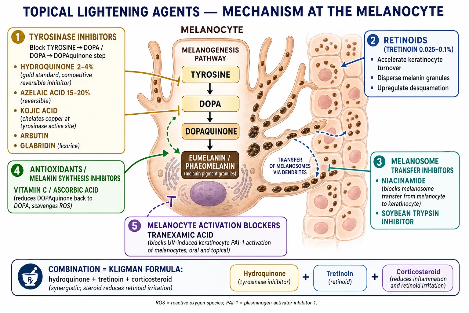
Topical therapy is the mainstay of epidermal PIH and the first pharmacological step for most patients. The agents act at one of four points: inhibition of tyrosinase (the rate-limiting enzyme of melanin synthesis), acceleration of epidermal turnover (to shed pigmented keratinocytes faster), blockade of melanosome transfer (from melanocyte to keratinocyte), and antioxidant / anti-inflammatory effects that reduce the stimulus for further melanogenesis.[2][3][7]
Hydroquinone — the gold standard
Hydroquinone 2 to 4% cream, applied once nightly to the affected areas for 8 to 12 weeks (extendable up to 6 months under supervision), is the gold-standard tyrosinase inhibitor and the benchmark against which all other agents are compared. It competitively and reversibly inhibits tyrosinase, the enzyme that converts tyrosine to DOPA and DOPA to DOPAquinone, and also partially suppresses melanocyte RNA and DNA synthesis. It is most effective for epidermal PIH; dermal PIH responds poorly because topicals penetrate the dermis inefficiently.[2][7]
Cautions. Hydroquinone is irritant and can itself cause a contact dermatitis that paradoxically worsens PIH (the "irritation-darkening" cycle). The most important long-term caution is exogenous ochronosis — a blue-black, reticulate, photo-distributed pigmentation with histological ochre-coloured banana-shaped dermal fibres that arises from prolonged, unsupervised use of concentrations above 4% for months to years. To mitigate this risk, hydroquinone should be (a) used for a defined course of 8 to 12 weeks, extendable under dermatological supervision; (b) followed by a "hydroquinone holiday" of 1 to 2 months on a non-hydroquinone maintenance agent; and (c) combined with a topical retinoid and a mild corticosteroid (Kligman's formula) which both enhance efficacy and reduce the irritation that drives ochronosis. Hydroquinone is avoided in pregnancy.[2][7]
Tretinoin and other retinoids
Tretinoin 0.025 to 0.1% cream accelerates epidermal turnover (shedding pigmented keratinocytes faster), disperses existing melanin granules, and has a mild direct anti-inflammatory effect. It is applied nightly, is comedolytic (so doubles as acne therapy when acne is the PIH trigger), and is most useful in combination rather than as monotherapy. The principal adverse effect is retinoid dermatitis — erythema, scaling and burning that can paradoxically worsen PIH — so it must be introduced gradually (every other night, then nightly, with a ceramide moisturiser). Adapalene 0.1 to 0.3% is a better-tolerated alternative and is pregnancy-category C (tretinoin is also avoided in pregnancy).[3][7]
Azelaic acid — the versatile, pregnancy-safe agent
Azelaic acid 15 to 20% gel or cream, applied twice daily for 3 to 6 months, is a dicarboxylic acid with three useful properties: it is a reversible tyrosinase inhibitor (selectively toxic to hyperactive melanocytes), it has a direct anti-inflammatory and anti-acne effect (it suppresses neutrophilic ROS and reduces follicular bacterial load), and it is safe in pregnancy and lactation. These three properties make azelaic acid the agent of choice for PIH in (a) pregnant or breastfeeding patients, (b) patients with coexisting acne or rosacea, and (c) patients who cannot or will not use hydroquinone. Mild transient irritation and depigmentation of normally-pigmented skin at the application site are the principal adverse effects.[3][7]
Other topical agents
- Kojic acid (1 to 4%) — a fungal-derived tyrosinase inhibitor that chelates copper at the enzyme's active site; a common cosmeceutical ingredient but a notable sensitiser (contact allergy).
- Arbutin — a naturally occurring beta-D-glucopyranoside of hydroquinone that releases hydroquinone in the skin; a milder, less regulated alternative available in many over-the-counter preparations.
- Licorice extract (glabridin) — a botanical tyrosinase inhibitor with additional anti-inflammatory properties; widely used in cosmeceuticals.
- Niacinamide (nicotinamide) 4 to 5% — a vitamin B3 derivative that blocks the transfer of melanosomes from melanocyte to keratinocyte, reduces epidermal barrier water loss and has a mild anti-acne and anti-rosacea effect; well tolerated and pregnancy-safe.[9]
- Vitamin C (L-ascorbic acid) 5 to 15% — an antioxidant that reduces DOPAquinone back to DOPA (short-circuiting melanin synthesis) and scavenges the reactive oxygen species generated by UV; unstable in solution and best used in stabilised, low-pH formulations.
- Tranexamic acid (topical, 2 to 5%, or micro-needled) — inhibits UV-induced melanocyte activation by blocking the keratinocyte plasminogen-activator-inhibitor-1 / plasmin pathway; emerging evidence supports efficacy in epidermal and mixed PIH.[10]
- Cysteamine, thiamidol, rucinol, licorice, soybean trypsin inhibitor, oligopeptide-68 — newer agents with growing evidence, particularly thiamidol (a potent synthetic tyrosinase inhibitor).[11]
Combination therapy — Kligman's formula
The principle of combination therapy is to attack melanogenesis at multiple points simultaneously while counteracting the irritation of any single agent. The prototype is Kligman's formula (Kligman and Willis, 1975): hydroquinone 4% + tretinoin 0.025 to 0.05% + a low-potency corticosteroid (originally dexamethasone 0.1%; modern commercial preparations use fluocinolone acetonide 0.01% with hydroquinone 4% and tretinoin 0.05%). The hydroquinone blocks tyrosinase, the tretinoin accelerates turnover and disperses melanin, and the corticosteroid suppresses the inflammation and the retinoid-induced irritation that would otherwise worsen PIH. Triple-combination cream applied nightly for 8 weeks is the most effective topical regimen for epidermal PIH and melasma, but is not for long-term unsupervised use (ochronosis risk, steroid atrophy, telangiectasia).[2][7]
STAR-P
Management — Procedural Therapies
When topical therapy has been optimised for 3 to 6 months and epidermal or mixed PIH persists — or when dermal PIH is present from the outset — procedural options are considered. All procedures carry a paradoxical-worsening risk in Fitzpatrick IV-VI skin, where the procedure itself can become a new inflammatory insult and generate more PIH than it removes. The cardinal principles in skin of colour are: lower concentrations or fluences, shorter contact times, test spots, and a conservative escalation.[1][3]
Chemical peels
Glycolic acid (an alpha-hydroxy acid, AHA, 20 to 70%), salicylic acid (a beta-hydroxy acid, BHA, 20 to 30%), Jessner's solution (resorcinol, salicylic acid, lactic acid, ethanol), mandelic acid (a large-molecule AHA with a favourable safety profile in skin of colour) and lactic acid are the principal agents. Peels are most effective for epidermal PIH, where they accelerate the removal of pigmented keratinocytes. In Fitzpatrick IV-VI skin, begin with lower concentrations (e.g., glycolic acid 20 to 35%), shorter contact times and longer intervals (2 to 4 weeks), and pre-treat with a topical tyrosinase inhibitor for 2 to 4 weeks ("priming"). The two principal complications are post-peel hyperpigmentation (the procedure-induced PIH it was meant to treat) and, with over-aggressive peeling, scarring and hypopigmentation.[3][7]
Laser therapy
Laser therapy is reserved for dermal PIH or for refractory epidermal PIH that has failed topical therapy and peels. The principal devices are: [1]
- Q-switched (quality-switched) lasers — ruby (694 nm), alexandrite (755 nm) and Nd:YAG (1064 nm) — nanosecond-pulse lasers that fragment melanin into particles small enough for dermal clearance. The 1064 nm Nd:YAG, with its deeper penetration and lower melanin absorption, has the most favourable safety profile in skin of colour and is the workhorse device.[8]
- Picosecond lasers (755 nm alexandrite, 1064 nm Nd:YAG) — newer devices with picosecond pulses that fragment melanin by a photoacoustic rather than purely photothermal mechanism, delivering equivalent or superior pigment clearance with less collateral thermal damage and a lower paradoxical-worsening rate; increasingly preferred in skin of colour.[8]
Laser must be performed by a clinician experienced in skin of colour, with test spots at least 4 to 6 weeks before full-face treatment, conservative fluences, and rigorous pre- and post-treatment photoprotection and topical priming with a tyrosinase inhibitor.[1][8]
Microneedling
Microneedling (collagen-induction therapy) creates controlled micro-injuries that enhance the penetration of topical agents (notably tranexamic acid) and may itself remodel pigment; it has a relatively favourable safety profile in skin of colour because it does not rely on thermal energy, and can be combined with topical tyrosinase inhibitors ("mesotherapy"). [1]
Management — Systemic Therapy
Systemic therapy for PIH is limited. Oral tranexamic acid (typically 250 mg twice daily for 3 to 6 months, with photoprotection) is the principal emerging systemic option; it inhibits UV-induced melanocyte activation by blocking the plasmin/plasminogen pathway in keratinocytes, and is supported by randomised controlled trial evidence in melasma (a closely related disorder) and a growing observational literature in dermal and mixed PIH. Tranexamic acid is contraindicated in patients with a history of thromboembolism, an acquired or inherited thrombophilia, active anticoagulation, or pregnancy, and should be used with caution in smokers and patients on combined oral contraceptives.[10][11]
There is no role for systemic corticosteroids, oral retinoids (other than isotretinoin to control the underlying acne), or oral "skin-lightening" supplements in routine PIH management; the evidence base for oral vitamin C, oral glutathione and oral niacinamide is weak and these agents are not recommended. [1]
Management by Site, Scenario and Subtype
STEP 1 — Photoprotection: broad-spectrum SPF 50+ daily, tinted iron-oxide in skin of colour, sun-avoidance behaviour. Non-negotiable.
STEP 2 — Treat the trigger: control acne (topical retinoid + benzoyl peroxide ± isotretinoin), eczema (emollient + corticosteroid/calcineurin inhibitor), psoriasis, lichen planus; withdraw a fixed-drug culprit. Counsel: do not pick or squeeze.
STEP 3 — Topical lightening (8-12 weeks): hydroquinone 2-4% nightly OR azelaic acid 15-20% BD (pregnancy / acne) OR tretinoin 0.025-0.1% nightly; combination triple-cream (Kligman) for stubborn epidermal PIH; maintenance with niacinamide, vitamin C, kojic acid.
STEP 4 — Chemical peels (epidermal PIH after step 3): glycolic / salicylic / Jessner's / mandelic, low concentration and primed with tyrosinase inhibitor in Fitzpatrick IV-VI.
STEP 5 — Laser / energy device (dermal or refractory): Q-switched Nd:YAG 1064 nm or picosecond; test spots; experienced operator.
STEP 6 — Consider oral tranexamic acid 250 mg BD for dermal / mixed / refractory PIH (screen for thromboembolic contraindications).
Acne-induced PIH (the most common presentation) is best prevented by early, aggressive acne control — because the most effective treatment for PIH is never to generate it. Once present, a combination of a topical retinoid (tretinoin or adapalene, which treats both acne and PIH) with azelaic acid or hydroquinone, plus photoprotection and counselling against excoriation, is first-line. Oral isotretinoin for severe nodulocystic acne both clears the acne and prevents the PIH that new lesions would cause, though isotretinoin itself can transiently cause photosensitivity and must be paired with rigorous photoprotection.[4]
Atopic-dermatitis PIH is managed by first extinguishing the eczema (emollients, topical corticosteroids, calcineurin inhibitors, and — in moderate-to-severe cases — dupilumab), then introducing a gentle lightening agent (azelaic acid or niacinamide preferred over hydroquinone, because the atopic skin barrier is fragile and easily irritated). The pigment is often diffuse and slow to resolve, and patient counselling is critical. [1]
Procedural PIH (after peels, lasers, cryotherapy, dermabrasion, microneedling) is prevented by conservative parameters in skin of colour, pre-treatment with a tyrosinase inhibitor for 2 to 4 weeks ("priming"), rigorous post-procedure photoprotection, and prompt treatment of any post-procedural inflammation. Once procedural PIH has occurred, the same topical ladder applies; patience is essential because iatrogenic PIH is often dermal and slow. [1]
Fixed-drug-eruption residual pigmentation is characteristically well-circumscribed, round or oval, and recurs at the same anatomical site on each re-exposure to the culprit drug (commonly NSAIDs, paracetamol, sulfonamides, tetracyclines, barbiturates, phenolphthalein). The key intervention is identification and lifelong avoidance of the culprit drug; the residual pigment is treated with topical lightening agents but is often dermal and refractory. [1]
Burn and trauma PIH depth is dictated by the depth of the original injury; deep partial- and full-thickness burns produce dermal PIH (and sometimes true post-inflammatory hypopigmentation or scarring) that may require laser. [1]
Complications and Pitfalls
The complications of PIH are mostly iatrogenic — the consequences of treating it badly.[2][7]
- Exogenous ochronosis — the blue-black, reticulate, photo-distributed pigmentation with confetti-like islands of normal skin caused by prolonged unsupervised hydroquinone. Prevent by defined courses, hydroquinone holidays, combination with retinoid plus steroid, and avoidance of concentrations above 4% for prolonged periods. Once established, ochronosis is very difficult to reverse — discontinuation of hydroquinone, topical retinoids, and Q-switched laser are the mainstays.
- Retinoid dermatitis paradoxically worsening PIH — the erythema and irritation of aggressive retinoid use is itself a low-grade inflammation that can drive more pigment. Prevent by gradual introduction, every-other-night initiation, and concurrent ceramide moisturiser.
- Post-procedure PIH — over-aggressive peels or lasers in skin of colour generate more pigment than they remove. Prevent by conservative parameters, test spots, priming, and post-procedure photoprotection.
- Misdiagnosis of a systemic cause. The most dangerous pitfall is labelling generalised pigmentation as "post-inflammatory" and missing Addison's disease, haemochromatosis or vitamin B12 deficiency. Any pigmentation with mucosal, palmar-crease or flexural accentuation, or with systemic symptoms, warrants a cortisol, ferritin and B12 work-up.
- Confusing melasma with PIH. Melasma is hormonally driven, symmetrical, sun-exposed, and has no preceding inflammation; the distinction matters because the two conditions are managed with overlapping but not identical regimens (melasma is more chronic, often requires maintenance, and oral tranexamic acid has stronger evidence).
- Psychosocial impact. Facial PIH, especially in adolescents and in skin of colour, can produce measurable anxiety, depression and quality-of-life impairment. Addressing this explicitly, setting realistic expectations, and offering a structured plan are part of management. [1]
Special Populations
- Paediatric PIH. Common after atopic dermatitis, insect bites, impetigo, varicella and acne in Fitzpatrick IV-VI children. Azelaic acid and photoprotection are the safest agents; hydroquinone and tretinoin are generally avoided in young children. Counselling the parents that epidermal paediatric PIH will fade over months, provided the trigger is controlled, is central.
- PIH in pregnancy. Distinguish from melasma of pregnancy (chloasma) — symmetrical, sun-exposed, hormonally driven. Hydroquinone and tretinoin are avoided in pregnancy; the safe agents are azelaic acid, niacinamide, vitamin C and rigorous photoprotection with a tinted iron-oxide sunscreen. Many PIH regimens can be deferred until the post-partum period.
- PIH in skin of colour (Fitzpatrick IV-VI). The defining clinical scenario. PIH is more frequent, more severe, more visible and more persistent; the risk of procedure-induced worsening is higher; and the management must be calibrated to darker skin (conservative peels, test-spot lasers, tinted sunscreens for HEV protection, priming with tyrosinase inhibitors).[1][8]
- Immunocompromised patients. PIH may follow opportunistic infections — herpes zoster, secondary syphilis (the pigmented macules of pityriasis rosea-like secondary syphilis), HIV-related eosinophilic folliculitis, or drug eruptions from antiretrovirals. Recognise and treat the underlying infection; the PIH is then managed on the standard ladder.
Prognosis and Follow-Up
PIH prognosis is dictated by depth, phototype, sun exposure, ongoing inflammation and treatment adherence.[2][5]
- Epidermal PIH resolves over 3 to 12 months with rigorous photoprotection and appropriate topical therapy; clearance is the rule.
- Dermal PIH resolves over 6 to 24 months, is often incomplete, and may be permanent; topical therapy is less effective, and laser may be required for meaningful improvement.
- Mixed PIH is intermediate: the epidermal component responds to topicals, and the dermal component persists until it is treated with a procedure. [1]
Patients should be reviewed at 3 months to (a) assess response, (b) reassess depth (a mixed lesion may now look purely dermal once the epidermal component has cleared), (c) check adherence and photoprotection technique, and (d) reconsider the diagnosis if there has been no improvement at all. Photoprotection must be continued indefinitely after clearance, because UV and HEV re-exposure will darken any residual pigment and can re-trigger melanogenesis. Patient counselling on the slow, cumulative nature of improvement is essential — many patients abandon an effective regimen prematurely because they expect results in weeks rather than months. [1]
Evidence, Guidelines and Regional Differences
The evidence base for PIH treatment is moderate but heterogeneous. The 2024 systematic reviews by Mar et al. (skin of colour) and Kashetsky et al. (treatment outcomes) and the 2020 review by Shenoy and Madan confirm that topical hydroquinone, tretinoin, azelaic acid and triple-combination creams have the strongest evidence for epidermal PIH, that chemical peels and the Q-switched/picosecond lasers have evidence in dermal and refractory disease, and that the evidence base is weaker in skin of colour (Fitzpatrick IV-VI), where paradoxical worsening is a real and under-reported outcome. The Wang et al. (Part I) and Ko et al. (Part II) JAAD reviews (2023) are the current authoritative overviews of pathogenesis and management.[1][3][6][7]
Hydroquinone regulation varies markedly. In the United States, hydroquinone 2% is available over the counter and 4% by prescription; in the European Union, hydroquinone is banned in cosmetics since 2001 and is available only on prescription for defined indications; and in several African and Asian countries (including Japan, Rwanda, Ivory Coast, Kenya, Ghana, South Africa and others) hydroquinone is restricted or banned in skin-lightening products because of ochronosis and the unregulated use of dangerously high concentrations in the skin-lightening ("bleaching") industry. The practical consequence: when a patient from one of these regions presents with pigmentation, ask specifically about prior over-the-counter or informal skin-lightening product use — many "bleaching creams" sold outside formal pharmacies contain hydroquinone above 4%, corticosteroids, mercury or other agents that themselves cause pigmentation, and the resulting ochronosis or steroid damage may be the true diagnosis.
In Australia and New Zealand, oral tranexamic acid for melasma and dermal/mixed PIH is used off-label (it is not TGA/PDF-registered for this indication) and the randomised Australian evidence (Bala et al., 2022) supports its use in moderate-to-severe melasma; it is contraindicated in thromboembolic disease, anticoagulation and pregnancy.[10]
Visible-light photoprotection is a developing evidence area: tinted iron-oxide sunscreens have been shown in controlled studies to reduce visible-light-induced pigmentation in Fitzpatrick IV-VI skin, and the most recent pigmentary-disorder guidelines now recommend tinted over non-tinted sunscreen in skin of colour.[1][11]
Exam Pearls
Red Flags
Exam application bank (NEET-PG / INICET)
One-line answer
Post-inflammatory hyperpigmentation (PIH) = acquired excess melanin deposited in the skin as a sequela of cutaneous inflammation or injury (acne, eczema, psoriasis, insect bites, burns, chemical peels, lasers, cryotherapy, drug eruptions). It is one of the most common pigmentary disorders worldwide and is markedly more frequent, more conspicuous and more persistent in skin of colour (Fitzpatrick IV-VI). Three anatomic types exist: epidermal PIH (melanin retained in basal and suprabasal keratinocytes; light-brown to tan; brightens under Wood's lamp; responds to topical therapy; resolves over months), dermal PIH (melanin incontinence into the upper dermis with uptake by melanophages; grey-blue to slate; does NOT brighten under Wood's lamp; topicals penetrate poorly; persists for years and may be permanent), and mixed. Erythema from the vasodilatory phase of inflammation resolves in days to
Worked stems (answer without another resource)
Stem 1 — Classic presentation. Map symptoms to mechanism; name the first investigation and first treatment step with dose/route if drug therapy is standard. [1]
Stem 2 — Unstable / complicated. List red flags that force immediate resuscitation, theatre, ICU, antidote, or reperfusion — and what you do in the first 15 minutes. [1]
Stem 3 — Atypical group. Elderly, pregnancy, child, or immunocompromised: how presentation and thresholds change. [1]
Stem 4 — Differential trap. Name the three closest mimics and one discriminator for each. [1]
Stem 5 — Disposition. Who goes home with safety-netting, who is admitted, who needs HDU/ICU/theatre, and what follow-up is mandatory. [1]
Rapid viva checklist
- Definition + classification
- Pathophysiology chain
- Bedside signs / criteria
- Score with exact components (if any)
- Emergency bundle
- Definitive therapy with doses
- Complications of disease and of treatment
- Special populations
- Guideline/trial name if classic
- Three exam traps
Coverage self-check
If you cannot answer any stem above from this page alone, re-read the matching section — the page is intended to be self-sufficient for final-prof and NEET-PG/INICET questions on Post-inflammatory hyperpigmentation.
[1]References
- [1]Mar K, Khalid B, Maazi M, et al. Treatment of Post-Inflammatory Hyperpigmentation in Skin of Colour: A Systematic Review J Cutan Med Surg, 2024.PMID 39075672
- [2]Shenoy A, Madan R. Post-Inflammatory Hyperpigmentation: A Review of Treatment Strategies J Drugs Dermatol, 2020.PMID 32845587
- [3]Kashetsky N, Feschuk A, Pratt ME, et al. Post-inflammatory hyperpigmentation: A systematic review of treatment outcomes J Eur Acad Dermatol Venereol, 2024.PMID 37843491
- [4]Elbuluk N, Grimes P, Chien A, et al. The Pathogenesis and Management of Acne-Induced Post-inflammatory Hyperpigmentation Am J Clin Dermatol, 2021.PMID 34468934
- [5]Maghfour J, Olayinka J, Hamzavi IH, et al. A Focused review on the pathophysiology of post-inflammatory hyperpigmentation Pigment Cell Melanoma Res, 2022.PMID 35306737
- [6]Wang RF, Ko D, Friedman BJ, et al. Disorders of hyperpigmentation. Part I. Pathogenesis and clinical features of common pigmentary disorders J Am Acad Dermatol, 2023.PMID 35151757
- [7]Ko D, Wang RF, Ozog D, et al. Disorders of hyperpigmentation. Part II. Review of management and treatment options for hyperpigmentation J Am Acad Dermatol, 2023.PMID 35158001
- [8]Levin MK, Ng E, Bae YS, et al. Treatment of pigmentary disorders in patients with skin of color with a novel 755 nm picosecond, Q-switched ruby, and Q-switched Nd:YAG nanosecond lasers: A retrospective photographic review Lasers Surg Med, 2016.PMID 26922302
- [9]Madaan P, Sikka P, Malik DS. Cosmeceutical Aptitudes of Niacinamide: A Review Recent Adv Antiinfect Drug Discov, 2021.PMID 34844552
- [10]Bala HR, Nguyen J, Ross A, et al. Randomised, Placebo-Controlled, Double-Blind Study of Oral Tranexamic Acid in the Treatment of Moderate-to-Severe Melasma in an Australian Cohort Indian J Dermatol, 2022.PMID 36578715
- [11]Lane BN, Hamzavi IH, Lim HW, et al. Concurrent management of vitiligo and acquired disorders of hyperpigmentation: a comprehensive literature review and current practice gaps Int J Dermatol, 2025.PMID 39817312
- [12]Narayan RV, Thakur V, Bishnoi A, et al. Acquired dermal macular hyperpigmentation: a unified spectrum of dermal pigmentary dermatoses Clin Exp Dermatol, 2026.PMID 41700520