Dermatology · Medicine
Pseudofolliculitis barbae (razor bumps)
Also known as Pseudofolliculitis barbae (PFB) · Razor bumps · Shaving bumps · Folliculitis barbae traumatica · Sycosis barbae traumatica
Pseudofolliculitis barbae (PFB, razor bumps) is a chronic, recurrent inflammatory disorder of hair-bearing skin caused by ingrown hairs — a sharply cut, tightly curled hair shaft re-enters the skin after shaving and provokes a foreign-body granulomatous reaction. It overwhelmingly affects men of African descent with tightly curled facial hair (up to 60-80% of Black men who shave) and is a particular problem in the military and other clean-shaven occupations; it also affects women in the pubic, axillary and leg areas. Distribution: beard, especially the anterior neck, mandibular angle, chin and submental area. The word 'pseudo' is deliberate: this is NOT a primary folliculitis but a foreign-body reaction to an extrafollicularly or transfollicularly penetrating hair. The management ladder is: AVOID shaving (grow beard, electric clippers, chemical depilatories) → MODIFIED shaving (single-blade, with-the-grain, Bump Fighter) → topical retinoid/antibiotic/corticosteroid → eflornithine 13.9% and/or oral tetracycline → long-pulsed Nd:YAG 1064 nm laser hair removal, the most effective long-term cure in skin of colour.
On this page & tools
Your progress
Saved locally on this device.
Exam tags
Red flags
Overview & Definition
Pseudofolliculitis barbae (PFB), best known to patients as razor bumps, is a chronic, recurrent inflammatory disorder of hair-bearing skin in which a sharply cut, tightly curled hair shaft re-enters the skin after shaving and provokes a foreign-body granulomatous reaction.[1][7] It is one of the commonest dermatoses of men of African descent and is a defining occupational dermatosis of clean-shaven populations such as the military.[4]
The word "pseudo" is deliberate and is the source of the most common exam trap: this is NOT a primary folliculitis. There is no primary infection of the follicle and no initial bacterial pathogen. The follicle is a bystander; the true event is mechanical penetration of the skin by the patient's own hair shaft, with the resulting inflammation being a foreign-body reaction to keratin.[5][11] Secondary bacterial colonisation by Staphylococcus aureus may follow, but it is a complication, not the cause. Recognising this distinction explains why antibiotics alone are insufficient and why the cornerstones of management are mechanical — stopping the close shave, removing the trigger, or permanently destroying the culpable follicle.[3]
Clinically, PFB presents with firm, hyperpigmented, follicular papules and pustules in the shaving distribution of the beard — especially the anterior neck, mandibular angle, chin and submental area — together with pin-point entrapped (ingrown) hairs, post-inflammatory hyperpigmentation (PIH) and, in a subset with a keloidal diathesis, keloidal scarring that may be permanent.[1][7]
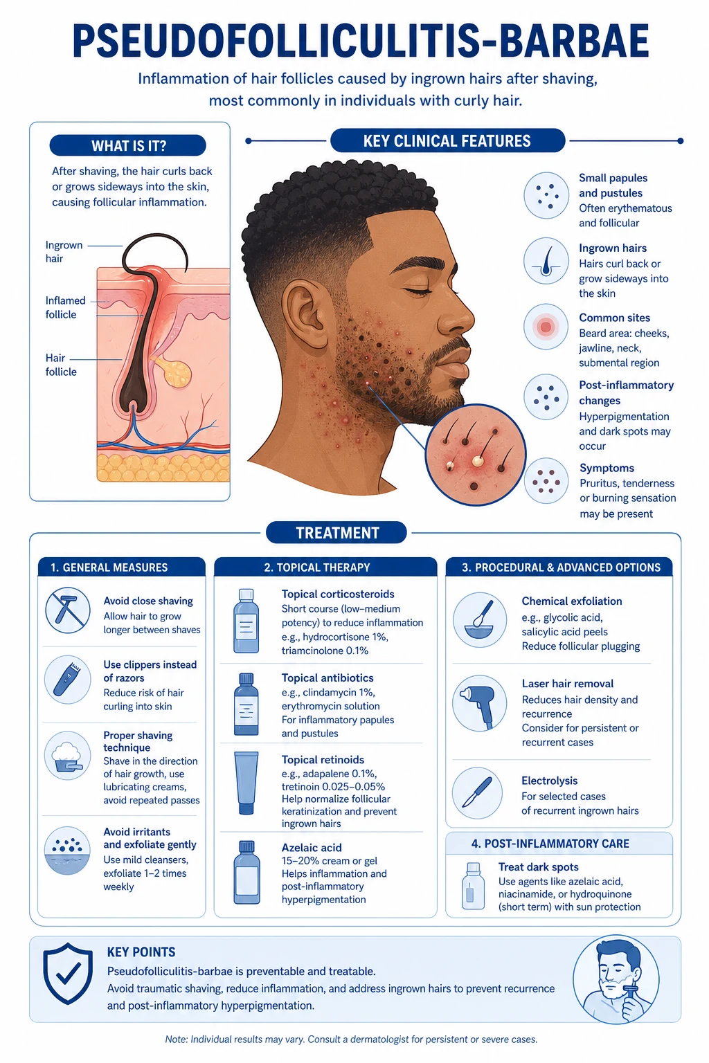
PFB is the beard-area sibling of acne keloidalis nuchae (AKN): the two conditions share an identical core mechanism (a closely clipped, tightly curled hair piercing the dermis and triggering a foreign-body granulomatous reaction), but they differ in site (PFB = beard/anterior neck; AKN = nape of the neck/occipital scalp) and trigger (PFB = shaving the beard; AKN = close clipping of the nape).[5] Recognising this kinship helps candidates remember the shared management principles: trigger avoidance, topical retinoid-antibiotic-steroid combinations, oral anti-inflammatory tetracyclines, and definitive long-pulsed Nd:YAG 1064 nm laser hair removal for skin of colour.[6]
Classification
PFB is classified along three practical axes that map directly onto management. [1]
By mechanism of hair re-entry — the fundamental classification:[11][12]
By site — most commonly the beard and anterior neck, but PFB occurs wherever tightly curled hair is closely removed:[8]
- Beard PFB (the classic form) — anterior neck, mandibular angle, chin, submental area.
- Pubic PFB — increasingly common in both sexes with the trend for pubic hair removal; women over-represented.
- Axillary and leg PFB — women who shave; may be mistaken for deodorant-related folliculitis or keratosis pilaris.
- Nape/occipital PFB — overlaps with acne keloidalis nuchae; the boundary between the two is blurred at the back of the neck. [1]
By severity — there is no universally adopted PFB severity score, but a pragmatic three-grade scheme guides escalation: [1]
- Mild — scattered papules, no scarring, controlled by shaving modification.
- Moderate — confluent papulopustules, PIH, occasional keloidal papule; needs topical medical therapy.
- Severe — extensive papulopustules, keloidal plaques, secondary infection, occupational/psychological impact; needs systemic therapy and/or laser/surgery. [1]
Epidemiology & Risk Factors
Demographic. PFB is overwhelmingly a disease of men of sub-Saharan African descent with tightly curled facial hair. The prevalence among Black men who shave is 45-83%, while among Caucasian men it is only around 3-5%.[1][9] It is the commonest dermatological condition presenting in Black men in many settings. Onset is typically in the late teens to early twenties, coinciding with the start of regular shaving.[7]
Hair and skin type — the master risk factor. The strongest predisposing factor is tightly curled ("woolly") hair with an elliptical (flat) cross-section, growing from a curved follicle at a low, acute angle to the skin surface — the hair type characteristic of people of African descent. When such a hair is cut short, the sharp tip curves back toward and into the skin. By contrast, straight hair has a circular cross-section, grows from a straight follicle at a perpendicular angle, and exits the skin surface without re-entering.[1][11] Skin of colour is also an independent risk factor for the keloidal and hyperpigmented complications of any follicular inflammation, because melanocyte reactivity produces prominent PIH and keloidal diathesis produces exaggerated fibrosis.[5]
Shaving-habits risk factors. These are modifiable and are the targets of prevention:[2][3]
- Multi-blade razors — the first blade pulls the hair up and the next blade cuts it below the skin surface (the "hysteresis" effect); the retracted tip then curves back into the dermis. This is the single most important iatrogenic risk factor.
- Shaving against the grain (against the direction of hair growth) cuts the hair even shorter and favours re-entry.
- Pulling the skin taut while shaving causes the cut hair to retract below the surface after the blade passes.
- Very close shaving and high shaving frequency (daily) increase the re-entry rate.
- Dry shaving or shaving with irritant foams increases inflammation. [1]
Occupational risk. PFB is a defining occupational dermatosis of the military, police, fire service and any uniformed service that mandates a clean-shaven face (for respirator/gas-mask seal or grooming regulations). In the US military it is among the commonest dermatological causes of clinic attendance by Black servicemen, and current policy accommodates it with shaving profiles/waivers permitting a short stubble or beard.[4][9]
Women. Although the beard form is overwhelmingly male, PFB in women is increasingly recognised as pubic, axillary and leg hair removal becomes near-universal; tightly curled pubic hair behaves exactly like tightly curled beard hair when shaved.[8] Latina, Mediterranean and Black women with curly hair and hirsutism are the groups most affected.
Genetics and co-morbidity. A personal or family history of keloids marks a keloidal diathesis and predicts severe scarring. PFB can coexist with the follicular occlusion tetrad (hidradenitis suppurativa, dissecting cellulitis of the scalp, acne conglobata, pilonidal sinus) and with acne keloidalis nuchae.[5]
Pathophysiology
The lesion begins with a mechanical event — a close shave — and proceeds through a stereotyped four-step cascade.[1][5]
Step 1 — The close shave creates a sharp tip below the skin surface. A blade cuts the tightly curled hair shaft obliquely, producing a sharp bevelled tip. With a multi-blade razor, the first blade lifts the hair and a subsequent blade severs it below the skin surface; on release, the elastic hair shaft retracts into the follicle, leaving the tip below the stratum corneum.[2]
Step 2 — Extrafollicular or transfollicular penetration. As the cut hair grows out, the natural curl of the shaft drives the sharp tip into the skin by one of two routes:[11][12]
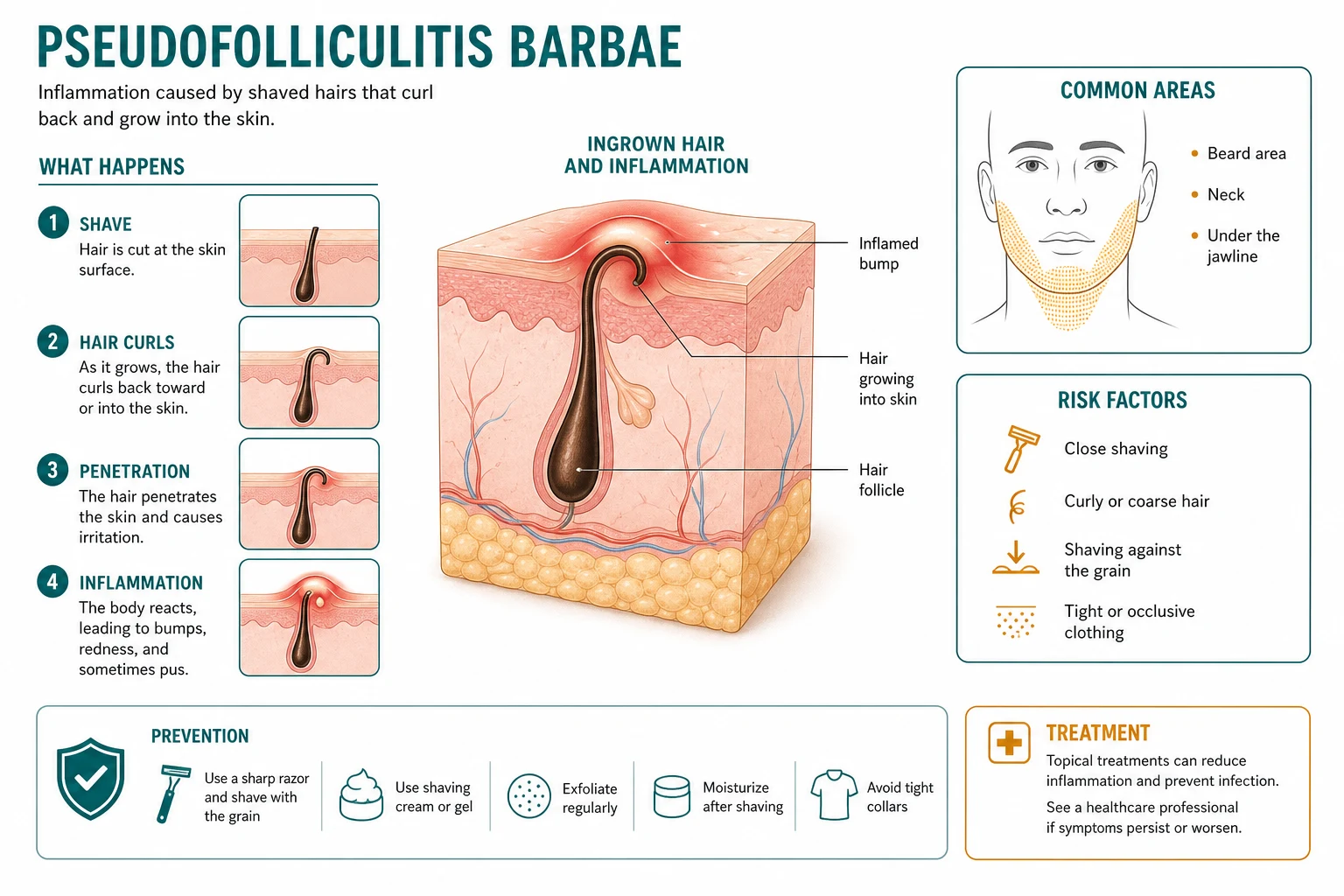
- Extrafollicular penetration (about 70%) — the tip exits the follicle, grows a short distance, then curves back and pierces the skin surface a millimetre or two from its own follicular orifice, drawn back by the natural curl of the hair.
- Transfollicular penetration (about 30%) — the tip never exits the follicle but instead pierces the follicular wall and grows into the surrounding dermis while still attached to the follicle. [1]
Step 3 — Foreign-body granulomatous reaction. Once the hair shaft (keratin) lies within the dermis, the immune system treats it as foreign material. A granulomatous inflammatory infiltrate assembles around the embedded shaft — multinucleated foreign-body giant cells, histiocytes/macrophages, lymphocytes and plasma cells — producing the clinically evident firm papule, often topped by a pustule when there is acute neutrophilic inflammation.[1][7]
Step 4 — Fibrosis and keloidal scarring. With chronicity and repeated shaving-triggered flares, the granulomatous inflammation drives dense fibrosis. In individuals with a keloidal diathesis (over-represented in skin of colour) this progresses to keloidal-type collagen deposition — the firm, lobulated, permanent keloidal papules and plaques that are the most troublesome complication.[5]
Why the beard and not elsewhere? The beard combines tightly curled hair, a high follicle density, daily close shaving, and high visibility — all amplifying both the trigger and the consequence. The anterior neck and mandibular area are worst because the hair there is curliest and shaved closest, and the skin is thinnest and most trauma-prone. The upper lip is characteristically spared, because the hair there is shorter, finer and often cut less aggressively.[1]
Why does growing a beard cure early disease? If the patient stops shaving, the hair grows out past the skin surface and can no longer re-enter; the foreign-body reaction resolves, the papules flatten, and only established fibrosis or keloid remains. This single fact — that the disease is trigger-dependent — is the conceptual key to all of management.[3]
Role of Staphylococcus aureus. Secondary staphylococcal colonisation/infection is common in pustular and crusted lesions, contributes to purulence and pain, and may progress to cellulitis or abscess; it is not the primary driver, which is mechanical.[7]
Why the multi-blade razor is the enemy. The "lift-and-cut" hysteresis mechanism deliberately cuts the hair below the skin surface for a closer shave — exactly the configuration that guarantees re-entry of a curved tip. Single-blade razors, by contrast, cut at the surface and leave a slightly longer shaft that is less likely to re-enter.[2]
Clinical Presentation
Site. PFB is confined to the shaving area of the beard, with a characteristic distribution:[1][7]
- Anterior neck — the single most common and most severe site.
- Mandibular angle and jawline.
- Chin and submental area.
- Cheeks (lower).
- Upper lip is characteristically SPARED — a useful discriminator from acne vulgaris. [1]
The distribution is bilateral and symmetric, restricted to skin the razor actually traverses, and worsens within 24-72 hours of each shave. [1]
Lesion morphology. [1]
- Firm, hyperpigmented, follicular papules 1-3 mm, often with an erythematous halo.
- Pustules atop the papules when there is acute inflammation or secondary infection.
- Pin-point entrapped (ingrown) hairs — the diagnostic clue: a tiny dark hair tip visible just under the skin surface or emerging from and re-entering a papule, sometimes best seen with side-lighting or a magnifying loupe.
- Post-inflammatory hyperpigmentation (PIH) — tan to dark-brown macules marking resolved papules; prominent and persistent in skin of colour.
- Keloidal papules and plaques — firm, dome-shaped, lobulated, skin-coloured to hyperpigmented nodules in those with keloidal diathesis.
- Occasionally hypertrophic scars, sinus tracts or abscesses in severe disease. [1]
Symptoms. Most patients complain of pruritus and burning shortly after shaving, pain from inflamed or infected lesions, the cosmetic disfigurement of papules, PIH and keloids, and the functional problem that shaving — often professionally required — provokes the rash. Many describe a vicious cycle: shave → bumps → shave again to look "clean" → worse bumps.[2]
Course. Chronic, recurrent and lifelong while shaving continues, punctuated by post-shave flares. Without intervention most patients progress over years to established PIH and keloidal scarring. Spontaneous resolution is rare; it usually requires either stopping shaving or permanent hair removal.[1]
Atypical presentations. [1]
- Women — pubic, axillary, leg papules after shaving; often misattributed to "infected hair follicles" or deodorant; the pubic form may be complicated by abscess and sinus.[8]
- Lighter skin types with curly hair (Latino, Mediterranean, Middle Eastern, South Asian) — less prominent PIH, keloidal quality subtler, diagnosis often missed.
- Transplant/immunosuppressed patients — more severe and refractory; ciclosporin and other immunosuppressants may amplify the inflammatory and keloidal response.
- Adolescents starting to shave — early papular disease, preventable by anticipatory counselling on technique.
Differential Diagnosis
The differential is high-yield exam territory. The key discriminators are site (shaving distribution), the entrapped hair, the absence of comedones, and the trigger relationship to shaving.[1][5]
Other considerations. [1]
- Contact dermatitis to shaving cream, aftershave or fragrance — eczematous, pruritic, sharply demarcated to the product application area; patch testing positive; resolves with allergen avoidance and topical corticosteroid. May coexist with PFB and confuse the picture.[3]
- Gram-negative folliculitis — arises in acne patients on long-term tetracycline; pustules around the nose and mouth; culture grows Gram-negatives. Rare in pure PFB.
- Rosacea and periorificial dermatitis — central face papulopustules without comedones and without entrapped hairs; no relationship to shaving; telangiectasia and flushing in rosacea.
- Irritant dermatitis from over-vigorous shaving or astringents — erythema, burning, scaling rather than papules; resolves with emollients and technique modification.
- Sycosis (barbae) vulgaris — deep bacterial folliculitis of the beard (S. aureus), with crusting and hairs that pull out easily; positive culture; responds to oral antistaphylococcal antibiotic. Distinct from PFB but the two coexist when PFB is secondarily infected.
Clinical & Bedside Assessment
Diagnosis is clinical. A confident bedside diagnosis rests on site (beard shaving distribution, anterior neck worst, upper lip spared), morphology (follicular papules/pustules with entrapped hairs and PIH) and demographic (man of African descent with tightly curled hair who shaves).[1]
History. Ask specifically about the shaving habit, because it is both the diagnosis and the treatment:[2][3]
- Type of razor — multi-blade cartridge razor (the worst offender), single-blade, electric shaver, clippers.
- Direction of shave — with or against the grain.
- Technique — does he pull the skin taut? Pre-shave preparation? Post-shave products?
- Frequency — daily shaving is high-risk.
- Occupation — any clean-shaven requirement (military, police, hospitality, food handling)?
- Onset and pattern — does the rash flare within a day or two of each shave and improve with shaving breaks?
- Past history — keloids, acne, the follicular occlusion tetrad, previous treatments tried.
- Psychosocial impact — distress, occupational consequences, relationships. [1]
Examination. Inspect the whole beard, neck, nape, scalp, axillae and groin (to detect coexisting AKN, hidradenitis suppurativa or follicular occlusion tetrad). Note the distribution, lesion morphology, presence of entrapped hairs, PIH, keloids, and any secondary infection (pustules, crusting, cellulitis, abscess). Use side-lighting or a magnifying loupe to reveal the pin-point entrapped hair shafts that confirm the diagnosis. Dermoscopy shows entrapped hair shafts, perifollicular inflammation and PIH, and helps exclude tinea (no broken hair shafts, no fungal involvement) and comedones.[7]
Severity assessment. There is no universally adopted severity score for PFB. Use the pragmatic three-grade scheme (mild/moderate/severe), the area and density of papules, the presence of keloids or secondary infection, and the DLQI / occupational impact to grade and justify escalation. [1]
Investigations
Investigations are usually unnecessary. PFB is a clinical diagnosis. Reserve tests for atypical, severe, complicated or refractory disease.[1][3]
Skin biopsy (4 mm punch) only when: [1]
- The morphology or site is atypical and a mimicker (discoid lupus, sarcoidosis, tinea, early squamous cell carcinoma in a long-standing keloid) is plausible.
- The disease is refractory to a correct management ladder and an alternative diagnosis is being entertained.
- A non-healing ulcer within a keloidal plaque raises suspicion of Marjolin's ulcer (squamous cell carcinoma) — biopsy is mandatory. [1]
Histology. An embedded hair shaft within the dermis, surrounded by a granulomatous infiltrate with multinucleated foreign-body giant cells, lymphocytes, histiocytes and plasma cells; with chronicity, fibrosis and, in keloidal variants, keloidal-type collagen. The findings are diagnostic.[7]
Bacterial swab (culture and sensitivity) from any pustule, crust or discharging sinus — typically grows Staphylococcus aureus (including MRSA in endemic areas); guides antibiotic choice for secondary infection. Swab pustules that are unusually painful, rapidly spreading, or associated with cellulitis.[1]
Fungal studies (skin scrapings for KOH microscopy, and fungal culture) only if tinea barbae is in the differential — a boggy, boggy, scaling plaque with hair loss and animal/farm contact. Negative in PFB. [1]
Patch testing when contact dermatitis to shaving products is suspected (eczematous, pruritic, product-restricted distribution). [1]
Blood tests before systemic therapy: FBC, U&E, LFTs if an oral tetracycline is contemplated (baseline and not routinely repeated); glucose/HbA1c only if long-term corticosteroid use is planned (rare). [1]
Management — Resuscitation
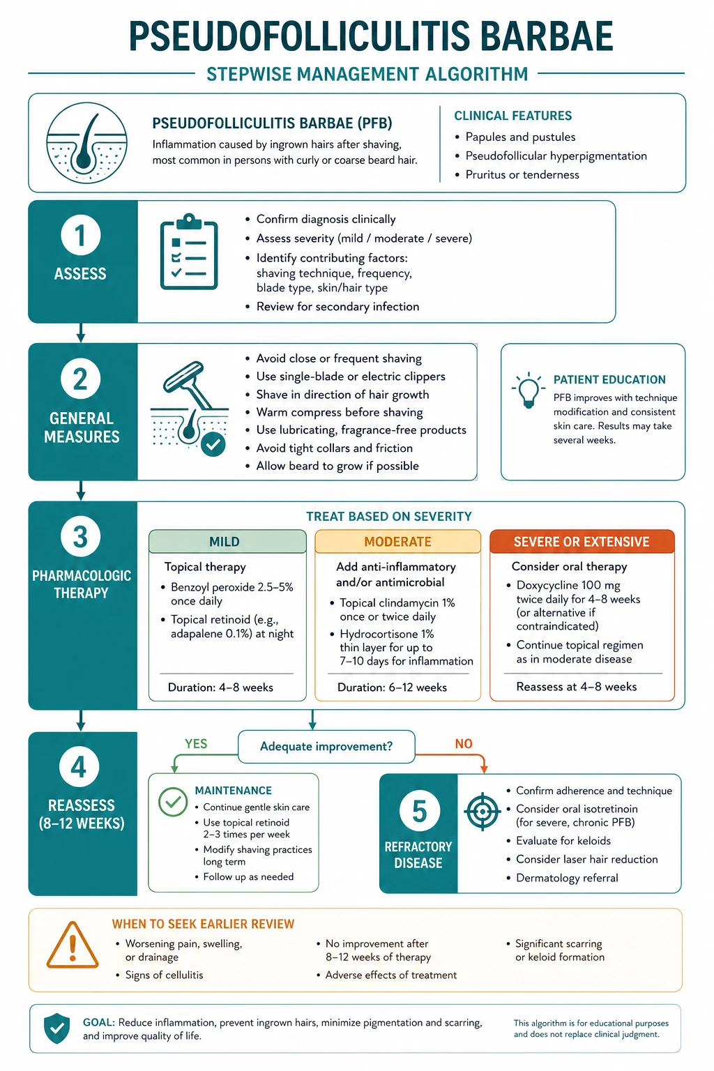
PFB is not a dermatological emergency, but several scenarios require prompt action.[1]
Acute inflammatory flare. The immediate measure is to stop shaving for at least 30 days, apply warm compresses, and use a short course (1-2 weeks) of a topical corticosteroid (e.g. hydrocortisone 1% cream twice daily, or a slightly more potent agent such as betamethasone valerate 0.1% for a limited period in moderate flares) to settle inflammation. Gently release obviously entrapped hairs with a sterile needle — do not pluck, which worsens re-entry.[2][3]
Acute secondary bacterial infection / abscess. A painful, expanding, cellulitic plaque or a fluctuant abscess is treated by a bacterial swab, a 7-10 day course of an oral anti-staphylococcal antibiotic guided by local sensitivities (flucloxacillin 500 mg four times daily, or clindamycin 300 mg four times daily if MRSA is suspected/confirmed), analgesia, and incision and drainage of any abscess.[1]
Occupational crisis. A patient who cannot meet a clean-shaven standard (military inspection, food-handling shift) needs a formal shaving profile/waiver letter and a rapid plan for definitive treatment (laser).[4]
Psychological distress. The cosmetic and occupational burden is significant; offer reassurance, an explicit plan and, where appropriate, psychological support.[2]
Management — Definitive & Stepwise
The therapeutic ladder is trigger-based and proceeds from avoidance, through modification, to topical medical therapy, systemic therapy, and definitive permanent hair removal.[1][2][3]
Tier 1 — Avoid or modify the shave (cornerstone; first-line for all patients)
The first-line shaving bundle for every PFB patient
Chemical depilatories deserve a specific note because they are the single most useful pharmacological substitute for shaving:[2][9]
[1]Pre- and post-shave technique (for those who must shave).[3]
- Pre-shave: hydrate the beard with warm water or a hot towel for 3-5 minutes to soften the hair; use a moisturising shaving gel (not a drying foam).
- Shave: single-blade, with the grain, light pressure, do not stretch the skin, rinse the blade after each stroke, and do not shave the same area repeatedly.
- Post-shave: rinse with cool water, apply a mild astringent or alum block, then a fragrance-free moisturiser; avoid alcohol-based aftershaves, which irritate and worsen PIH. [1]
Tier 2 — Topical medical therapy (for established papulopustular disease, PIH, and as adjunct)
The core topical triple — retinoid + antibiotic + corticosteroid — targets the three drivers of established PFB (follicular plugging, staphylococcal colonisation, and active inflammation), and the retinoid additionally accelerates resolution of post-inflammatory hyperpigmentation. Every topical agent must carry agent + concentration + frequency + rationale.[1][2][3]
- Benzoyl peroxide 2.5-5% wash or gel — antibacterial and mildly keratolytic; useful for the pustular component; warn about bleaching fabrics and irritancy.
- Glycolic acid (alpha-hydroxy acid) lotion 5-8% — exfoliates, reduces follicular plugging and improves PIH; the original report (Perricone, Cutis 1993) showed clear benefit.[10]
- Topical antibiotic alternative — erythromycin 2% solution twice daily if clindamycin is unavailable or resisted.
Tier 3 — Eflornithine and systemic therapy
Oral anti-inflammatory tetracyclines are reserved for moderate-to-severe inflammatory disease that has not responded to topical therapy and shaving modification:[1][7]
- Tetracycline 500 mg twice daily orally for 4-8 weeks, or
- Doxycycline 100 mg twice daily orally (preferred — better tolerated, less frequent dosing, fewer interactions) for 4-8 weeks, then taper to the lowest effective dose. [1]
These act primarily by anti-inflammatory mechanisms (inhibition of neutrophil chemotaxis and matrix metalloproteinases) rather than antibacterial ones. Warn about photosensitivity (especially doxycycline), gastrointestinal upset, oesophagitis (take with water, upright), contraindication in pregnancy and children under 12 (dental discolouration), and interaction with warfarin and the oral contraceptive pill (doxycycline less so than older tetracyclines, but still counsel). A short course of oral prednisolone (0.5 mg/kg/day tapering over 1-2 weeks) is occasionally used for an acute severe inflammatory flare, but is not routine. [1]
Tier 4 — Definitive permanent hair removal (the long-term cure)
Laser hair removal is the single most effective long-term treatment for PFB because it permanently destroys the culpable hair follicles, removing the trigger entirely.[1][6][9]
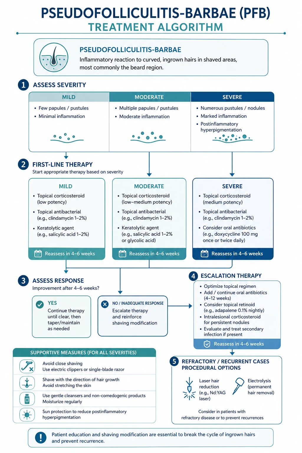
Electrolysis — insertion of a fine needle into each follicle with galvanic or thermolytic current to destroy it — is an alternative permanent method. It is technique- and operator-dependent, slow, painful, and carries a higher risk of PIH and scarring in skin of colour, so it is generally second-line to laser. Useful for the few residual hairs after laser, or for patients in whom laser is contraindicated.[9]
Keloid scar management
For established keloidal papules and plaques:[1][2]
- Intralesional triamcinolone acetonide 10-40 mg/mL (10 mg/mL for softer/early lesions; up to 40 mg/mL for firm established keloids) injected into the body of the keloid every 4-6 weeks for several sessions until flattened. Limit total dose per visit (e.g. 10-20 mg of triamcinolone) to avoid systemic effects.
- Silicone gel sheeting worn overnight for months.
- Pulsed dye laser (585-595 nm) for erythematous keloids.
- Intralesional 5-fluorouracil (50 mg/mL, alone or combined with triamcinolone) for refractory keloids.
- Surgical excision of keloids is generally avoided in PFB (high recurrence, may worsen keloid) unless combined with adjuvant therapy. [1]
Escalation triggers
- No response to tier 1 (shaving modification) after 6-8 weeks → add tier 2 topical therapy.
- Persistent papulopustules or PIH despite tiers 1-2 after 8-12 weeks → add tier 3 (eflornithine ± oral tetracycline) and plan laser.
- Keloidal scarring, occupational crisis, or patient preference for permanent cure → tier 4 (laser hair removal) as definitive treatment.
- Suspected or confirmed secondary infection (cellulitis, abscess) at any stage → swab + oral antibiotic ± I&D. [1]
Specific Subtypes & Scenarios
PFB in women (pubic, axillary, leg). Increasingly common with the near-universal trend for pubic hair removal. Tightly curled pubic hair behaves exactly like tightly curled beard hair when shaved. Management is identical in principle: stop shaving (or switch to trimming/depilatory), topical retinoid-antibiotic-steroid for inflammation, and Nd:YAG laser as definitive treatment (especially attractive for women who do not want to remove pubic hair at all). The pubic form may be complicated by abscess and sinus and overlaps with hidradenitis suppurativa.[8]
PFB in the military and clean-shaven occupations. The conflict between a medical need to stop close shaving and an occupational requirement to be clean-shaven is the core problem. Current US military policy accommodates PFB with shaving profiles/waivers permitting a short stubble (typically up to 1/4 inch) or, in some branches, a beard for documented PFB refractory to treatment. The definitive solution is laser hair removal, which both cures the dermatosis and may restore the ability to achieve a close seal for respirators in some cases.[4][9]
PFB coexisting with acne keloidalis nuchae. Common, because both target men of African descent with tightly curled hair. Examine the nape in every PFB patient and the beard in every AKN patient. The management ladder is shared (trigger avoidance, topical retinoid-antibiotic-steroid, oral tetracycline, Nd:YAG laser), applied to the relevant site.[5]
PFB in the follicular occlusion tetrad. Coexisting hidradenitis suppurativa, dissecting cellulitis, acne conglobata or pilonidal sinus warrants coordinated dermatology input and consideration of broader systemic therapy (including biologics for severe HS overlap).[5]
PFB in transplant/immunosuppressed patients. May be more severe, refractory, and keloidal; ciclosporin has been implicated as an amplifier. Treat the follicular inflammation aggressively, prevent secondary infection, and prefer laser over oral retinoids.[1]
PFB in adolescents starting to shave. Anticipatory counselling on single-blade, with-the-grain technique and avoidance of multi-blade razors can prevent onset in predisposed (curly-haired) teenagers. [1]
Complications & Pitfalls
Cutaneous complications. [1]
- Post-inflammatory hyperpigmentation (PIH) — the commonest sequela in skin of colour; persistent, cosmetically distressing, responds slowly to tretinoin and sun protection.
- Keloidal scarring — in those with keloidal diathesis; firm, lobulated, permanent; difficult to treat (intralesional triamcinolone, silicone, laser).
- Secondary bacterial infection — S. aureus pustules, cellulitis, abscess; rarely bacteraemia in severe/immunosuppressed.
- Sinus tracts and chronic draining papules in severe neglected disease. [1]
Psychological and occupational complications. Depression, anxiety, social withdrawal, impaired quality of life (high DLQI), and occupational jeopardy — inability to meet military/police grooming standards or maintain a food-handling job.[4]
Complications of intralesional corticosteroid. Skin atrophy, telangiectasia, hypopigmentation (clinically important in skin of colour), and rare systemic absorption with repeated high-dose injection.[2]
Complications of laser. PIH, blistering, crusting, hypopigmentation, and paradoxical hypertrichosis (rare, more with low-fluence treatment). Minimised by using long-pulsed Nd:YAG 1064 nm with appropriate cooling in trained hands.[6]
Classic management pitfalls. [1]
- Recommending a multi-blade razor — the worst possible advice; it cuts below the surface and guarantees re-entry.
- Recommending against-the-grain shaving — cuts the hair shorter, promotes re-entry.
- Plucking/epilating the ingrown hairs — traumatises the follicle and worsens inflammation and scarring.
- Choosing the wrong laser for skin of colour (ruby, alexandrite) — high risk of PIH and scarring; Nd:YAG 1064 nm is the safe choice.
- Using topical corticosteroid indefinitely on the face — causes atrophy, telangiectasia, periorificial dermatitis.
- Mistaking PFB for acne vulgaris and treating only with acne regimens, ignoring the shaving trigger.
- Failing to issue a shaving profile when occupation mandates shaving, leaving the patient caught between disease and discipline. [1]
Prognosis & Disposition
Natural history. PFB is chronic, recurrent and lifelong while the patient continues to shave in the predisposing way. Each shave provokes a flare; cumulative damage produces PIH and keloidal scarring. Spontaneous resolution is rare.[1]
Effect of treatment. Early disease (papules, no scarring) is fully reversible with shaving modification alone — within 4-6 weeks of stopping the close shave, the papules resolve and only established PIH remains. Established PIH improves over months with tretinoin, glycolic acid and strict sun protection. Keloidal scarring can be flattened but not fully erased. Laser hair removal is the one intervention that produces durable long-term remission by removing the trigger permanently; maintenance top-up sessions may be needed.[6][9]
Recurrence prevention. The single most effective measure to prevent recurrence is permanent removal of the culpable follicles by laser (or, where laser is unavailable, lifelong adherence to single-blade with-the-grain shaving or chemical depilatories, and avoidance of multi-blade razors).[6]
Disposition. Most patients are managed in primary care or routine dermatology. Refer to a laser/dermatology service for definitive hair removal, to a scalp/plastics service for excision of refractory keloidal plaques (rarely needed in PFB), and to occupational health when the dermatosis conflicts with a clean-shaven requirement.[4]
Safety-net. Advise the patient to return urgently if they develop a spreading cellulitis, an abscess, or a non-healing ulcer within a keloidal plaque (possible Marjolin's ulcer).[1]
Special Populations
Pregnancy. Avoid tetracyclines (tetracycline, doxycycline — dental and bone effects on the fetus) and systemic retinoids (highly teratogenic). Favour physical measures (stop shaving, chemical depilatories, electric clippers), topical clindamycin or erythromycin (both considered safe), glycolic acid, and laser hair removal (considered safe in pregnancy as the energy does not penetrate beyond the dermis; defer if the patient prefers). Intralesional triamcinolone for keloids is acceptable in single small doses. Defer oral prednisolone to a brief taper only for severe flares.[1]
Adolescents. Anticipatory counselling before they start shaving is the most effective prevention: recommend single-blade, with-the-grain technique, pre-shave hydration, and avoidance of multi-blade razors. Treat established disease as in adults; laser is acceptable and effective in older teenagers.[7]
Women. The pubic, axillary and leg forms are increasingly common; laser hair removal is first-line definitive because shaving the body is more easily abandoned than shaving the face. Address coexisting hirsutism if present.[8]
Immunosuppressed/transplant patients. PFB may be more severe, refractory and keloidal; ciclosporin is an amplifier. Treat the follicular inflammation aggressively, prevent secondary infection, and prefer laser over oral retinoids.[1]
Patients with a keloidal diathesis. Early, aggressive prevention is essential — any follicular inflammation in these patients scars. Combine shaving modification with prompt topical therapy and early laser. [1]
Evidence, Guidelines & Regional Differences
Guidelines. PFB lacks a single authoritative global guideline; recommendations are synthesised from the American Academy of Dermatology (and its skin-of-colour guidance), the British Association of Dermatologists and NICE Clinical Knowledge Summaries (UK), the European Academy of Dermatology and Venereology consensus statements (Europe), and the Indian Association of Dermatologists, Venereologists and Leprologists (IADVL) guidance (India). All converge on the same shaving-modification-first ladder with Nd:YAG laser as the definitive option in skin of colour.[1][3]
Laser hair removal in skin of colour. Shokeir et al. (J Cosmet Dermatol 2021) randomised PFB patients to topical eflornithine, long-pulsed Nd:YAG laser, or the combination, and found all three effective, with the combination superior — the strongest current evidence supporting Nd:YAG as first-line in skin of colour.[6] Alexis, Heath and Halder (Dermatol Clin 2014) reviewed both PFB and AKN and confirmed that prevention and effective treatment are within reach through a combination of trigger avoidance and laser, with Nd:YAG 1064 nm the safest wavelength in Fitzpatrick IV-VI.[5]
Glycolic acid. Perricone (Cutis 1993) reported two studies showing topical glycolic acid substantially improved PFB, establishing the alpha-hydroxy acid as a useful adjunct that also helps PIH.[10]
Military policy. Garcia-Zuazaga (Mil Med 2003) and Jung, Lannan and Weiss (Cutis 2023) documented the evolution of US military policy on PFB — from a condition that limited service to one accommodated by shaving profiles and waivers, with laser increasingly offered as definitive treatment that can preserve operational fitness.[9][4]
PFB in women. Nguyen, Patel and Viola (Br J Dermatol 2015) provided the modern clinical perspective on female PFB, highlighting the rising prevalence with pubic hair removal and the under-recognition of the condition in women.[8]
Regional deltas. [1]
Exam Pearls
[1]SHAVE — the pathophysiology in five steps
TREAT — the management ladder
Red Flags
Exam application bank (NEET-PG / INICET)
One-line answer
Pseudofolliculitis barbae (PFB, razor bumps) is a chronic, recurrent inflammatory disorder of hair-bearing skin caused by ingrown hairs — a sharply cut, tightly curled hair shaft re-enters the skin after shaving and provokes a foreign-body granulomatous reaction. It overwhelmingly affects men of African descent with tightly curled facial hair (up to 60-80% of Black men who shave) and is a particular problem in the military and other clean-shaven occupations; it also affects women in the pubic, axillary and leg areas. Distribution: beard, especially the anterior neck, mandibular angle, chin and submental area. The word 'pseudo' is deliberate: this is NOT a primary folliculitis but a foreign-body reaction to an extrafollicularly or transfollicularly penetrating hair. The management ladder is: AVOID shaving (grow beard, electric clippers, chemical depilatories) → MODIFIED shaving (single-bl
Worked stems (answer without another resource)
Stem 1 — Classic presentation. Map symptoms to mechanism; name the first investigation and first treatment step with dose/route if drug therapy is standard. [1]
Stem 2 — Unstable / complicated. List red flags that force immediate resuscitation, theatre, ICU, antidote, or reperfusion — and what you do in the first 15 minutes. [1]
Stem 3 — Atypical group. Elderly, pregnancy, child, or immunocompromised: how presentation and thresholds change. [1]
Stem 4 — Differential trap. Name the three closest mimics and one discriminator for each. [1]
Stem 5 — Disposition. Who goes home with safety-netting, who is admitted, who needs HDU/ICU/theatre, and what follow-up is mandatory. [1]
Rapid viva checklist
- Definition + classification
- Pathophysiology chain
- Bedside signs / criteria
- Score with exact components (if any)
- Emergency bundle
- Definitive therapy with doses
- Complications of disease and of treatment
- Special populations
- Guideline/trial name if classic
- Three exam traps
Coverage self-check
If you cannot answer any stem above from this page alone, re-read the matching section — the page is intended to be self-sufficient for final-prof and NEET-PG/INICET questions on Pseudofolliculitis barbae (razor bumps).
References
- [1]Patel TS, Dalia Y Pseudofolliculitis Barbae JAMA Dermatol, 2022.PMID 35475875
- [2]Nussbaum D, Friedman A Pseudofolliculitis Barbae: A Review of Current Treatment Options J Drugs Dermatol, 2019.PMID 30909328
- [3]Ogunbiyi A Pseudofolliculitis barbae; current treatment options Clin Cosmet Investig Dermatol, 2019.PMID 31354326
- [4]Jung I, Lannan FM, Weiss A Treatment and Current Policies on Pseudofolliculitis Barbae in the US Military Cutis, 2023.PMID 38290080
- [5]Alexis AF, Heath CR, Halder RM Folliculitis keloidalis nuchae and pseudofolliculitis barbae: are prevention and effective treatment within reach? Dermatol Clin, 2014.PMID 24680005
- [6]Shokeir H, Samy N, Taymour M Pseudofolliculitis barbae treatment: Efficacy of topical eflornithine, long-pulsed Nd-YAG laser versus their combination J Cosmet Dermatol, 2021.PMID 33629488
- [7]Quarles FN, Brody H, Johnson BA, Badreshia S Pseudofolliculitis barbae Dermatol Ther, 2007.PMID 17803606
- [8]Nguyen TA, Patel PS, Viola KV Pseudofolliculitis barbae in women: a clinical perspective Br J Dermatol, 2015.PMID 25557018
- [9]Garcia-Zuazaga J Pseudofolliculitis barbae: review and update on new treatment modalities Mil Med, 2003.PMID 12901468
- [10]Perricone NV Treatment of pseudofolliculitis barbae with topical glycolic acid: a report of two studies Cutis, 1993.PMID 8261811
- [11]Halder RM Pseudofolliculitis barbae and related disorders Dermatol Clin, 1988.PMID 3048823
- [12]Dunn JF Jr Pseudofolliculitis barbae Am Fam Physician, 1988.PMID 3046269