Dermatology · Medicine
Pyoderma gangrenosum
Also known as Pyoderma gangrenosum (PG) · PG · Dermatitis gangrenosa · Brocq's pyoderma gangrenosum
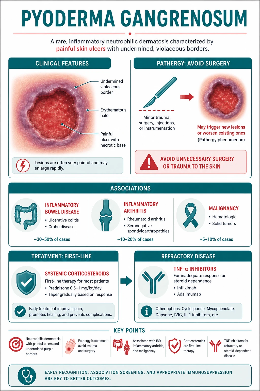
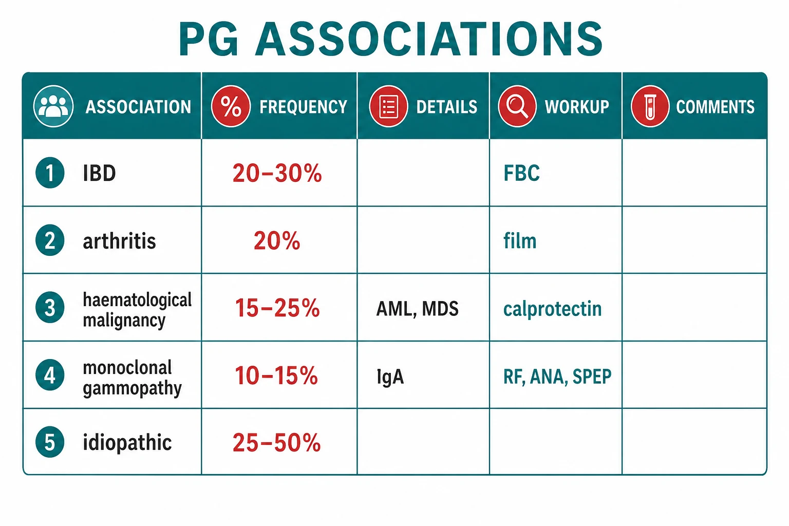
Pyoderma gangrenosum (PG) is a rare, sterile, autoinflammatory NEUTROPHILIC DERMATOSIS presenting as rapidly enlarging, deeply PAINFUL skin ulcers with characteristic UNDERMINED VIOLACEOUS (purple) BORDERS and an absolute hallmark of PATHERGY (new lesions at sites of trauma or surgery — for which surgical debridement is CONTRAINDICATED). Associated with systemic disease in ~50% of cases: inflammatory bowel disease (UC Crohn, 20-30%), seropositive/seronegative arthritis (20%), haematological malignancy (AML, MDS — particularly the bullous variant, 15-25%), and IgA monoclonal gammopathy (10-15%). It is a DIAGNOSIS OF EXCLUSION with NON-SPECIFIC histology (sterile dermal neutrophilic infiltrate); the role of biopsy and culture is to exclude mimics (infection, vasculitis, malignancy). First-line therapy is super-potent topical corticosteroid (clobetasol) for localised disease and systemic prednisolone 0.5-1 mg/kg/day (often with ciclosporin 3-5 mg/kg/day) for classic PG. Infliximab (5 mg/kg IV at weeks 0, 2, 6) is the biologic of choice, particularly for PG with IBD. AVOID surgical debridement — pathergy worsens PG. Cribriform scarring is pathognomonic.
On this page & tools
Your progress
Saved locally on this device.
Exam tags
Red flags
Overview & Definition
Pyoderma gangrenosum (PG) is a rare, sterile, autoinflammatory neutrophilic dermatosis that presents as one or more rapidly enlarging, deeply painful skin ulcers with a characteristic undermined, violaceous (purple) border.[3] Despite its name — coined by Louis Brocq in 1908 ("pyoderma" implies pus-forming) — the lesion is not infectious: cultures are sterile and antibiotics alone do not heal it. PG is instead driven by aberrant innate immune activation with sterile neutrophilic infiltration of the skin.[4][3]
Two features distinguish PG from ordinary skin ulcers and dominate every examination question on the topic: [1]
[1]PG belongs to the family of neutrophilic dermatoses — a group of autoinflammatory skin conditions sharing a final common pathway of sterile neutrophilic skin infiltration:[4]
- Sweet syndrome (acute febrile neutrophilic dermatosis — tender erythematous plaques, no ulceration).
- Pyoderma gangrenosum (painful ulceration with undermined border).
- Subcorneal pustular dermatosis (Sneddon-Wilkinson).
- Erythema elevatum diutinum.
- Bowel-associated dermatitis-arthritis syndrome (BADAS).
- Neutrophilic eccrine hidradenitis (chemotherapy-associated). [1]
PG is a diagnosis of exclusion — there is no pathognomonic laboratory test or histological feature; the diagnosis is made clinically after infection, vasculitis, malignancy, and venous/arterial disease have been ruled out.[3][7]
Classification
PG is classified into five clinical variants that differ in morphology, association, behaviour, and treatment response. Distinguishing the variant at first presentation directs the workup (e.g. a bullous lesion mandates haematological malignancy screening). [1]
PG variants — frequency and signature association
| Variant | Morphology | Typical site | Hallmark association | First-line treatment |
|---|---|---|---|---|
| Classic ulcerative | Deep, painful ulcer with undermined violaceous border; starts as a papulopustule | Lower legs (shin), trunk | IBD, RA | Systemic corticosteroid ± ciclosporin |
| Bullous (atypical superficial) | Rapidly spreading superficial, haemorrhagic, painful bullae | Upper limbs, dorsal hands | Haematological malignancy (AML, MDS) | Treat underlying malignancy; systemic steroid cautiously |
| Pustular | Multiple discrete painful pustules on otherwise normal skin | Extensor limbs, trunk | Active IBD flare | Treat IBD; dapsone/colchicine useful |
| Vegetative | Solitary superficial verrucous ulcer with raised granulating border | Trunk | Often none | Topical clobetasol usually sufficient |
| Peristomal | Undermined ulcer adjacent to a stoma; worsened by appliance | Around ileostomy/colostomy/urostomy | IBD after colectomy | Appliance refit + topical tacrolimus ± intralesional triamcinolone; infliximab if severe |
The now-abandoned term "malignant pyoderma" describes an aggressive ulcerative form on the head and neck of young adults; most such cases are now reclassified as PG variant of cutaneous lymphoma or Mondor-like disease and warrant biopsy.[3]
Epidemiology & Risk Factors
PG is rare — annual incidence 3-10 per million population, with a lifetime prevalence of about 1 in 100,000.[3]
Epidemiology at a glance
Risk factors and triggers: [1]
- Systemic autoimmune / autoinflammatory disease — the dominant risk factor (see associations).
- Stomas and skin trauma — peristomal PG occurs in 1-2% of patients with inflammatory bowel disease who undergo colectomy.[2]
- Surgery — post-surgical PG appears 4-7 days after the operation (especially after abdominopelvic, breast, or cardiac surgery); often misdiagnosed as wound infection and "treated" with debridement, which worsens it.
- Drugs — rarely, pegylated G-CSF, isotretinoin, propylthiouracil.
- Genetic predisposition — familial PG with PSTPIP1 / CD2BP1 mutations, presenting as PAPA (pyogenic arthritis, PG, acne), PASH (PG, acne, suppurative hidradenitis), PAPASH, PsAPASH syndromes.[9]
The "IMAH" mnemonic summarises the four systemic disease categories searched for at every PG workup: [1]
IMAH — systemic associations of pyoderma gangrenosum
Idiopathic PG (no associated disease) accounts for 25-50% of cases and remains a diagnosis of careful exclusion.[3]
Pathophysiology
PG is best understood as a skin-limited autoinflammatory disease driven by aberrant innate immune activation. The neutrophil, not the lymphocyte, is the effector cell. [1]
The cytokine cascade. An external trigger (skin trauma, IBD flare, malignancy-derived cytokines) activates skin-resident innate immune cells (keratinocytes, dendritic cells, macrophages) to release IL-1β, IL-17, IL-23, TNF-α, IL-8 (CXCL8) and G-CSF.[3] This cytokine cocktail recruits and activates neutrophils in a runaway positive feedback loop:
- IL-1β — drives pyroptosis, fever, further neutrophil recruitment.
- IL-23 → IL-17 axis — sustains the neutrophilic infiltrate (Th17 axis); explains the efficacy of ustekinumab (IL-12/23 blockade) and the IL-17 biologics.
- TNF-α — central upstream amplifier; its blockade by infliximab/adalimumab is the rationale for biologic therapy.
- IL-8 (CXCL8) — the dominant neutrophil chemoattractant; overexpressed in PG skin.
- G-CSF — both drives neutrophil release and is itself a recognised drug trigger of PG. [1]
Neutrophil dysfunction. Patients with PG demonstrate abnormal neutrophil trafficking — including defective CD18 integrin expression, reactive oxygen species dysregulation, and altered skin-homing — that leads to inappropriate neutrophil accumulation in otherwise normal skin.[3]
Pathergy. The defining clinical-pathological link. Trauma provokes a localised neutrophilic infiltrate that, in a healthy person, resolves within hours; in PG it escalates into a destructive ulcer. This explains why: [1]
- PG appears at surgical wounds, venepuncture sites, intravenous cannulae, tattoos, insect bites.
- Surgical debridement of a PG ulcer causes the ulcer to enlarge dramatically and new lesions to appear along the surgical incision.
- Skin grafting without immunosuppression fails — the graft donor site (a new wound) itself becomes PG. [1]
Genetics and autoinflammatory syndromes. Rare familial PG is associated with mutations in PSTPIP1 (CD2BP1), a cytoskeleton-regulating protein that interacts with PEST-type phosphatases; the resulting over-production of IL-1β links PG to the autoinflammatory syndromes PAPA (Pyogenic arthritis, PG, Acne), PASH (PG, Acne, Suppurative hidradenitis), PAPASH, and PsAPASH.[9] Recognition of these syndromes matters because IL-1 blockade (anakinra, canakinumab) can be highly effective.
Why histology is non-specific. PG produces a sterile dermal neutrophilic infiltrate with central necrosis and a mixed lymphohistiocytic infiltrate at the periphery — a pattern shared with Sweet syndrome, BADAS, and other neutrophilic dermatoses. There is no pathognomonic histological feature. The role of biopsy is to exclude vasculitis, infection (special stains for mycobacteria, fungi), and malignancy (SCC).[3][4]
Clinical Presentation
The classic PG ulcer evolves in two phases: [1]
- Pre-ulcerative phase (24-72 h): a small, tender papule, pustule, or nodule — often mistaken for an insect bite or folliculitis. It rapidly becomes haemorrhagic and breaks down.
- Ulcerative phase: the lesion evolves into a deep, painful ulcer with the signature undermined, overhanging, violaceous (purple-grey) border, a ragged necrotic base with purulent exudate, and surrounding dusky erythema. The ulcer enlarges concentrically — often 1-2 cm per day in untreated disease — and is exquisitely painful (out of proportion to apparent size). [1]
Distribution: [1]
- Lower extremities (shin, ankle, dorsum of foot) — most common site.
- Trunk, peristomal areas, abdomen (post-surgical PG).
- Upper limbs, head and neck, hands (bullous variant).
- Mucous membranes are typically spared (helps distinguish PG from Behçet disease). [1]
Pathergy at presentation. A careful history almost always elicits the trigger: the ulcer appeared at the site of a recent venepuncture, intravenous cannula, insect bite, scratch, or surgical wound. Post-surgical PG classically declares itself 4-7 days after the operation — the wound fails to heal, becomes exquisitely painful and purple rather than pink, and enlarges dramatically when "debrided" as a presumed infection.[3]
Systemic features. Many patients feel systemically well, but fever, malaise, arthralgia, and myalgia may accompany active widespread disease or herald an underlying IBD/malignancy flare. [1]
Variant-specific presentation is detailed under Specific Subtypes & Scenarios below. [1]
Differential Diagnosis
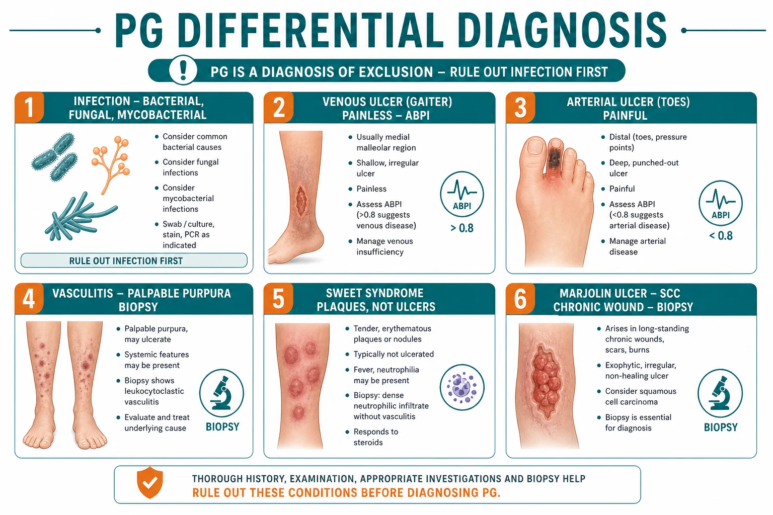
PG is a diagnosis of exclusion — and the price of misdiagnosis is high. Treating an infected ulcer with high-dose steroids is catastrophic; treating PG as infection with serial debridement equally so. The list of mimics is long:[3][7]
| Mimic | Distinguishing feature | Key test |
|---|---|---|
| Venous ulcer | Gaiter distribution; shallow, irregular; mildly painful; varicose veins; ABPI normal/high | Clinical + ABPI >0.8 |
| Arterial ulcer | Distal (toes, forefoot); punched-out; rest pain; cold, hairless skin; ABPI below 0.5 | ABPI + arterial Doppler |
| Infective ulcer | Culture-positive; rapid progression with systemic toxicity; undermined edge rare | Wound swab + tissue culture (bacterial, mycobacterial, fungal); special stains on biopsy |
| Necrotising fasciitis | Rapid spread, severe pain out of proportion, systemic sepsis, crepitus — surgical emergency | Clinical + LRINEC score + urgent surgical exploration |
| Vasculitic ulcer (ANCA, cryoglobulin, rheumatoid) | Palpable purpura; nailfold infarcts; biopsy shows leucocytoclastic vasculitis | Biopsy + DIF; ANCA, complements, cryoglobulins |
| Marjolin's ulcer (SCC in chronic wound) | Arises in a long-standing scar, burn, chronic venous ulcer; rolled everted edge; biopsy the EDGE | Punch biopsy of ulcer edge |
| Factitial / self-inflicted ulcer | Bizarre geometric shapes; patient denies; psychiatric history; sharp demarcation on covered skin | Detailed psychosocial history |
| Behçet disease | Oral + genital aphthae, uveitis, positive pathergy, erythema nodosum, HLA-B51 | International Study Group criteria |
| Sweet syndrome | Tender erythematous plaques/pseudovesicles (not true ulceration); fever; leukaemia association | Biopsy — dense dermal neutrophils, no vasculitis |
| Cutaneous lymphoma / pseudolymphoma | Slow-growing plaque or ulcer; atypical lymphoid infiltrate; EORTC cutaneous lymphoma classification | Biopsy + immunophenotyping |
| Dissecting cellulitis / acne keloidalis nuchae | Occipital scalp, neck; boggy nodules; sinus tracts; keloidal scarring; follicular occlusion | Clinical + culture |
| Crohn's cutaneous disease (metastatic) | "Knife-cut" fissures, ulceration of inframammary/inguinal folds; granulomas on biopsy | Biopsy + IBD workup |
| Antiphospholipid syndrome | Livedo, retiform purpura, multiple small ulcers; thrombosis history | aCL, anti-β2-GPI, lupus anticoagulant |
Clinical & Bedside Assessment
A structured bedside assessment serves two purposes: to confirm the diagnosis (using the Delphi and PARACELSUS tools once mimics are excluded) and to screen for associated systemic disease. [1]
Step 1 — confirm the morphology. Document ulcer site, size, depth, edge character (undermined, violaceous, overhanging), exudate, surrounding erythema, and number of lesions. Photograph with a ruler in frame. Measure pain on a 0-10 VAS. [1]
Step 2 — elicit and document pathergy. Ask specifically about new lesions at surgical wounds, cannulae, venepuncture, insect bites, scratches, tattoos. A formal pathergy test (skin prick with sterile needle, read at 24-48 h for a pustule) is positive in ~40-70% of PG patients; it is more often used in Behçet disease but supports PG when present.[3]
Step 3 — screen for associated disease. Take a focused history covering: [1]
- Bowel: diarrhoea, bloody stool, abdominal pain, weight loss → IBD.
- Joints: morning stiffness, synovitis, inflammatory back pain → inflammatory arthritis.
- Constitutional: fever, night sweats, weight loss, fatigue, bleeding/bruising → haematological malignancy.
- Stoma: appliance fit, leakage, mucocutaneous trauma → peristomal PG. [1]
Step 4 — apply the diagnostic criteria. Two validated instruments help standardise diagnosis once mimics are excluded: [1]
Cheng 2018 Delphi criteria — ulcerative PG
The PARACELSUS score of Dissemond 2019 uses eight diagnostic categories (Painful, Asymmetric distribution, Rapid progression, Association with systemic disease, Central clearing, Exclusion of mimics, Liquefactive necrosis, Serpiginous or undermined border, Undermined violaceous border, Size) plus four exclusionary items (infection, vasculitis, malignancy, drug-induced). A score ≥6/10 supports PG.[8]
Step 5 — quantify severity. The Pyoderma Gangrenosum Area Score (PGAS) is a body-surface-area-weighted score that, with the pain VAS, allows objective monitoring of treatment response.[3]
Step 6 — exclude mimics at the bedside. Examine pulses and ABPI (arterial insufficiency); assess for varicose veins and oedema (venous disease); palpate for lymphadenopathy and hepatosplenomegaly (haematological malignancy); inspect oral and genital mucosa (Behçet, Crohn). [1]
Investigations
There is no diagnostic test for PG. Investigations are designed to (a) exclude mimics and (b) detect associated systemic disease. [1]
Histopathology. A punch biopsy (preferably from the active advancing edge) is essential. The findings are non-specific: a dense sterile dermal neutrophilic infiltrate with central necrosis, mixed lymphohistiocytic infiltrate at the periphery, and a sometimes-suppurative or granulomatous component. Crucially, the biopsy excludes leucocytoclastic vasculitis, lymphoma, and infection (special stains for mycobacteria — Ziehl-Neelsen, Fite — and fungi — PAS, Grocott — must be ordered). Direct immunofluorescence excludes IgA vasculitis and bullous disorders.[3][4]
Microbiology. Send a wound swab AND a deep tissue biopsy for: [1]
- Bacterial culture (aerobic and anaerobic) — including group A streptococcus, Pseudomonas, Staphylococcus aureus.
- Atypical mycobacteria and mycobacterial culture (including TB) — particularly if immunocompromised.
- Fungal culture (deep rather than superficial).
- Tissue PCR for Leishmania, atypical mycobacteria, herpes viruses if the clinical context suggests. [1]
A positive superficial swab may simply reflect colonisation; deep tissue culture is more reliable. [1]
Blood tests — associated disease screen: [1]
| Test | Rationale |
|---|---|
| FBC + differential + blood film | AML, MDS, hairy cell leukaemia, polycythaemia — particularly with bullous variant |
| ESR, CRP | Disease activity marker; supports inflammation |
| Serum protein electrophoresis (SPEP) + urine Bence-Jones | IgA monoclonal gammopathy (MGUS or myeloma) |
| Serum free light chains, β2-microglobulin, serum IgA | Myeloma workup if paraprotein detected |
| Rheumatoid factor, anti-CCP, ANA | Rheumatoid arthritis, connective tissue disease |
| HLA-B27 | Seronegative spondyloarthropathy |
| Stool calprotectin | Sensitive screen for IBD; if elevated, refer for colonoscopy |
| Faecal culture, C. diff toxin | Exclude infectious diarrhoea |
| ANCA (p-ANCA, c-ANCA), anti-Ro/La, complements | Vasculitis screen — ANCA-associated, lupus, cryoglobulinaemia |
| Hepatitis B surface antigen, hepatitis C antibody, HIV | Pre-biologic workup; exclusion of viral vasculitis |
| QuantiFERON-TB Gold / IGRA | Pre-anti-TNF screen for latent TB |
| Interleukin profiles (research) | Not routine |
Imaging: [1]
- Chest X-ray — underlying sarcoidosis, TB, malignancy.
- Plain X-ray of any painful bone beneath a PG ulcer — exclude osteomyelitis.
- Doppler ultrasound of leg arteries and veins / ABPI — exclude venous and arterial insufficiency.
- CT-PET / whole-body MRI — search for occult malignancy in bullous variant with abnormal FBC/film.
- Bone marrow aspiration / trephine biopsy — when FBC/film or SPEP suggest haematological malignancy. [1]
Endoscopy. Colonoscopy with biopsies is indicated when stool calprotectin is elevated or any bowel symptoms exist — ulcerative colitis and Crohn's are the strongest associations.[2]
Specialist review. Multidisciplinary involvement is mandatory: dermatology leads, with gastroenterology, rheumatology, haematology, wound-care / stoma nurse, and psychology.[5]
Management — Resuscitation
PG is not a "fast" dermatological emergency like Stevens-Johnson syndrome or necrotising fasciitis, but the resuscitative phase before definitive immunosuppression is critical because the wrong first action can be disastrous. [1]
[1]Step 1 — exclude infection first. Before any corticosteroid or biologic is given, take a wound swab AND deep tissue biopsy for bacterial, mycobacterial, and fungal culture. Initiate empirical broad-spectrum antibiotics only if there is clinical sepsis, rapid progression with systemic toxicity, or strong suspicion of secondary infection. Stopping an immunosuppressant in established PG with secondary infection is balanced against the risk of PG rebound — involve microbiology and dermatology early. [1]
Step 2 — wound care. Aim for a moist, low-bioburden, non-traumatic environment: [1]
- Non-adherent dressings — silicone foams (Mepilex), soft silicones, hydrofibres (Aquacel) — to avoid trauma at dressing changes (pathergy).
- Cadexomer iodine or silver dressings — for high-bioburden, exuding ulcers.
- Topical anaesthetics (e.g., EMLA under occlusion before dressing change) reduce procedural pain.
- Compression bandaging is used only if venous insufficiency is documented AND arterial supply is adequate (ABPI ≥0.8); never as a default for a painful leg ulcer. [1]
Step 3 — analgesia. PG pain is often excruciating and is the single most cited quality-of-life issue. Use a stepwise approach: [1]
- Regular paracetamol 1 g QDS.
- NSAID (e.g., ibuprofen 400 mg TDS) if renal function and ulcer risk permit — short-term only.
- Short-acting opioid (e.g., oral morphine solution 5-10 mg every 4 h PRN, or oxycodone) — frequently required; titrate.
- Neuropathic agent (gabapentin 300 mg OD titrated, or pregabalin 25-75 mg BD) for the neuropathic / burning component that opioids under-treat.
- Patient-controlled analgesia or ketamine infusion in severe, hospitalised disease. [1]
Step 4 — decide disposition. Admit if any of: [1]
- Ulcer grows >5 cm in 24 hours.
- Bullous variant (likely AML/MDS) or rapidly progressive systemic upset.
- Pain uncontrolled orally.
- Suspected secondary infection / sepsis.
- Need for intravenous pulse methylprednisolone or biologic induction. [1]
Step 5 — institute multidisciplinary team. Dermatology, gastroenterology (if IBD), haematology (if abnormal FBC/SPEP), rheumatology (if arthritis), stoma nurse (peristomal), tissue viability nurse, and psychology. A clearly documented management plan reduces the risk of inadvertent surgical debridement by a covering team.[5]
Management — Definitive & Stepwise
Definitive therapy is determined by severity (localised vs extensive vs rapidly progressive) and variant. The overall ladder is summarised below. [1]
Mild / localised disease (single small or vegetative ulcer): [1]
- Super-potent topical corticosteroid — clobetasol propionate 0.05% cream/ointment BD under occlusion for 2 weeks, then taper.
- Topical calcineurin inhibitor — tacrolimus 0.1% ointment OD-BD (steroid-sparing, useful on face/folds/peristomal).
- Intralesional triamcinolone acetonide 10-40 mg/mL injected at the active border every 2-4 weeks. [1]
Moderate disease (multiple small ulcers or one large slowly enlarging ulcer): [1]
- Add oral prednisolone 0.5-1 mg/kg/day (max ~80 mg) as a single morning dose; taper after response.
- Consider concurrent ciclosporin 3-5 mg/kg/day in two divided doses for synergistic, steroid-sparing effect.
- Continue optimised wound care and analgesia. [1]
Severe / rapidly progressive / refractory disease: [1]
- IV methylprednisolone pulse 0.5-1 g/day for 3-5 days, then oral prednisolone 1 mg/kg/day.
- Ciclosporin 3-5 mg/kg/day added early (effective within 1-2 weeks in classic PG).
- Infliximab 5 mg/kg IV at weeks 0, 2, 6, then 8-weekly — biologic of choice for refractory PG, especially with IBD; treats both diseases.[1][6]
- Adalimumab 80 mg SC, then 40 mg alternate weeks — subcutaneous alternative.
- Other biologics: ustekinumab (IL-12/23), secukinumab / brodalumab (IL-17A), canakinumab (IL-1β — particularly for PAPA / autoinflammatory syndromes), vedolizumab (gut-selective for IBD), tocilizumab (IL-6).
- JAK inhibitors (tofacitinib, baricitinib, upadacitinib) — emerging evidence.[1]
Steroid-sparing agents (for maintenance, refractory disease, or when steroids cannot be tapered): [1]
- Azathioprine 1-3 mg/kg/day — check TPMT activity first.
- Mycophenolate mofetil 2 g/day in two divided doses.
- Methotrexate 10-25 mg/week oral/SC — avoid in pregnancy.
- Dapsone 100-150 mg/day — check G6PD first; monitor methaemoglobinaemia and haemolysis; useful for pustular variant.
- Colchicine 0.5-0.6 mg BD — particularly for pustular variant and arthritis overlap.
- IVIG 2 g/kg/cycle over 2-5 days, 4-weekly — useful in refractory disease or when conventional immunosuppression is contra-indicated.
- Tocilizumab, rituximab — for refractory disease in selected patients. [1]
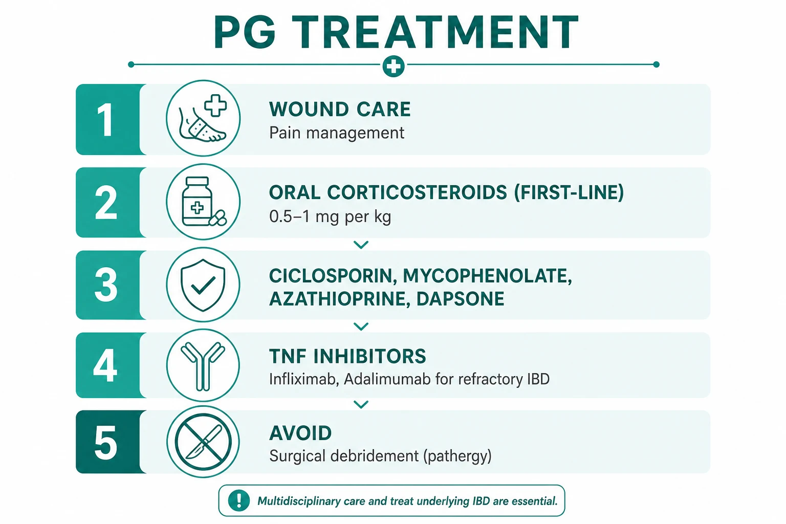
Surgical approach. Surgical debridement is absolutely CONTRAINDICATED in active PG because of pathergy. The only acceptable surgical procedure is meshed split-thickness skin grafting or cellular therapy (cultured keratinocyte / skin substitute) performed only once the PG is immunologically controlled and under cover of concomitant systemic immunosuppression; even then, donor-site pathergy may occur.[3]
Monitoring response. Use the PGAS and serial photography weekly during the active phase, with pain VAS and CRP/ESR trends. Expect ~30% improvement by 2 weeks and ~50% by 4-6 weeks on effective therapy.[5] Failure to respond by 2-4 weeks, continued rapid enlargement, or multiple lesions are all triggers to escalate to a biologic.
Drug-specific monitoring. [1]
| Agent | Pre-treatment | On-treatment monitoring | Specific adverse effects |
|---|---|---|---|
| Prednisolone | Bone density, glucose, BP, weight | Glucose, BP, weight, DEXA at 3 months | Osteoporosis, diabetes, hypertension, infection, peptic ulcer, cataracts, mood |
| Ciclosporin | BP, creatinine, electrolytes, LFTs, lipids | BP + creatinine every 2 weeks for 3 months, then monthly; trough levels in selected patients | Nephrotoxicity, hypertension, gum hyperplasia, hyperkalaemia, tremor, hirsutism |
| Infliximab / adalimumab | TB (IGRA), hepatitis B/C, HIV; cardiac function | Vigilance for infection; FBC; LFTs | Reactivation of TB / HBV; infusion reaction; heart failure; demyelination; lymphoma |
| Azathioprine | TPMT genotype / activity; FBC; LFTs | FBC weekly ×4 weeks, then monthly; LFTs | Myelosuppression, hepatotoxicity, pancreatitis |
| Mycophenolate | FBC; LFTs; pregnancy test | FBC monthly | Myelosuppression, teratogenic (avoid in pregnancy) |
| Methotrexate | FBC; LFTs; renal; chest X-ray | FBC + LFTs monthly; folic acid 5 mg weekly apart from MTX | Hepatotoxicity, pneumonitis, mucositis, teratogenic |
| Dapsone | G6PD; FBC; methaemoglobin; LFTs | FBC weekly ×4 weeks; methaemoglobin at 1 month | Haemolysis, methaemoglobinaemia, agranulocytosis, hypersensitivity syndrome |
| Colchicine | FBC; LFTs; renal | FBC | Diarrhoea, myelosuppression in renal impairment |
| IVIG | IgA level; renal function; hepatitis screen | Renal function, viscosity symptoms in elderly | Headache, aseptic meningitis, thrombosis, renal failure, infusion reaction |
Escalation triggers include failure to halt ulcer enlargement within 2-4 weeks of effective-dose corticosteroid, rapid progression >5 cm/24 h, multiple lesions, bullous variant, severe peristomal PG impairing stoma function, or PG associated with active IBD not controlled by gastroenterology. [1]
Specific Subtypes & Scenarios
Classic ulcerative PG. The most common presentation — a deep painful ulcer on the lower leg with undermined violaceous border and positive pathergy. First-line is oral prednisolone 0.5-1 mg/kg/day; ciclosporin 3-5 mg/kg/day added if rapid progression or inadequate response at 1-2 weeks; infliximab if refractory.[3]
Bullous (atypical superficial) PG. Rapidly enlarging, superficial, painful, haemorrhagic bullae typically on the dorsal hands, forearms, or upper arms. The morphological mimic is Sweet syndrome but the differential that matters is acute myeloid leukaemia, myelodysplastic syndrome, and other myeloproliferative neoplasms. Workup is mandatory: FBC, peripheral blood film, bone marrow aspirate / trephine. Treatment priority is to treat the underlying malignancy — PG often resolves with chemotherapy; systemic corticosteroid is used cautiously (immunosuppression in marrow-failure patients risks sepsis).[3][4]
Pustular PG. Multiple discrete painful pustules on otherwise normal skin, often during an IBD flare. Treatment of the underlying IBD (corticosteroid, anti-TNF) usually settles the skin; dapsone (100-150 mg/day, after G6PD check) and colchicine 0.5-0.6 mg BD are particularly useful for the neutrophilic pustular component. [1]
Vegetative PG. A solitary, superficial, verrucous, granulating ulcer with elevated borders, usually on the trunk, often without systemic association. Super-potent topical corticosteroid (clobetasol 0.05%) or topical tacrolimus is usually sufficient; systemic therapy is rarely needed.[3]
Peristomal PG. Occurs around an ileostomy, colostomy, or urostomy — classically in patients with UC after colectomy. Pathogenesis is mucocutaneous trauma from the appliance and pathergy. Management is multi-pronged: [1]
- Stoma appliance refit by a specialist stoma nurse — eliminate leaks, friction, mucocutaneous trauma.
- Topical tacrolimus 0.1% ointment (preferred over steroid: no skin atrophy with chronic use).
- Intralesional triamcinolone 10-40 mg/mL at the active border every 2-4 weeks.
- Topical crushed tacrolimus or clobetasol in stomahesive powder for ulcers under the wafer.
- Systemic therapy (prednisolone, ciclosporin, infliximab) for refractory or extensive peristomal PG.
- Surgical stoma relocation is rarely required and itself risks pathergy.[2][5]
Post-surgical PG. Appears 4-7 days postoperatively (abdominopelvic, breast, cardiac surgery); frequently mislabelled as wound infection. The clinical clues are intense pain out of proportion, violaceous rather than pink wound margins, and dramatic deterioration with debridement. Management: stop debridement; confirm diagnosis; exclude infection; start systemic corticosteroid ± ciclosporin.[3]
PG in pregnancy. Rare. Use topical corticosteroids and oral prednisolone; avoid ciclosporin (especially first trimester), methotrexate and mycophenolate (teratogenic); biologics (infliximab, adalimumab) are generally continued if PG/IBD is severe, with multidisciplinary team input. PG often improves in later pregnancy and may flare postpartum. [1]
PG in children. Rare (~4% of cases). Same treatment ladder, with weight-based dosing; prefer topical and lower-dose systemic therapy; dapsone and colchicine are useful adjuncts. Psychological impact of disfiguring leg ulcers in school-aged children requires specific support.[3]
PG in the elderly. Increased susceptibility to corticosteroid adverse effects (osteoporosis, hyperglycaemia, infection, fluid retention). Screen aggressively for underlying malignancy. Prefer topical and lower-dose systemic therapy; consider earlier biologic to avoid steroid toxicity. [1]
PG in the immunocompromised (HIV, transplant, post-chemotherapy). Exclude infection with even greater rigour (multiple biopsies, special stains, tissue PCR). Use lower-dose immunosuppression, prefer biologic over dual cytotoxic therapy, and involve infectious diseases. [1]
Complications & Pitfalls
Local complications: [1]
- Cribriform ("meshed") scarring — a pathognomonic pattern of multiple small punched-out scars from healing pustules — remains after the ulcer heals.[7]
- Secondary infection — Staphylococcus aureus, Pseudomonas, Streptococcus pyogenes — may progress to cellulitis, sepsis.
- Chronic non-healing — particularly with delayed or inadequate immunosuppression.
- Limb-threatening tissue loss in extensive disease; amputation is occasionally required.
Systemic complications: [1]
- Sepsis from secondary infection — the leading cause of PG-related death.
- Thromboembolism — PG is a prothrombotic state; venous thromboembolism is recognised, particularly in bullous variant with malignancy.
- Malnutrition and anaemia from chronic inflammation and wound exudate protein loss.
- Depression, anxiety, and opioid dependence from chronic pain, disfigurement, and isolation. [1]
Pitfalls: [1]
- Misdiagnosis as infection or venous ulcer delays immunosuppression; risks inappropriate debridement which worsens PG.
- Over-diagnosis of PG (treating an infected ulcer, vasculitic ulcer, or SCC with high-dose steroids) is equally catastrophic — hence the insistence on tissue culture and biopsy before steroids.
- Long-term corticosteroid toxicity if prednisolone is not tapered and steroid-sparing agent introduced.
- Ciclosporin nephrotoxicity if BP/creatinine not monitored.
- TNF inhibitor reactivation of latent TB or hepatitis B — always screen with IGRA and serology before starting.
- Pregnancy exposure to methotrexate / mycophenolate — confirm contraception and pregnancy test before prescription.
- Donor-site pathergy if skin grafting is attempted without immunosuppressive cover — the donor site itself becomes PG. [1]
Prognosis & Disposition
PG is a chronic relapsing-remitting disease. Without effective immunosuppression it is progressive and disabling; with modern therapy, the prognosis is favourable but not benign.[3]
Time course. Mean time to complete healing with appropriate therapy is 3-9 months, with substantial pain relief within 1-2 weeks of starting systemic corticosteroid. Recurrence rate is 20-40% over 5 years — higher in patients with active IBD or persistent haematological malignancy. [1]
Predictors of poor prognosis: [1]
- Large ulcer size (>20 cm) at presentation.
- Multiple lesions.
- Delay >6 weeks to appropriate therapy.
- Bullous variant with underlying AML/MDS.
- Active uncontrolled IBD.
- Older age and significant comorbidity.
- Continued pathergic trauma (recurrent stoma leaks, surgical procedures). [1]
Mortality. Overall mortality attributable to PG itself is low, but 3-30% depending on series — driven predominantly by underlying malignancy, secondary sepsis, and thromboembolism, not by skin failure per se.[3]
Tapering and stopping therapy. Once complete healing is achieved, immunosuppression is continued for a further 1-3 months and then tapered slowly (steroids over 4-8 weeks, biologics stretched to longer intervals) to avoid rebound flare. Long-term low-dose maintenance (azathioprine, mycophenolate, or anti-TNF) is reserved for patients with multiple recurrences or persistent underlying disease. [1]
Disposition. Stable PG is managed in the outpatient dermatology clinic with shared care from gastroenterology / haematology / rheumatology / stoma therapy. A documented flare action plan — early presentation, photography, swab, and escalation pathway — reduces avoidable admissions.[5]
Evidence, Guidelines & Regional Differences
Landmark papers. [1]
- Brocq (1908). Original description of PG as a distinct entity. The name persists despite being a misnomer (the lesion is sterile).
- Maverakis et al. (2020) — Nature Reviews Disease Primers (PMID 33033263).[3] The definitive modern overview of pathophysiology, classification, diagnosis, and management; the most cited reference in PG.
- Cheng et al. (2018) — JAMA Dermatology Delphi consensus (PMID 29450466).[7] The first international consensus on diagnostic criteria for ulcerative PG: 2 major + 2 minor criteria, providing a reproducible diagnostic standard.
- Dissemond et al. (2019) — PARACELSUS score (PMID 29388188).[8] An 8-category, 10-point score with exclusionary items; useful in difficult cases.
- Dissemond et al. (2023) — Drugs (PMID 37610614).[5] Comprehensive treatment-options review; standard current reference for therapy.
- Maronese et al. (2022) — American Journal of Clinical Dermatology (PMID 35606650).[1] Updated systematic review of established and emerging pharmacological treatments, including JAK inhibitors, IL-17, and IL-1 biologics.
- Rogler et al. (2021) — Gastroenterology (PMID 34358489).[2] Authoritative review of extraintestinal manifestations of IBD; locates PG among the IBD-associated neutrophilic dermatoses and supports infliximab as the dual-disease biologic.
- DeFilippis et al. (2015) — British Journal of Dermatology (PMID 25350484).[9] Genetics of PG and the autoinflammatory PSTPIP1-related syndromes (PAPA, PASH, PAPASH).
Evidence quality. PG is rare, so most treatment data are from case series, retrospective cohorts, and small randomised trials; the field relies heavily on consensus and Delphi methods. Randomised controlled trial evidence is strongest for ciclosporin versus prednisolone (Brocq 2005 small RCT showing equivalence) and for infliximab in IBD-associated PG. [1]
Guidelines & regional differences. [1]
Controversies. [1]
- The role of surgery: debridement is universally condemned in active PG, but grafting under immunosuppressive cover (meshed split-thickness skin graft, cultured keratinocytes, bioengineered skin substitutes) is increasingly used once the inflammation is controlled — with careful patient selection and informed consent.
- The optimal biologic: infliximab has the strongest evidence but is being superseded in some centres by ustekinumab (cleaner safety profile) or the IL-17 biologics; IL-1 blockade (anakinra, canakinumab) is preferred for PAPA / autoinflammatory-syndrome PG.
- Diagnostic criteria: Cheng 2018 and PARACELSUS overlap substantially but differ in operational detail; both are accepted.
- Maintenance therapy duration after first remission is debated — most guidelines recommend 1-3 months, but many clinicians maintain low-dose steroid-sparing therapy for 12 months in patients with underlying IBD or malignancy. [1]
Exam Pearls
[1]Red Flags
Exam application bank (NEET-PG / INICET)
One-line answer
Pyoderma gangrenosum (PG) is a rare, sterile, autoinflammatory NEUTROPHILIC DERMATOSIS presenting as rapidly enlarging, deeply PAINFUL skin ulcers with characteristic UNDERMINED VIOLACEOUS (purple) BORDERS and an absolute hallmark of PATHERGY (new lesions at sites of trauma or surgery — for which surgical debridement is CONTRAINDICATED). Associated with systemic disease in ~50% of cases: inflammatory bowel disease (UC > Crohn, 20-30%), seropositive/seronegative arthritis (20%), haematological malignancy (AML, MDS — particularly the bullous variant, 15-25%), and IgA monoclonal gammopathy (10-15%). It is a DIAGNOSIS OF EXCLUSION with NON-SPECIFIC histology (sterile dermal neutrophilic infiltrate); the role of biopsy and culture is to exclude mimics (infection, vasculitis, malignancy). First-line therapy is super-potent topical corticosteroid (clobetasol) for localised disease and systemic
Worked stems (answer without another resource)
Stem 1 — Classic presentation. Map symptoms to mechanism; name the first investigation and first treatment step with dose/route if drug therapy is standard. [1]
Stem 2 — Unstable / complicated. List red flags that force immediate resuscitation, theatre, ICU, antidote, or reperfusion — and what you do in the first 15 minutes. [1]
Stem 3 — Atypical group. Elderly, pregnancy, child, or immunocompromised: how presentation and thresholds change. [1]
Stem 4 — Differential trap. Name the three closest mimics and one discriminator for each. [1]
Stem 5 — Disposition. Who goes home with safety-netting, who is admitted, who needs HDU/ICU/theatre, and what follow-up is mandatory. [1]
Rapid viva checklist
- Definition + classification
- Pathophysiology chain
- Bedside signs / criteria
- Score with exact components (if any)
- Emergency bundle
- Definitive therapy with doses
- Complications of disease and of treatment
- Special populations
- Guideline/trial name if classic
- Three exam traps
Coverage self-check
If you cannot answer any stem above from this page alone, re-read the matching section — the page is intended to be self-sufficient for final-prof and NEET-PG/INICET questions on Pyoderma gangrenosum.
[1]References
- [1]Maronese CA, Pimentel MA, Li MM, et al. Pyoderma Gangrenosum: An Updated Literature Review on Established and Emerging Pharmacological Treatments Am J Clin Dermatol, 2022.PMID 35606650
- [2]Rogler G, Singh A, Kavanaugh A, et al. Extraintestinal Manifestations of Inflammatory Bowel Disease: Current Concepts, Treatment, and Implications for Disease Management Gastroenterology, 2021.PMID 34358489
- [3]Maverakis E, Marzano AV, Le ST, et al. Pyoderma gangrenosum Nat Rev Dis Primers, 2020.PMID 33033263
- [4]Delaleu J, Lepelletier C, Calugareanu A, et al. Neutrophilic dermatoses Rev Med Interne, 2022.PMID 35870984
- [5]Dissemond J, Marzano AV, Hampton PJ, et al. Pyoderma Gangrenosum: Treatment Options Drugs, 2023.PMID 37610614
- [6]Tan MG, Tolkachjov SN. Treatment of Pyoderma Gangrenosum Dermatol Clin, 2024.PMID 38423680
- [7]Cheng MY, Chen CB, Wang YJ, et al. Diagnostic Criteria of Ulcerative Pyoderma Gangrenosum: A Delphi Consensus of International Experts JAMA Dermatol, 2018.PMID 29450466
- [8]Dissemond J, Fritz K, Bültemann A, et al. The PARACELSUS score: a novel diagnostic tool for pyoderma gangrenosum Br J Dermatol, 2019.PMID 29388188
- [9]DeFilippis EM, Feldman SR, Huang WW. The genetics of pyoderma gangrenosum and implications for treatment: a systematic review Br J Dermatol, 2015.PMID 25350484