Dermatology · Medicine
Rosacea
Also known as Acne rosacea · Erythematotelangiectatic rosacea · Papulopustular rosacea · Phymatous rosacea · Ocular rosacea · Facial flushing · Rhinophyma
Rosacea is a chronic inflammatory facial dermatosis characterised by centrofacial erythema, flushing, papules, pustules, telangiectasia and, in some patients, phymatous change and ocular involvement. Fellowship-level assessment requires mastery of the 2017 ROSCo phenotype approach, the TLR2–KLK5–cathelicidin axis, neurovascular and Demodex mechanisms, severity instruments (IGA, CEA, lesion counts, DLQI), phenotype-directed topical and systemic therapy (ivermectin, brimonidine, doxycycline 40 mg modified-release), procedural options for erythema/telangiectasia and rhinophyma, ocular rosacea management, and recognition of mimics and treatment pitfalls.
On this page & tools
Your progress
Saved locally on this device.
Exam tags
Red flags
Definition & Classification
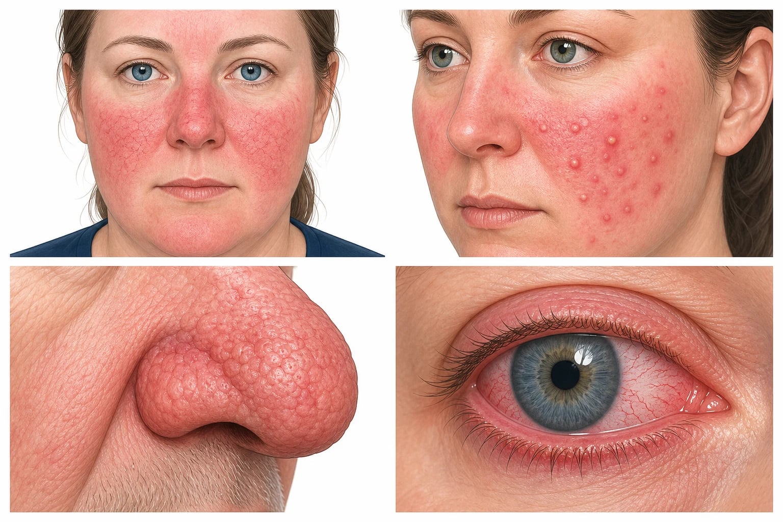
Rosacea is a chronic, relapsing inflammatory dermatosis that predominantly affects the ** centrofacial convexities** — cheeks, nose, chin and glabella. The clinical signature is persistent centrofacial erythema with periodic intensification, accompanied by flushing, papules, pustules, telangiectasia, skin sensitivity or, in some patients, phymatous change and ocular inflammation.[1]
Traditional subtypes (NRS, 2002)
The National Rosacea Society originally classified rosacea into four subtypes, still useful for descriptive communication and historical continuity. Each overlaps with the others in real patients, and the boundaries are porous, but the categories remain an efficient shorthand on ward rounds, in clinic letters and in exam viva answers. Recognising the dominant subtype (and recognising when more than one is active) remains the first step in phenotype-led management. [1]
| Subtype | Defining features | Sex / age tendency | Clinical pearls |
|---|---|---|---|
| Subtype one — Erythematotelangiectatic rosacea (ETR) | Flushing and persistent centrofacial erythema; telangiectasia; stinging, burning or tight sensation | Adults 20–50; slight female excess | Often the EARLIEST phenotype; skin sensitivity is characteristic; minimal or absent papules; treatment centres on trigger avoidance, gentle skincare, brimonidine or oxymetazoline and laser/IPL |
| Subtype two — Papulopustular rosacea (PPR) | Persistent erythema with transient dome-shaped erythematous papules and pinpoint pustules | Adults 30–50; women > men in clinic populations | Most commonly DRIVES treatment escalation; comedones are ABSENT (key acne discriminator); ivermectin 1% cream and doxycycline 40 mg modified-release are mainstays |
| Subtype three — Phymatous rosacea | Skin thickening, nodularity, glandular hyperplasia, often with fibrosis | Men > women; usually > 40 years | Rhinophyma is the canonical lesion — sebaceous hyperplasia of the distal nose; medical therapy is largely ineffective once established; CO₂/Er:YAG laser or surgical sculpting is definitive |
| Subtype four — Ocular rosacea | Blepharitis, meibomian gland dysfunction, conjunctival hyperaemia, dry gritty burning eyes, recurrent styes | Any adult; may precede skin disease | Frequently UNDERTREATED; up to 50% of rosacea patients have eye findings on slit-lamp; ophthalmology input if pain, photophobia, blurred vision or corneal change |
Phenotype approach (ROSCO, 2017)
The Global ROSacea COnsensus (ROSCO) panel moved away from rigid subtypes toward a phenotype-led approach. Two phenotypes are considered diagnostic: [1]
- Persistent centrofacial erythema that intensifies periodically
- Phymatous changes [1]
Other major phenotypes include papules/pustules, flushing, telangiectasia and ocular features. Patients commonly have overlapping phenotypes that fluctuate over time, so treatment is matched to the predominant feature(s) rather than a subtype label.[1]
Subtypes in clinical detail
Erythematotelangiectatic rosacea (ETR)
ETR is the vascular-dominant subtype. Patients have persistent centrofacial erythema of the cheeks, nose, chin and glabella that intensifies with triggers and gradually settles to a baseline rather than fully resolving. Flushing is the cardinal symptom — sudden, episodic, deep redness that may last minutes to hours and is often preceded by warmth or a burning sensation. Fine linear telangiectasia (often more visible on the ala nasi and on the cheek below the malar eminence) and skin sensitivity (stinging or burning after application of water, sunscreen, makeup, wind or heat) are characteristic. Importantly, papules and pustules are minimal or absent, which distinguishes ETR from PPR. ETR is often the EARLIEST phenotype a patient develops and may evolve into PPR with superimposed inflammation; some patients remain purely ETR for years. Treatment centres on trigger avoidance, photoprotection, gentle non-soap cleansers, fragrance-free moisturisers, brimonidine 0.33% gel or oxymetazoline 1% cream for residual background erythema, and pulsed dye laser or IPL for telangiectasia. Topical metronidazole, ivermectin or azelaic acid help when there is any inflammatory component.[1][2][4]
Papulopustular rosacea (PPR)
PPR is the inflammatory-dominant subtype and the one most likely to bring the patient to the dermatologist. It comprises persistent centrofacial erythema punctuated by dome-shaped erythematous papules and small pustules, often with a few background telangiectasia. The comedones are ABSENT — the single most useful bedside discriminator from acne vulgaris. The papules and pustules of PPR arise in crops over days, may be tender and resolve over a week or two, often leaving transient erythematous macules but not pitted scars. Distribution is centrofacial but may extend onto the perioral skin, lateral cheeks and sometimes the upper neck. Skin sensitivity, burning and stinging are common, and patients frequently report intolerance to cosmetic products. Standard care is topical ivermectin 1% cream once daily for 12 to 16 weeks (with superior lesion reduction versus vehicle and metronidazole in pivotal trials), with topical metronidazole 0.75–1% or azelaic acid 15–20% as alternatives; oral doxycycline 40 mg modified-release once daily is added for moderate-to-severe disease. Concomitant brimonidine can be used for residual erythema.[1][8][10][15]
Phymatous rosacea
Phymatous rosacea is defined by cutaneous thickening and hyperplasia of sebaceous glands and connective tissue, most often producing rhinophyma — bulbous, nodular enlargement of the distal nose with patulous follicular ostia, telangiectasia and a violaceous or ruddy hue. Less common sites include the chin (gnathophyma), forehead (metophyma), ears (otophyma) and eyelids. Phymatous change is more common in men, typically over the age of 40, with longer disease duration and a higher cumulative inflammatory load. Histology shows sebaceous hyperplasia, perivascular and perifollicular inflammation, fibrosis, and (in established disease) vascular ectasia. Medical therapy alone has limited effect once tissue remodelling is established; long-term oral tetracyclines and low-dose isotretinoin may slow or partially regress disease, but the definitive treatment is physical — CO₂ laser ablation, Er:YAG laser, electrosurgery, dermabrasion or cold-steel surgical sculpting, often in combination. Recurrence is possible if background inflammation is not controlled.[2][4]
Ocular rosacea
Ocular rosacea affects up to half of all rosacea patients when examined with slit-lamp, although only a minority volunteer eye symptoms. The clinical spectrum includes blepharitis, meibomian gland dysfunction, conjunctival hyperaemia, dry eye, foreign-body sensation, recurrent styes, episcleritis and, at the severe end, keratitis with corneal vascularisation, ulceration and (rarely) perforation. Ocular symptoms may precede skin disease in up to 20% of patients and are easily missed unless actively asked about — every consultation should include the question "are your eyes dry, gritty, red or light-sensitive?". Management combines lid hygiene (warm compresses, lid massage, dilute baby-shampoo or commercial lid wipes), preservative-free artificial tears, oral doxycycline 40 mg modified-release for meibomian gland dysfunction, and topical cyclosporine or steroids under ophthalmology supervision for inflammatory flares. Persistent symptoms, any pain, photophobia or change in vision mandate urgent ophthalmology referral. Untreated disease can progress to irreversible visual loss.[1][2][4]
Triggers — what sets off a flare?
Triggers are remarkably consistent across studies and trials, and avoidance is a powerful adjunct to pharmacotherapy. Patients should be encouraged to keep a brief symptom diary for at least two to four weeks after diagnosis to personalise the list. The ROSCO 2019 update and the National Rosacea Society's patient data have converged on a small set of common offenders:
- Ultraviolet radiation (UVA + UVB) — the single most consistently reported trigger; broad-spectrum SPF 30–50 with high UVA rating, applied liberally and reapplied every two hours outdoors, is essential for every phenotype.
- Heat and hot environments — saunas, hot showers, steamy kitchens, hot baths, exertion in warm weather, and even cooking over hot stoves.
- Cold wind and abrupt temperature change — a frequent winter trigger; barrier creams and physical protection (scarves, balaclava in extreme conditions) help.
- Alcohol — particularly red wine (histamine, resveratrol, congeners), beer, and spirits; white wine and clear spirits are often better tolerated but not always.
- Spicy foods and hot drinks — capsaicin activates TRPV1 on cutaneous sensory neurons; even black coffee at high temperature may trigger flushing.
- Strenuous exercise — desirable for general health but may precipitate flushing; dividing activity, cooling with a damp cloth and avoiding peak heat helps.
- Emotional stress and anxiety — adrenergic flushing mediated via TRPA1; addressing underlying anxiety and depression is itself therapeutic for the skin.
- Drugs and cosmetics — topical steroids (a top pitfall), vasodilators (nicotinic acid, hydralazine, sildenafil), some acne topicals (benzoyl peroxide, retinoids), abrasive exfoliants, alcohol-based toners, fragrances and astringents; opt for fragrance-free, non-soap cleansers and minimal skincare routines.
- Hormonal change — perimenopausal flushing, pregnancy, thyroid disease; the differential with true menopausal flushing must be considered in older women.
- Microbial factors — flares may follow upper respiratory infections, Helicobacter pylori eradication failure or marked increases in Demodex density; targeted antimicrobials (ivermectin, oral tetracyclines, sometimes metronidazole) treat the underlying driver. [1]
Epidemiology & Risk Factors
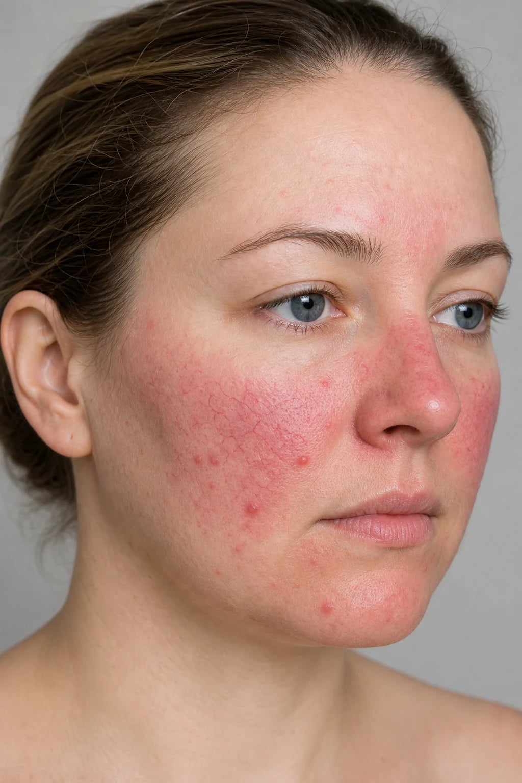
Rosacea primarily affects adults aged 30–50 years, with a female predominance for erythematotelangiectatic and papulopustular phenotypes, while phymatous change (especially rhinophyma) is more common in men. It is most prevalent in fair-skinned individuals of Celtic or Northern European descent (Fitzpatrick types I–II), although it is increasingly recognised and underdiagnosed in darker skin phototypes where erythema may be subtler and post-inflammatory hyperpigmentation more prominent. [1]
Risk factors and triggers [1]
- Genetics — family history, HLA associations.
- Demodex folliculorum density — strongly associated with papulopustular disease.
- Environmental triggers — ultraviolet radiation, heat, cold wind, exercise, alcohol (especially red wine), spicy foods, hot drinks.
- Neurovascular stimuli — stress, emotional flushing, menopausal flushing.
- Drugs and topical products — topical or systemic corticosteroids, nicotinic acid, vasodilators, some cosmetics.
- Comorbidities — migraine, cardiovascular disease, gastrointestinal disorders (notably H. pylori and small intestinal bacterial overgrowth in some studies), anxiety and depression. [1]
Pathophysiology
Rosacea is no longer viewed as a single pathway disease. Current models integrate innate immune dysregulation, neurovascular hyper-reactivity, microbiome alterations and barrier dysfunction in a genetically predisposed host.[7]
The TLR2–KLK5–cathelicidin axis
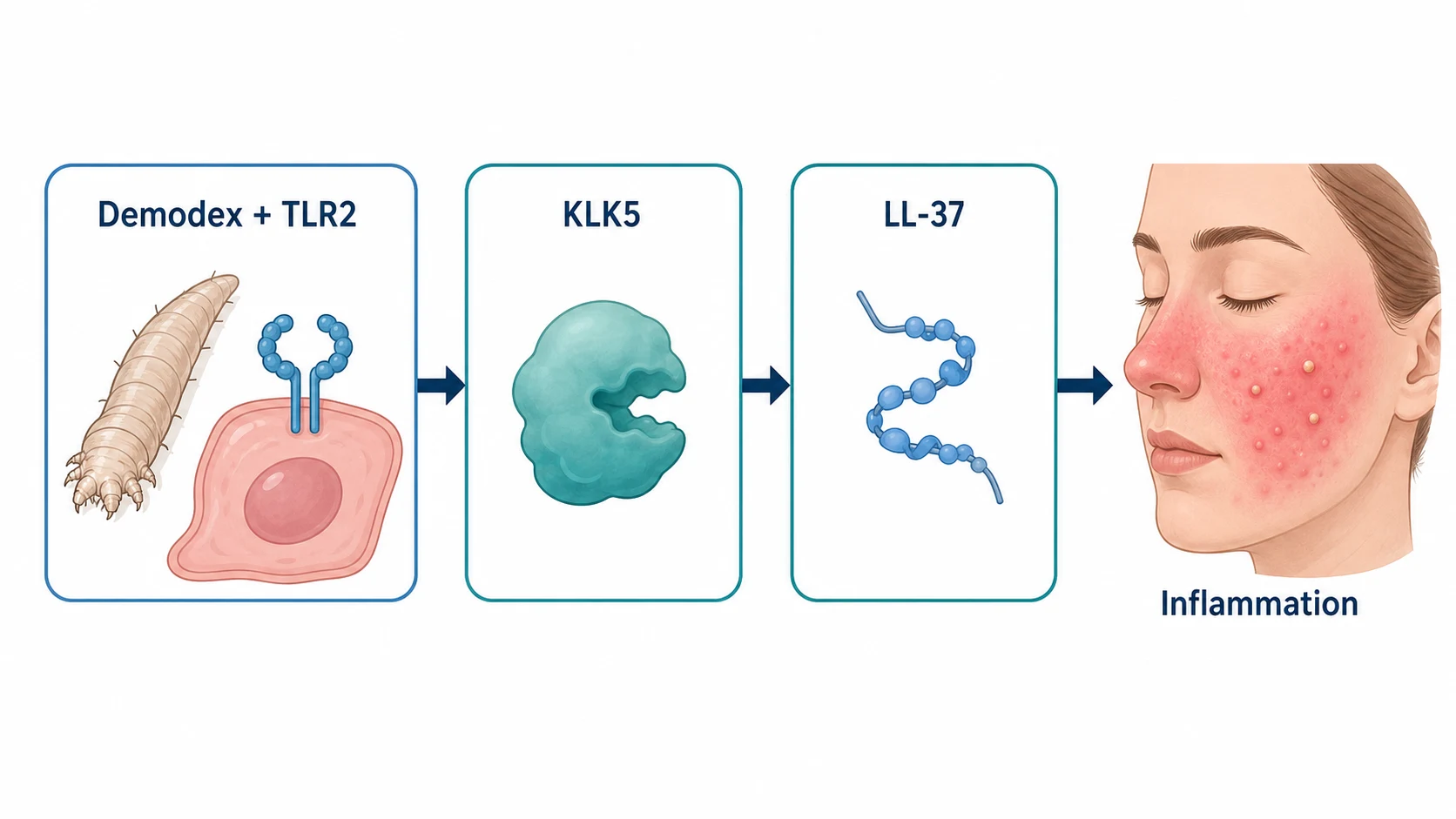
The central innate immune pathway is: [1]
- Trigger — environmental factors or increased Demodex density upregulate Toll-like receptor 2 (TLR2) on keratinocytes and sebocytes.
- Serine protease release — TLR2 activation increases kallikrein 5 (KLK5) release.
- Cathelicidin processing — KLK5 cleaves the cathelicidin precursor CAMP into the pro-inflammatory peptide LL-37.
- Inflammation and angiogenesis — LL-37 drives cytokine release, neutrophil recruitment, vasodilation and angiogenesis, producing erythema, papules and pustules. [1]
Neurovascular and barrier mechanisms
- Transient receptor potential (TRP) channels (TRPV1, TRPA1) mediate heat, capsaicin and alcohol-induced flushing and neurogenic inflammation.
- Neuropeptides (substance P, CGRP, VIP) promote vasodilation and mast-cell activation.
- Skin barrier impairment permits increased penetration of irritants and microbial antigens.
- Demodex overgrowth may initiate or amplify TLR2/LL-37 signalling; this underpins the efficacy of ivermectin. [1]
Clinical Presentation
Diagnostic phenotypes
Persistent centrofacial erythema
- Dull or bright red erythema of cheeks, nose, chin and glabella.
- Periodically intensifies with triggers.
- May be accompanied by burning, stinging or dryness. [1]
Papules and pustules
- Dome-shaped erythematous papules and pinpoint pustules on an erythematous background.
- Typically comedones are absent — a key distinction from acne vulgaris.
- Centrofacial distribution; may extend to perioral area. [1]
Flushing and telangiectasia
- Transient or persistent facial redness.
- Fine linear telangiectasia on cheeks and nose. [1]
Phymatous change
- Thickened, sebaceous, nodular skin, especially of the nose (rhinophyma).
- More common in older men. [1]
Ocular features
- Blepharitis, meibomian gland dysfunction, conjunctival injection, dry eye, foreign-body sensation, photophobia.
- Keratitis and corneal ulceration are sight-threatening complications. [1]
Severity and impact
Symptoms range from mild cosmetic concern to severe pain, psychological distress and vision threat. Quality of life is often disproportionate to objective severity because of the visibility of facial redness. [1]
Differential Diagnosis
[1]Rosacea versus acne vulgaris — the bedside discriminator
The most common examination pitfall in a patient presenting with centrofacial papules and pustules is conflating papulopustular rosacea with acne vulgaris. Although they share inflammatory facial lesions, the natural history, distribution, lesion morphology, age of onset, scarring pattern and treatment ladder differ enough that confident distinction is expected at fellowship and FRCDerm/ABD vivas. The comparison below captures the features the examiner will probe: [1]
[1] [1] [2]Clinical & Bedside Assessment
Severity instruments
| Tool | Use | Notes |
|---|---|---|
| Investigator Global Assessment (IGA) | Overall disease severity | 5-point scale from clear to severe |
| Clinician Erythema Assessment (CEA) | Erythema severity | 5-point scale |
| Patient Self-Assessment (PSA) | Patient-reported erythema | Useful for brimonidine/oxymetazoline trials |
| Inflammatory lesion count | Papules + pustules | Primary endpoint in many PPR trials |
| Dermatology Life Quality Index (DLQI) | Quality of life | ≥10 indicates severe impact |
Practical severity categorisation
- Mild — occasional flushing, few papules/pustules, minimal impact; topical therapy and trigger avoidance usually suffice.
- Moderate — persistent erythema, multiple papules/pustules, noticeable impact; topical ± oral therapy.
- Severe — extensive papules/pustules, marked erythema, phymatous change, ocular involvement or major psychosocial impact; systemic therapy, combinations and procedures. [1]
Investigations
Rosacea is a clinical diagnosis; no laboratory test is pathognomonic. Investigations are directed at: [1]
- Excluding mimics — ANA, anti-Ro/La, CK if lupus/dermatomyositis suspected; 5-HIAA if carcinoid suspected.
- Demodex density — skin surface biopsy or standardized skin scraping (not routine; research tool).
- Ocular assessment — slit-lamp examination, Schirmer test, meibomian gland expression; refer if pain, photophobia or visual change.
- Skin biopsy — only if diagnosis uncertain or to exclude granulomatous rosacea, lupus or lymphoma. Histology may show dilated vessels, perivascular/perifollicular lymphohistiocytic infiltrate, solar elastosis and, occasionally, Demodex mites. [1]
Management — General Measures
Trigger avoidance and skincare
- Sun protection — broad-spectrum SPF 30–50 daily; UV is a universal trigger.
- Gentle skincare — non-soap cleanser, fragrance-free moisturiser, barrier repair; avoid astringents, scrubs and irritant cosmeceuticals.
- Identify triggers — keep a symptom diary for alcohol, heat, spicy food, exercise, stress and cosmetic products.
- Cooling measures — tepid water, fans, avoidance of hot environments.
- Psychological support — visible facial redness causes significant anxiety and depression; screen and refer when needed. [1]
Management — Topical Therapy
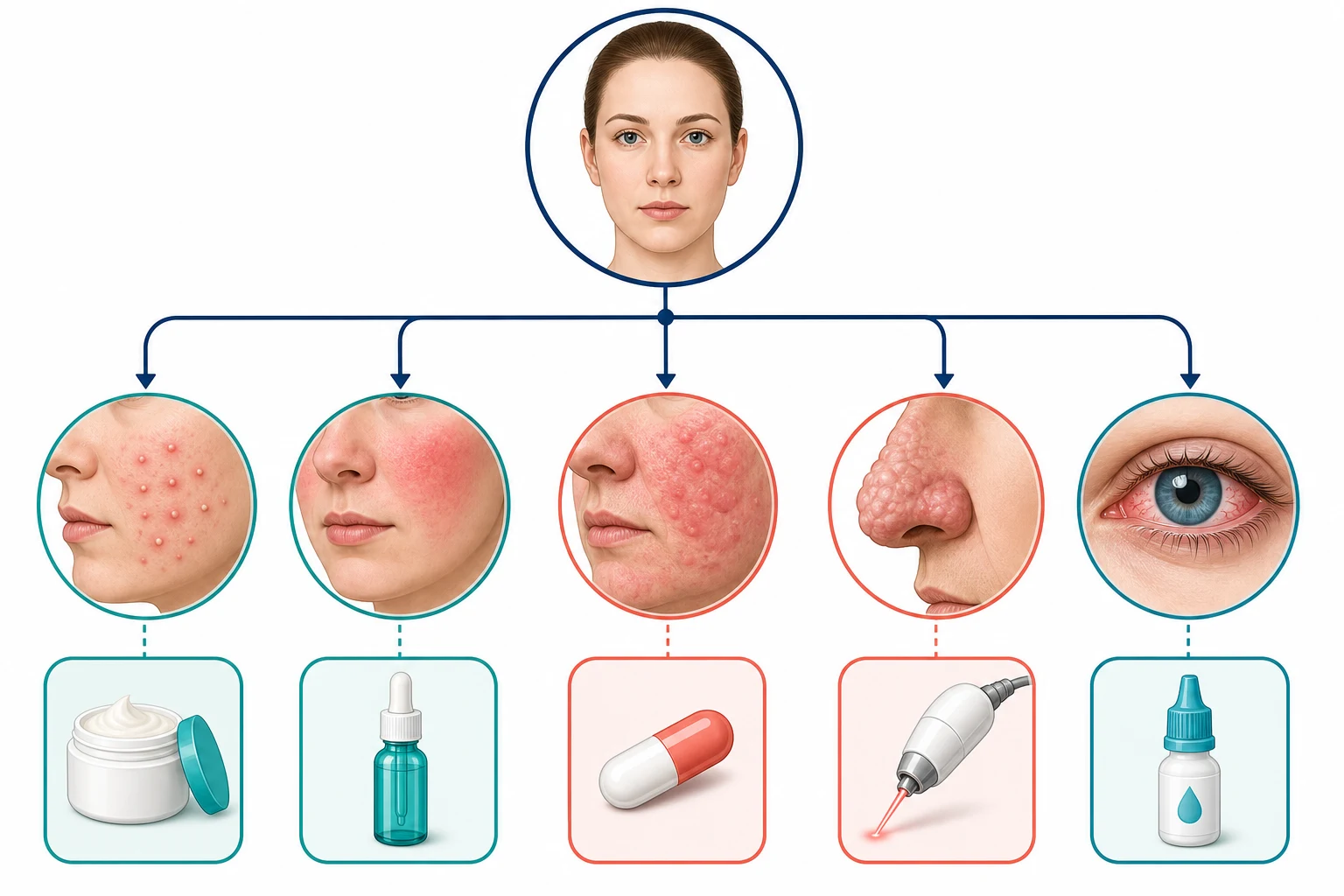
Treatment choice follows the predominant phenotype, with dose, frequency and duration anchored to phenotype severity, prior treatment response and patient factors (pregnancy, age, intolerance, drug interactions): [1]
Core pharmacotherapy at a glance — topical, systemic and procedural doses
| Agent | Indication | Mechanism / notes |
|---|---|---|
| Metronidazole 0.75–1% | Papulopustular | Anti-inflammatory and antimicrobial; traditional first-line |
| Azelaic acid 15–20% | Papulopustular, erythema | Anti-inflammatory, keratolytic; may sting on application |
| Ivermectin 1% cream | Papulopustular | Anti-inflammatory and anti-Demodex; once daily; pivotal trials showed superior lesion reduction versus vehicle[8] |
| Brimonidine 0.33% gel | Persistent erythema | α₂-adrenergic agonist vasoconstrictor; onset ~30 min; effect ~12 h; rebound erythema possible[12] |
| Oxymetazoline 1% cream | Persistent erythema | α-adrenergic vasoconstrictor; longer duration than brimonidine in some studies |
Combination topical therapy
Patients with both erythema and papulopustular disease often need combination therapy. Trials of ivermectin 1% cream plus brimonidine 0.33% gel showed that simultaneous treatment of inflammatory lesions and erythema is safe and effective.[11]
Management — Systemic Therapy
Oral tetracyclines
Doxycycline 40 mg modified-release (MR) once daily is the best-established systemic therapy for papulopustular rosacea. At this dose it has anti-inflammatory effects without antimicrobial activity, reducing inflammatory lesions and erythema with a favourable safety profile. Phase III trials established efficacy; long-term studies support sustained benefit and low resistance risk.[15][17]
- Alternatives — minocycline 50–100 mg daily, lymecycline 408 mg daily, oxytetracycline 500 mg twice daily.
- DOMINO trial showed doxycycline 40 mg MR was non-inferior to minocycline 100 mg for papulopustular rosacea with fewer gastrointestinal adverse effects.[18]
- Biomarker studies show doxycycline 40 mg MR reduces cathelicidin and serine protease activity, supporting its mechanistic rationale.[16]
Other systemic options
- Low-dose isotretinoin (10–20 mg/day, or 0.1–0.3 mg/kg/day) for severe, refractory papulopustular or phymatous rosacea. Teratogenic; requires pregnancy prevention and lipid/liver monitoring.
- Metronidazole or clarithromycin occasionally used if tetracyclines contraindicated.
- Carvedilol (oral β-blocker with α-blocking activity) has been reported for severe refractory flushing and erythema, though evidence is limited. [1]
Severe disease and combination therapy
For severe papulopustular rosacea, ivermectin 1% cream combined with doxycycline 40 mg MR produced greater improvement than doxycycline plus vehicle, supporting a topical–systemic combination strategy.[10]
Management — Procedural Therapy
| Modality | Indication | Notes |
|---|---|---|
| Pulsed dye laser (PDL, 585–595 nm) | Erythema, telangiectasia | Targets oxyhaemoglobin; bruising common |
| Nd:YAG (1064 nm) | Deeper telangiectasia, resistant vessels | Longer wavelength penetrates deeper |
| Intense pulsed light (IPL) | Erythema, telangiectasia, background redness | Broadband light; multiple sessions needed |
| CO₂ or Er:YAG laser | Rhinophyma | Reshapes and debulks nasal tissue |
| Electrosurgery / dermabrasion | Rhinophyma | Established surgical alternatives |
Management by Special Scenario
Ocular rosacea
- Lid hygiene — warm compresses, lid massage, diluted baby shampoo or dedicated lid wipes.
- Artificial tears for dry eye.
- Oral tetracyclines (doxycycline 40 mg MR or standard tetracycline) improve meibomian gland dysfunction and reduce inflammation.
- Ophthalmology referral for any pain, photophobia, visual change, corneal opacity or ulceration. [1]
Rhinophyma
- Medical therapy has limited effect once tissue hyperplasia is established.
- CO₂ laser, Er:YAG laser, electrosurgery, dermabrasion or scalpel sculpting are effective; often performed by dermatologic surgeons.
- Recurrence is common if background inflammation persists. [1]
Pregnancy and lactation
- Avoid tetracyclines, isotretinoin and oral retinoids in pregnancy.
- Topical azelaic acid, metronidazole and ivermectin are generally considered safe; use lowest effective potency and limited areas.
- Brimonidine and oxymetazoline are topical vasoconstrictors with limited systemic absorption; discuss risk/benefit. [1]
Children
- Rosacea is uncommon in children; consider steroid-induced dermatitis, periorificial dermatitis or Demodex as differentials.
- Tetracyclines are contraindicated in children under 8 years (tooth discolouration); erythromycin may be used if systemic therapy essential. [1]
Complications, Emergencies & Pitfalls
Ocular complications
Untreated ocular rosacea can progress to keratitis, corneal ulceration, neovascularisation and visual impairment. Any ocular pain, photophobia or visual disturbance is an ophthalmology emergency. [1]
Phymatous rosacea
Rhinophyma causes functional problems (nasal obstruction, difficulty wearing glasses) and significant psychological morbidity. Early control of inflammation may reduce progression. [1]
Corticosteroid-induced rosacea-like dermatitis
A common iatrogenic pitfall. Potent topical steroids on the face produce rebound erythema, telangiectasia and papulopustules. Management is gradual steroid withdrawal plus standard rosacea therapy; patients may flare initially. [1]
Psychosocial comorbidity
Rosacea is associated with anxiety, depression, social phobia and reduced workplace productivity. Dermatologists should proactively screen and signpost support. [1]
[2] [4]Comorbidities
Rosacea is increasingly recognised as a systemic inflammatory condition with associations beyond the skin: [1]
| Comorbidity | Notes |
|---|---|
| Migraine | Higher prevalence, especially with phymatous disease |
| Cardiovascular disease | Some studies link rosacea to dyslipidaemia, hypertension and coronary disease |
| Gastrointestinal disease | Associations with H. pylori, small intestinal bacterial overgrowth and inflammatory bowel disease reported |
| Neuropsychiatric disease | Anxiety, depression and reduced quality of life |
| Autoimmune disease | Higher rates of type 1 diabetes, coeliac disease and multiple sclerosis in some cohorts |
These associations should prompt holistic risk-factor review rather than exhaustive investigation in every patient. [1]
[4]Prognosis & Follow-Up
Rosacea is chronic and relapsing; there is no cure. Treatment goals are control of inflammation, reduction of flares, improvement of quality of life and prevention of complications. [1]
- Initial topical therapy — review at 8–12 weeks.
- Systemic therapy — review at 6–12 weeks; many patients can step down or stop after 3–6 months, using topical maintenance.
- Maintenance — long-term topical ivermectin, azelaic acid or intermittent brimonidine is often needed.
- Relapse — common after stopping therapy; treat-to-target with planned re-treatment is more effective than reactive rescue. [1]
Evidence, Guidelines & Regional Differences
Major guidelines
- National Rosacea Society Expert Committee (2019 update) — phenotype-based management; topical ivermectin, azelaic acid and metronidazole for papulopustular disease; brimonidine for erythema; doxycycline 40 mg MR for moderate-to-severe disease.[2]
- ROSCO (2017) — diagnostic and severity recommendations; persistent centrofacial erythema and phymatous change as diagnostic phenotypes.[1]
- BAD rosacea guideline (2021) — UK consensus emphasising trigger avoidance, skincare, topical/systemic therapy and laser/IPL for appropriate phenotypes.
Landmark trials
- Yamasaki 2007 — cathelicidin/LL-37 central to rosacea inflammation.[5]
- Yamasaki 2011 — TLR2 upregulation increases KLK5 and drives LL-37 processing.[6]
- Stein 2014 (IVERMECTIN) — pivotal phase III trials of ivermectin 1% cream in papulopustular rosacea.[8]
- Fowler 2013 (BRIMONIDINE) — pivotal phase III trials establishing brimonidine gel for facial erythema.[12]
- Del Rosso 2007 (DOXYCYCLINE) — pivotal phase III trials of doxycycline 40 mg MR.[15]
- DOMINO 2017 — doxycycline 40 mg MR non-inferior to minocycline 100 mg with better tolerability.[18]
Exam Pearls
Red Flags [1]
Exam application bank (NEET-PG / INICET)
One-line answer
Rosacea is a chronic inflammatory facial dermatosis characterised by centrofacial erythema, flushing, papules, pustules, telangiectasia and, in some patients, phymatous change and ocular involvement. Fellowship-level assessment requires mastery of the 2017 ROSCo phenotype approach, the TLR2–KLK5–cathelicidin axis, neurovascular and Demodex mechanisms, severity instruments (IGA, CEA, lesion counts, DLQI), phenotype-directed topical and systemic therapy (ivermectin, brimonidine, doxycycline 40 mg modified-release), procedural options for erythema/telangiectasia and rhinophyma, ocular rosacea management, and recognition of mimics and treatment pitfalls. [1]
Worked stems (answer without another resource)
Stem 1 — Classic presentation. Map symptoms to mechanism; name the first investigation and first treatment step with dose/route if drug therapy is standard. [1]
Stem 2 — Unstable / complicated. List red flags that force immediate resuscitation, theatre, ICU, antidote, or reperfusion — and what you do in the first 15 minutes. [1]
Stem 3 — Atypical group. Elderly, pregnancy, child, or immunocompromised: how presentation and thresholds change. [1]
Stem 4 — Differential trap. Name the three closest mimics and one discriminator for each. [1]
Stem 5 — Disposition. Who goes home with safety-netting, who is admitted, who needs HDU/ICU/theatre, and what follow-up is mandatory. [1]
Rapid viva checklist
- Definition + classification
- Pathophysiology chain
- Bedside signs / criteria
- Score with exact components (if any)
- Emergency bundle
- Definitive therapy with doses
- Complications of disease and of treatment
- Special populations
- Guideline/trial name if classic
- Three exam traps
Coverage self-check
If you cannot answer any stem above from this page alone, re-read the matching section — the page is intended to be self-sufficient for final-prof and NEET-PG/INICET questions on Rosacea.
[1]References
- [1]Tan J, Almeida LMC, Bewley A, et al. Updating the diagnosis, classification and assessment of rosacea: recommendations from the global ROSacea COnsensus (ROSCO) panel Br J Dermatol, 2017.PMID 27718519
- [2]Thiboutot D, Anderson R, Cook-Bolden F, et al. Standard management options for rosacea: The 2019 update by the National Rosacea Society Expert Committee J Am Acad Dermatol, 2020.PMID 32035944
- [3]Barakji YA, Oumeish O, Oumeish I, Parish JL. Assessment of Frequency of Rosacea Subtypes in Patients With Rosacea: A Systematic Review and Meta-analysis JAMA Dermatol, 2022.PMID 35385049
- [4]Del Rosso J, Baldwin H, Bhatia N, et al. A Review of the Diagnostic and Therapeutic Gaps in Rosacea Management: Consensus Opinion Dermatol Ther (Heidelb), 2024.PMID 38194021
- [5]Yamasaki K, Di Nardo A, Bardan A, et al. Increased serine protease activity and cathelicidin promotes skin inflammation in rosacea Nat Med, 2007.PMID 17676051
- [6]Yamasaki K, Kanada K, Macleod DT, et al. TLR2 expression is increased in rosacea and stimulates enhanced serine protease production by keratinocytes J Invest Dermatol, 2011.PMID 21107351
- [7]Wang H, Yin J, Li J, et al. Advances in the pathogenesis of rosacea Front Immunol, 2025.PMID 41646975
- [8]Stein L, Kircik L, Fowler J, et al. Efficacy and safety of ivermectin 1% cream in treatment of papulopustular rosacea: results of two randomized, double-blind, vehicle-controlled pivotal studies J Drugs Dermatol, 2014.PMID 24595578
- [9]Taieb A, Khemis A, Ruzicka T, Bachelez H. Maintenance of remission following successful treatment of papulopustular rosacea with ivermectin 1% cream vs. metronidazole 0.75% cream: 36-week extension of the ATTRACT randomized study J Eur Acad Dermatol Venereol, 2016.PMID 26691278
- [10]Schaller M, Kemény L, Havlickova B, et al. A randomized phase 3b/4 study to evaluate concomitant use of topical ivermectin 1% cream and doxycycline 40-mg modified-release capsules, versus topical ivermectin 1% cream and placebo in the treatment of severe rosacea J Am Acad Dermatol, 2020.PMID 31150711
- [11]Gold LS, Del Rosso JQ, Draelos ZD, et al. Treatment of Rosacea With Concomitant Use of Topical Ivermectin 1% Cream and Brimonidine 0.33% Gel: A Randomized, Vehicle-controlled Study J Drugs Dermatol, 2017.PMID 28915286
- [12]Fowler J Jr, Jackson M, Moore A, et al. Efficacy and safety of once-daily topical brimonidine tartrate gel 0.5% for the treatment of moderate to severe facial erythema of rosacea: results of two randomized, double-blind, and vehicle-controlled pivotal studies J Drugs Dermatol, 2013.PMID 23839181
- [13]Fowler J, Tan J, Jackson M, et al. Once-daily topical brimonidine tartrate gel 0·5% is a novel treatment for moderate to severe facial erythema of rosacea: results of two multicentre, randomized and vehicle-controlled studies Br J Dermatol, 2012.PMID 22050040
- [14]Layton AM, Cunliffe WJ, Hughes K, et al. Brimonidine gel 0.33% rapidly improves patient-reported outcomes by controlling facial erythema of rosacea: a randomized, double-blind, vehicle-controlled study J Eur Acad Dermatol Venereol, 2015.PMID 26416154
- [15]Del Rosso JQ, Webster GF, Jackson M, et al. Two randomized phase III clinical trials evaluating anti-inflammatory dose doxycycline (40-mg doxycycline, USP capsules) administered once daily for treatment of rosacea J Am Acad Dermatol, 2007.PMID 17367893
- [16]Di Nardo A, Holmes AD, Muto Y, et al. Improved clinical outcome and biomarkers in adults with papulopustular rosacea treated with doxycycline modified-release capsules in a randomized trial J Am Acad Dermatol, 2016.PMID 26951940
- [17]Del Rosso JQ, Baldwin H, Bhatia N, et al. Long-term inflammatory rosacea management with subantibiotic dose oral doxycycline 40 mg modified-release capsules once daily Dermatol Ther, 2022.PMID 34713539
- [18]van der Linden MMD, van Rappard DC, Daams JG, et al. DOMINO, doxycycline 40 mg vs. minocycline 100 mg in the treatment of rosacea: a randomized, single-blinded, noninferiority trial, comparing efficacy and safety Br J Dermatol, 2017.PMID 27797396
- [19]Moore A, Kempers S, Murakawa G, et al. Long-term safety and efficacy of once-daily topical brimonidine tartrate gel 0.5% for the treatment of moderate to severe facial erythema of rosacea: results of a 1-year open-label study J Drugs Dermatol, 2014.PMID 24385120
- [20]Fowler J, Tan J, Bhatt R, Schöpf T. Treatment of facial erythema in patients with rosacea with topical brimonidine tartrate: correlation of patient satisfaction with standard clinical endpoints of improvement of facial erythema J Eur Acad Dermatol Venereol, 2015.PMID 25074756