Dermatology · Medicine
Scabies
Also known as Classical scabies · Crusted (Norwegian) scabies · Scabetic infestation
Scabies is an infestation of the skin by the mite Sarcoptes scabiei var. hominis, transmitted by skin-to-skin contact, producing intense nocturnal pruritus and characteristic burrows in web-spaces, wrists, and genitalia. Fellowship-level assessment demands mastery of the classical distribution and burrows, the contrasting hyperkeratotic, highly contagious crusted (Norwegian) scabies of immunocompromise, dermoscopic and microscopic diagnosis, first-line permethrin 5% and oral ivermectin (including the two-dose regimen and mass drug administration), treatment of contacts and environmental decontamination, post-scabetic itch, and recognition of treatment failure and emerging resistance.
On this page & tools
Your progress
Saved locally on this device.
Exam tags
Red flags
Overview
Scabies is one of the commonest dermatological conditions worldwide, yet it remains under-recognised in everyday clinical practice. The disease is caused by the obligate human ectoparasite Sarcoptes scabiei var. hominis, an arachnid that completes its entire life cycle within the superficial layers of human skin. Transmission requires close, prolonged skin-to-skin contact, and the infestation is characterised by an intensely pruritic, papular eruption with a distinctive anatomical distribution. The hallmark of the disease is the burrow, a short, serpiginous track in the stratum corneum created by the egg-laying female mite. Fellowship-level competence requires not only recognition of the classical presentation, but also an understanding of the spectrum that includes crusted (Norwegian) scabies, nodular scabies, bullous scabies, scabies incognito, and animal-transmitted variants. A secure grasp of the 2020 International Alliance for the Control of Scabies (IACS) diagnostic criteria, the two-dose permethrin or ivermectin regimens, the simultaneous treatment of all contacts, environmental decontamination, post-scabetic itch, and the causes of treatment failure is essential for both clinical practice and high-stakes examinations.[1][4][6]
1. Definition and classification
Definition
Scabies is a parasitic infestation of the skin caused by the mite Sarcoptes scabiei var. hominis. The adult female mite burrows into the stratum corneum, deposits eggs and faecal pellets known as scybala, and evokes a host immune response that is responsible for the clinical signs and symptoms. The disease is not caused by the mechanical presence of the mite alone, but by the delayed hypersensitivity reaction to mite-derived antigens. It is a disease of poverty, overcrowding, and close human contact, and it is increasingly recognised as a public-health priority.[1][6]
Classification
Several clinical variants are recognised, each with important implications for diagnosis, treatment, and public-health control. [1]
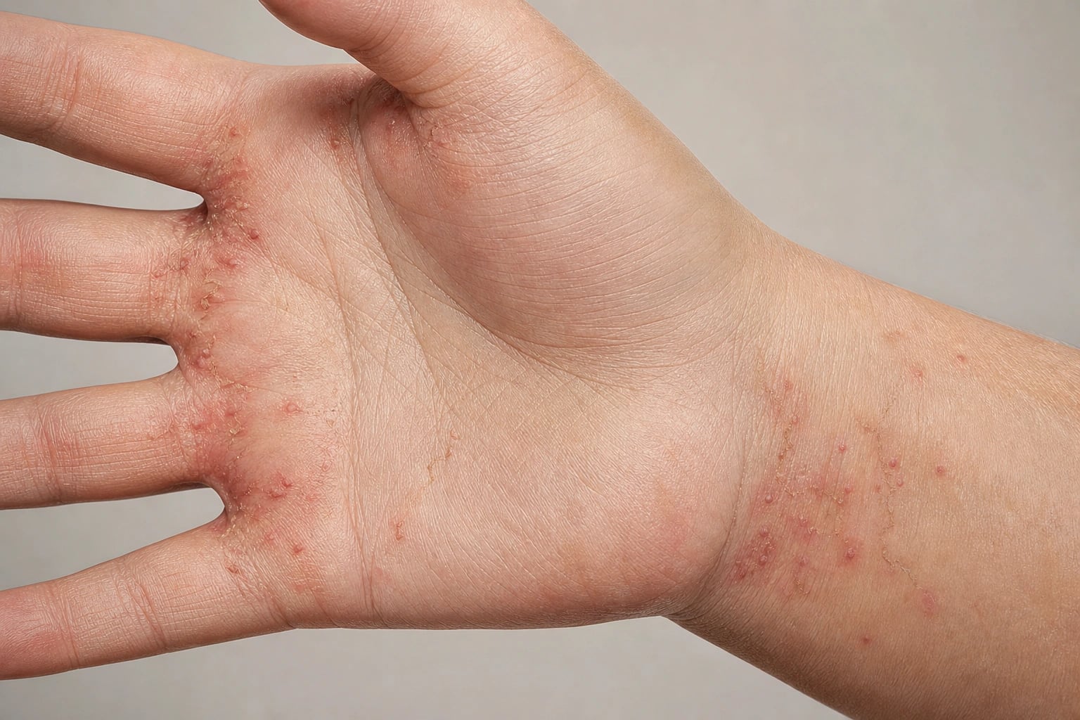
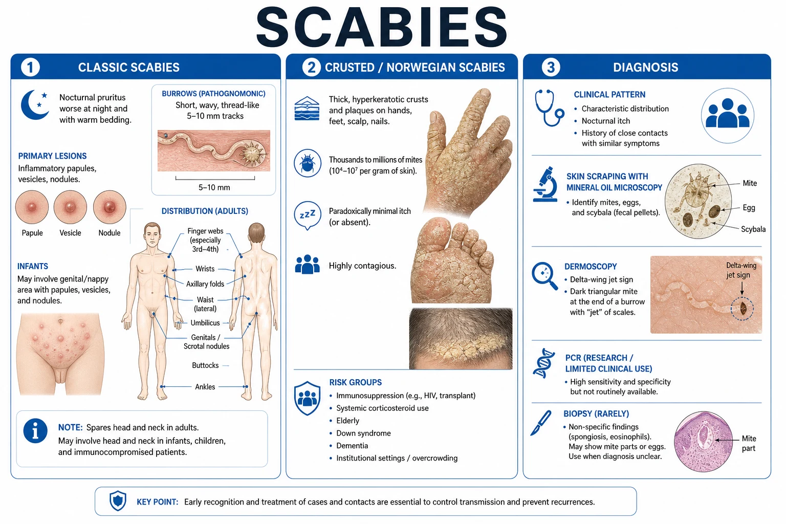
Classical scabies is the most common form. A typical adult carries only 10-15 mites on the entire body at any one time. The burden is low because the host immune response limits replication, but the inflammatory response produces intense pruritus and a widespread eruption. The distribution is characteristic, with a predilection for the finger web spaces, flexor wrists, lateral fingers, axillae, periumbilical skin, waistline, areolae, genitalia, buttocks, and, in infants, the palms, soles, head, and neck.[1][4]
Crusted (Norwegian) scabies is a hyperkeratotic, highly contagious variant in which mite counts reach thousands to millions. It develops in hosts with impaired cellular immunity, particularly T-cell responses, and is typified by thick, waxy, psoriasiform crusts on the hands, feet, scalp, and nails. Itch is often minimal or absent, and because of the enormous mite load, transmission can occur through contaminated bedding, clothing, and fomites. Crusted scabies is the engine of institutional outbreaks and demands combined, multi-dose therapy and strict isolation.[9][10][11]
Nodular scabies presents as persistent, firm, pruritic, reddish-brown nodules, most commonly on the scrotum, penis, axillae, buttocks, and groin. These nodules represent a prolonged hypersensitivity reaction to retained mite antigens, and they may persist for weeks or months after successful eradication. They are not evidence of active infestation unless new burrows are present.[1][12]
Bullous scabies is a rare variant, usually in older adults, in which tense bullae develop on the hands and feet. The histology shows eosinophilic spongiosis and a subepidermal cleft, and the clinical picture can closely mimic bullous pemphigoid. Direct immunofluorescence is negative or shows only non-specific deposits, in contrast to the linear IgG and C3 of bullous pemphigoid.[1][12]
Scabies incognito describes a presentation modified by previous topical or systemic corticosteroid therapy. The inflammatory response is suppressed, burrows are absent or atypical, and the distribution can be truncal or facial. This variant is a diagnostic trap because the signs are masked while the infestation continues to spread. A careful history of steroid exposure and a high index of suspicion are required.[1][4]
Animal-transmitted scabies, also known as sarcoptic mange, is acquired from dogs, cats, pigs, or other animals. The animal mite cannot complete its life cycle on human skin, so the infestation is transient and self-limiting. It produces a pruritic, papular eruption without true burrows, and the key intervention is treatment of the affected animal.[1][12]
Distinguishing scabies from other ectoparasites
Scabies must be distinguished from pediculosis (lice), which affects hair-bearing sites and presents with nits attached to hair shafts; Demodex infestation, which produces follicular papules and pustules on the face and is associated with rosacea; trombiculosis, which presents with intensely pruritic papules on the ankles and waist after outdoor exposure; and Cheyletiella infestation, which produces a papular urticarial eruption in contacts with dogs, cats, or rabbits. The presence of burrows, the nocturnal pruritus, and the classical distribution in close contacts point to scabies.[1][12]
2. Epidemiology and risk factors
Scabies is a neglected tropical disease with a global point prevalence estimated at roughly 200 million and an annual incidence of around 455 million. It is most prevalent in low-resource tropical and subtropical settings, where overcrowding, limited access to water, and poor sanitation favour transmission. Children are disproportionately affected, and in some communities the point prevalence in school-age children exceeds 20 percent. The disease also clusters in closed settings such as prisons, refugee camps, long-term care facilities, hospitals, and boarding schools.[1][6][14]
Scabies at a glance
Populations at highest risk
The highest risk is seen in children living in overcrowded households in low-resource countries, Indigenous communities in remote areas, prisoners, residents of care homes, people with HIV or other immunosuppressive conditions, and those with intellectual or physical disability. Crusted scabies is especially common in patients with Down syndrome, lepromatous leprosy, haematological malignancy, transplant recipients, and individuals receiving long-term systemic corticosteroids or biologics. Sexual transmission can occur in adults, particularly with crusted scabies, and the infection should be considered in the context of sexually transmitted infection screening.[1][9][10]
Modifiable and non-modifiable risk factors
Modifiable factors include overcrowding, poverty, poor hygiene, limited access to healthcare, and household transmission from untreated contacts. Non-modifiable factors include age, genetic background, immunosuppression, Down syndrome, and intellectual disability. In endemic settings, transmission is driven by household density and social mixing; in institutional outbreaks, the driver is often a single undiagnosed case of crusted scabies in a vulnerable resident. Endemic settings require community-wide control strategies, whereas institutional outbreaks are best managed with coordinated mass treatment and strict environmental decontamination.[1][14]
3. Pathophysiology and life cycle
Life cycle of Sarcoptes scabiei
Sarcoptes scabiei var. hominis is an eight-legged arachnid, approximately 0.3 to 0.4 mm in length, visible only under magnification or dermoscopy. The gravid female mite burrows into the stratum corneum at a rate of roughly 0.5 to 5 mm per day, excavating a tunnel in which she lays two to three eggs daily over a lifespan of 30 to 60 days. Eggs hatch within three to four days, and the larvae migrate to the skin surface. They mature through nymphal stages into adults over a further seven to ten days, and the total egg-to-adult cycle is approximately 10 to 14 days. Males die after mating, and fertilised females re-enter the skin to begin new burrows. The entire life cycle is confined to human skin, and the mite can survive off the host for only 24 to 72 hours under normal environmental conditions.[1][6][4]
Why classical scabies carries so few mites
In immunocompetent hosts, the cutaneous immune response limits the mite population to roughly 10 to 15 mites. This control is mediated by T-helper 1 (Th1) cells, interferon-gamma, interleukin-2, and cytotoxic effector mechanisms. A balance is struck between the mite's capacity to reproduce and the host's ability to contain it. The disease therefore reflects an inflammatory reaction to a small number of parasites rather than a heavy burden of infestation. This explains why the pruritus can be far more severe than the visible lesions, and why scratching can obscure the original burrows.[1][6]
Crusted scabies: loss of immune control
In crusted scabies, the host immune response is defective, often involving T-cell dysfunction or an impaired Th1 response. As a result, mites proliferate unchecked and colonise hyperkeratotic plaques in enormous numbers. The diminished inflammatory response also explains why itch is often minimal in crusted scabies despite the massive mite load. This variant is the source of large outbreaks because the crusts shed highly infectious mites into the environment. The pathophysiology is fundamentally one of failed host containment rather than enhanced parasite virulence.[9][10][1]
Immunology of the itch
The pruritus of scabies is predominantly a type IV delayed hypersensitivity reaction to proteins in the mite saliva, eggs, and faecal pellets (scybala). On first exposure, sensitisation takes 4 to 6 weeks, during which time the patient may be asymptomatic despite active infestation. On re-exposure, memory T cells mount a rapid recall response within 1 to 3 days. A type I IgE-mediated component also contributes, with elevated total IgE and peripheral eosinophilia in some patients. This dual hypersensitivity explains both the delayed onset of symptoms and the intensity of itch after re-infestation. Cytokines such as interleukin-2, interferon-gamma, and tumour necrosis factor are involved, and the response is polarised toward Th1 in early infection with a possible Th2 shift in chronic or heavily infested cases.[1][4][6]
Mite immune evasion
The mite has evolved mechanisms to subvert host immunity. Secreted products include proteases, protease inhibitors, and serpins that modulate complement activation and dampen inflammatory signalling. These molecules help the mite survive within the stratum corneum and may contribute to the relatively low mite burden in classical scabies. Understanding these immune-evasion strategies is important for vaccine development and for explaining why some individuals remain heavily infested despite apparently normal immune function.[1][7]
4. Clinical presentation
Classical scabies in adults
The cardinal symptom is intense pruritus, classically worse at night and after a hot bath. The primary lesion is the burrow: a 5 to 10 mm, grey-white or dark, wavy, thread-like track in the stratum corneum, often ending in a tiny vesicle or papule that represents the mite. Burrows are most easily found in the finger web spaces, particularly between the fingers, on the flexor wrists, along the lateral fingers, and around the ulnar styloid. Other characteristic sites include the axillae, periumbilical skin, waistline, inframammary folds, areolae, nipples, genitalia, and buttocks. The head and neck are usually spared in adults because the thicker stratum corneum and higher sebum production are less favourable for burrowing. Secondary changes include excoriations, crusting, eczematisation, and lichenification. In long-standing cases, the skin may show no burrows because repeated scratching has removed the superficial stratum corneum.[1][4][12]
Scabies in infants and young children
Infants and young children have a thinner stratum corneum and lower sebum production, allowing mites to colonise the palms, soles, head, neck, and face. Vesicles and pustules are more common, and the eruption can be widespread and eczematous. The presence of involvement above the neck in a child should not be misinterpreted as an alternative diagnosis; it is simply a feature of infantile scabies. The nails may be involved, and infants may be irritable, feed poorly, and sleep badly because of the pruritus.[1][12]
Scabies in the elderly
In older adults, especially those in care homes, scabies is frequently missed. The presentation may be less inflammatory, with a more generalised, subtle pruritus, and the classical distribution may be absent. The head and neck may be involved, and crusted scabies should be considered in any elderly resident with unexplained pruritus, hyperkeratosis, or nail dystrophy. The diagnosis is often delayed until several contacts become symptomatic, by which time an outbreak is already established.[1][4]
Crusted (Norwegian) scabies
Crusted scabies presents with thick, waxy, psoriasiform, yellow-to-brown crusts, most prominently on the hands, feet, wrists, ankles, elbows, knees, scalp, ears, and nails. Nail dystrophy is common, with subungual hyperkeratosis and accumulation of debris. Itch may be minimal or absent, and the patient may appear relatively well despite carrying millions of mites. Systemic signs are usually absent unless there is secondary bacterial infection. The diagnosis is easily missed if the clinician is not alert to the possibility, and the patient may be treated for psoriasis or eczema for months before the true nature of the eruption is recognised. Crusted scabies is a dermatological emergency because of its extreme contagiousness and the risk of secondary sepsis.[9][10][11]
Nodular scabies
Nodular scabies presents as firm, erythematous to brownish nodules, typically 5 to 20 mm in diameter, on the scrotum, penile shaft, groin, axillae, buttocks, and areolae. The nodules are thought to be a persistent cell-mediated immune response to retained mite antigens. They are intensely pruritic and may persist for months after successful mite eradication. They can be mistaken for genital warts, lymphogranuloma venereum, syphilitic gummata, or insect-bite reactions. Treatment usually requires intralesional or potent topical corticosteroids after the mite has been eliminated.[1][12]
Bullous scabies
Bullous scabies is uncommon and typically affects older adults. It presents with tense bullae on the hands, feet, and lower legs, sometimes with haemorrhagic bullae. The condition mimics bullous pemphigoid clinically and histologically, showing eosinophilic spongiosis and subepidermal clefting. Direct immunofluorescence lacks the linear IgG and C3 deposits typical of bullous pemphigoid, and the clinical context of pruritus, burrows, and contact history supports the diagnosis. A skin scraping for mites and dermoscopy can be decisive.[1][12]
Scabies incognito
Scabies incognito is scabies that has been partially suppressed by topical or systemic corticosteroids. Burrows are absent or scant, and the eruption may be atypical, truncal, or facial. The diagnosis requires a careful drug history, a high index of suspicion, and sometimes a therapeutic trial. This variant is a common cause of treatment failure and prolonged diagnostic delay in patients who have been empirically treated for eczema or psoriasis.[1][4]
Secondary bacterial infection
Scratching breaches the skin barrier and introduces Staphylococcus aureus and group A Streptococcus. Impetigo, folliculitis, cellulitis, ecthyma, and furunculosis may develop. In endemic settings, scabies-associated skin infection is a major risk factor for post-streptococcal glomerulonephritis and possibly acute rheumatic fever. Recognition of secondary infection is important because it may require systemic antibiotics alongside anti-scabetic therapy.[1][6][14]
5. Differential diagnosis
The differential diagnosis of scabies depends on the clinical pattern. A burrow in a typical site with nocturnal pruritus and affected contacts is pathognomonic. When burrows are absent, the differential broadens. [1]
Common mimics of classical scabies
Atopic dermatitis produces pruritic, erythematous, scaly patches, but the distribution is usually flexural in children and extensor or flexural in adults, and burrows are absent. Contact dermatitis follows exposure to an irritant or allergen and is usually localised to the site of contact. Papular urticaria, caused by insect bites, produces grouped pruritic papules, often on exposed areas, and lacks burrows. Pediculosis affects hair-bearing sites, and nits are visible on hair shafts. Folliculitis presents with pustules centred on follicles. Prurigo nodularis shows discrete, hyperkeratotic nodules, but the distribution is not the classical scabetic pattern. Drug eruptions can mimic many patterns, but they are temporally related to drug exposure. Dermatitis herpetiformis produces grouped vesicles on the extensor surfaces, especially the elbows, knees, and buttocks, and is associated with coeliac disease.[1][4][12]
Mimics of crusted scabies
Crusted scabies must be distinguished from psoriasis, particularly pustular and plaque psoriasis; eczema, especially hyperkeratotic eczema of the hands and feet; hyperkeratotic tinea pedis or manuum; Darier disease; and cutaneous T-cell lymphoma, including Sézary syndrome. The clue is the combination of hyperkeratotic plaques, a background of contact pruritus, and often a history of immunosuppression or institutional residence. A skin scraping for mites is the fastest way to confirm the diagnosis.[1][9][10]
Mimics of nodular scabies
Nodular scabies on the genitalia can be mistaken for syphilitic gummata, scrotal lymphogranuloma venereum, genital warts, or insect-bite reactions. A history of scabies in the patient or contacts, the presence of typical burrows elsewhere, and response to scabetic treatment help distinguish it. In persistent cases, a skin biopsy may show a dense lymphohistiocytic infiltrate with eosinophils but no mite parts.[1][12]

Classical scabies
- Mite burden: 10-15 mites
- Excoriated papules and burrows
- Intense, nocturnal pruritus
- Distribution: finger webs, wrists, genitalia; head spared in adults
- Requires prolonged skin-to-skin contact
- Intact Th1 host response
- Permethrin or ivermectin, two doses 7-14 days apart
Crusted (Norwegian) scabies
- Mite burden: thousands to millions
- Hyperkeratotic crusted plaques and nail dystrophy
- Minimal or absent pruritus
- Distribution: hands, feet, scalp, nails; often generalised
- Highly contagious via skin and fomites
- Impaired T-cell immunity
- Combined daily permethrin plus multi-dose ivermectin
6. Clinical bedside assessment
A systematic examination is essential. The patient should be examined in good light, with particular attention to the following sites: the finger web spaces, especially between the index and middle fingers; the flexor wrists and ulnar styloids; the lateral aspects of the fingers; the palms and soles in infants; the axillae; the periumbilical skin; the waistline; the inframammary folds and areolae; the genitalia, including the scrotum and penile shaft; and the buttocks. In infants, the head, neck, face, palms, and soles must also be inspected. A magnifying lens or dermatoscope is invaluable. The examiner should look for burrows, vesicles, pustules, excoriations, nodules, and signs of secondary infection. In crusted scabies, the scalp, ears, and nails should be examined for thick crusts and subungual debris.[1][4][12]
Identifying the burrow
The burrow is the pathognomonic lesion. It is a short, wavy, grey-white or dark line, 5 to 10 mm long, lying in the stratum corneum. At the end of the burrow there is often a tiny vesicle or papule that represents the mite or an egg. Burrows are easiest to find in the finger web spaces and on the flexor wrists, where the stratum corneum is thin and the mite can move freely. If burrows are not immediately visible, a dermoscope should be used.[1][4]
Bedside adjuncts
The burrow ink test (also called the fountain-pen or felt-tip test) is a simple bedside technique. A washable felt-tip pen is used to draw across a suspicious burrow. The ink is then wiped off with an alcohol swab. If a burrow is present, ink remains trapped in the track, highlighting the serpiginous line. The adhesive-tape test involves pressing a strip of clear adhesive tape over a suspected burrow or papule, then removing it and examining the tape under a microscope for mites, eggs, or scybala. These techniques are quick, inexpensive, and can be performed in any clinic, but they are less sensitive than dermoscopy or skin scraping in experienced hands.[1][4][12]
Recognising scabies of the clean and scabies incognito
In well-washed patients, the skin may appear relatively clean, and burrows may be subtle. In patients using topical corticosteroids, the eruption is flattened, atypical, and often misdiagnosed as eczema. The diagnosis depends on the history of nocturnal pruritus, the distribution, and the presence of affected contacts. A high index of suspicion and a low threshold for diagnostic scraping or therapeutic trial are appropriate.[1][4]
7. Investigations
2020 IACS Consensus Criteria
The 2020 International Alliance for the Control of Scabies Consensus Criteria stratify the diagnosis into three levels. Confirmed scabies (level A) requires direct visualisation of the mite, eggs, or scybala by microscopy, dermoscopy, or histology. Clinical scabies (level B) is diagnosed by the presence of a burrow, or typical lesions at typical sites with a history of contact, or typical lesions at typical sites with a dermoscopic second-tier sign. Suspected scabies (level C) includes atypical lesions with a history of contact, or typical lesions at typical sites without other supportive features. These criteria are used for both clinical practice and epidemiological surveillance, and they allow for diagnosis in settings where microscopy is unavailable.[3][1][5]
Dermoscopy
Dermoscopy is the most useful bedside investigation. The characteristic sign is the delta-wing jet, a dark, triangular structure at the end of a serpiginous burrow that corresponds to the head and anterior legs of the mite viewed en face. The burrow itself appears as a white, slightly scaly, serpiginous track. Dermoscopy can also reveal eggs as small white or brown dots, and scybala as small dark dots. It is highly specific, increases diagnostic confidence, and can be used by clinicians with minimal training.[1][3][4]
Skin scraping and microscopy
Definitive diagnosis is made by microscopic examination of skin scrapings collected in mineral oil or a non-mineral oil medium. A #15 surgical blade is used to scrape a suspected burrow, with the material placed on a glass slide and examined under low power. The sensitivity in classical scabies is approximately 50 percent, but it is very high in crusted scabies because of the heavy mite load. The procedure can be uncomfortable and requires skill. Findings include the adult mite, eggs, egg casings, and scybala. In crusted scabies, multiple scrapings from the hands, feet, and other crusted areas are usually positive.[1][3][12]
Histopathology
A skin biopsy is rarely needed, but it can be helpful in atypical cases such as bullous scabies, nodular scabies, or scabies incognito. Histology may show a mite within the stratum corneum, an eosinophilic spongiotic dermatitis, a subepidermal cleft, and a dermal infiltrate with lymphocytes and eosinophils. Direct immunofluorescence is negative or shows only non-specific deposits, which helps to exclude bullous pemphigoid and other autoimmune blistering diseases.[1][12]
Emerging diagnostics
Molecular diagnosis by polymerase chain reaction (PCR) of skin scrapings or adhesive tape is increasingly available in research and reference laboratories. Reflectance confocal microscopy allows non-invasive, in vivo visualisation of the mite and burrow. Video-dermoscopy and artificial intelligence-assisted image analysis are being evaluated for use in large-scale screening and in low-resource settings where expert dermatologists are scarce. These tools are not yet standard of care but may transform scabies control programmes in the future.[1][7]
Crusted scabies severity grading
Crusted scabies is graded by the extent of crusting and mite density. A practical severity classification adapted from Davis and colleagues and reflected in current guidelines distinguishes mild, limited crusting (Grade 1); moderate, more extensive crusting with variable mite density (Grade 2); and severe, generalised, thick hyperkeratotic crusting with very high mite density (Grade 3). The grade guides the intensity and duration of therapy, with higher grades requiring more frequent applications of scabicide and more doses of ivermectin. Baseline tests should include HIV testing in atypical or crusted cases, and urea, electrolytes, and anti-streptococcal antibodies if post-streptococcal glomerulonephritis is suspected.[5][9][10]
8. Management: resuscitation and public-health response
Scabies is rarely life-threatening in its classical form, but it can become a public-health emergency. The first priority in crusted scabies is isolation with contact precautions and the immediate institution of combined topical and oral therapy. The second priority is the simultaneous treatment of all close contacts, and the third is rigorous environmental decontamination. Secondary bacterial infection should be treated with appropriate systemic antibiotics. In endemic settings, the downstream complications of post-streptococcal glomerulonephritis and acute rheumatic heart disease justify community-wide mass drug administration programmes. The Skin Health Intervention Fiji Trial (SHIFT) demonstrated that mass drug administration with ivermectin can reduce both scabies and impetigo prevalence in endemic communities, providing high-level evidence for public-health control.[1][6][14]
Exam application bank (NEET-PG / INICET)
One-line answer
Scabies is an infestation of the skin by the mite Sarcoptes scabiei var. hominis, transmitted by skin-to-skin contact, producing intense nocturnal pruritus and characteristic burrows in web-spaces, wrists, and genitalia. Fellowship-level assessment demands mastery of the classical distribution and burrows, the contrasting hyperkeratotic, highly contagious crusted (Norwegian) scabies of immunocompromise, dermoscopic and microscopic diagnosis, first-line permethrin 5% and oral ivermectin (including the two-dose regimen and mass drug administration), treatment of contacts and environmental decontamination, post-scabetic itch, and recognition of treatment failure and emerging resistance.
Worked stems (answer without another resource)
Stem 1 — Classic presentation. Map symptoms to mechanism; name the first investigation and first treatment step with dose/route if drug therapy is standard. [1]
Stem 2 — Unstable / complicated. List red flags that force immediate resuscitation, theatre, ICU, antidote, or reperfusion — and what you do in the first 15 minutes. [1]
Stem 3 — Atypical group. Elderly, pregnancy, child, or immunocompromised: how presentation and thresholds change. [1]
Stem 4 — Differential trap. Name the three closest mimics and one discriminator for each. [1]
Stem 5 — Disposition. Who goes home with safety-netting, who is admitted, who needs HDU/ICU/theatre, and what follow-up is mandatory. [1]
Rapid viva checklist
- Definition + classification
- Pathophysiology chain
- Bedside signs / criteria
- Score with exact components (if any)
- Emergency bundle
- Definitive therapy with doses
- Complications of disease and of treatment
- Special populations
- Guideline/trial name if classic
- Three exam traps
Coverage self-check
If you cannot answer any stem above from this page alone, re-read the matching section — the page is intended to be self-sufficient for final-prof and NEET-PG/INICET questions on Scabies.
[1]9. Management: definitive stepwise therapy
First-line treatment of classical scabies
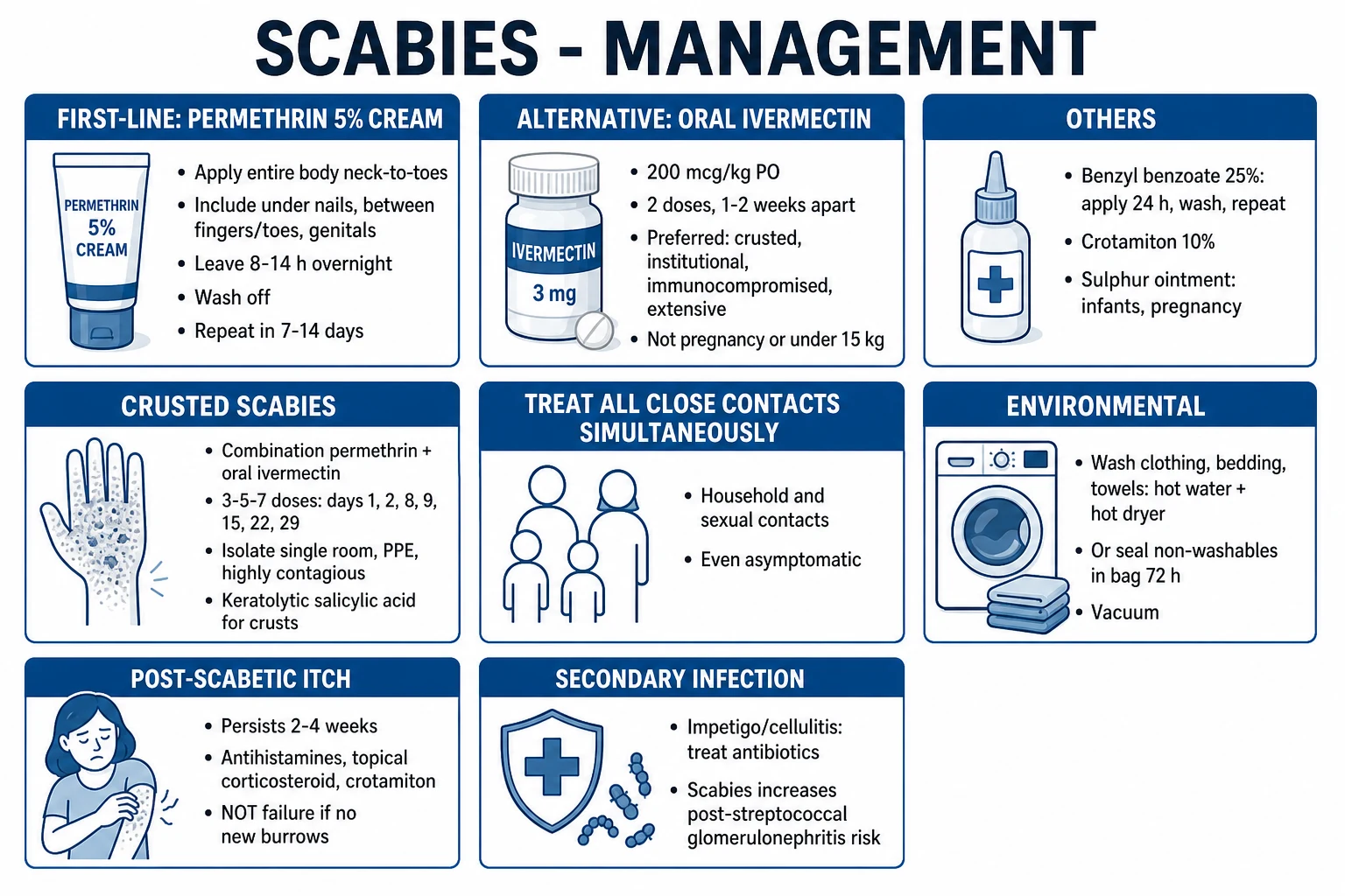
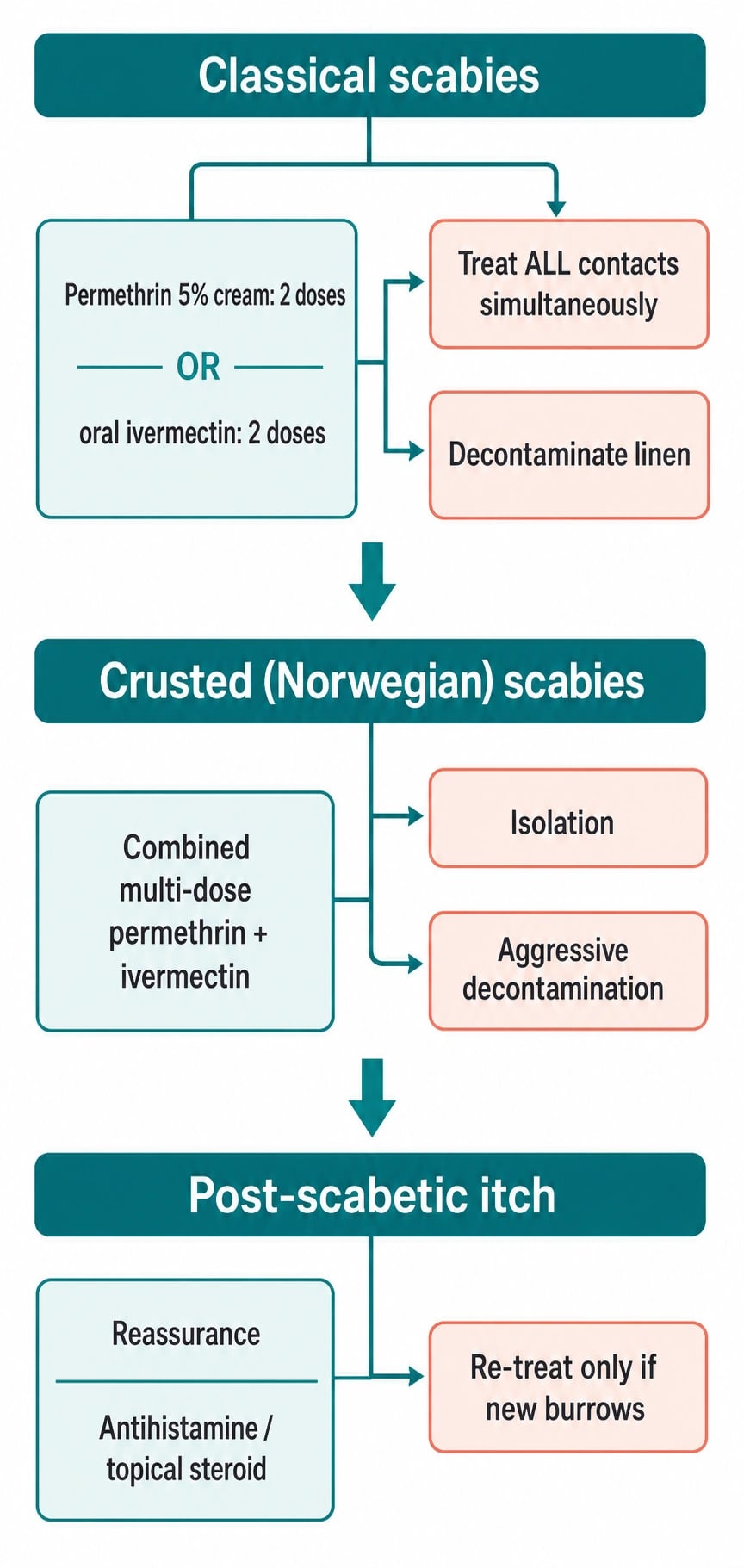
The two recommended first-line treatments are permethrin 5 percent cream and oral ivermectin 200 micrograms per kilogram. Permethrin 5 percent cream is applied to the entire body from the neck down to the soles of the feet, including the finger and toe webs, under the nails, the genitalia, the buttocks, and any skin folds. It is left on for 8 to 14 hours, usually overnight, and then washed off. The application should be repeated at 7 to 14 days. Permethrin is safe in children older than 2 months and in pregnancy, and it is generally well tolerated. Oral ivermectin is given as a single dose of 200 micrograms per kilogram, repeated at 7 to 14 days. It is particularly useful in outbreaks, crusted scabies, when topical therapy is impractical, or when adherence is uncertain. Ivermectin should be avoided in pregnancy and in children weighing less than 15 kilograms, depending on local guidance.[2][4][5][12]
Why the second dose is mandatory
Neither permethrin nor ivermectin reliably kills scabies eggs. The first dose eliminates adult and larval mites, but eggs deposited in burrows survive and hatch over the next 3 to 4 days. The second dose, given at 7 to 14 days, catches these newly hatched mites before they mature and reproduce. Failure to complete the second dose is a leading cause of apparent treatment failure. This principle is central to every scabies regimen and is a frequent examination point.[1][2][4]
Why do we repeat scabies treatment at day 7-14?
The repeat dose is not a precaution or a sign that the first treatment failed. It is pharmacologically necessary because permethrin and ivermectin are not reliably ovicidal. Eggs hatch after the first dose, and the second dose kills the larvae before they can mature and lay eggs of their own. Skipping the second dose is one of the commonest causes of persistent scabies.
Alternatives
When permethrin and ivermectin are unavailable or contraindicated, several alternatives exist. Benzyl benzoate 25 percent lotion is effective but can be irritant; it is usually diluted in children and applied to the whole body for 24 hours before washing off. Precipitated sulphur 6 to 10 percent ointment is safe in pregnancy, lactation, and infants, but it is messy, has an unpleasant odour, and requires repeated application. Malathion 0.5 percent lotion is applied for 8 to 12 hours and repeated after 7 days. Crotamiton 10 percent is less effective than permethrin but can help with residual pruritus. Lindane is no longer recommended because of neurotoxicity and the availability of safer alternatives. The choice of alternative depends on availability, cost, patient age, pregnancy status, and local resistance patterns.[2][13][14]
Treatment of contacts
All household members, close physical contacts, and sexual partners must be treated at the same time as the index case, even if they are asymptomatic. Many contacts are in the incubation period and will become symptomatic only after they have re-infested the treated index case. This phenomenon of "ping-pong" re-infestation is avoided by simultaneous treatment. In institutional outbreaks, the entire affected unit or dormitory may require mass treatment. The definition of a contact depends on the setting: household members, bed partners, carers, and anyone with prolonged skin-to-skin contact are considered exposed.[1][2][4]
Environmental decontamination
Mites survive only 24 to 72 hours away from human skin, so decontamination can be limited to items used in the previous 72 hours. Bedding, clothing, and towels should be washed in hot water at 50 degrees Celsius or higher and then tumble-dried on a hot cycle. Items that cannot be washed can be sealed in a plastic bag for 7 days at room temperature, after which the mites will have desiccated. Vacuuming of mattresses, carpets, and upholstered furniture is reasonable. In crusted scabies, environmental contamination is far more significant, and aggressive cleaning of the patient's room and belongings is essential.[1][2][4]
Treatment of crusted scabies
Crusted scabies requires combined, multi-dose therapy. Topical permethrin is applied daily or every 2 to 3 days for 7 days, then twice weekly until the patient is cured. Oral ivermectin is given at 200 micrograms per kilogram on days 1, 2, 8, 9, 15, 22, and 29, for a total of 7 doses in the first month. Keratolytics such as salicylic acid 5 to 10 percent or urea 10 to 40 percent are applied to the crusts to improve penetration of the scabicide. The patient should be isolated under contact precautions until the mite burden is substantially reduced. Close contacts and carers must be treated simultaneously. The duration of therapy is dictated by clinical response, and relapses are common, so prolonged follow-up is needed.[9][10][5]
Adjunctive symptomatic therapy
Itch is often the dominant symptom. Oral antihistamines, especially sedating agents such as hydroxyzine at night, can help sleep. Emollients and barrier creams repair the damaged stratum corneum and reduce irritation. Topical corticosteroids of mild to moderate potency can be used for post-scabetic eczematisation and nodular scabies after the mite has been eradicated. In nodular scabies, potent topical corticosteroids or intralesional triamcinolone may be required. Antibiotics are indicated for secondary bacterial infection, and the choice should be guided by local resistance patterns; anti-streptococcal coverage is important when impetigo is present.[2][4][12]
10. Specific subtypes and clinical scenarios
Crusted scabies in immunocompromise
Crusted scabies is most strongly associated with immunosuppression. Risk groups include patients with untreated HIV infection, solid-organ or haematopoietic transplant recipients, patients with HTLV-1 infection, those with leukaemia or lymphoma, and individuals receiving long-term systemic corticosteroids or biologics. Down syndrome, leprosy, severe intellectual disability, and advanced age are also recognised risk factors. The impaired T-cell response permits uncontrolled mite proliferation, and the resulting hyperkeratotic crusts harbour millions of mites. Management requires early recognition, isolation, combined topical and oral therapy, and treatment of all contacts. HIV testing should be offered to any adult with crusted scabies and no obvious risk factor, because the presentation may be the first clue to underlying immunosuppression.[9][10][1]
Infantile scabies
Infants present with a more widespread eruption than adults, with involvement of the palms, soles, head, neck, and face. Vesicles and pustules are common, and the child may be irritable and feed poorly. Family members are usually infested, and the history of nocturnal pruritus in parents or siblings is a valuable clue. Permethrin 5 percent cream is safe in infants older than 2 months. For younger infants, sulphur ointment or cautious permethrin use according to local guidance is appropriate. All family members must be treated at the same time, and clothing, bedding, and soft toys should be decontaminated.[1][12]
Bullous scabies
Bullous scabies should be considered in older adults with pruritic blisters on the hands and feet. The key differential is bullous pemphigoid. Direct immunofluorescence and dermoscopy are helpful. A positive skin scraping for scabies or a therapeutic trial of scabicidal therapy can confirm the diagnosis. Treatment follows standard scabies protocols, with care to avoid ivermectin in those with low body weight or contraindications.[1][12]
Nodular scabies
Nodular scabies can persist for months after successful mite eradication. The nodules represent a persistent cell-mediated hypersensitivity reaction and are not evidence of active infestation unless new burrows appear. Treatment is symptomatic, with potent topical corticosteroids, intralesional corticosteroids, or topical calcineurin inhibitors. It is important to reassure the patient that persistence of nodules does not mean treatment failure, provided the mite has been eliminated.[1][12]
Scabies incognito
Scabies incognito occurs when the inflammatory response is suppressed by topical or systemic corticosteroids. The eruption is atypical, often truncal or facial, and burrows are absent. The patient may have been treated for eczema or psoriasis for weeks. The diagnosis is made by a careful history, the presence of affected contacts, and sometimes a skin scraping or therapeutic trial. The key is to stop the corticosteroid and treat the scabies, while warning the patient that the rash may temporarily worsen as the immune response returns.[1][4]
Animal-transmitted scabies
Sarcoptic mange from dogs, cats, and other animals can cause a transient pruritic eruption in humans. The animal mite cannot reproduce on human skin, so the infestation is self-limiting and usually resolves within days to weeks. True burrows are not formed, and the eruption consists of pruritic papules or urticarial plaques on contact areas. The diagnosis is supported by a history of animal contact, and the management is treatment of the animal by a veterinarian. Human treatment is usually not necessary unless symptoms are severe.[1][12]
11. Complications and pitfalls
Secondary bacterial infection
The most common complication is secondary bacterial infection due to scratching. Staphylococcus aureus and group A Streptococcus cause impetigo, folliculitis, cellulitis, ecthyma, and furunculosis. In endemic paediatric populations, scabies-associated pyoderma is a major cause of morbidity and drives the need for mass drug administration programmes. The affected skin should be swabbed if infection is suspected, and systemic antibiotics with anti-staphylococcal and anti-streptococcal activity should be prescribed.[1][6][14]
Post-streptococcal glomerulonephritis
Acute post-streptococcal glomerulonephritis can develop 1 to 3 weeks after scabies-associated streptococcal skin infection. The presentation includes haematuria, proteinuria, oedema, hypertension, and renal impairment. It is one of the most serious complications of scabies in endemic settings and provides the rationale for community-wide scabies control. Anti-DNase B and antistreptolysin O titres may be elevated. Management is supportive, with blood-pressure control and renal monitoring; the prognosis in children is usually good, though chronic kidney disease can occur.[1][6][14]
Acute rheumatic fever and rheumatic heart disease
Although the link between streptococcal pharyngitis and acute rheumatic fever is stronger than the link with skin streptococci, scabies-associated pyoderma is nevertheless recognised as a contributing factor to rheumatic heart disease in high-burden settings. Control of scabies and skin infection is therefore a component of broader strategies to reduce the burden of cardiovascular disease in children in low-resource countries.[1][14]
Bacteraemia, sepsis, and necrotising infection
In crusted scabies, heavy bacterial colonisation of the crusts can lead to bacteraemia, sepsis, and, rarely, necrotising soft-tissue infection. These complications are most likely in immunocompromised hosts and in settings where diagnosis is delayed. Early recognition of crusted scabies, combined anti-scabetic and antibiotic therapy, and meticulous wound care reduce the risk.[9][10]
Causes of treatment failure
Treatment failure can result from poor adherence, incorrect application of topical therapy, failure to treat all contacts simultaneously, fomite re-infestation, permethrin resistance, misdiagnosis, premature cessation of therapy, or failure to give the second dose. The rise of permethrin resistance in some endemic regions has increased the need for ivermectin-based regimens or combination therapy. A systematic review of treatment failure has identified adherence and contact tracing as modifiable determinants of success. A structured approach to apparent failure includes checking adherence, confirming that all contacts were treated, decontaminating the environment, repeating the regimen, and rotating to oral ivermectin or combination therapy if resistance is suspected.[8][1][7]
[1]12. Prognosis and disposition
Classical scabies carries an excellent prognosis when the correct treatment is applied correctly and all contacts are treated simultaneously. Cure is usually achieved with two doses of permethrin or ivermectin, combined with environmental decontamination. The most important counselling point is that pruritus can persist for 2 to 6 weeks after the mite has been eradicated. This post-scabetic itch reflects residual hypersensitivity and does not indicate ongoing infestation unless new burrows appear. Patients can be reassured that the itch will gradually settle, and symptomatic treatment with antihistamines, emollients, and mild topical corticosteroids is usually sufficient.[1][2][4]
Crusted scabies has a more guarded prognosis. Relapses are common, and patients may require repeated courses of combined therapy and prolonged skin care for hyperkeratosis and nail dystrophy. Follow-up is recommended at 2 to 4 weeks, and again at 6 weeks for crusted scabies, with further review if there is incomplete response. At discharge, all contacts should have been treated, linen and clothing decontaminated, and the patient given a written safety-net advice about post-scabetic itch and the criteria for re-treatment.[9][10][5]
Public-health notification is required in many jurisdictions for institutional outbreaks. Schools, care homes, hospitals, and prisons should have outbreak management plans that include early identification, coordinated mass treatment, and environmental decontamination. In some countries, scabies is a notifiable disease, or specific guidance from public-health agencies applies to outbreaks in closed settings.[1][14]
13. Special populations
Pregnancy and lactation
Permethrin 5 percent cream is the treatment of choice in pregnancy. It is poorly absorbed, has a long safety record, and is not associated with teratogenicity. Sulphur ointment is also considered safe. Oral ivermectin is generally avoided in pregnancy because of limited safety data and its category C status in many regulatory frameworks. During lactation, permethrin is safe, and ivermectin is secreted in breast milk in low amounts but should be used with caution. Benzyl benzoate should be avoided in neonates because of the risk of absorption and irritation.[2][4][5]
Infants and small children
Permethrin 5 percent cream is approved for use in infants older than 2 months in many countries. For infants under 2 months, sulphur 6 to 10 percent ointment is preferred because of its safety profile. Benzyl benzoate can be used if diluted appropriately. Oral ivermectin is often deferred until the child weighs at least 15 kilograms, though local guidelines vary. The child's entire body surface area, including the head and neck, should be treated, and close contacts should be treated simultaneously. Parental education on application technique and the need for a second dose is crucial.[2][5][12]
Elderly
Scabies should be considered in any elderly patient with unexplained pruritus, particularly in care homes. The presentation may be atypical, with minimal burrows and more generalised dryness. Crusted scabies is common in this group, and the diagnosis is often delayed until an outbreak is established. A low threshold for diagnostic scraping and treatment, combined with environmental control, is recommended. Drug interactions and renal function should be considered when prescribing systemic agents.[1][4]
Immunocompromised patients
Patients with HIV, transplant recipients, those with haematological malignancy, and those on biologics or long-term systemic corticosteroids are at high risk for crusted scabies. A high index of suspicion is required, and HIV testing should be offered when crusted scabies is diagnosed without a clear cause. These patients need combined, multi-dose therapy and prolonged follow-up. Prophylactic treatment of close contacts and carers is essential because of the high contagiousness of crusted scabies.[9][10][1]
Down syndrome and intellectual disability
Individuals with Down syndrome and severe intellectual or physical disability have an increased risk of crusted scabies, possibly due to immune dysregulation and institutional living. Outbreaks are common in residential facilities. Management requires a coordinated approach involving dermatologists, infection-control teams, carers, and families. Patient comfort, consent, and practical application technique must be addressed.[1][9][10]
Indigenous and remote populations
Scabies is highly prevalent in many Indigenous and remote communities. Mass drug administration with ivermectin or permethrin is a key control strategy and has been shown to reduce scabies and pyoderma prevalence. The World Health Organization and IACS have developed guidance for community-wide treatment, including criteria for starting and stopping mass drug administration. These programmes require community engagement, health-worker training, and integration with other skin-health initiatives.[1][14]
14. Evidence, guidelines, and regional practice
European guideline (EADV 2017)
The European Academy of Dermatology and Venereology guideline recommends permethrin 5 percent cream as first-line therapy for classical scabies, applied from neck to toe and repeated after 7 days. Oral ivermectin is recommended as an alternative first-line option, particularly for crusted scabies, outbreaks, and when topical therapy is impractical. The guideline also addresses contact treatment, environmental decontamination, and the management of special populations including pregnancy, children, and immunocompromised patients.[2]
American Academy of Dermatology guidance (AAD 2024)
American guidance similarly recommends permethrin 5 percent cream or oral ivermectin 200 micrograms per kilogram, with a repeat dose at 7 to 14 days. It emphasises the treatment of all close contacts, the use of combination therapy for crusted scabies, and the importance of recognising scabies in institutional and immunocompromised patients. The AAD has also highlighted the need to address emerging permethrin resistance through surveillance and combination approaches.[12][7]
Turkish and International practice guideline (Uzun 2024)
The 2024 International Journal of Dermatology guideline provides a comprehensive, practical algorithm for diagnosis and treatment. It grades crusted scabies severity and recommends combination therapy with daily topical permethrin plus multiple doses of ivermectin. It also provides detailed advice on special populations, resistance management, and the role of alternatives such as sulphur, benzyl benzoate, and crotamiton.[5]
2020 IACS diagnostic criteria
The IACS criteria provide a standardised framework for diagnosis in clinical and research settings. Confirmed scabies requires direct visualisation of the parasite or its products; clinical scabies uses a combination of typical lesions, typical distribution, and supportive findings; and suspected scabies covers atypical presentations with contact history. These criteria have improved comparability across studies and are used to define cases for public-health surveillance and clinical trials.[3][1]
Mass drug administration
Mass drug administration with ivermectin has been evaluated in several endemic settings, most notably the Skin Health Intervention Fiji Trial (SHIFT). These studies have shown that community-wide treatment reduces scabies prevalence and, importantly, the prevalence of impetigo and associated post-streptococcal complications. The WHO and IACS have outlined criteria for MDA implementation, including baseline prevalence thresholds and monitoring endpoints. MDA is now an accepted strategy in high-burden, remote, or closed settings.[1][14][6]
Treatment failure and resistance
A 2024 systematic review and meta-analysis of scabies treatment failure identified poor adherence, inadequate contact tracing, and fomite re-infestation as the most important modifiable factors. Permethrin resistance, mediated by mutations in the voltage-gated sodium channel, has been reported in several endemic regions and is a growing concern. Strategies to address resistance include ensuring correct application, rotating to oral ivermectin, using combination topical and oral therapy, and, in the future, moxidectin and other agents with longer half-lives. Moxidectin is under investigation and may allow single-dose treatment because of its prolonged presence in skin.[8][1][7]
Regional variation
In the United Kingdom, the British Association of Dermatologists and Public Health England recommend two doses of permethrin 1 week apart, with treatment of all household contacts. In Australia and New Zealand, coordinated mass treatment is standard for institutional outbreaks. In India, the Indian Council of Medical Research recommends permethrin for classical scabies and combined permethrin plus ivermectin for crusted or resistant cases, with MDA in highly endemic tribal areas. The core principles of correct application, simultaneous contact treatment, and repeat dosing are universal.[2][5][14]
15. Exam pearls and minutiae
Distribution of scabies: WAFGS-P
WAFGS-P
Flexor wrists, ulnar styloid, finger webs
Axillae, inframammary folds, nipples, periumbilical skin
Interdigital spaces, especially index-middle web
Scrotal nodules, penile shaft, groin folds
Periumbilical, waistline, buttocks
Head, neck, palms and soles commonly involved in infants
Treatment escalation: PPI
PPI
Apply neck-to-soles, leave 8-14 hours, repeat at 7-14 days
Add oral ivermectin 200 microgram/kg when topical is impractical, in outbreaks, or for resistance
Permethrin plus multi-dose ivermectin for crusted scabies
Contact tracing: ABCDE
ABCDE
Treat everyone who sleeps in the same bed
Intimate and sexual contacts regardless of symptoms
Carers, household members, prolonged skin-to-skin contacts
Everyone living in the same household
Same day treatment to prevent ping-pong re-infestation
Summary
Scabies is a parasitic skin infestation caused by Sarcoptes scabiei var. hominis, transmitted by prolonged skin-to-skin contact, and characterised by intense nocturnal pruritus and burrows in the finger web spaces, wrists, and genitalia. The clinical spectrum includes classical scabies, crusted (Norwegian) scabies, nodular scabies, bullous scabies, scabies incognito, and animal-transmitted variants. Diagnosis is usually clinical, supported by dermoscopy, skin scraping, and the 2020 IACS criteria. First-line therapy is permethrin 5 percent cream or oral ivermectin, each given in two doses 7 to 14 days apart, with simultaneous treatment of all contacts and environmental decontamination. Crusted scabies requires combined daily topical permethrin and multi-dose ivermectin, keratolytics, and isolation. Post-scabetic itch is common and does not indicate failure; treatment failure should prompt review of adherence, contacts, decontamination, and resistance. A structured, public-health-minded approach is essential for individual cure and community control. [1]
References
- [1]Fernando DD, Mounsey KE, Bernigaud C, et al. Scabies Nat Rev Dis Primers, 2024.PMID 39362885
- [2]Salavastru CM, Chosidow O, Boffa MJ, et al. European guideline for the management of scabies J Eur Acad Dermatol Venereol, 2017.PMID 28639722
- [3]Engelman D, Yoshizumi J, Hay RJ, et al. The 2020 International Alliance for the Control of Scabies Consensus Criteria for the Diagnosis of Scabies Br J Dermatol, 2020.PMID 32034956
- [4]Sunderkötter C, Wohlrab J, Hamm H. Scabies: Epidemiology, Diagnosis, and Treatment Dtsch Arztebl Int, 2021.PMID 34615594
- [5]Uzun S, Durdu M, Yürekli A, et al. Clinical practice guidelines for the diagnosis and treatment of scabies Int J Dermatol, 2024.PMID 38922701
- [6]Heukelbach J, Feldmeier H. Scabies Lancet, 2006.PMID 16731272
- [7]Tavoletti G, Avallone G, Sechi A, et al. Scabies: An updated review from epidemiology to current controversies and future perspectives Travel Med Infect Dis, 2025.PMID 40754226
- [8]Mbuagbaw L, Sadeghirad B, Morgan RL, et al. Failure of scabies treatment: a systematic review and meta-analysis Br J Dermatol, 2024.PMID 37625798
- [9]Sun YJ, Gong HB. Crusted Scabies N Engl J Med, 2025.PMID 40768718
- [10]Selvaraj R, Purushothaman S, Indra Couppoussamy K. Crusted scabies Indian J Dermatol Venereol Leprol, 2023.PMID 37317720
- [11]Kolar KA, Rapini RP. Crusted (Norwegian) scabies Am Fam Physician, 1991.PMID 1718155
- [12]Thomas C, Coates SJ, Engelman D, et al. Ectoparasites: Scabies J Am Acad Dermatol, 2020.PMID 31310840
- [13]Ertugrul G, Aktas H. Comparison of sulfur ointment and permethrin treatments in scabies Dermatol Ther, 2022.PMID 36193643
- [14]Widaty S, Miranda E, Cornain EF, et al. Scabies: update on treatment and efforts for prevention and control in highly endemic settings J Infect Dev Ctries, 2022.PMID 35298417