Dermatology · Medicine
Sclerotherapy for vascular lesions
Also known as Injection sclerotherapy · Foam sclerotherapy · Polidocanol sclerotherapy · STS sclerotherapy · Sclerosant therapy veins
Board-level procedural leaf on sclerotherapy for dermatologic vascular targets: telangiectasia, reticular veins, selected tributaries after reflux assessment, and specialist foam use in low-flow venous malformations. Covers detergent vs osmotic sclerosants, liquid vs foam, contraindications including pregnancy, hyperpigmentation and ulceration, ultrasound when treating larger veins, and comparison with vascular laser adjuncts.
On this page & tools
Your progress
Saved locally on this device.
Exam tags
Red flags
Definition & Scope
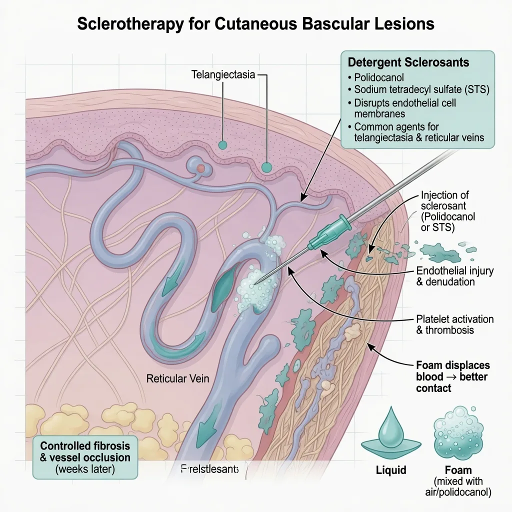
Sclerotherapy is the percutaneous injection of a chemical agent into a vascular channel to produce endothelial injury and durable occlusion.[1][4] In dermatology and aesthetic phlebology the usual targets are spider telangiectasias, reticular veins, and selected small tributaries after appropriate venous assessment; foam sclerotherapy also has a role for low-flow venous malformations in specialist centres.[3][6][8]
This leaf does not replace full chronic venous disease (CVD) pathways for truncal saphenous reflux — ESVS CVD guidelines place injection therapy within a broader diagnostic and treatment hierarchy.[9]
Indications by Target
| Target (teaching) | Typical setting | Key prerequisite |
|---|---|---|
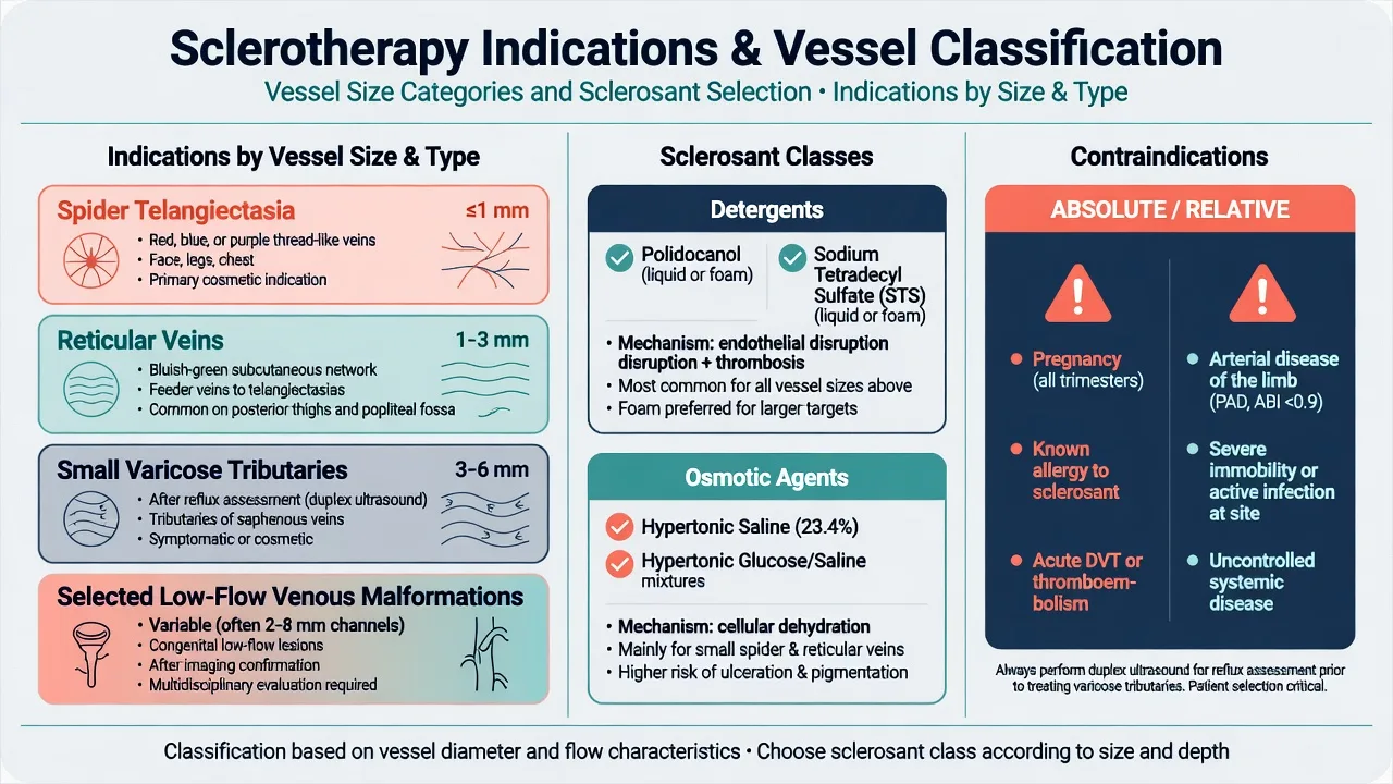
| Spider telangiectasia | Cosmetic dermatology/phlebology | Realistic multi-session consent |
| Reticular veins | Often feed spiders | Map feeders; consider combination liquid/foam strategies |
| Small varicose tributaries | After reflux work-up | Duplex; treat axial reflux first when indicated |
| Low-flow venous malformation | Multidisciplinary / IR-vascular | Imaging; not high-flow AVM |
Combination foam for reticular plus liquid for telangiectatic channels is a practical pattern described in contemporary dermatologic surgery series.[3]
Sclerosant Classes
Comparative reviews group agents by mechanism:[1]
| Class | Examples (teaching) | Mechanism sketch | Practical note |
|---|---|---|---|
| Detergents | Polidocanol, STS | Endothelial membrane injury | Workhorses for most cosmetic veins |
| Osmotic | Hypertonic saline / glucose systems | Cellular dehydration | Pain and ulcer risk if extravasated |
| Chemical irritants | Historical agents | Direct irritation | Mostly of historical board interest |
Liquid vs foam: foam displaces blood and increases wall contact for larger channels; liquid is often preferred for the finest telangiectasias.[1][3][4] Exact concentration/volume algorithms are training- and product-label-dependent — exams test principles, not memorised uncited millilitre tables.
Liquid sclerosant
- Often chosen for finest telangiectasias
- Less blood displacement than foam
- Familiar office technique for spiders
Foam sclerosant
- Displaces blood → more wall contact
- Useful for reticular / larger channels
- Own risk profile (counsel systemic rare events)
Pathophysiology of Benefit & Harm
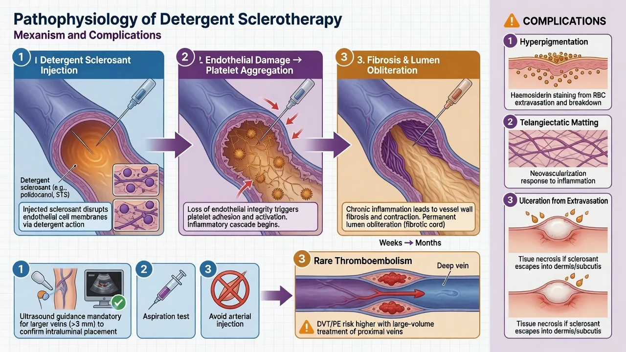
Intended sequence: endothelial denudation → platelet activation / local thrombosis → organisation and fibrosis → lumen obliteration over weeks.[1][4]
Common adverse pathways: [1]
- Hyperpigmentation — haemosiderin staining after polidocanol and other agents; systematic review synthesises risk context.[2]
- Telangiectatic matting — fine red blush near treated areas
- Ulceration — usually extravasation or arteriolar injury
- Thrombophlebitis / trapped coagulum — local discomfort; may need micro-evacuation in practice
- Systemic rare events — visual, neurologic, or thromboembolic concerns summarised in safety reviews, especially relevant to foam technique and patient selection.[7]
Pre-Procedure Assessment
- Goals — cosmetic vs symptomatic CVD (heaviness, swelling, eczema, ulcer risk).[8][9]
- History — prior DVT/PE, thrombophilia, arterial disease, migraine with aura (foam caution in many protocols), medications, pregnancy/breastfeeding, allergy to sclerosants.
- Exam — distribution, feeder reticulars, skin quality, arterial pulses/ABI if indicated.
- Duplex ultrasound — when varicose tributaries, asymmetry, or CVD symptoms suggest saphenous or perforator reflux.[8][9]
- Photography and written consent listing staining, matting, ulcer, need for multiple sessions, and recurrence.
Exam application bank (NEET-PG / INICET)
One-line answer
Board-level procedural leaf on sclerotherapy for dermatologic vascular targets: telangiectasia, reticular veins, selected tributaries after reflux assessment, and specialist foam use in low-flow venous malformations. Covers detergent vs osmotic sclerosants, liquid vs foam, contraindications including pregnancy, hyperpigmentation and ulceration, ultrasound when treating larger veins, and comparison with vascular laser adjuncts.
Worked stems (answer without another resource)
Stem 1 — Classic presentation. Map symptoms to mechanism; name the first investigation and first treatment step with dose/route if drug therapy is standard. [1]
Stem 2 — Unstable / complicated. List red flags that force immediate resuscitation, theatre, ICU, antidote, or reperfusion — and what you do in the first 15 minutes. [1]
Stem 3 — Atypical group. Elderly, pregnancy, child, or immunocompromised: how presentation and thresholds change. [1]
Stem 4 — Differential trap. Name the three closest mimics and one discriminator for each. [1]
Stem 5 — Disposition. Who goes home with safety-netting, who is admitted, who needs HDU/ICU/theatre, and what follow-up is mandatory. [1]
Rapid viva checklist
- Definition + classification
- Pathophysiology chain
- Bedside signs / criteria
- Score with exact components (if any)
- Emergency bundle
- Definitive therapy with doses
- Complications of disease and of treatment
- Special populations
- Guideline/trial name if classic
- Three exam traps
Coverage self-check
If you cannot answer any stem above from this page alone, re-read the matching section — the page is intended to be self-sufficient for final-prof and NEET-PG/INICET questions on Sclerotherapy for vascular lesions.
Expanded exam teaching (depth pass)
Clinical reasoning
For Sclerotherapy for vascular lesions, examiners test whether you can prioritise life threats, choose the right first test, and give specific therapy (agent, dose, route, timing). Generic phrases without numbers score poorly.
Mechanism → feature map
Build a short chain: cause → pathophysiologic intermediate → clinical feature → complication. Every major symptom in the classic vignette should sit on that chain.
Investigation strategy
- Bedside/first-line tests that change immediate management
- Confirmatory or staging tests
- What a normal result does not exclude
- When not to delay treatment for imaging (unstable patient)
Management ladder
- Resuscitation / ABC / sepsis or haemorrhage bundle as relevant
- Specific antidote / procedure / antimicrobial / reperfusion / surgery
- Supportive care and monitoring targets
- Definitive long-term therapy and secondary prevention
- Disposition and safety-net advice
Special populations
Always prepare one line each for children, pregnancy, elderly, renal/hepatic impairment, and immunocompromised patients when the topic allows.
Pitfalls that fail candidates
- Treating the number not the patient
- Missing pregnancy status when relevant
- Imaging before stabilisation
- Wrong empiric cover or wrong antidote timing
- Incomplete counselling on recurrence, adherence, or red-flag return
Board-level procedural leaf on sclerotherapy for dermatologic vascular targets: telangiectasia, reticular veins, selected tributaries after reflux assessment, and specialist foam use in low-flow venous malformations. Covers detergent vs osmotic sclerosants, liquid vs foam, contraindications including pregnancy, hyperpigmentation and ulceration, ultrasound when treating larger veins, and comparison with vascular laser adjuncts. [1]
Structured revision sheet
Must-know numbers and names
List every score, size threshold, dose, and time window from this topic on a blank page from memory, then check against the sections above.
Three classic MCQ angles
- Most likely diagnosis given a vignette
- Next best step in management
- Most appropriate investigation
Three classic SAQ angles
- Pathophysiology in five steps
- Management algorithm with doses
- Complications and prevention
Clinical station flow
Greet → focused history → targeted exam → investigations → explain diagnosis → emergency care → definitive plan → safety-net / follow-up → answer examiner questions on mechanism and pitfalls.
Technique Principles (Office Teaching Level)
Practical sclerotherapy teaching emphasises vessel selection, gentle precise cannulation, appropriate agent form, and compression/aftercare rather than heroic volumes.[4]
- Treat feeders thoughtfully so spiders are less likely to refill.
- Use the lowest effective concentration for the vessel diameter (principle).
- Aspirate / confirm intravascular position before injecting; stop if blanching pattern suggests extravasation risk.
- Compression after lower-limb sessions per local protocol.
- Stage sessions; counsel that clearance is often incomplete after one visit.[3][4]
Contraindications (Exam List)
| Absolute / strong relative | Why |
|---|---|
| Pregnancy | Fetal/maternal risk framing; elective delay[5] |
| Known allergy to planned sclerosant | Anaphylaxis risk |
| Acute DVT / PE | Thrombotic disease active |
| Critical arterial disease of the limb | Ischaemic ulcer risk |
| Local infection at site | Spread / poor healing |
| Inability to ambulate / high VTE risk without mitigation | Safety reviews emphasise selection[7] |
Comparators: Laser, Combination, Surgery
Randomised and comparative work shows foam sclerotherapy and Nd:YAG are both used for leg telangiectasia, with different comfort and clearance trade-offs depending on vessel and study design.[10][11] CLaCS (cryo-laser cryo-sclerotherapy concepts / laser combined with injection) is an evolving combination strategy studied in randomised settings for reticular veins and telangiectasias.[12]
Refer or prioritise ablation/surgery when duplex shows significant truncal reflux or advanced CVD — injection of spiders alone will not fix axial disease.[8][9]
Low-Flow Venous Malformations
Foam sclerotherapy can reduce bulk and symptoms of low-flow VMs in children and adults when performed in experienced multidisciplinary programmes with appropriate imaging and airway/compartment awareness.[6] High-flow arteriovenous malformations are not routine office foam targets.
Aftercare & Follow-Up
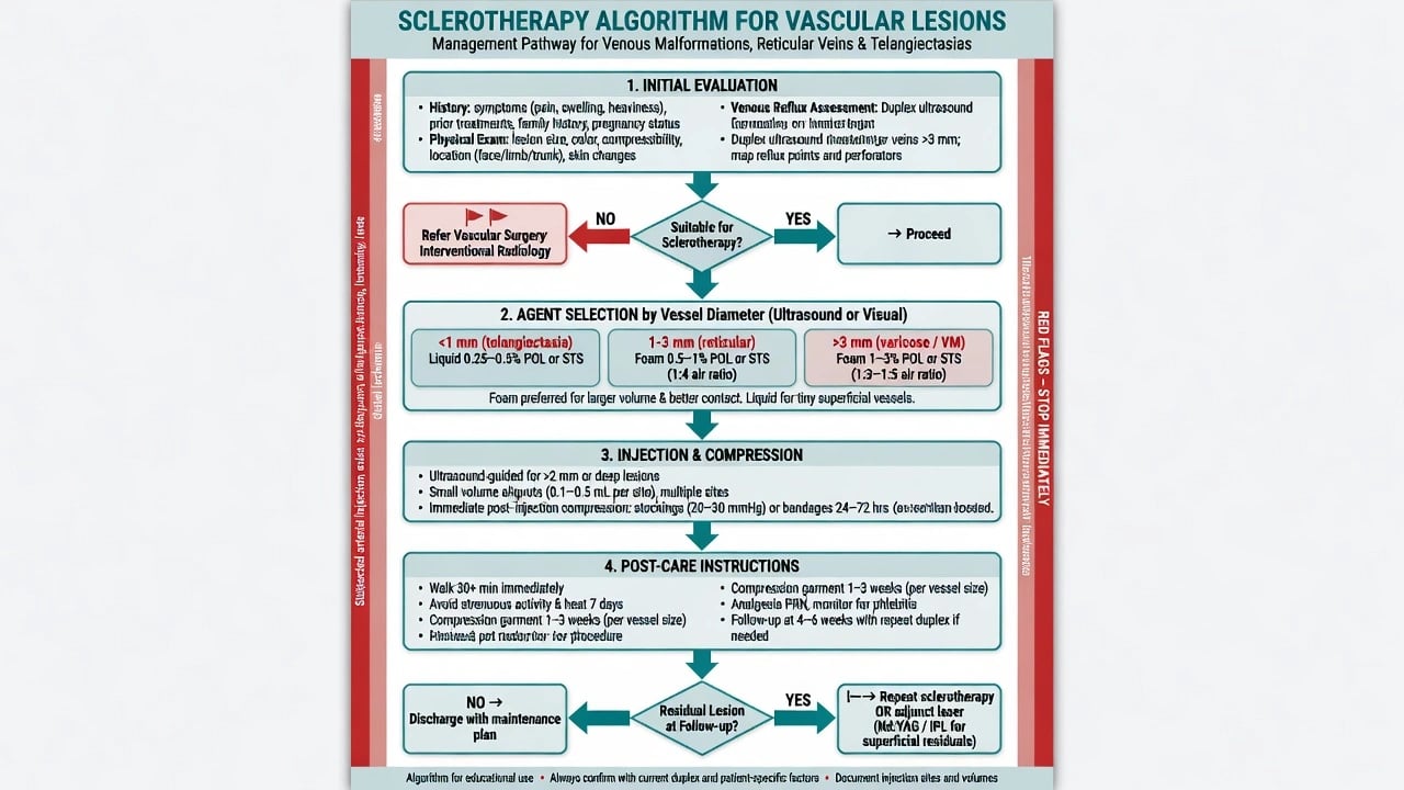
- Expect bruising and firm cords; staining may take months to fade.[2]
- Sun protection on treated legs reduces pigment emphasis in counselling.
- Review for residual feeders; plan retreatments at safe intervals.
- Document complications honestly and adjust technique.
Regional & Exam Notes
MBBS/NEET-PG: mechanism, common agents, pregnancy contraindication, pigmentation/ulcer. MRCP: CVD assessment hierarchy. IADVL/FACD/FRCDerm/ABD: foam vs liquid, matting, polidocanol staining evidence, when to image, VM vs cosmetic spider distinction. [1]
Bottom Line
Sclerotherapy is targeted chemical endovenous ablation for the right vessel in the right patient: screen pregnancy and thrombosis risk, image when reflux is plausible, match detergent/foam strategy to diameter, counsel staining and ulcer risk, and refer axial venous disease beyond cosmetic spiders.[1][2][7][9]
Exam anchors
References
- [1]Duffy DM. Sclerosants: a comparative review Dermatol Surg, 2010.PMID 20590708
- [2]Bossart S, Daneluzzi C, Cazzaniga S, et al. Skin hyperpigmentation after sclerotherapy with polidocanol: A systematic review J Eur Acad Dermatol Venereol, 2023.PMID 36196455
- [3]Verma KK, Friedmann DP. Combination Foam and Liquid Sclerotherapy for Lower Extremity Reticular and Telangiectatic Veins: A Single-Center Retrospective Study Dermatol Surg, 2024.PMID 38860828
- [4]Stücker M, Dörler M. [Practical Aspects of Sclerotherapy] Wien Med Wochenschr, 2016.PMID 27379853
- [5]Reich-Schupke S, Leiste A, Moritz R, et al. Sclerotherapy in an undetected pregnancy - a catastrophe? Vasa, 2012.PMID 22825857
- [6]Markovic JN, Nag U, Shortell CK. Safety and efficacy of foam sclerotherapy for treatment of low-flow vascular malformations in children J Vasc Surg Venous Lymphat Disord, 2020.PMID 32284312
- [7]Yiannakopoulou E. Safety Concerns for Sclerotherapy of Telangiectases, Reticular and Varicose Veins Pharmacology, 2016.PMID 27104778
- [8]Hamdan A. Management of varicose veins and venous insufficiency JAMA, 2012.PMID 23268520
- [9]Wittens C, Davies AH, Bækgaard N, et al. Editor's Choice - Management of Chronic Venous Disease: Clinical Practice Guidelines of the European Society for Vascular Surgery (ESVS) Eur J Vasc Endovasc Surg, 2015.PMID 25920631
- [10]Parlar B, Blazek C, Cazzaniga S, et al. Treatment of lower extremity telangiectasias in women by foam sclerotherapy vs. Nd:YAG laser: a prospective, comparative, randomized, open-label trial J Eur Acad Dermatol Venereol, 2015.PMID 25069999
- [11]Ianosi G, Ianosi S, Calbureanu-Popescu MX, et al. Comparative study in leg telangiectasias treatment with Nd:YAG laser and sclerotherapy Exp Ther Med, 2019.PMID 30679981
- [12]Fonseca MM, Mocelin FJ, Grill MH, et al. Nd:Yag laser combined with injection sclerotherapy in the treatment of reticular veins and telangiectasias (CLaCS method): A triple-blind randomized clinical trial comparing two sclerosing agents associated with same laser patterns Phlebology, 2023.PMID 36657386