Dermatology · Medicine
Seborrhoeic Dermatitis
Also known as Seborrheic dermatitis · Seborrhoeic eczema · Dandruff · Pityriasis capitis · Cradle cap
Seborrhoeic dermatitis is a chronic, relapsing inflammatory dermatosis of sebaceous-rich skin driven by interplay between Malassezia yeast, host immune responses, and epidermal barrier dysfunction. Fellowship-level assessment requires mastery of the bimodal age distribution (infantile cradle cap at 3 months and adult peak 20-40 years), scalp and facial morphology, the Malassezia-host immune axis, differential diagnosis from psoriasis and tinea capitis, the topical antifungal and anti-inflammatory ladder (ketoconazole, ciclopirox, calcineurin inhibitors, roflumilast 0.3% foam), site-specific therapy, systemic itraconazole pulse for refractory disease, and recognition of HIV, Parkinson disease, and infantile immunodeficiency associations.
On this page & tools
Your progress
Saved locally on this device.
Exam tags
Red flags
Definition & Classification
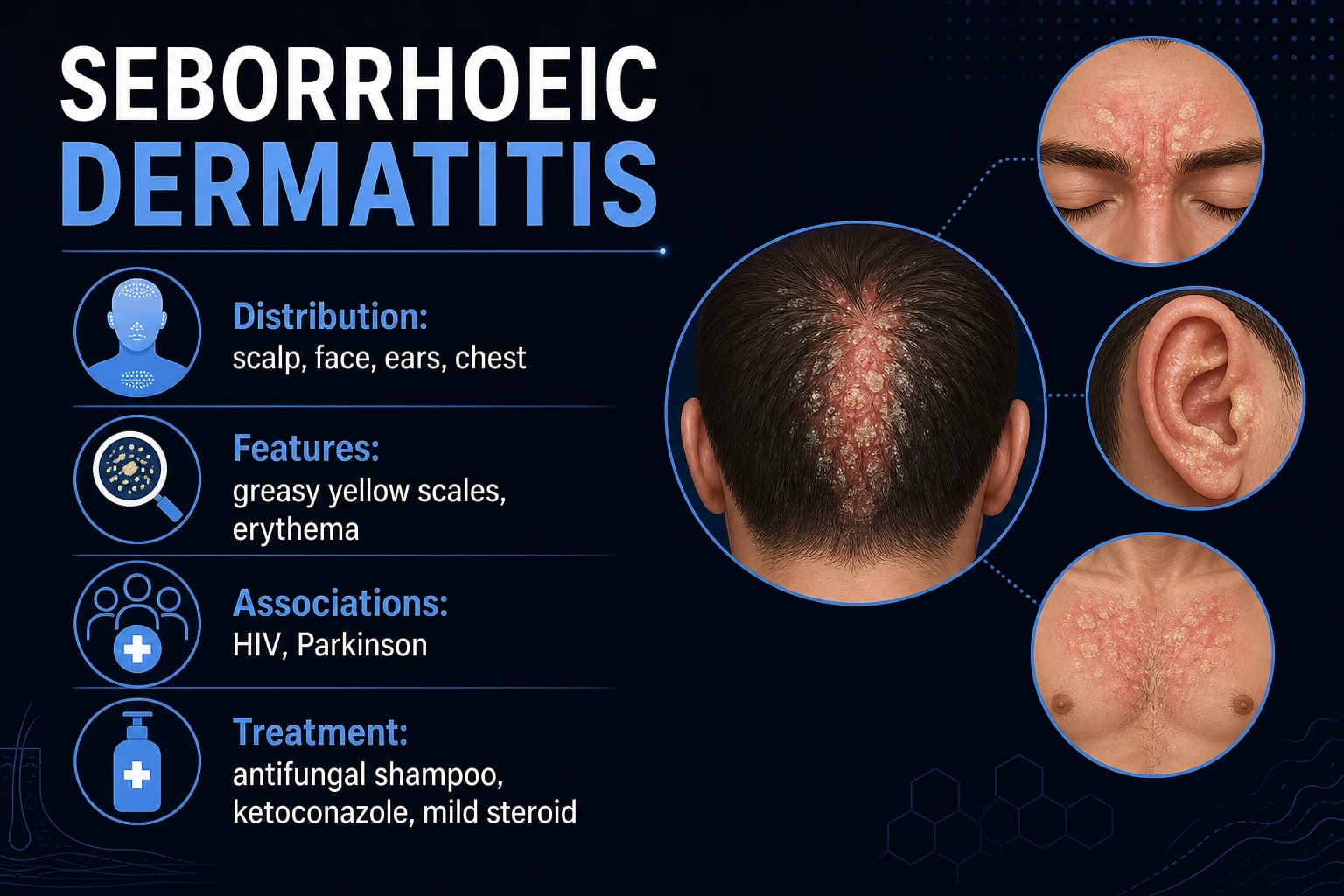
Seborrhoeic dermatitis is a common, chronic, relapsing inflammatory skin disease characterised by erythema with greasy, yellowish scale in areas with a high density of sebaceous glands. It sits on a clinical spectrum that includes pityriasis capitis (dandruff) at the mild end and erythroderma at the severe end, with infantile cradle cap as a distinct early-life phenotype.[1][2][9]
The disease is classified primarily by age of onset and distribution:[1][2][20][21]
MALAS
GREASY
- Fine, white-to-grey, non-inflammatory scalp scale
- No erythema; itch variable
- Mild end of the seborrhoeic spectrum
- M. restricta and M. globosa driven
- Erythema + greasy yellow scale
- Scalp, face, retro-auricular, trunk
- Chronic, relapsing, fluctuating
- Same Malassezia axis as dandruff
- First 3 months; vertex/anterior scalp
- Thick, yellow, adherent, non-itchy
- Often self-limiting by 6-12 months
- Red flag: failure to thrive → immunodeficiency
Epidemiology & Risk Factors
A 2024 systematic review and meta-analysis estimated a global pooled prevalence of 4.38% (95% CI 3.58-5.17%), with higher rates in adults (5.64%) than children (3.70%) and very low neonatal prevalence (0.23%).[3] Most clinical series report an adult prevalence in the 1-3% range, with dandruff (pityriasis capitis) affecting up to 50% of the adult population at some point. The condition is bimodal, peaking in infancy and again after puberty, with a second peak in the sixth decade.[2]
Risk factors and clinical associations [1]
- Immunodeficiency — HIV/AIDS is the classic association; seborrhoeic dermatitis may be severe, widespread, and refractory, and can be the presenting feature of undiagnosed HIV.[8]
- Neurological disease — Parkinson disease (prevalence up to 60%), stroke, head injury, epilepsy, spinal cord injury, facial nerve palsy, and severe depression. In Parkinson disease, severity may parallel motor decline.[7]
- Iatrogenic immunosuppression — organ transplant recipients, long-term systemic corticosteroids, biologic therapy, chemotherapy.
- Psychological stress, fatigue, and sleep deprivation — commonly exacerbate disease and trigger flares.
- Seasonal variation — often worse in winter and in low-humidity environments; improves in summer with UV exposure.
- Genetic susceptibility — familial clustering is described; polymorphisms affecting lipid metabolism and innate immunity may explain variable disease expression.[4]
- Infant-specific factors — maternal androgen stimulation of fetal sebaceous glands; rare associations with Leiner disease, Netherton syndrome, severe combined immunodeficiency, Omenn syndrome, and hyper-IgE syndrome.[21]
- Prevalence 30-40%
- Severe, widespread, refractory
- May be the presenting feature
- Optimise ART plus aggressive skin therapy
- Prevalence 19-60%
- Facial predilection, blepharitis
- Severity may track motor decline
- Autonomic + sebaceous dysfunction
- Transplant, biologics, chemotherapy
- Often under-recognised
- Reduce immunosuppression if possible
- Topical antifungal first-line
Pathophysiology
The pathogenesis is multifactorial and increasingly understood as a dysregulated host-microbe interaction rather than simple yeast overgrowth. The classical "Malassezia is necessary but not sufficient" model explains why most adults carry the yeast as a commensal yet only a minority develop disease.[4][5][6]
The Malassezia axis
The three species most commonly implicated are M. restricta, M. globosa, and M. furfur (the last is the classic cause of pityriasis versicolor). All are lipophilic, dimorphic yeasts that colonise human skin as commensals from infancy onward, with density highest in sebaceous-rich sites.[4][5][6]
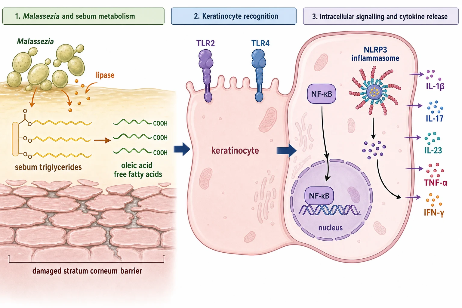
- Lipase activity — Malassezia secrete lipases that hydrolyse sebum triglycerides into free fatty acids, including oleic acid. Oleic acid penetrates the stratum corneum, disrupts barrier lipids, and acts as a danger signal in susceptible individuals.
- Hyphal transformation — the yeast-to-hyphae switch is associated with disease activity and inflammatory cytokine release.
- Pigments — Malassezia produce pityriacitrin (UV-protective) and other indole pigments, contributing to the hypopigmentation and hyperpigmentation of pityriasis versicolor.
- Cell wall components — β-glucans and lipoproteins activate keratinocyte TLR2/TLR4 and downstream NF-κB signalling. [1]
Immune dysregulation
Lesional skin shows upregulation of TLR2/TLR4-NF-κB signalling, NLRP3 inflammasome activation, and release of IL-1β, IL-6, IL-8, IL-17, IL-23, TNF-α, and IFN-γ, reflecting a mixed Th1/Th17 response with innate immune amplification. Healthy carriers mount tolerance to Malassezia antigens; patients with seborrhoeic dermatitis show an aberrant Th1/Th17-skewed response, with reduced Treg function.[4][5]
Sebaceous activity and altered lipid composition
Sebaceous glands provide the triglyceride substrate that Malassezia metabolise. Androgen-driven sebum secretion peaks in infancy (maternal androgens), post-puberty, and again in the sixth decade — matching the bimodal epidemiology. Patients with seborrhoeic dermatitis show altered sebum composition, with increased free fatty acids, squalene, and cholesterol relative to controls, and decreased ceramides in the stratum corneum.[4]
Barrier dysfunction
Impaired epidermal barrier integrity — measured as increased transepidermal water loss (TEWL) — permits enhanced penetration of Malassezia products and sustains inflammation. Lipid metabolism and innate-immunity polymorphisms (e.g., CARD14, IL-36RN, DEFB1) may explain inter-individual variation in susceptibility.[4]
Neurogenic inflammation
Substance P is upregulated in lesional skin and is the leading mechanistic explanation for the strong association with Parkinson disease and other CNS disorders. Substance P is a tachykinin released by unmyelinated C-fibres; it promotes mast-cell degranulation, vasodilation, pruritus, and sebaceous secretion. In Parkinson disease, dopaminergic degeneration is accompanied by disinhibited substance P release, autonomic dysfunction, and altered sebum composition — a triple hit.[7]
Clinical Presentation
Adult disease
Adult seborrhoeic dermatitis is the commonest form and runs a chronic, relapsing course with seasonal fluctuation. The morphology is erythema with greasy yellow-white scale.[1][2]
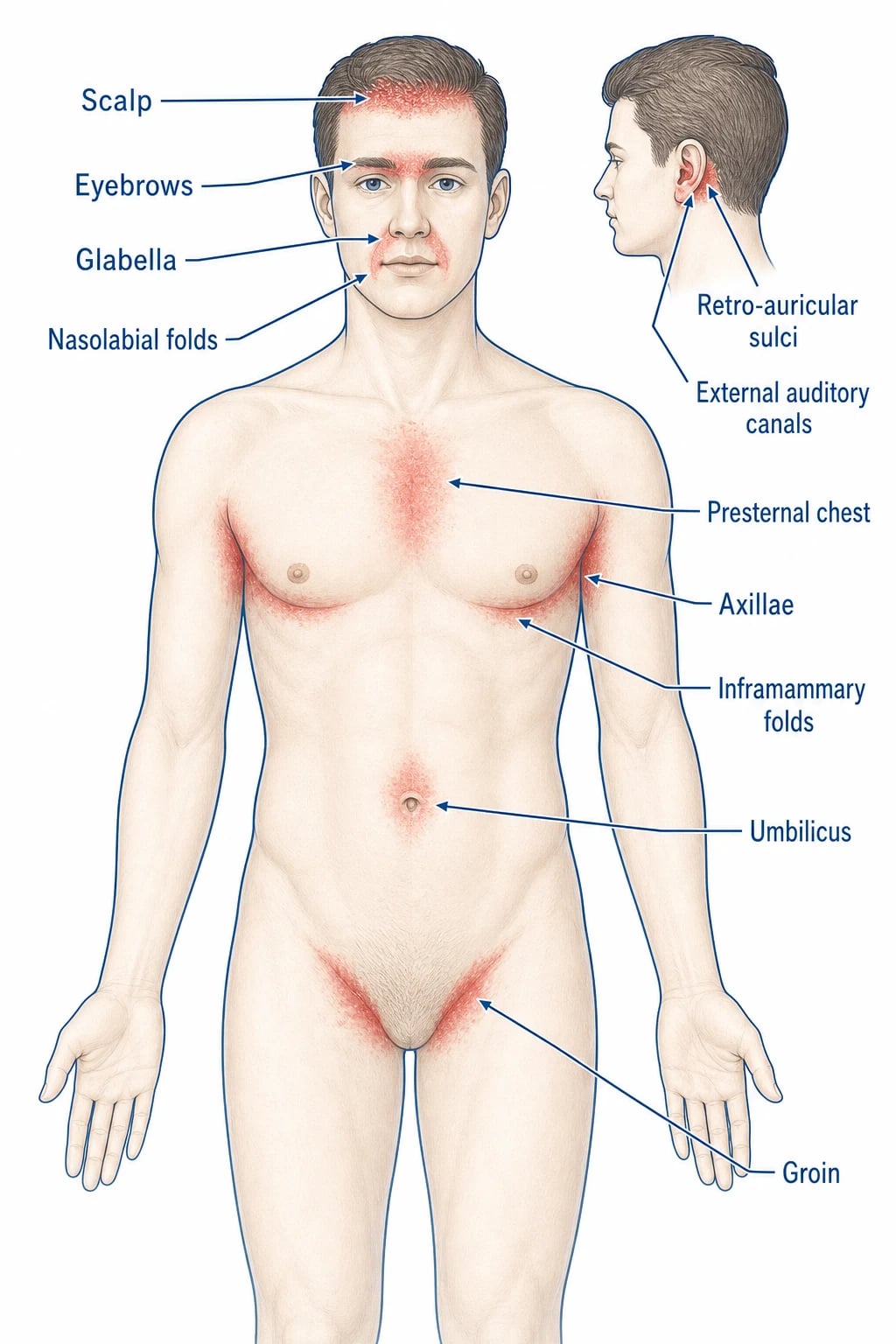
- Scalp — diffuse or patchy dandruff; fine white-to-yellow greasy scale; may be pruritic. Erythema may extend across the hairline to the forehead and post-auricular skin. Pityriasis sicca is a dry, scaly form without overt erythema.
- Face — salmon-pink erythema with yellowish scale in the eyebrows, glabella, and nasolabial folds; blepharitis with eyelid margin scaling and madarosis; retro-auricular fissuring and involvement of the external auditory canal (otitis externa-like picture).
- Trunk — well-demarcated erythematous patches with greasy scale over the presternal area and upper back; "petaloid" lesions are ring-shaped or flower-petal shaped plaques with a peripheral collarette of scale.
- Intertriginous sites — sharply marginated, beefy-red patches in axillae, inframammary folds, groin, and umbilicus, often with maceration and minimal scale.
- Seborrhoeic blepharitis — eyelid margin scaling, crusting, collarettes around lash bases, conjunctival injection, and symptomatic dry eye. [1]
Infantile disease
- Cradle cap — thick, yellow, greasy, adherent scale on the vertex and anterior scalp; may be associated with erythema but is usually non-itchy and the infant is comfortable.[21]
- Facial and flexural involvement is common; napkin-area involvement can mimic irritant dermatitis or candidiasis (look for satellite pustules in the latter).
- The infant is usually comfortable and thriving; systemic upset (failure to thrive, diarrhoea, recurrent infection) should prompt evaluation for immunodeficiency (LAD type 1, Netherton syndrome, severe combined immunodeficiency, Omenn syndrome, hyper-IgE syndrome, Leiner disease).[20][21]
HIV and erythrodermic phenotypes
In advanced HIV, seborrhoeic dermatitis may be severe, widespread, and refractory, sometimes progressing to erythroderma. The disease can be the presenting feature of undiagnosed HIV, and the severity often tracks the CD4 count.[8]
Parkinson-disease phenotype
Facial-predominant disease with prominent seborrhoeic blepharitis and retro-auricular involvement; severity may track motor decline and respond to optimisation of dopaminergic therapy.[7]
[1] [2] [7] [8] [21]Differential Diagnosis
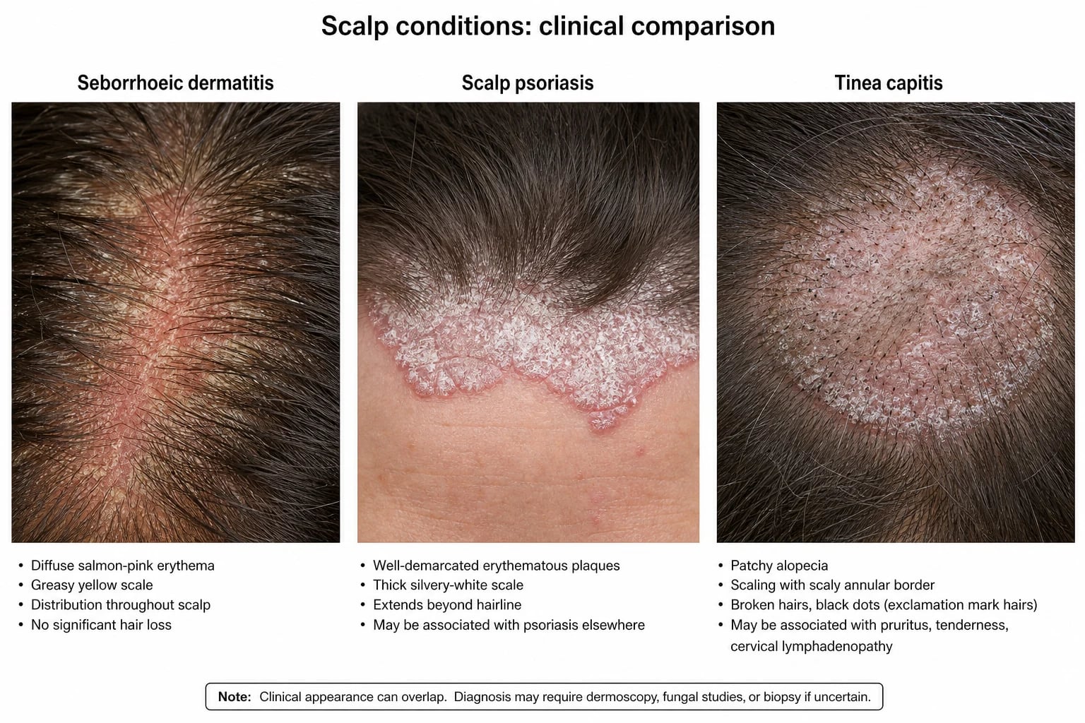
The differential diagnosis is site-specific. The four most-tested mimics at fellowship level are psoriasis, tinea capitis, atopic dermatitis, and rosacea. [1]
- Greasy yellow scale
- Sebaceous-rich sites: scalp, nLF, eyebrows
- M. globosa / M. restricta
- KOH: yeast and short hyphae ('spaghetti and meatballs')
- Thick, silvery, well-demarcated plaques
- Extends beyond hairline; Auspitz sign
- Nail pitting; family history
- KOH negative
- Patchy alopecia, broken hairs
- Kerion (boggy inflammatory mass)
- Cervical lymphadenopathy in children
- KOH/culture: septate hyphae
- Pruritus, xerosis, flexural predilection
- Spares nasolabial folds
- Personal/family atopy
- Personal/family history of asthma or hay fever
Clinical & Bedside Assessment
A focused history and examination is the cornerstone. The history should establish chronicity, seasonality, triggers, treatments already tried, HIV risk factors, and neurological symptoms. The examination should map the full distribution of disease — scalp, face, retro-auricular, external auditory canal, chest, back, intertriginous sites, and nails — and look for diagnostic clues (vermilion-border sparing, nail pitting, broken hairs, scarring).[1][2]
Dermoscopy
Dermoscopy of seborrhoeic dermatitis shows linear branching vessels in a yellowish background with follicular plugs and yellow scales — a pattern that helps distinguish it from psoriasis (regularly distributed dotted vessels on a light-red background) and from tinea (comma-shaped or corkscrew hairs).[9]
Investigations
Seborrhoeic dermatitis is usually a clinical diagnosis. Investigations are reserved for atypical, refractory, or diagnostically challenging cases.[1][2][9]
- Skin scrapings for KOH — to exclude dermatophyte infection (tinea capitis, tinea corporis) or to confirm Malassezia overgrowth (pityriasis versicolor). Malassezia KOH shows yeast clusters and short hyphae ("spaghetti and meatballs" or "bananas and grapes").[9]
- Fungal culture — when KOH is negative but clinical suspicion of tinea is high; allows species identification (e.g., Trichophyton tonsurans, Microsporum canis).
- Skin biopsy — when diagnosis is uncertain, the disease is refractory, or there is suspicion of CTCL, pemphigus, lupus, or Langerhans cell histiocytosis. Histology shows psoriasiform hyperplasia, follicular plugging, parakeratosis, spongiosis, and a superficial perivascular lymphocytic infiltrate; fungal stains (PAS) may show Malassezia yeasts in the stratum corneum.[9]
- HIV test — for new, severe, widespread, or refractory adult seborrhoeic dermatitis, especially with risk factors. In some regions this is a routine first-line test.
- Patch testing — when contact dermatitis is suspected as a trigger or comorbidity.
- Wood light examination — to distinguish from erythrasma (coral-red fluorescence due to porphyrin production by Corynebacterium minutissimum).
- Blood tests — usually not needed; in selected cases (suspected immunodeficiency, systemic disease) FBC, LFT, U&E, and HIV serology.
- Severity scoring — the Seborrhoeic Dermatitis Area and Severity Index (SDSI) is a research tool that grades erythema, scale, and BSA. The Dermatology Life Quality Index (DLQI) captures patient-reported impact.
Management — Resuscitation
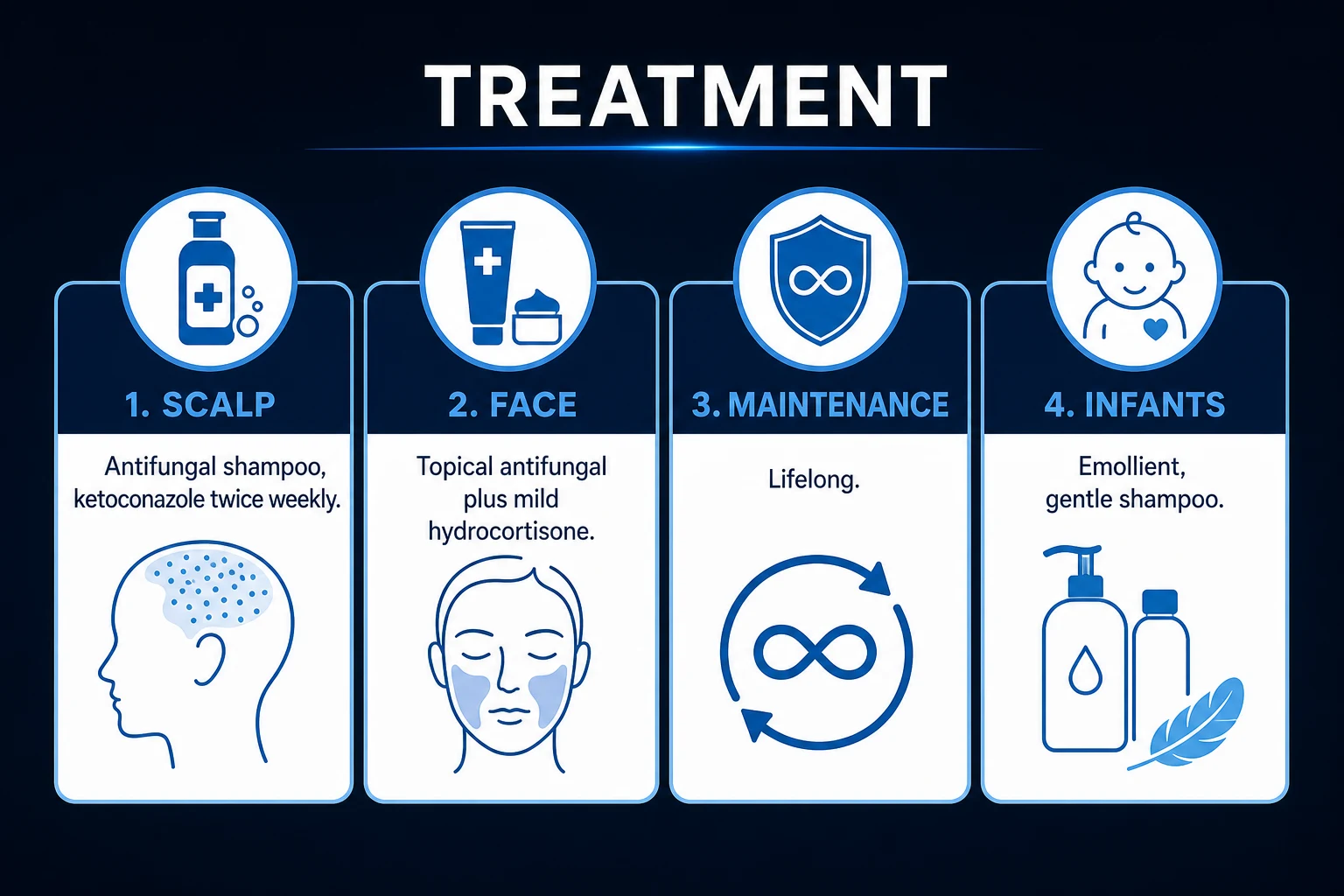
Seborrhoeic dermatitis is rarely a dermatological emergency. The exceptions are erythroderma and severe HIV-associated disease.[1][2][8]
- Erythroderma — admit for supportive care: temperature regulation, fluid and electrolyte replacement, mid-potency topical corticosteroids (e.g., betamethasone valerate 0.1% BD), systemic antifungal (itraconazole pulse), and management of secondary infection. Dermatology emergency review.
- Severe HIV-associated disease — optimise ART, screen for opportunistic infections, use potent topical and systemic antifungals; consider dermatology-specialist referral.
- Secondary bacterial infection — impetiginised crusts, folliculitis, or frank cellulitis. Treat with topical or oral antibiotics guided by swabs and clinical severity.
- Infantile erythroderma — admit urgently; consider immunodeficiency; supportive care; treat underlying cause. [1]
Management — Topical Antifungals
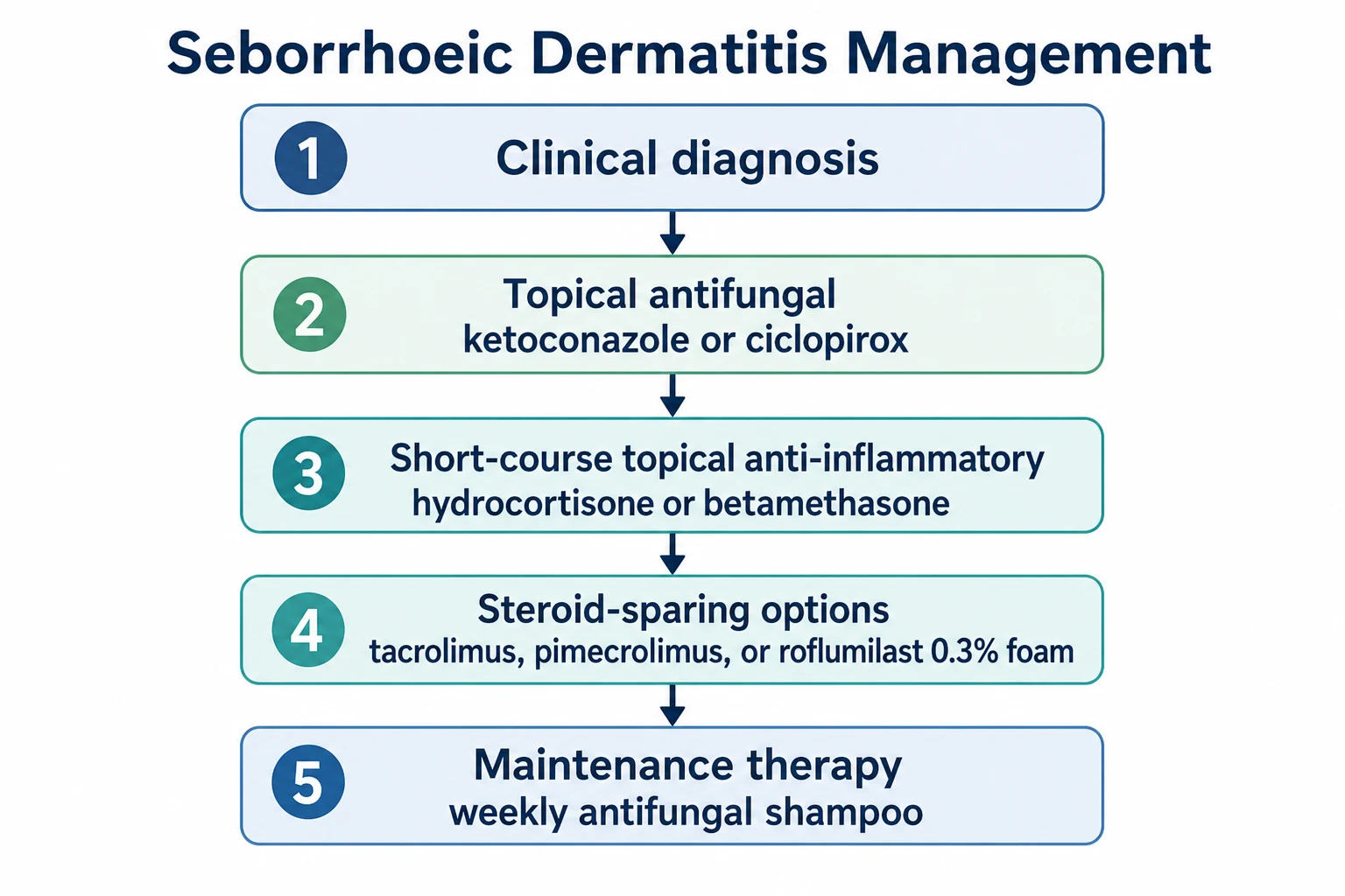
Antifungals remain the cornerstone of therapy for both scalp and facial disease, targeting Malassezia and exerting additional anti-inflammatory effects.[11][12][13][16]
Scalp / hair-bearing areas
Face / trunk / intertriginous
- Ketoconazole 2% cream — once or twice daily for 2-4 weeks; good first-line facial therapy; long-term safety for intermittent use is excellent.[11]
- Ciclopirox 1% cream or gel — alternative; useful for resistant or intertriginous disease.
- Miconazole 2% + hydrocortisone 1% (Daktacort) — combined antifungal and low-potency steroid; useful when inflammation and yeast overgrowth coexist.
- Selenium sulfide 2.5% lotion — alternative for truncal/petaloid disease.
- Zinc pyrithione 1% cream or wash — OTC option; useful for maintenance.
Management — Topical Anti-inflammatories
Corticosteroids
- Mild disease (face/intertrigo): hydrocortisone 1% cream or ointment once or twice daily for 1-2 weeks.
- Moderate scalp/trunk disease: betamethasone valerate 0.1% or mometasone furoate 0.1%, short courses (2-4 weeks).
- Avoid potent steroids on the face and in intertriginous sites because of atrophy, telangiectasia, striae, perioral dermatitis, and rebound flares. Potent steroids can also flourish Malassezia by removing competing skin flora. [1]
Calcineurin inhibitors
- Tacrolimus 0.1% ointment and pimecrolimus 1% cream are effective facial/intertriginous alternatives that do not cause skin atrophy — a critical advantage for chronic facial disease.[14][15]
- Evidence shows comparable efficacy to low-potency corticosteroids, with potential for more prolonged remission on the face. Pimecrolimus is comparable to betamethasone valerate 0.1% cream in facial disease.[14][15]
- Cautions: black-box warning in the US (theoretical malignancy risk; causality unproven); avoid in active skin infection; do not use with occlusion.
Combination antifungal + steroid
- Ketoconazole 2% + desonide 0.05% (Ketozal-D, Acuatim-D) — combination for facial disease when inflammation and yeast overgrowth coexist.
- Miconazole 2% + hydrocortisone 1% (Daktacort) — useful for napkin-area seborrhoeic dermatitis and intertriginous disease.
- Hydrocortisone 1% + clotrimazole 1% (Canesten HC, Lotriderm equivalents) — combination antifungal and steroid for facial and intertriginous disease. [1]
Management — Systemic Antifungals
Systemic antifungals are reserved for refractory or widespread disease when topical therapy has failed, and for HIV-associated severe disease. [1]
Oral ketoconazole
- Dose: 200 mg once daily for 2-4 weeks, often with a 2-week break.
- Mechanism: inhibits fungal lanosterol 14α-demethylase; anti-inflammatory at high doses.
- Monitoring: baseline and 2-4 weekly LFTs.
- Caveats: hepatotoxicity (1 in 10,000 symptomatic hepatitis), adrenal suppression at >400 mg/day, and CYP3A4 inhibition (multiple drug interactions). The FDA in 2013 and EMA in 2013 restricted oral ketoconazole for superficial fungal infections because of the risk-benefit profile; in many regions it is reserved for endemic mycoses (blastomycosis, histoplasmosis, coccidioidomycosis, paracoccidioidomycosis). Itraconazole is now preferred for refractory seborrhoeic dermatitis. [1]
Oral itraconazole pulse
- Dose: 200 mg twice daily for 1 day per week for 1-3 months ("pulse therapy").
- Mechanism: broad-spectrum azole; accumulates in keratin and sebum.
- Efficacy: multiple open-label and small RCT studies support efficacy in refractory seborrhoeic dermatitis.
- Monitoring: baseline LFTs; repeat at 4 weeks and end of treatment. Avoid in heart failure (negative inotropic effect). Multiple CYP3A4 interactions. [1]
Oral fluconazole
- Dose: 200-300 mg once weekly for 4-8 weeks.
- Mechanism: inhibits fungal lanosterol 14α-demethylase; excellent bioavailability.
- Efficacy: open-label series support weekly fluconazole for refractory seborrhoeic dermatitis.
- Monitoring: baseline LFTs; caution with CYP2C9 and CYP3A4 substrates. [1]
Management — Emerging and Novel Therapies
Roflumilast 0.3% foam
Roflumilast is a phosphodiesterase-4 (PDE4) inhibitor that reduces pro-inflammatory cytokines (TNF-α, IL-17, IL-23, IL-4, IL-13) by elevating intracellular cAMP. A 0.3% foam formulation is FDA-approved for adolescents and adults with seborrhoeic dermatitis (2023 phase 2a; 2024 phase 3 STRATUM; 2026 long-term open-label 52-week data).[17][18][19]
- Phase 2a: roflumilast 0.3% foam demonstrated rapid and significant improvement in erythema, scaling, and pruritus versus vehicle.[17]
- Phase 3 STRATUM: replicated efficacy and safety in adolescent and adult patients with seborrhoeic dermatitis affecting scalp, face, and trunk.[18]
- Long-term (52-week open-label): sustained benefit and safety; no new safety signals.[19]
It is particularly useful for patients who need steroid-sparing therapy, who cannot tolerate topical antifungals alone, or who have disease in cosmetically sensitive areas (face, scalp with hair). [1]
Other emerging therapies
- Topical 0.3% roflumilast cream and 0.3% roflumilast scalp solution — same drug, different formulation, in development.
- Topical lotamilast (RVT-502) — another PDE4 inhibitor in trials.
- Low-dose oral isotretinoin (0.1-0.3 mg/kg/day) — case series suggest efficacy in refractory seborrhoeic dermatitis by reducing sebaceous gland activity.
- Topical crisaborole 2% ointment — a PDE4 inhibitor approved for atopic dermatitis; off-label use for seborrhoeic dermatitis has been reported. [1]
Management by Site / Scenario
- Ketoconazole 2% shampoo 2-3×/wk (leave 3-5 min)
- Add topical corticosteroid solution/foam for erythema
- Maintenance: weekly antifungal shampoo
- Ketoconazole 2% cream BD 2-4 wk
- Hydrocortisone 1% for 1-2 wk if inflamed
- Pimecrolimus 1% / tacrolimus 0.1% for sensitive areas
- Ketoconazole 2% cream + hydrocortisone 1%
- Calcineurin inhibitor for fissures
- Avoid prolonged potent steroids
- Ketoconazole 2% cream BD
- Mild-to-moderate potency steroid 1-2 wk
- Ciclopirox 1% as alternative
- Warm compresses, lid hygiene
- Dilute baby shampoo lid scrubs
- Low-potency steroid or calcineurin inhibitor; ophthalmology input if refractory
- Ketoconazole 2% cream
- Low-potency hydrocortisone 1% 1-2 wk
- Calcineurin inhibitor; barrier protection
Special Populations
Infants (cradle cap)
- Conservative first-line: emollients (mineral oil, petroleum jelly, olive oil) applied to soften scale, then gentle brushing with a soft brush after 15-30 minutes. Baby shampoo washes are usually sufficient.[20][21]
- Persistent disease: short-course ketoconazole 2% cream or mild topical corticosteroid (hydrocortisone 1%). The Cochrane 2019 review found limited evidence for any specific intervention; emollients and gentle scale-removal remain the cornerstone.[20]
- Red flags — failure to thrive, recurrent infection, diarrhoea, or widespread exfoliative erythroderma: evaluate for immunodeficiency (LAD type 1, Netherton syndrome, severe combined immunodeficiency, Omenn syndrome, hyper-IgE syndrome, Leiner disease).[21]
HIV/AIDS
- Seborrhoeic dermatitis may be more severe, widespread, and refractory; it can be the presenting feature of undiagnosed HIV and can herald progression of disease.[8]
- Optimise ART; use potent topical antifungals and anti-inflammatories; systemic itraconazole pulse for refractory disease; consider low-dose isotretinoin in severe cases. Long-term maintenance therapy is often required.
Parkinson disease
- Prevalence of seborrhoeic dermatitis in Parkinson disease ranges from ~19% to ~60%, and severity may correlate with motor symptom burden.[7]
- Proposed mechanisms: autonomic dysfunction, altered sebum composition, and dopaminergic therapy effects (levodopa may actually improve skin disease by reducing substance P release).
- Management: regular antifungal shampoo, topical ketoconazole cream, gentle skin care, and treatment of the underlying neurological disease.
Pregnancy
- Topical antifungals (ciclopirox, ketoconazole 2%) are generally safe.[10]
- Calcineurin inhibitors (pimecrolimus, tacrolimus) acceptable; minimal systemic absorption.
- Avoid oral ketoconazole and itraconazole (teratogenicity, embryotoxicity).
- Topical corticosteroids — low-to-moderate potency, short courses, are safe; avoid potent steroids over large areas and prolonged use.
Skin of colour
- Post-inflammatory hyperpigmentation is the dominant cosmetic concern; treat aggressively and early to limit dyspigmentation.
- The granulomatous variant (Facial Afro-Caribbean Childhood Eruption, FACE) is more common in children with skin of colour and may be misdiagnosed as sarcoidosis or lupus.
- Topical antifungals and calcineurin inhibitors are safe and effective; check for rosacea-like or lupus-like overlap presentations. [1]
Complications & Pitfalls
Disease-related complications
- Secondary bacterial infection — impetiginised crusts, folliculitis, or frank cellulitis.
- Otitis externa — extension of retro-auricular and external auditory canal disease.
- Blepharitis, conjunctivitis, keratitis — eyelid margin involvement can affect the ocular surface.
- Erythroderma — rare but reported, especially in advanced HIV.
- Post-inflammatory hyper- or hypopigmentation — common in skin of colour.
- Psychosocial impact — embarrassment, low self-esteem, social withdrawal, depression. [1]
Treatment-related pitfalls
- Potent topical steroid overuse on the face — atrophy, telangiectasia, striae, perioral dermatitis, rebound flares, tachyphylaxis.
- Oral ketoconazole — hepatotoxicity (1 in 10,000 symptomatic hepatitis), adrenal suppression, CYP3A4 interactions. Restricted in many regions.
- Oral itraconazole — heart failure (negative inotropy), hepatotoxicity, CYP3A4 interactions.
- Inadequate contact time with antifungal shampoo — must be left on for 3-5 minutes to allow Malassezia killing.
- Misdiagnosis — missing tinea capitis (especially in children, where antifungal shampoo alone is inadequate), CTCL (in adults with treatment-resistant disease), or HIV (in adults with severe refractory disease). [1]
Classic errors
- Prescribing a potent topical steroid for "severe eczema" on the face — exacerbates seborrhoeic dermatitis, induces perioral dermatitis, and risks steroid rosacea.
- Treating infantile "cradle cap" with prolonged potent steroids — risk of systemic absorption, hypothalamic-pituitary-adrenal axis suppression, and iatrogenic Cushing syndrome.
- Stopping antifungal shampoo once symptoms resolve — relapse is common; maintenance therapy is the rule.
- Forgetting to ask about HIV risk factors in an adult with new severe disease. [1]
Prognosis & Follow-Up
Seborrhoeic dermatitis is chronic and relapsing; there is no cure. Goals are symptom control, clearance of scale and erythema, and prevention of relapse.[1][2]
- Cradle cap — usually self-limiting; resolves in 6-12 months in most infants.
- Adult disease — chronic relapsing-remitting; severity fluctuates with stress, season, and comorbid disease (HIV, Parkinson disease). With consistent therapy, 80-90% achieve good control.
- Follow-up — review at 4-8 weeks after initiating therapy, then as needed.
- Maintenance — weekly antifungal shampoo and intermittent topical anti-inflammatory use reduce relapse.
- Lifestyle — gentle cleansers, avoidance of harsh soaps, stress management, adequate sleep, and avoidance of known triggers.
- Safety-net advice — never use potent topical steroids on the face; switch to non-fluoride toothpaste if peri-oral; use sunscreen; treat early to limit post-inflammatory dyspigmentation. [1]
Evidence, Guidelines & Regional Differences
There is no single international guideline equivalent to the AAD-NPF psoriasis guidelines; management is largely consensus-based but supported by multiple high-quality RCTs and a 2024 systematic review of global prevalence.[3]
- Ketoconazole and ciclopirox are widely available first-line antifungals globally; the Danby 1993 RCT established that ketoconazole 2% shampoo and selenium sulfide 2.5% shampoo are both significantly superior to placebo for dandruff.[13]
- Calcineurin inhibitors may be preferred on the face and in maintenance regimens to limit steroid adverse effects; the Rigopoulos 2004 RCT showed pimecrolimus 1% cream is comparable to betamethasone valerate 0.1% for facial seborrhoeic dermatitis,[14] and the Papp 2012 RCT showed tacrolimus 0.1% ointment is comparable to hydrocortisone 1% ointment.[15]
- Roflumilast 0.3% foam represents the first novel FDA-approved topical for seborrhoeic dermatitis in many years and is becoming available in an increasing number of markets.[17][18][19]
- Oral itraconazole pulse is widely used in India and Europe for refractory disease (200 mg BD for 1 day/week for 1-3 months).
- Oral ketoconazole is restricted in many regions (FDA 2013, EMA 2013) because of hepatotoxicity, adrenal suppression, and CYP3A4 interactions; itraconazole is now preferred for refractory disease.
- Cochrane 2019 — limited high-quality evidence for any specific intervention in infantile seborrhoeic dermatitis; emollients and gentle scale-removal remain the cornerstone.[20]
Regional deltas
- US (AAD/JAAD): adult and adolescent roflumilast 0.3% foam FDA-approved 2023/2024; mineral-oil and low-potency steroid cradle-cap regimens.
- European (EADV/EDF): ciclopirox and ketoconazole shampoo first-line; tacrolimus/pimecrolimus preferred for facial/intertriginous disease; itraconazole pulse for refractory disease.
- UK (BAD): no specific NICE guideline; consensus-based; emollient + antifungal shampoo + topical corticosteroid ladder.
- Indian (IADVL): high prevalence in adult men with HIV/immunosuppression; itraconazole pulse widely used; cost-driven choice of selenium sulfide over ketoconazole.
- ANZ (ACD/ACDerm): ciclopirox preferred in pregnancy; roflumilast emerging; mineral-oil emollient for cradle cap. [1]
Exam Pearls
[1]Exam application bank (NEET-PG / INICET)
One-line answer
Seborrhoeic dermatitis is a chronic, relapsing inflammatory dermatosis of sebaceous-rich skin driven by interplay between Malassezia yeast, host immune responses, and epidermal barrier dysfunction. Fellowship-level assessment requires mastery of the bimodal age distribution (infantile cradle cap at 3 months and adult peak 20-40 years), scalp and facial morphology, the Malassezia-host immune axis, differential diagnosis from psoriasis and tinea capitis, the topical antifungal and anti-inflammatory ladder (ketoconazole, ciclopirox, calcineurin inhibitors, roflumilast 0.3% foam), site-specific therapy, systemic itraconazole pulse for refractory disease, and recognition of HIV, Parkinson disease, and infantile immunodeficiency associations.
Worked stems (answer without another resource)
Stem 1 — Classic presentation. Map symptoms to mechanism; name the first investigation and first treatment step with dose/route if drug therapy is standard. [1]
Stem 2 — Unstable / complicated. List red flags that force immediate resuscitation, theatre, ICU, antidote, or reperfusion — and what you do in the first 15 minutes. [1]
Stem 3 — Atypical group. Elderly, pregnancy, child, or immunocompromised: how presentation and thresholds change. [1]
Stem 4 — Differential trap. Name the three closest mimics and one discriminator for each. [1]
Stem 5 — Disposition. Who goes home with safety-netting, who is admitted, who needs HDU/ICU/theatre, and what follow-up is mandatory. [1]
Rapid viva checklist
- Definition + classification
- Pathophysiology chain
- Bedside signs / criteria
- Score with exact components (if any)
- Emergency bundle
- Definitive therapy with doses
- Complications of disease and of treatment
- Special populations
- Guideline/trial name if classic
- Three exam traps
Coverage self-check
If you cannot answer any stem above from this page alone, re-read the matching section — the page is intended to be self-sufficient for final-prof and NEET-PG/INICET questions on Seborrhoeic Dermatitis.
References
- [1]Gupta AK, Bluhm R. Seborrheic dermatitis J Eur Acad Dermatol Venereol, 2004.PMID 14678527
- [2]Tucker D, Masood S. Seborrheic Dermatitis 2026.PMID 31869171
- [3]Polaskey MT, Kwatra SG, Chen JG, et al. The Global Prevalence of Seborrheic Dermatitis: A Systematic Review and Meta-Analysis JAMA Dermatol, 2024.PMID 38958996
- [4]Adalsteinsson JA, Kaushik S, Muzumdar S, et al. An update on the microbiology, immunology and genetics of seborrheic dermatitis Exp Dermatol, 2020.PMID 32125725
- [5]Piacentini F, Kerrouche N, Bouslama L, Benhadou F. Seborrheic Dermatitis: Exploring the Complex Interplay with Malassezia Int J Mol Sci, 2025.PMID 40141293
- [6]Chang CH, Wu YH, Hsu CK, Lee CH. More yeast, more problems?: reevaluating the role of Malassezia in seborrheic dermatitis Arch Dermatol Res, 2024.PMID 38472524
- [7]Tomic S, Knezevic A, Stefanovski I, et al. Seborrheic Dermatitis Is Related to Motor Symptoms in Parkinson's Disease J Clin Neurol, 2022.PMID 36367060
- [8]Forrestel AK, Kovarik CL, Mosam A, et al. Diffuse HIV-associated seborrheic dermatitis - a case series Int J STD AIDS, 2016.PMID 27013615
- [9]Janniger CK. Seborrheic dermatitis Am Fam Physician, 1995.PMID 7604759
- [10]Gupta AK, Versteeg SG. Topical Treatment of Facial Seborrheic Dermatitis: A Systematic Review Am J Clin Dermatol, 2017.PMID 27804089
- [11]Carr MM, Pryce DM, Ive FA. Treatment of seborrhoeic dermatitis with ketoconazole: II. Response of seborrhoeic dermatitis of the face, scalp and trunk to topical ketoconazole Br J Dermatol, 1987.PMID 2950915
- [12]Green CA, Farr PM, Shuster S. Treatment of seborrhoeic dermatitis with ketoconazole: I. Response of seborrhoeic dermatitis of the scalp to topical ketoconazole Br J Dermatol, 1987.PMID 2950914
- [13]Danby FW, Maddin WS, Margesson LJ, et al. A randomized, double-blind, placebo-controlled trial of ketoconazole 2% shampoo versus selenium sulfide 2.5% shampoo in the treatment of moderate to severe dandruff J Am Acad Dermatol, 1993.PMID 8245236
- [14]Rigopoulos D, Ioannides D, Kalogeromitros D, et al. Pimecrolimus cream 1% vs. betamethasone 17-valerate 0.1% cream in the treatment of seborrhoeic dermatitis. A randomized open-label clinical trial Br J Dermatol, 2004.PMID 15541087
- [15]Papp KA, Papp A, Dahmer B, Clark CS. Single-blind, randomized controlled trial evaluating the treatment of facial seborrheic dermatitis with hydrocortisone 1% ointment compared with tacrolimus 0.1% ointment in adults J Am Acad Dermatol, 2012.PMID 22101215
- [16]Shin H, Kwon OS, Won CH, et al. Clinical efficacies of topical agents for the treatment of seborrheic dermatitis of the scalp: a comparative study J Dermatol, 2009.PMID 19335686
- [17]Zirwas MJ, Miller JL, Marks CJ. Efficacy of Roflumilast Foam, 0.3%, in Patients With Seborrheic Dermatitis: A Double-blind, Vehicle-Controlled Phase 2a Randomized Clinical Trial JAMA Dermatol, 2023.PMID 37133856
- [18]Blauvelt A, Stein Gold L, Bhatia N, et al. Roflumilast foam 0.3% for adolescent and adult patients with seborrheic dermatitis: A randomized, double-blinded, vehicle-controlled, phase 3 trial J Am Acad Dermatol, 2024.PMID 38253129
- [19]Alexis AF, Stein Gold L, Bhatia N, et al. Long-Term Safety and Efficacy of Roflumilast Foam 0.3% in Patients with Seborrheic Dermatitis: A Phase II, Open-Label Trial of up to 52 Weeks Am J Clin Dermatol, 2026.PMID 41175336
- [20]Victoire A, Magin P, Coughlan J, van der Kruis J. Interventions for infantile seborrhoeic dermatitis (including cradle cap) Cochrane Database Syst Rev, 2019.PMID 30828791
- [21]Nobles T, Harberger S, Williams J. Cradle Cap(Archived) 2026.PMID 30285358