Dermatology · Medicine
Seborrhoeic keratosis
Also known as Seborrhoeic keratosis (SK) · Seborrheic keratosis · Basal cell papilloma · Senile wart
Seborrhoeic keratosis (SK, basal cell papilloma, senile wart) = the most common benign epidermal tumour — the 'barnacles of ageing'. Well-defined, oval, 'stuck-on' waxy/verrucous plaques (tan-brown-black) on face, trunk and extremities of older adults. Clonal proliferation of basaloid keratinocytes driven by somatic FGFR3 activating mutations. Dermoscopy: milia-like cysts, comedo-like openings, fissures and ridges (brain-like), fingerprint structures, sharp demarcation; NO pigment network, NO arborising vessels. Five histological variants (acanthotic, hyperkeratotic, reticulated, clonal, irritated). Benign — NOT premalignant. Sign of Leser-Trélat: sudden eruption of numerous SKs with pruritus is paraneoplastic (gastric adenocarcinoma, AML). Management: leave alone if asymptomatic; cryotherapy, curettage, shave excision, laser ablation, or topical hydrogen peroxide 40% for cosmetic removal; biopsy any atypical lesion to exclude melanoma.
On this page & tools
Your progress
Saved locally on this device.
Exam tags
Red flags
Overview & Definition
Seborrhoeic keratosis (SK), also called basal cell papilloma or the older and now-discouraged term senile wart, is the most common benign tumour of the human epidermis and one of the most frequent of all skin lesions.[1][4] It arises from a clonal proliferation of immature basaloid keratinocytes within the basal and suprabasal epidermis, producing a well-demarcated, exophytic, waxy or verrucous plaque that appears to be "stuck on" to the skin surface — as if it could be lifted off. The colloquial name "the barnacles of ageing" captures both its extraordinary prevalence in older adults and its raised, roughened, crusty morphology.[3]
A defining and frequently examined point is that seborrhoeic keratosis is not premalignant: it does not transform into squamous cell carcinoma, basal cell carcinoma, or melanoma. It is a true benign neoplasm with a recurrent somatic driver mutation, not a degenerative or reactive change.[1][7] The clinical importance of SK therefore lies less in its own biological behaviour than in three examiner-tested problems: distinguishing a pigmented or atypical SK from melanoma, recognising the irritated variant that mimics squamous cell carcinoma, and identifying the paraneoplastic Sign of Leser-Trélat when numerous lesions erupt suddenly.[5][8]
Classification & Histological Variants
Clinically, SK is a single entity, but several clinical variants are recognised and examined, and five histological subtypes exist (often coexisting within one lesion).[1][3]
Acanthotic (common)
Most frequent histological pattern
- Thickened epidermis of basaloid cells
- Papillomatosis + hyperkeratosis
- Many horn cysts
- Most raised 'stuck-on' plaque
Hyperkeratotic
Predominantly orthokeratosis
- Marked hyperkeratosis
- Less papillomatosis
- Rough scaly surface
- Extremities; resembles wart
Reticulated / adenoid
Interlacing cords
- Double-layered basaloid strands
- Lace-like network
- Often on face/neck/sun-exposed sites
- May merge with solar lentigo
Clonal
Nests within epidermis
- Intraepidermal nests of basaloid cells
- Can mimic Bowen disease
- Correlate with dermoscopy
Irritated
Inflamed variant
- Squamous eddies + apoptosis
- Lichenoid inflammatory infiltrate
- Tender, crusted; mimics SCC
- Biopsy if diagnosis uncertain
The named clinical variants — dermatosis papulosa nigra and stucco keratosis — are discussed under Specific Subtypes. [1]
Epidemiology & Risk Factors
Seborrhoeic keratosis is essentially universal in older adults. Prevalence rises steeply with age: lesions are uncommon before 30, become frequent in the 40s, and are present in the great majority of people over 50–60 years, with estimates as high as 80–100 per cent in elderly cohorts.[1][4] They accumulate throughout life — new lesions continue to appear while existing ones slowly enlarge — which is the basis of the "barnacles of ageing" metaphor.
Epidemiology at a glance
Established risk factors are age (the dominant determinant), a family history of multiple lesions (an autosomal dominant pattern of multiple SKs is described), and chronic sun exposure (lesions are more numerous on sun-exposed skin, though they also occur on covered areas such as the back, so ultraviolet light is a cofactor rather than the cause).[1] There is no sex predilection. The historic suggestion of a viral (human papillomavirus) aetiology has been refuted — SK is not a viral wart.[3]
Age, genetics and the natural accumulation of lesions
The relationship with age is the single strongest epidemiological signal. SKs are rare before the age of 30, become common in the fifth decade, and by the seventh and eighth decades are present in almost every individual examined closely.[1][4] The number of lesions per person also rises with age: a younger patient may have a handful, whereas an elderly patient can carry a hundred or more scattered across the trunk and face. Once a lesion appears it persists for life — it does not regress spontaneously — and new lesions continue to emerge, so the burden accumulates.
A familial tendency is well recognised. Some families show an autosomal dominant pattern of multiple seborrhoeic keratoses segregating across generations, with affected individuals developing large numbers of lesions relatively early in adult life. No single germline gene has been definitively linked, but the pattern supports a heritable predisposition on top of the somatic FGFR3 driver.[1]
Sunlight plays a partial role: lesions are more numerous and appear earlier on chronically sun-exposed skin (face, scalp, dorsa of hands, forearms), but they also arise on sun-protected sites such as the central back and intertriginous folds, confirming that UV is a cofactor rather than the cause. The relationship distinguishes SK from purely UV-driven lesions such as actinic keratosis, which is confined to sun-damaged skin and is premalignant.[3]
Pathophysiology
Cellular origin and the "stuck-on" growth pattern
SK is a true benign neoplasm arising from a clonal expansion of immature basaloid keratinocytes — small, basal-cell-like keratinocytes that retain their proliferative capacity and accumulate within the epidermis rather than maturing and shedding normally.[1][7] The tumour grows outward (exophytic/papillomatous), lifting above the surrounding skin surface. This outward growth is what gives the lesion its hallmark "stuck-on" appearance: it sits on top of the skin as though pasted there, with the dermis and adnexal structures undisturbed beneath it.
The pigmentation of an SK does not come from melanocytic proliferation. Melanocytes in the lesion transfer melanin into the surrounding basaloid keratinocytes, which retain pigment as they pile up; the denser the basaloid cell mass, the darker the lesion can appear. This is why a deeply pigmented SK can mimic melanoma clinically despite having no atypical melanocytes. [1]
Molecular genetics — FGFR3 and the neoplastic nature of SK
The pivotal molecular discovery is that seborrhoeic keratoses harbour somatic activating mutations in FGFR3 (fibroblast growth factor receptor 3), found in roughly 40–60 per cent of lesions.[1][7] The same gene, when mutated in the germline, causes achondroplasia and thanatophoric dysplasia; in SK the mutation is somatic (confined to the lesion). Activated FGFR3 drives basaloid keratinocyte proliferation and survival through the PI3K–AKT and RAS–MAPK signalling cascades, producing the self-limited hyperplasia that characterises the lesion.
A crucial point that examiners test is why FGFR3 mutation alone does not produce malignancy. Experimental work shows that activating FGFR3 mutations cause only mild epidermal hyperplasia and are insufficient, by themselves, to drive either benign tumour formation or malignant transformation; additional cooperating oncogenic events are required.[7] This explains both why SK is benign and why it never progresses to carcinoma. Lower-frequency mutations in PIK3CA, HRAS, KRAS and EGFR are also found in some lesions, reinforcing that SK is a genetically driven neoplasm rather than a degenerative change.[1]
Relationship to epidermal naevi
SK shares its FGFR3 mutational signature with epidermal naevi, the congenital/linear counterpart. Both are benign proliferations of basaloid keratinocytes driven by the same somatic FGFR3 mutation acquired during embryogenesis (naevi) or adult life (SK). This shared genetics is a high-yield examination point linking a common adult lesion to a developmental condition.[1][7]
Clinical Presentation
Morphology and distribution
The classic seborrhoeic keratosis is a well-defined, oval or round, "stuck-on" waxy or verrucous plaque that looks and feels as though it has been glued onto the skin surface.[1][4] Colours range from flesh-coloured through tan, yellow-brown, dark brown to almost black, and a single lesion may be uniformly or variably pigmented. The surface is rough, granular and slightly greasy, often dotted with tiny dark pinpoint plugs (comedo-like openings) and firm white grains (milia-like cysts). Lesions measure from a few millimetres to several centimetres and enlarge slowly over years. Crucially, skin lines are preserved over the surface (unlike a viral wart, which disrupts them).
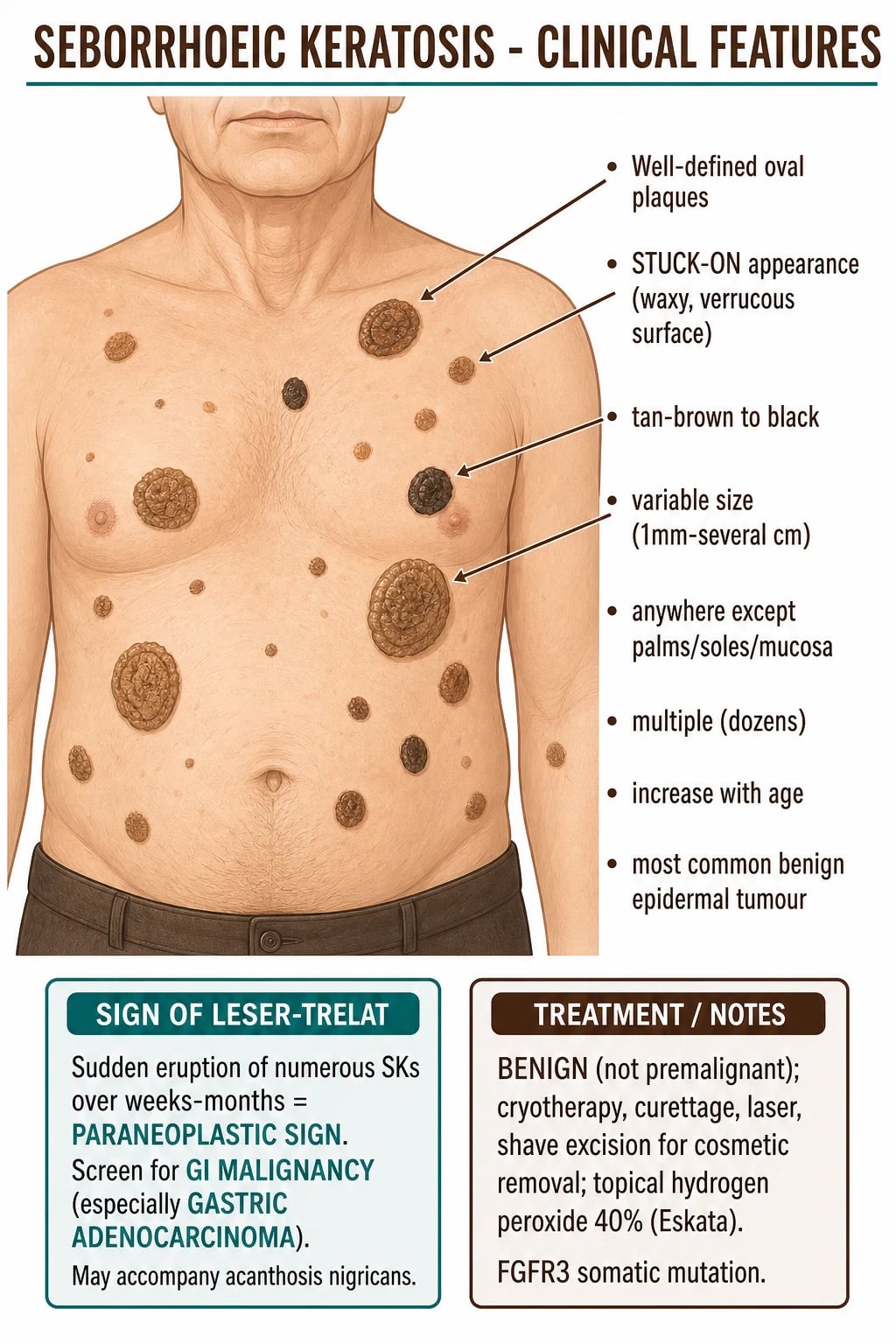
The distribution favours sun-exposed and seborrhoeic (oil-rich) areas — the face (especially temples and hairline), scalp, chest, upper back, abdomen and the upper extremities — but lesions can appear almost anywhere except the palms, soles and mucosal surfaces, which are characteristically spared.[1] Lesions are almost always multiple; an individual may carry dozens, and they tend to be symmetrically distributed. A solitary seborrhoeic keratosis in a young adult is unusual enough to warrant dermoscopy and biopsy to exclude another diagnosis.
Morphology in detail and evolution over time
A useful way to think about SK clinically is to trace how an individual lesion evolves. It often begins as a flat, slightly tan macule or thin plaque that is easily mistaken for a solar lentigo. Over months to years it thickens, develops a waxy, roughened surface, and takes on the unmistakable raised "stuck-on" quality. With further ageing the surface becomes more verrucous and fissured, sometimes adopting a cerebriform or brain-like topography, and the colour often darkens as pigment accumulates within the expanding mass of basaloid cells. The same lesion may therefore look quite different at different stages of its life, which is why dermoscopy is so valuable: it confirms the diagnosis regardless of the stage.[1][3]
The texture is distinctive. Running a finger across the surface reveals a rough, granular, slightly greasy feel, and the lesion often appears greasy or waxy under oblique light — the basis for the older term "seborrhoeic". Tiny dark pinpoint plugs (comedo-like openings) and firm white grains (milia-like cysts) may be visible to the naked eye or with a hand lens, especially on a thick lesion. Unlike a viral wart, skin lines are preserved across the surface of an SK rather than being disrupted, and unlike a solar lentigo the lesion is palpably raised. [1]
Site-specific patterns are worth knowing. On the face, lesions are often flatter and more variably pigmented, and they overlap morphologically with solar lentigo and lentigo maligna — dermoscopy is essential. On the trunk, lesions are typically larger, thicker, more numerous and more classically "stuck-on". On the scalp, they can be hidden in hair and become irritated by combing. Intertriginous lesions (axillae, inframammary, groin) are frequently macerated and inflamed. Lesions are never found on the palms, soles, or mucosa, and a lesion in these sites should not be diagnosed as an SK.[1][4]
Symptoms
Most SKs are asymptomatic and are brought to medical attention for cosmetic reasons, or because the patient has noticed a new "mole" or growth. When symptoms occur they are usually mild — itching, a catching sensation on clothing or jewellery, or transient irritation from friction. Pain, bleeding, rapid growth, or ulceration are not features of a typical SK and should prompt dermoscopy and biopsy to exclude malignancy. [1]
Irritated seborrhoeic keratosis
An irritated (inflamed) SK becomes erythematous, swollen, crusted and tender, most often because of chronic friction from clothing, a bra strap, or a belt. Histologically it shows squamous eddies (whorls of keratinocytes) and a lichenoid inflammatory infiltrate.[3] Because the clinical picture — a crusted, keratotic, tender lesion — overlaps with squamous cell carcinoma, the safe rule is to biopsy any lesion where the diagnosis is not secure.
Atypical presentations
The examiner deliberately tests atypical presentations. In darker phototypes (Fitzpatrick IV–VI), the dermatosis papulosa nigra variant produces many small, smooth, dark-brown-to-black facial papules (see Specific Subtypes). In the immunocompromised, SKs are not increased in number, but any new pigmented lesion must be biopsied to exclude squamous cell carcinoma or melanoma. In younger adults (under 30), a genuine solitary SK is uncommon enough that melanoma and other diagnoses should be actively excluded before the label is accepted. [1]
Differential Diagnosis
The differential of a "stuck-on" pigmented plaque is one of the highest-yield areas for this topic, because the single most dangerous error in dermatology is labelling a melanoma as a seborrhoeic keratosis.[6]
Melanoma
The must-not-miss
- Irregular border, colour, shape; changing
- Dermoscopy: atypical pigment network, streaks, blue-white veil, atypical vessels
- ABCDE positive; history of change
- Biopsy any dark/irregular/atypical 'SK'
Pigmented BCC
Pigmented basal cell carcinoma
- Pearly papule with pigment and arborising vessels
- Slow growth; may ulcerate
- Dermoscopy: arborising telangiectasia, blue-grey ovoid nests
- Biopsy if any vessel pattern suggests BCC
Solar lentigo
'Liver spot' / senile lentigo
- Flat macule — no surface change
- 'Moth-eaten' irregular border
- Dermoscopy: fingerprint network, moth-eaten border
- SK is raised; lentigo is flat
Viral wart
Common wart
- Rough firm papule; thrombosed capillaries (black dots)
- Skin lines disrupted over surface
- Palmar/plantar possible (SK spares these)
- HPV-driven; not stuck-on waxy
Actinic keratosis
Premalignant
- Rough gritty erythematous patch on sun-damaged skin
- Felt more easily than seen
- Premalignant — SK is not
- Biopsy if thickening/induration (SCC)
Melanocytic naevus
Mole
- Pigmented, may be raised
- Dermoscopy: pigment network, globules
- Symmetrical, uniform
- Distinguish by dermoscopy
Two principles govern the differential. First, a typical SK with classic dermoscopy in an asymptomatic older adult is a clinical diagnosis. Second, any dark, irregular, changing, bleeding, or ulcerated lesion must be biopsied — dermoscopy is supportive, never a substitute for histology when suspicion exists, because a minority of melanomas display milia-like cysts or comedo-like openings that mimic SK.[6]
Distinguishing each mimic — what the examiner wants
Melanoma is the lesion that must never be missed. The clinical clue is asymmetry, border irregularity, colour variegation, diameter greater than 6 mm, and evolution (the ABCDE criteria), together with any history of change, bleeding or itching.[6] On dermoscopy, melanoma shows an atypical pigment network, streaks, regression structures, a blue-white veil, and atypical (dotted/linear) vessels — precisely the features absent in SK. A nodular melanoma can be uniformly dark and raised, which is the most dangerous mimic of all: if there is any doubt, biopsy.
Pigmented basal cell carcinoma typically shows a pearly, translucent papule with focal pigment and arborising (tree-like) telangiectatic vessels, and may ulcerate centrally. Dermoscopy reveals the pathognomonic arborising telangiectasia and blue-grey ovoid nests or multiple blue-grey globules. SK lacks these vessels, so any telangiectasia on a presumed SK should trigger biopsy.[6]
Solar lentigo ("liver spot", senile lentigo) is the flat cousin of SK and shares the same patient demographic. The decisive difference is that a solar lentigo is a flat macule with no surface change and a "moth-eaten" border, and on dermoscopy shows a fingerprint network and moth-eaten edge without milia-like cysts or comedo-like openings. An SK, by contrast, is palpably raised. The two lesions exist on a spectrum and a single lesion may show features of both (a "lentigo-like" early SK), which is why early flat lesions can be difficult.[1]
A viral wart is rough and firm, with thrombosed capillaries appearing as black dots on the surface; crucially, skin lines are disrupted over a wart but preserved over an SK. Warts also occur on the palms and soles, which SK never does. The historic confusion between SK ("senile wart") and true viral warts is purely nominal — they are unrelated aetiologically.[3]
Actinic keratosis is a rough, gritty, erythematous patch on chronically sun-damaged skin that is "felt more easily than seen". It is premalignant, in contrast to SK, and any thickening, induration, tenderness, or rapid growth in a presumed actinic keratosis should be biopsied to exclude progression to squamous cell carcinoma.[4]
A melanocytic naevus (mole) may be raised and pigmented, but it grows within the dermis/epidermis rather than sitting on top of the skin, and dermoscopy shows a regular pigment network, globules, and a symmetrical architecture. A dermatofibroma is a firm dermal papule that dimples inward when pinched (the "dimple sign"), distinguishing it from the outward, stuck-on SK. An accessory nipple or sebaceous hyperplasia can occasionally mimic a facial SK and is excluded by dermoscopy and clinical correlation. [1]
Clinical & Bedside Assessment
The assessment of a suspected seborrhoeic keratosis is built around two questions: Is this really an SK? and, if there are many, is this a Sign of Leser-Trélat? [1]
Begin with inspection under good lighting, examining the lesion and the surrounding skin. The lesion's surface should be felt: a typical SK is rough, granular and slightly greasy, and the "stuck-on" quality is confirmed by gently stretching or pinching the skin, which shows the tumour sitting superficially on the surface rather than arising from within it. A solar lentigo, by contrast, is perfectly flat; a melanocytic naevus is raised but grows within the skin, not on top of it. [1]
Dermoscopy is the key bedside tool and should be performed on every pigmented lesion before a diagnosis of SK is accepted (see Investigations). Look specifically for the features that exclude melanoma — a sharp border, milia-like cysts, comedo-like openings, and a brain-like fissured architecture — and for the features that would demand biopsy, such as an atypical pigment network, streaks, a blue-white veil, or atypical vessels. [1]
Because SKs are multiple and coexist with other lesions, always examine the whole skin, not just the presenting lesion, and screen for any lesion that "does not belong" among the benign barnacles. Take a focused history for change: has the lesion grown, changed colour, bled, itched, or become painful? A positive answer to any of these demands escalation to dermoscopy and, if there is doubt, biopsy. [1]
When numerous SKs have appeared suddenly (over weeks to a few months), ask about pruritus, weight loss, GI symptoms, and fatigue, and screen for acanthosis nigricans — the combination defines the Sign of Leser-Trélat and mandates a search for underlying malignancy.[5][8]
Investigations
Dermoscopy — the diagnostic cornerstone
Dermoscopy is the primary diagnostic test for a typical SK and, critically, the tool that excludes melanoma and pigmented BCC.[1][6]
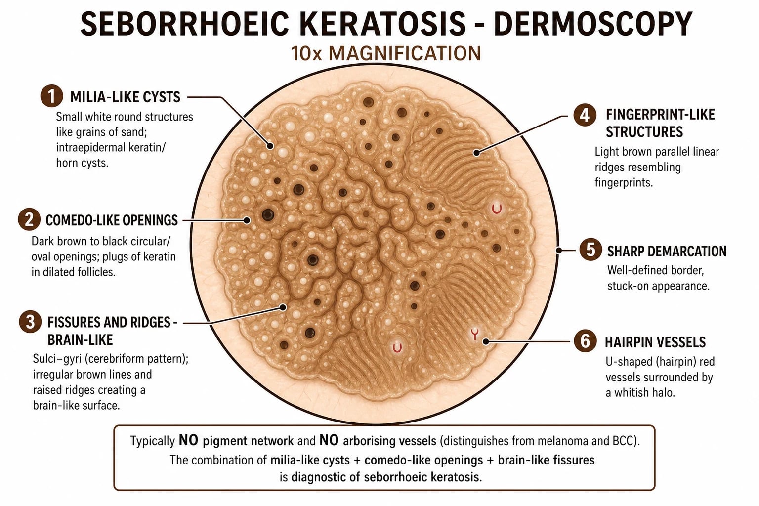
The classic dermoscopic features are:[1][3]
- Milia-like cysts — small, bright-white, round structures, like grains of sand, representing intraepidermal keratin (horn cysts) viewed through the surface.
- Comedo-like openings — dark brown to black plugged follicular openings filled with keratin, sometimes called "comedo-like plugs".
- Fissures and ridges — irregular linear depressions and raised ridges producing a "brain-like" or "sulci-and-gyri" appearance, typical of thicker lesions.
- Fingerprint-like structures — delicate parallel brown lines at the periphery of thin lesions, where basaloid rete ridges align.
- Moth-eaten border and sharp demarcation — the lesion ends abruptly at its edge.
- Network-like structures — a coarse, regular pigment pattern (not the atypical network of melanoma). [1]
Equally important are the features that are absent in SK: there is no atypical pigment network, no streaks/pseudopods, no blue-white veil, and no arborising (tree-like) telangiectatic vessels. The presence of any of these should prompt biopsy, because a small minority of melanomas produce milia-like cysts and can masquerade as SK.[6]
Dermoscopy by lesion type and the false-negative trap
The dermoscopic appearance of an SK varies with its thickness and stage. A thin, early SK may show only faint fingerprint-like structures and a moth-eaten border, making it almost indistinguishable from a solar lentigo — the distinction is that a lentigo is flat while the early SK is just palpable. A thick, mature SK displays the full repertoire of milia-like cysts, comedo-like openings and a brain-like (cerebriform) fissured surface. A heavily pigmented SK can show a regular network-like structure that must be distinguished from the atypical network of melanoma by its regularity and sharp border. [1]
The false-negative trap is the cardinal dermoscopy lesson of this topic.[6] Although the classic features are highly specific, dermoscopy is an imperfect test: a small but real proportion of melanomas (especially verrucous, papillomatous, or regressing melanomas) can display milia-like cysts, comedo-like openings or a fissured surface that mimics SK. Conversely, an SK can occasionally lack its hallmark features and look atypical. The examiner's rule is therefore absolute: dermoscopy is an aid to pattern recognition, never a licence to ignore clinical suspicion. A lesion that looks wrong clinically — changing, bleeding, irregular, darker than the patient's other SKs — must be biopsied regardless of a reassuring dermoscopic picture.
Dermoscopy of seborrhoeic keratosis — remember the four hallmarks
MCFS
White 'grains of sand' — intraepidermal keratin
Dark plugged follicular openings
Brain-like / sulci-and-gyri surface
Abrupt border; no pigment network
Biopsy — when and how
A typical SK in an asymptomatic older adult is a clinical and dermoscopic diagnosis and needs no histology. Biopsy is indicated whenever the diagnosis is not secure — specifically for any dark, irregular, changing, bleeding, or ulcerated lesion, any pigmented lesion in a younger patient, and any lesion that fails to behave as expected.[4][6] Because SK is superficial, a shave biopsy or a punch biopsy is usually sufficient; a full-thickness excisional biopsy is reserved for lesions where melanoma is genuinely suspected.
Histopathology
When biopsied, the histology confirms the diagnosis and excludes mimics. The cardinal features are:[1][3]
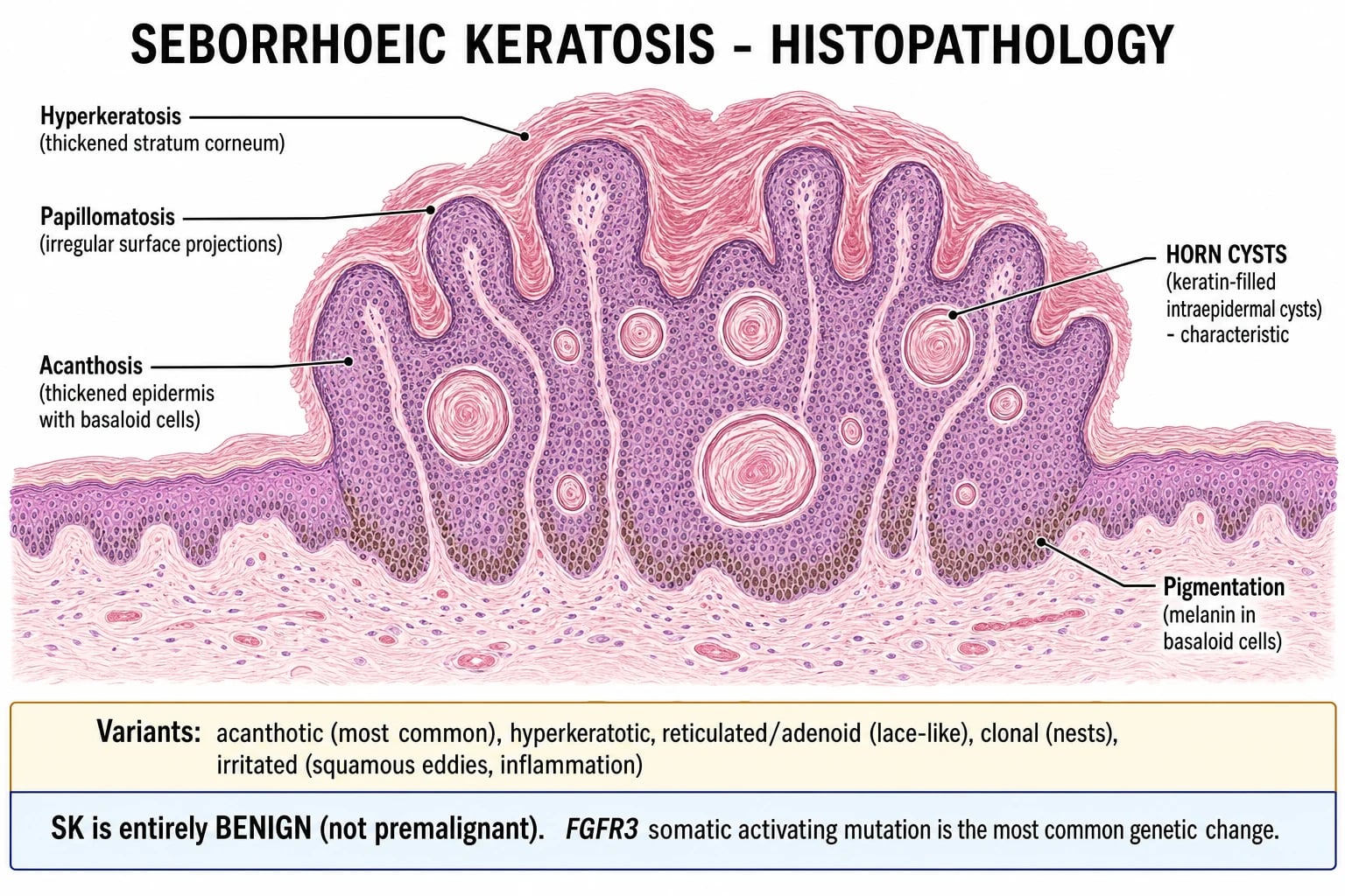
- Acanthosis — a thickened epidermis composed of basaloid (small, basal-cell-like) keratinocytes that extends above the level of the surrounding normal epidermis (the histological correlate of the "stuck-on" appearance).
- Papillomatosis — an irregular, exophytic surface contour.
- Hyperkeratosis — a thickened, often orthokeratotic stratum corneum.
- Horn cysts (true and pseudo-) — intraepidermal keratin-filled cysts; "pseudo-horn cysts" are invaginations of the surface stratum corneum cut in cross-section.
- Increased melanin within the basaloid cells, accounting for the pigmentation (no melanocytic proliferation).
- Irritated variant — squamous eddies (whorls of keratinocytes), apoptotic keratinocytes, and a lichenoid lymphocytic infiltrate; this pattern can be confused with squamous cell carcinoma in situ and must be correlated with the clinical picture. [1]
Management — Resuscitation
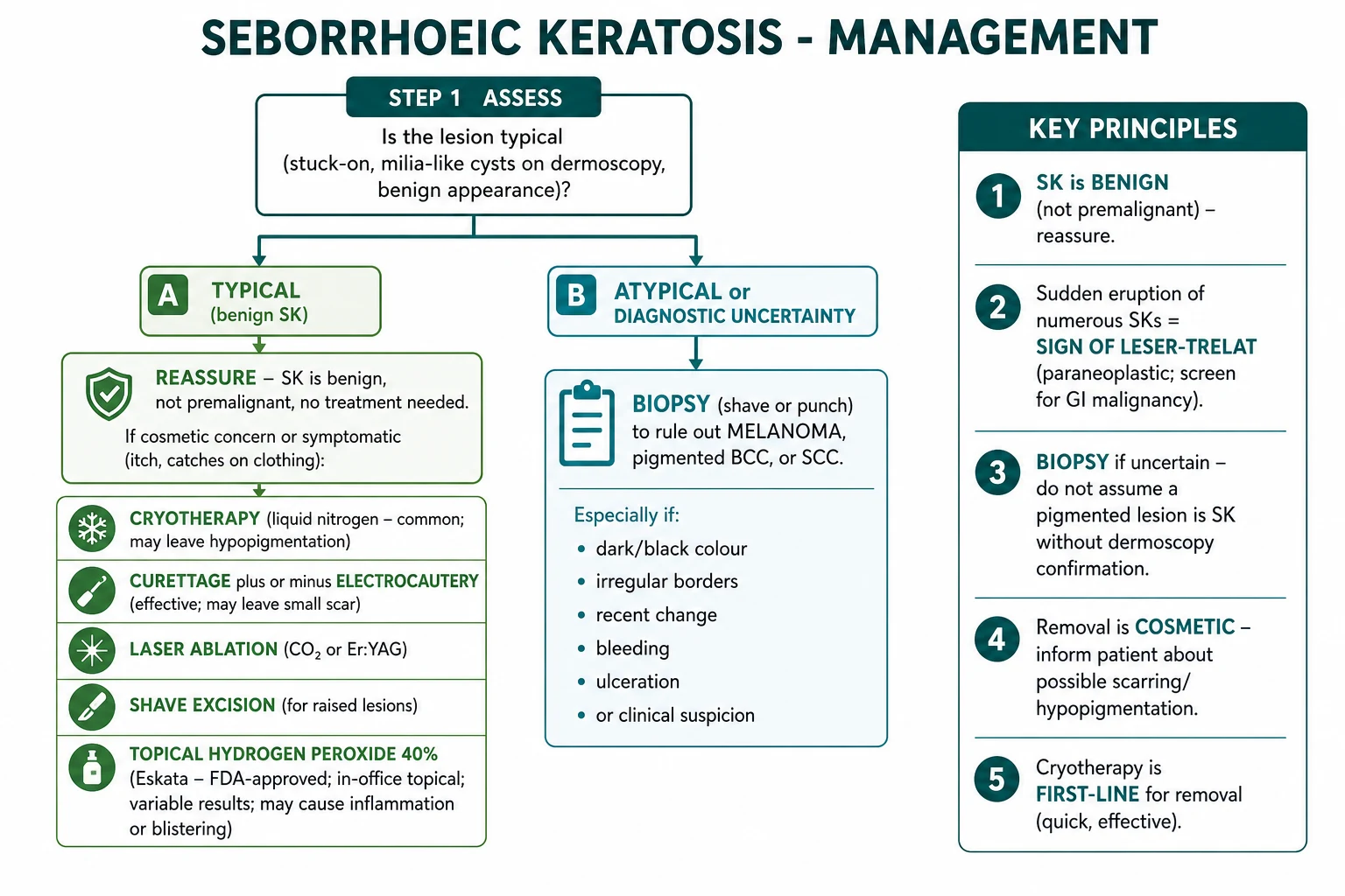
Seborrhoeic keratosis is not an emergency; a confirmed typical lesion needs no urgent treatment. The "resuscitation" step for this topic is really a diagnostic safety step: when a patient presents with a "changing mole" or a "new growth", the immediate priority is to exclude melanoma by dermoscopy, and to biopsy if there is any atypical feature — never to reassure on the basis of a presumed "seborrhoeic keratosis" without confirming.[6] Equally, when numerous lesions have erupted suddenly, the priority is to recognise the Sign of Leser-Trélat and begin a workup for occult malignancy rather than dismiss the eruption as "just age spots".[5][8]
Management — Definitive & Stepwise
The management of a confirmed seborrhoeic keratosis is built on a simple principle: leave it alone if it is asymptomatic, and remove it only for symptoms, diagnostic uncertainty, or cosmetic concern.[4]
Step 1 — Reassure and leave alone
For the asymptomatic typical lesion, the correct management is reassurance: explain that the lesion is benign, is not a cancer, and does not require removal.[4] This is both the safest and the most cost-effective option and should be the default.
Step 2 — Destructive and excisional treatments
When removal is desired — for irritation, diagnostic uncertainty, or cosmesis — several office-based options exist, each with trade-offs.[2][4]
Cryotherapy (LN2)
First-line destructive
- Liquid nitrogen, two freeze-thaw cycles
- Quick, cheap, no anaesthetic
- Risk: hypo/hyperpigmentation (dark skin)
- Poor for thick lesions; no histology
Curettage + electrocautery
Thick lesions; gives tissue
- Local anaesthetic; curette + cautery
- Provides specimen for histology
- Good for thicker raised lesions
- Risk: scarring, recurrence if incomplete
Shave excision
Pedunculated lesions
- Blade shave at the base
- Provides tissue for histology
- Good for pedunculated 'stuck-on' lesions
- Minimal scarring
Laser ablation
CO2 / Er:YAG
- Precise; multiple thin lesions
- Excellent for DPN on the face
- No histology — biopsy first if uncertain
- Risk: dyspigmentation in dark skin
Hydrogen peroxide 40%
Topical (Eskata/A-101)
- FDA-approved for raised SKs
- Applied four times (~1 min each) per session
- Modest efficacy; local erythema/burning/stinging
- Avoid facefold/intertriginous skin
Choosing between treatments
The choice of destructive modality is individualised:[2]
- Thin lesions on a light background → cryotherapy is simplest and first-line.
- Thick or pedunculated lesions → curettage with electrocautery or shave excision, both of which provide tissue for histology.
- Multiple thin facial lesions / dermatosis papulosa nigra → laser ablation (low-fluence CO2 or KTP) or light electrocautery, chosen to minimise dyspigmentation in darker skin.
- Patient preference for a non-procedural option → topical hydrogen peroxide 40%, accepting its modest efficacy and local skin reactions.
- Any atypical lesion → biopsy before any destructive treatment, because destruction without histology forfeits the chance to diagnose a melanoma. [1]
A 2023 systematic review of topical treatments found that hydrogen peroxide 40% had the best evidence among topical agents, with acceptable but modest efficacy and predictable local skin reactions; other topicals (trichloroacetic acid, potassium hydroxide) are less well supported.[2]
Technique notes for the destructive modalities
Cryotherapy with liquid nitrogen is performed using a spray or cotton-tipped applicator to deliver two freeze-thaw cycles, with a visible ice ball extending 1–2 mm beyond the lesion edge and a total freeze time of roughly 5–30 seconds depending on lesion thickness.[2][4] The lesion blister, crusts and separates over the following one to two weeks, leaving temporary hypopigmentation that may be permanent in darker skin. Cryotherapy is least effective for thick, deeply pigmented lesions, which often recur because the basaloid cells at the base are not destroyed.
Curettage with electrocautery requires local anaesthetic (e.g. lidocaine 1 per cent with adrenaline). The lesion is scraped off at its base with a dermal curette — a typical SK separates cleanly because it is confined to the epidermis — and the base is lightly electrocauterised for haemostasis and to destroy residual cells. The advantage over cryotherapy is that the curettings provide tissue for histology, which is invaluable when the clinical diagnosis is not certain, and the cosmetic result is often superior to cryotherapy for thicker lesions. [1]
Shave excision uses a blade to shave the lesion flush with the surrounding skin at its base; it suits pedunculated or clearly raised lesions and also provides tissue for histology. Bleeding is controlled with aluminium chloride or electrocautery. [1]
Laser ablation with the CO2 or Er:YAG laser vaporises the lesion layer by layer; it is precise and well suited to multiple thin lesions and to dermatosis papulosa nigra on the face, where a superior cosmetic result is expected. The disadvantage is that no tissue is sent for histology, so any lesion with atypical features must be biopsied first rather than ablated. [1]
Post-procedure care and follow-up
After any destructive treatment the patient should be advised on wound care — keep the site clean, apply a simple antiseptic or petroleum jelly, and expect crusting for one to two weeks. Warn specifically about dyspigmentation (hypopigmentation after cryotherapy; hyperpigmentation after any inflammatory modality in darker skin), about scarring after deep curettage, and about the possibility of recurrence if removal was incomplete.[2] Because new SKs continue to appear throughout life, follow-up is pragmatic rather than scheduled: the patient is reassured that the condition is benign and advised to return for review of any new lesion that changes colour, shape or size, bleeds, itches, or becomes painful.
Specific Subtypes & Scenarios
Dermatosis papulosa nigra
Dermatosis papulosa nigra (DPN) is a distinct clinical variant seen predominantly in individuals of African, Asian and South-Asian ancestry with darker phototypes (Fitzpatrick IV–VI).[1][4] It presents as multiple small (1–5 mm), smooth, round, dark-brown to black papules on the cheeks, temples, periorbital skin, upper chest and neck, often beginning in adolescence or early adulthood and with a female preponderance. Histologically the lesions are identical to SK.
Management of DPN demands special care to avoid dyspigmentation, which is the main complication of destructive treatment in darker skin. Cryotherapy risks cosmetically unacceptable hypopigmentation and is generally avoided in favour of light electrocautery, low-fluence CO2 laser, or KTP laser, which can remove the lesions with a lower risk of pigmentary change. Patients must be counselled that postinflammatory hyperpigmentation can follow any treatment and that new lesions will continue to appear. [1]
Stucco keratosis
Stucco keratosis presents as multiple small (1–5 mm), grey-white, scaly, "stuck-on" papules on the dorsa of the feet, ankles, hands and forearms of older adults.[3][4] The lesions are loosely attached and can often be scraped off easily. Histologically they resemble a hyperkeratotic SK. They are entirely benign; treatment, if desired, is by light curettage, cryotherapy, or emollients to reduce the chalky scale.
Irritated seborrhoeic keratosis
When an SK becomes inflamed — erythematous, crusted and tender — the usual trigger is chronic friction (bra strap, belt, collar). Histology shows squamous eddies and a lichenoid infiltrate, and the clinical overlap with squamous cell carcinoma means the safe rule is to biopsy any crusted keratotic lesion where the diagnosis is not secure.[3] A genuinely irritated SK that is histologically confirmed can be settled with a short course of a topical corticosteroid (e.g. clobetasol 0.05 per cent once daily for up to two weeks) and then removed if irritation keeps recurring.
Sign of Leser-Trélat
The Sign of Leser-Trélat is the sudden eruption (over weeks to a few months) of numerous seborrhoeic keratoses, accompanied by pruritus, and is a recognised paraneoplastic phenomenon.[5][8] It is often associated with acanthosis nigricans. The underlying malignancy is most often gastric adenocarcinoma, but colon, breast, lung and prostate cancers, and haematological malignancies (especially acute myeloid leukaemia), are also reported.[5]
The sign is rare and its existence as a true paraneoplastic marker is debated (because both SK and cancer are common in older adults), but the examination convention is clear: a convincing sudden eruption with pruritus warrants a structured workup — thorough history and examination, upper and lower gastrointestinal endoscopy, cross-sectional imaging (CT), a full blood count and film, and age-appropriate cancer screening.[8]
Complications & Pitfalls
Disease-related complications
In their natural state, seborrhoeic keratoses cause few problems: they are cosmetically objectionable, they may catch on clothing or jewellery and bleed, and an irritated lesion can be tender. The complications that matter are largely iatrogenic and diagnostic.[4]
Treatment-related complications
Every destructive modality carries risks. Cryotherapy can cause hypo- or hyperpigmentation — particularly problematic in darker phototypes, where hypopigmentation may be permanent and cosmetically devastating. Curettage and electrocautery can scar. Laser ablation similarly risks dyspigmentation and provides no histology, so a malignancy destroyed by laser is a malignancy missed. All modalities can be complicated by infection and by recurrence if the lesion is incompletely removed.[2]
The defining pitfalls
Prognosis & Disposition
The prognosis of seborrhoeic keratosis is uniformly benign: lesions slowly enlarge over years, new lesions continue to appear throughout life, and no SK ever undergoes malignant transformation.[1][4] Removal is curative for that individual lesion but does not alter the tendency for new lesions to arise elsewhere.
The prognosis of the Sign of Leser-Trélat is determined entirely by the underlying malignancy; the keratoses themselves are benign and may regress if the tumour is successfully treated.[5][8]
Typical lesions are managed in primary care. Dermatology referral is appropriate for atypical or extensive lesions, for cosmetic procedures beyond primary-care scope, and when melanoma cannot be confidently excluded. A confirmed or suspected Sign of Leser-Trélat requires gastroenterology/oncology referral for malignancy workup. The safety-net for every patient is clear advice to return if any lesion changes colour, shape or size, bleeds, itches persistently, or becomes painful. [1]
Special Populations
Older adults
Older adults are the typical population for SK. The priorities are to reassure, to remain vigilant for coexisting skin cancer (melanoma, BCC, SCC) hiding among the benign barnacles, and to avoid over-treatment of fragile elderly skin, where cryotherapy and curettage heal more slowly and dyspigmentation is more obvious.[4]
Darker phototypes
In darker phototypes, the dermatosis papulosa nigra variant is common, and the overriding concern is postinflammatory dyspigmentation after destructive treatment. Cryotherapy is best avoided on the face; light electrocautery, low-fluence CO2, or KTP laser are preferred, and every patient must be counselled about the risk of hyper- or hypopigmentation.[1]
Younger adults
A genuine solitary "stuck-on" lesion under the age of 30 is uncommon; the priority is to exclude melanoma and other diagnoses by dermoscopy and biopsy before accepting the label of seborrhoeic keratosis. Dermatosis papulosa nigra can begin in adolescence and is the main legitimate exception.[3]
Immunocompromised patients and pregnancy
SK is not increased in number in the immunocompromised, but any new atypical pigmented lesion should be biopsied to exclude SCC or melanoma. Pregnancy does not specifically affect SK, but any pigmented lesion arising or changing in pregnancy still requires dermoscopy to exclude melanoma. [1]
Evidence, Guidelines & Regional Differences
Key evidence
The molecular redefinition of SK as a true neoplasm followed the identification of somatic FGFR3 activating mutations in a substantial proportion of lesions, with subsequent work showing that FGFR3 activation produces only mild, self-limited hyperplasia and is insufficient to drive malignancy — explaining SK's benign behaviour.[1][7] The 2023 JDDG review (Barthelmann) consolidates the clinical, dermoscopic, histological and genetic criteria and the treatment ladder, and is the best single modern reference.[1] The 2023 systematic review of topical treatments (Natarelli) establishes hydrogen peroxide 40% as the topical agent with the best evidence, albeit with modest efficacy and frequent local reactions.[2] A 2026 case report documents the dramatic onset and resolution of the Leser-Trélat sign alongside treatment of the underlying tumour, illustrating the paraneoplastic link.[8]
Regional differences
[1] [1]Across Europe (EADV) there is greater uptake of CO2 and Er:YAG laser ablation and of hydrogen peroxide 40% in private practice. In South-Asian and African populations, where dermatosis papulosa nigra is common, light electrocautery, low-fluence CO2, or KTP laser are preferred over cryotherapy to minimise dyspigmentation, and patients are routinely counselled about postinflammatory hyperpigmentation.
Controversies
The main controversies are the choice of destructive modality (no single technique is superior across all lesions and skin types), the cost-effectiveness of hydrogen peroxide 40% given its modest efficacy, the risk of over-treating asymptomatic lesions that would never trouble the patient, and whether dermoscopy alone is ever sufficient to exclude melanoma in an atypical lesion. The existence of the Sign of Leser-Trélat as a true paraneoplastic marker is itself debated, because both SK and cancer are common in the elderly, but the examination convention is to investigate a convincing sudden eruption.[5][8]
Exam Pearls
Exam application bank (NEET-PG / INICET)
One-line answer
Seborrhoeic keratosis (SK, basal cell papilloma, senile wart) = the most common benign epidermal tumour — the 'barnacles of ageing'. Well-defined, oval, 'stuck-on' waxy/verrucous plaques (tan-brown-black) on face, trunk and extremities of older adults. Clonal proliferation of basaloid keratinocytes driven by somatic FGFR3 activating mutations. Dermoscopy: milia-like cysts, comedo-like openings, fissures and ridges (brain-like), fingerprint structures, sharp demarcation; NO pigment network, NO arborising vessels. Five histological variants (acanthotic, hyperkeratotic, reticulated, clonal, irritated). Benign — NOT premalignant. Sign of Leser-Trélat: sudden eruption of numerous SKs with pruritus is paraneoplastic (gastric adenocarcinoma, AML). Management: leave alone if asymptomatic; cryotherapy, curettage, shave excision, laser ablation, or topical hydrogen peroxide 40% for cosmetic remova
Worked stems (answer without another resource)
Stem 1 — Classic presentation. Map symptoms to mechanism; name the first investigation and first treatment step with dose/route if drug therapy is standard. [1]
Stem 2 — Unstable / complicated. List red flags that force immediate resuscitation, theatre, ICU, antidote, or reperfusion — and what you do in the first 15 minutes. [1]
Stem 3 — Atypical group. Elderly, pregnancy, child, or immunocompromised: how presentation and thresholds change. [1]
Stem 4 — Differential trap. Name the three closest mimics and one discriminator for each. [1]
Stem 5 — Disposition. Who goes home with safety-netting, who is admitted, who needs HDU/ICU/theatre, and what follow-up is mandatory. [1]
Rapid viva checklist
- Definition + classification
- Pathophysiology chain
- Bedside signs / criteria
- Score with exact components (if any)
- Emergency bundle
- Definitive therapy with doses
- Complications of disease and of treatment
- Special populations
- Guideline/trial name if classic
- Three exam traps
Coverage self-check
If you cannot answer any stem above from this page alone, re-read the matching section — the page is intended to be self-sufficient for final-prof and NEET-PG/INICET questions on Seborrhoeic keratosis.
Self-test: A 72-year-old man presents with dozens of new, itchy, dark-brown 'stuck-on' plaques that have appeared over six weeks. What is the diagnosis, and what must you do?
This is the Sign of Leser-Trélat — the sudden eruption of numerous seborrhoeic keratoses with pruritus, a recognised paraneoplastic phenomenon. Confirm that the individual lesions are indeed SKs by dermoscopy (and biopsy any atypical one), then perform a structured workup for occult malignancy: thorough history and examination, upper and lower gastrointestinal endoscopy (gastric adenocarcinoma is the classic association), CT imaging, a full blood count and film (acute myeloid leukaemia), and age-appropriate cancer screening. The keratoses themselves are benign and may regress if the underlying tumour is treated.[5][8]
References
- [1]Barthelmann S, Butsch F, Lang BM, et al. Seborrheic keratosis J Dtsch Dermatol Ges, 2023.PMID 36892019
- [2]Natarelli N, Krenitsky A, Hennessy K, et al. Efficacy and safety of topical treatments for seborrheic keratoses: a systematic review J Dermatolog Treat, 2023.PMID 36215682
- [3]Hafner C, Vogt T. Seborrheic keratosis J Dtsch Dermatol Ges, 2008.PMID 18801147
- [4]Wilson JL. Benign Skin Tumors Prim Care, 2025.PMID 40835288
- [5]Rowe B, Yosipovitch G. Paraneoplastic Itch Management Curr Probl Dermatol, 2016.PMID 27578084
- [6]Papageorgiou V, Apalla Z, Sotiriou E, et al. The limitations of dermoscopy: false-positive and false-negative tumours J Eur Acad Dermatol Venereol, 2018.PMID 29314288
- [7]Logié AR, Hafner C, Antal AS, et al. Activating FGFR3 mutations cause mild hyperplasia in human skin, but are insufficient to drive benign or malignant skin tumors Cell Cycle, 2014.PMID 24626198
- [8]Saikali S, Dey D, Sunkara T, et al. Dramatic onset and resolution of Leser-Trélat sign BMJ Case Rep, 2026.PMID 42225322