Dermatology · Medicine
Sézary syndrome
Also known as Sezary syndrome · Sézary syndrome · SS · Leukemic cutaneous T-cell lymphoma
Sézary syndrome (SS) is the aggressive leukaemic variant of cutaneous T-cell lymphoma defined by the triad of erythroderma, lymphadenopathy and circulating neoplastic Sézary cells (ISCL/EORTC B2 blood involvement). Malignant cells are skin-homing CD4+ memory T cells that typically lose CD7 and CD26, express CLA/CCR4, and show clonal TCR rearrangement. Most patients stage as TNMB IVA1 (T4 N0–2 M0 B2). Management is multimodal: intensive skin care and infection control, extracorporeal photopheresis, systemic immunomodulators (interferon, bexarotene, methotrexate), targeted agents (mogamulizumab anti-CCR4; brentuximab if CD30+), HDAC inhibitors, TSEBT, and allogeneic HSCT as the only potentially curative option in selected fit patients.
On this page & tools
Your progress
Saved locally on this device.
Exam tags
Red flags
Definition & Classification
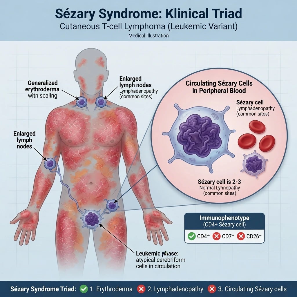
Sézary syndrome (SS) is an aggressive leukaemic cutaneous T-cell lymphoma (CTCL) within the WHO-EORTC spectrum, closely related to but distinct from mycosis fungoides (MF).[1][8] Classic teaching uses a diagnostic triad:
- Erythroderma (typically ≥80% body surface area)
- Lymphadenopathy
- Neoplastic circulating Sézary cells meeting B2 blood criteria [1]
SS accounts for a minority of CTCL (often cited around a few percent) but contributes disproportionately to CTCL morbidity and mortality.[2][8]
Erythrodermic MF can look identical on the skin yet lack B2 blood burden (B0/B1); blood staging is therefore decisive.[3][10]
Epidemiology
SS typically affects older adults, with a slight male predominance in many series. There is no single dominant environmental cause analogous to UV-driven keratinocyte cancers; pathogenesis is lymphoproliferative and immune-dysregulatory.[1][2] Diagnostic delay is common because erythroderma is attributed for months to eczema, psoriasis or drug eruption.[10]
Pathophysiology
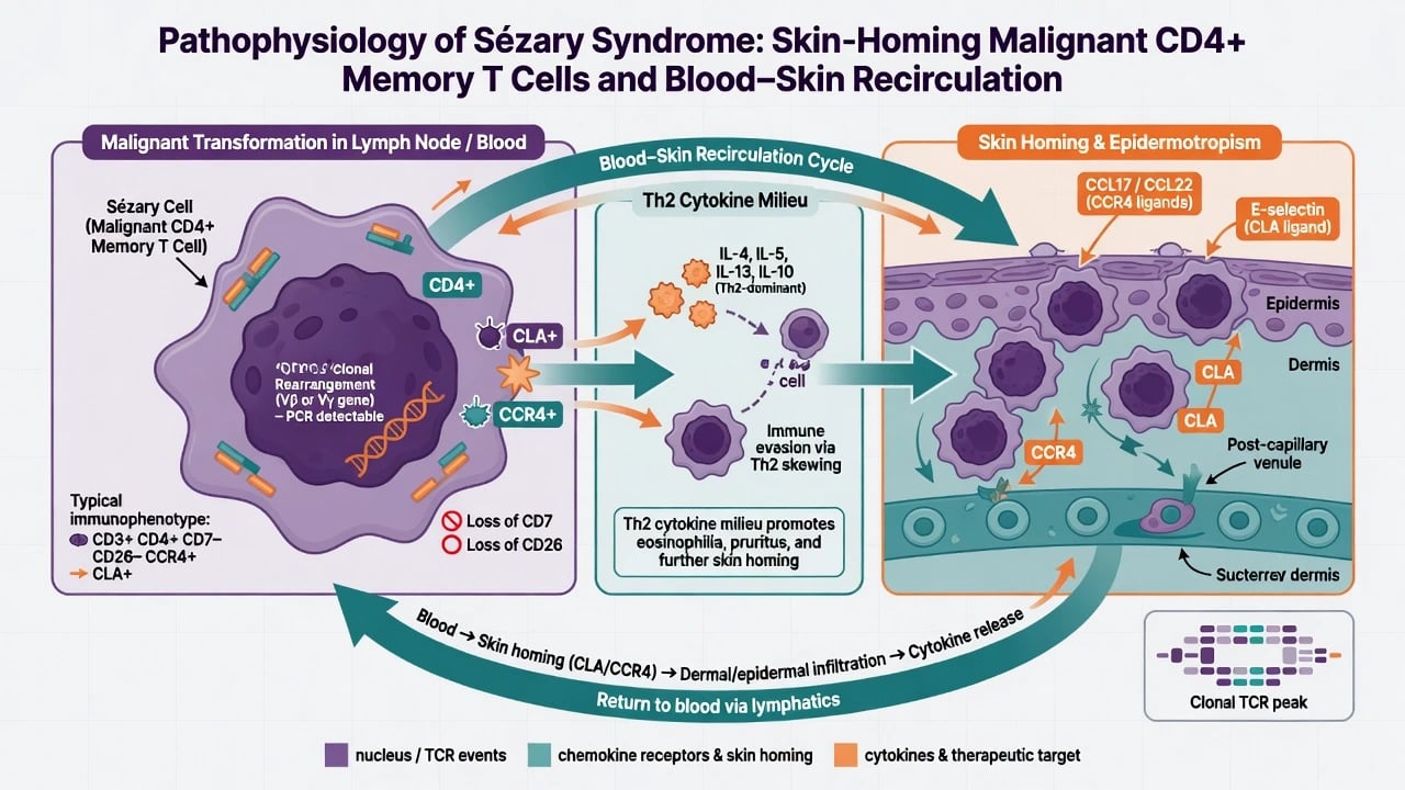
- Malignant cells are mature CD4+ memory T cells with skin-homing phenotype (CLA, CCR4).[8][1]
- Frequent loss of CD7 and CD26 on flow cytometry aids detection of the abnormal population.[10][2]
- Clonal T-cell receptor (TCR) gene rearrangement links skin and blood disease.[3]
- Th2 cytokine milieu (IL-4, IL-5, IL-13 and related signals) promotes pruritus, eosinophilia and impaired antimicrobial defence — partly explaining infection risk in erythroderma.[8]
- CCR4 expression underpins activity of mogamulizumab (anti-CCR4 monoclonal antibody).[6]
Clinical Presentation
- Generalised erythema and scale involving most of the body; ectropion, lagophthalmos risk, palmoplantar keratoderma, alopecia, and nail dystrophy are common accompaniments.[1][10]
- Intense pruritus often dominates quality of life.
- Lymphadenopathy — cervical, axillary and inguinal basins; nodes may be dermatopathic or frankly involved.[3]
- Systemic features — fatigue, temperature dysregulation, oedema, secondary infection, and occasionally B symptoms.
- Skin histology may be subtle compared with plaque MF; blood findings carry more diagnostic weight in classic SS.[10]
Differential Diagnosis of Erythroderma
| Differential | Distinguishing clues |
|---|---|
| Atopic erythroderma | Long atopic history; IgE; less often B2 clone/phenotype |
| Psoriasis | Prior plaques; nail pitting; histology |
| Drug-induced erythroderma / DRESS | Drug timeline; eosinophilia; visceral involvement |
| Pityriasis rubra pilaris | Islands of sparing, follicular keratoses, orange-red hue |
| Erythrodermic MF (B0/B1) | Same skin picture; blood does not meet B2 |
| Adult T-cell leukaemia/lymphoma | HTLV-1 serology; hypercalcaemia; flower cells |
| Idiopathic erythroderma | Exclusion diagnosis after full work-up |
Investigations
Blood (mandatory when SS suspected) [1]
- FBC and peripheral smear (cerebriform lymphocytes)
- Flow cytometry: CD3/CD4 population with CD7 and/or CD26 loss; CD4:CD8 ratio (often greater than 10 in B2 disease)
- Absolute Sézary cell count
- TCR clonality (PCR/NGS) on blood ± skin [1]
Skin [1]
- Biopsies (sometimes multiple) for histology, immunohistochemistry, and TCR studies — may show epidermotropism but can be non-specific in pure erythroderma.[10]
Staging body [1]
- Clinical node map; image-guided or excisional node assessment when indicated
- CT or PET-CT for nodal/visceral disease per centre protocol
- LDH, infection screen, viral serologies as relevant; baseline organ function before systemic therapy [1]
ISCL/EORTC TNMB Staging (Blood Is the Key)
Blood (B) categories — high yield
| B | Concept (teaching summary) |
|---|---|
| B0 | No significant blood involvement |
| B1 | Low blood burden (does not meet B2) |
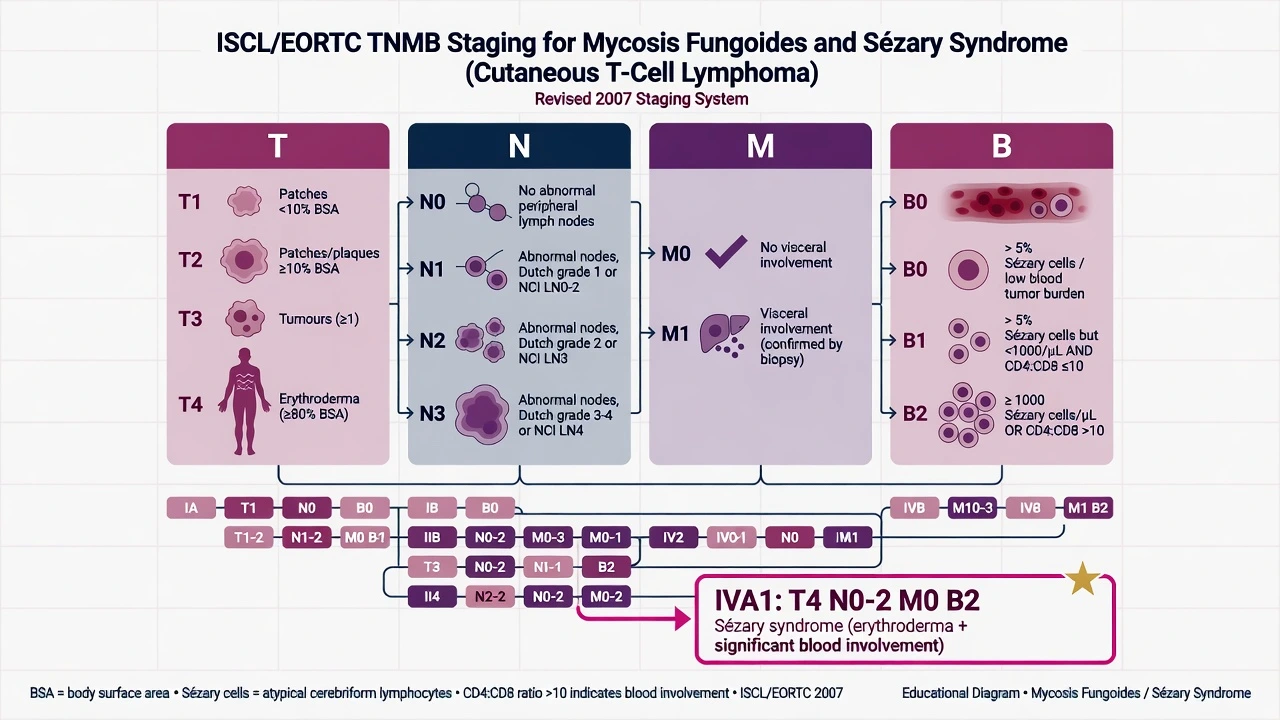
| B2 | High blood burden — e.g. ≥1000 Sézary cells/µL and/or CD4:CD8 greater than 10 with a matching T-cell clone (criteria operationalised in ISCL/EORTC documents) |
Updated ISCL/USCLC/EORTC work continues to refine blood definitions for trials, but the B2 concept remains central to SS diagnosis in clinical exams.[3][4]
Stage grouping
Classic SS with T4 N0–2 M0 B2 maps to stage IVA1.[3] Visceral disease (M1) advances stage further and worsens prognosis.[2]
[1]Management
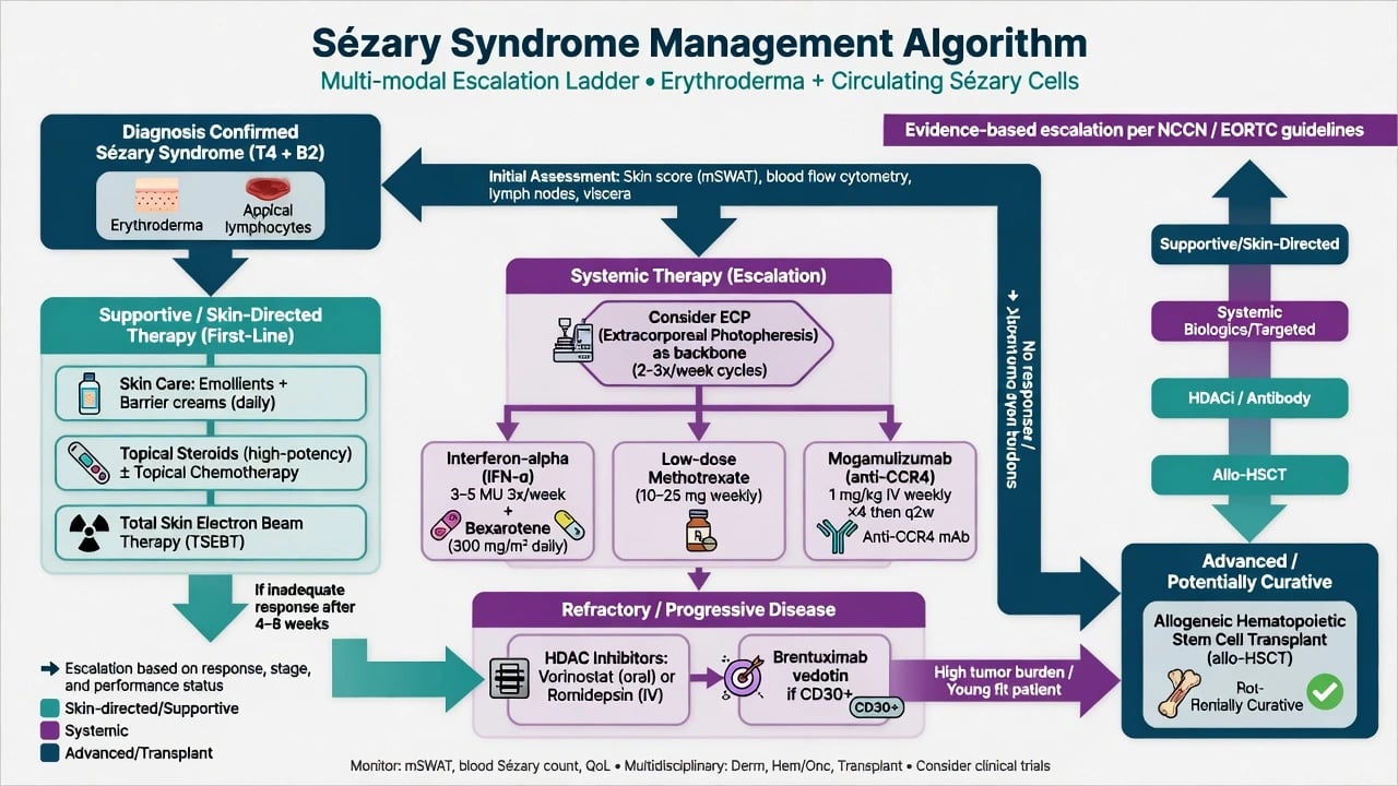
Management is rarely monotherapeutic. EORTC consensus recommendations organise stage-adapted multimodal care for MF/SS.[5]
Supportive / skin-directed foundations
- Emollients, bland skincare, temperature control
- Potent topical corticosteroids where tolerated
- Aggressive treatment of secondary infection (especially Staphylococcus aureus)
- Antipruritic strategies; nutrition and fluid balance in severe erythroderma
- Total skin electron beam therapy (TSEBT) may be used for extensive skin disease in selected pathways.[5]
Systemic and extracorporeal options
| Modality | Role |
|---|---|
| Extracorporeal photopheresis (ECP) | Backbone immunomodulatory therapy in many erythrodermic CTCL/SS pathways |
| Interferon-α | Systemic immunomodulation |
| Bexarotene | Oral rexinoid; monitor lipids/thyroid |
| Low-dose methotrexate | Cytostatic option in CTCL algorithms |
| Mogamulizumab (anti-CCR4) | Superior to vorinostat for progression-free survival in previously treated MF/SS (MAVORIC); particularly relevant in blood involvement |
| HDAC inhibitors (vorinostat, romidepsin) | Epigenetic therapy in relapsed/refractory disease |
| Brentuximab vedotin | If CD30 expressed (ALCANZA context in CD30+ CTCL) |
| Allogeneic HSCT | Only potentially curative modality for selected younger/fit patients with advanced/refractory disease |
ECP mechanisms and practical use in MF/SS are reviewed in dermatologic literature and remain exam-relevant.[9] MAVORIC established mogamulizumab versus vorinostat in previously treated CTCL, with meaningful activity in SS cohorts.[6] ALCANZA supports brentuximab in CD30-positive CTCL settings.[7]
Early MF (contrast)
- Patches/plaques, often B0
- Skin-directed therapy first
- Indolent course common
- Long survival if early stage
Sézary syndrome
- Erythroderma + B2 blood
- Systemic/multimodal from the start
- Infection and itch dominant
- Poorer prognosis; transplant in select cases
Multidisciplinary essentials
Dermatology + haemato-oncology ± radiation oncology ± transplant team; infection specialists when barrier failure is severe.[5][2]
Complications
- Sepsis and cutaneous infection through broken erythrodermic barrier
- Thermoregulatory failure, high-output cardiac strain, oedema
- Treatment toxicities: bexarotene metabolic effects; mogamulizumab-associated rash; cytopenias; transplant GVHD/mortality
- Progressive refractory disease and reduced performance status [1]
Prognosis
SS has a substantially worse prognosis than early-stage MF, with survival measured in years rather than decades for many patients; high blood burden, nodal involvement and poor performance status worsen outlook.[2][8] Goals often combine disease control with quality of life; curative intent is reserved for transplant candidates.
Special Populations & Access
- Elderly multimorbid patients may not tolerate multi-agent regimens or transplant — prioritise ECP, skin care, and least toxic systemic options.
- Resource-limited settings may lack ECP or mogamulizumab — still pursue accurate blood diagnosis, infection control, and referral pathways.[5]
Exam Pearls
SEZARY
SEZARY
T4 skin category
Part of classic triad
Circulating Sézary cells
CD4+ CD7− CD26−; CD4:CD8 often >10
Blood ± skin clonality
Not plain eczema
- Triad + B2 defines classic SS.[3]
- Separate erythrodermic MF (low blood burden) from SS.[10]
- ECP + systemic agents; mogamulizumab for advanced MF/SS after prior therapy.[9][6]
- Allo-HSCT = only potentially curative option in selected fit patients.[5]
- Always treat the skin barrier and infection, not only the clone.[1]
Exam application bank (NEET-PG / INICET)
One-line answer
Sézary syndrome (SS) is the aggressive leukaemic variant of cutaneous T-cell lymphoma defined by the triad of erythroderma, lymphadenopathy and circulating neoplastic Sézary cells (ISCL/EORTC B2 blood involvement). Malignant cells are skin-homing CD4+ memory T cells that typically lose CD7 and CD26, express CLA/CCR4, and show clonal TCR rearrangement. Most patients stage as TNMB IVA1 (T4 N0–2 M0 B2). Management is multimodal: intensive skin care and infection control, extracorporeal photopheresis, systemic immunomodulators (interferon, bexarotene, methotrexate), targeted agents (mogamulizumab anti-CCR4; brentuximab if CD30+), HDAC inhibitors, TSEBT, and allogeneic HSCT as the only potentially curative option in selected fit patients.
Worked stems (answer without another resource)
Stem 1 — Classic presentation. Map symptoms to mechanism; name the first investigation and first treatment step with dose/route if drug therapy is standard. [1]
Stem 2 — Unstable / complicated. List red flags that force immediate resuscitation, theatre, ICU, antidote, or reperfusion — and what you do in the first 15 minutes. [1]
Stem 3 — Atypical group. Elderly, pregnancy, child, or immunocompromised: how presentation and thresholds change. [1]
Stem 4 — Differential trap. Name the three closest mimics and one discriminator for each. [1]
Stem 5 — Disposition. Who goes home with safety-netting, who is admitted, who needs HDU/ICU/theatre, and what follow-up is mandatory. [1]
Rapid viva checklist
- Definition + classification
- Pathophysiology chain
- Bedside signs / criteria
- Score with exact components (if any)
- Emergency bundle
- Definitive therapy with doses
- Complications of disease and of treatment
- Special populations
- Guideline/trial name if classic
- Three exam traps
Coverage self-check
If you cannot answer any stem above from this page alone, re-read the matching section — the page is intended to be self-sufficient for final-prof and NEET-PG/INICET questions on Sézary syndrome.
Expanded exam teaching (depth pass)
Clinical reasoning
For Sézary syndrome, examiners test whether you can prioritise life threats, choose the right first test, and give specific therapy (agent, dose, route, timing). Generic phrases without numbers score poorly.
Mechanism → feature map
Build a short chain: cause → pathophysiologic intermediate → clinical feature → complication. Every major symptom in the classic vignette should sit on that chain.
Investigation strategy
- Bedside/first-line tests that change immediate management
- Confirmatory or staging tests
- What a normal result does not exclude
- When not to delay treatment for imaging (unstable patient)
Management ladder
- Resuscitation / ABC / sepsis or haemorrhage bundle as relevant
- Specific antidote / procedure / antimicrobial / reperfusion / surgery
- Supportive care and monitoring targets
- Definitive long-term therapy and secondary prevention
- Disposition and safety-net advice
Special populations
Always prepare one line each for children, pregnancy, elderly, renal/hepatic impairment, and immunocompromised patients when the topic allows.
Pitfalls that fail candidates
- Treating the number not the patient
- Missing pregnancy status when relevant
- Imaging before stabilisation
- Wrong empiric cover or wrong antidote timing
- Incomplete counselling on recurrence, adherence, or red-flag return
Sézary syndrome (SS) is the aggressive leukaemic variant of cutaneous T-cell lymphoma defined by the triad of erythroderma, lymphadenopathy and circulating neoplastic Sézary cells (ISCL/EORTC B2 blood involvement). Malignant cells are skin-homing CD4+ memory T cells that typically lose CD7 and CD26, express CLA/CCR4, and show clonal TCR rearrangement. Most patients stage as TNMB IVA1 (T4 N0–2 M0 B2). Management is multimodal: intensive skin care and infection control, extracorporeal photopheresi [1]
Structured revision sheet
Must-know numbers and names
List every score, size threshold, dose, and time window from this topic on a blank page from memory, then check against the sections above.
Three classic MCQ angles
- Most likely diagnosis given a vignette
- Next best step in management
- Most appropriate investigation
Three classic SAQ angles
- Pathophysiology in five steps
- Management algorithm with doses
- Complications and prevention
Clinical station flow
Greet → focused history → targeted exam → investigations → explain diagnosis → emergency care → definitive plan → safety-net / follow-up → answer examiner questions on mechanism and pitfalls.
References
- [1]Miyashiro D, Sanches JA. Mycosis fungoides and Sézary syndrome: clinical presentation, diagnosis, staging, and therapeutic management Front Oncol, 2023.PMID 37124514
- [2]Hristov AC, Tejasvi T, Wilcox RA. Mycosis Fungoides, Sézary Syndrome, and Cutaneous B-Cell Lymphomas: 2025 Update on Diagnosis, Risk-Stratification, and Management Am J Hematol, 2025.PMID 40495407
- [3]Olsen E, Vonderheid E, Pimpinelli N, et al. Revisions to the staging and classification of mycosis fungoides and Sezary syndrome: a proposal of the International Society for Cutaneous Lymphomas (ISCL) and the cutaneous lymphoma task force of the European Organization of Research and Treatment of Cancer (EORTC) Blood, 2007.PMID 17540844
- [4]Olsen EA, Whittaker S, Willemze R, et al. Primary cutaneous lymphoma: recommendations for clinical trial design and staging update from the ISCL, USCLC, and EORTC Blood, 2022.PMID 34758074
- [5]Latzka J, Assaf C, Bagot M, et al. EORTC consensus recommendations for the treatment of mycosis fungoides/Sézary syndrome - Update 2023 Eur J Cancer, 2023.PMID 37890355
- [6]Kim YH, Bagot M, Pinter-Brown L, et al. Mogamulizumab versus vorinostat in previously treated cutaneous T-cell lymphoma (MAVORIC): an international, open-label, randomised, controlled phase 3 trial Lancet Oncol, 2018.PMID 30100375
- [7]Prince HM, Kim YH, Horwitz SM, et al. Brentuximab vedotin or physician's choice in CD30-positive cutaneous T-cell lymphoma (ALCANZA): an international, open-label, randomised, phase 3, multicentre trial Lancet, 2017.PMID 28600132
- [8]Dummer R, Vermeer MH, Scarisbrick JJ, et al. Cutaneous T cell lymphoma Nat Rev Dis Primers, 2021.PMID 34446710
- [9]Zic JA. Extracorporeal Photopheresis in the Treatment of Mycosis Fungoides and Sézary Syndrome Dermatol Clin, 2015.PMID 26433848
- [10]Spicknall KE. Sézary syndrome-clinical and histopathologic features, differential diagnosis, and treatment Semin Cutan Med Surg, 2018.PMID 29719016