Dermatology · Medicine
Skin biopsy
Also known as Skin biopsy · Punch biopsy · Shave biopsy · Saucerisation · Excisional biopsy · Incisional biopsy · Curettage and electrodesiccation · Direct immunofluorescence biopsy · Alopecia biopsy
Skin biopsy is the diagnostic removal of a sample of living skin so it can be examined under the microscope. Five techniques cover almost every indication: punch biopsy (3-4 mm cylindrical blade → full-thickness epidermis, dermis and subcutaneous fat — the standard diagnostic procedure for inflammatory disease, alopecia and blistering disorders); shave biopsy / saucerisation (tangential cut with a flat blade → epidermis and upper dermis only — for raised benign lesions like seborrhoeic keratoses, viral warts and superficial BCC — NEVER for suspected melanoma); excisional biopsy (full-thickness elliptical excision along skin tension lines with 1-3 mm clinical margin — the diagnostic and sometimes therapeutic procedure for suspected melanoma and pigmented lesions); incisional biopsy (partial-thickness ellipse — for large lesions where complete excision is impractical, and for deep processes such as panniculitis); curettage and electrodesiccation (sharp curette followed by electrofulguration — a therapeutic rather than diagnostic modality for small low-risk BCC). Site selection (active edge of rash; edge of ulcer; fresh blister; comparison biopsy in alopecia; orientation along resting skin tension lines). Specimen handling is part of the procedure: 10% neutral buffered formalin for H&E, Michel's medium for direct immunofluorescence, glutaraldehyde for electron microscopy, sterile saline for microbiology. Fixative errors are unfixable: a DIF specimen placed in formalin destroys the immunoreactants and the test must be repeated.
On this page & tools
Your progress
Saved locally on this device.
Practise this topic
Exam tags
Red flags
Overview & Definition
A skin biopsy is the targeted removal of a sample of living skin so it can be examined under the microscope (and, increasingly, by molecular methods) to establish or refine a diagnosis, guide therapy, or complete staging. It is one of the most frequently performed outpatient procedures in dermatology and primary care — by volume second only to simple excision of benign skin lesions — and it underpins almost every decision in modern dermatopathology. The biopsy is not a single procedure but a small family: the choice of technique, the choice of site, the choice of fixative, and the choice of how the specimen is handled en route to the laboratory are all part of one integrated decision. [1]
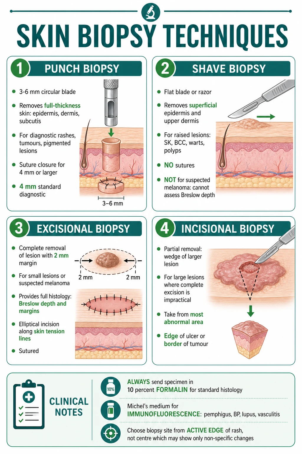
The depth of tissue each technique samples is the single most important diagnostic distinction. A punch biopsy, excisional biopsy and incisional biopsy all reach the subcutaneous fat, giving the dermatopathologist full thickness; a shave biopsy stops at the upper or mid-reticular dermis and so cannot answer questions that depend on deep extension (Breslow depth, margin assessment, panniculitis pattern). This is also why a shave biopsy of a suspected melanoma is a categorical error: the deepest tumour cell — the basis of Breslow thickness — is left behind in the patient, and the pathologist can only report what was transected.[3][4][5]
The classical answer set is four techniques (punch, shave, excisional, incisional). The modern answer set adds a fifth: curettage and electrodesiccation (C+E). Curettage is not primarily a diagnostic biopsy — it deliberately destroys tissue and forfeits margin control — but it is the same practitioner and the same procedural family, and examiners frequently ask why it is not appropriate for high-risk BCC or facial lesions.[1][2]
The second-tier layer, often ignored but routinely examined, is procedural quality: the choice of site (active edge vs centre, fresh vs old, ulcer rim vs ulcer base), the orientation (long axis of an elliptical excision along the resting skin tension lines), the layered closure (deep dermal buried absorbable + epidermal non-absorbable), the avoidance of crush artefact (lift the plug with a needle, never crush it with forceps), the avoidance of epinephrine in digits/nose/ear/penis, and the fixative discipline (H&E to formalin, DIF to Michel's medium, EM to glutaraldehyde). Each is a load-bearing step. Failure of any one derails the result. [1]
Classification — the five core techniques
The five skin biopsy techniques at a glance
| Technique | Tissue sampled | Commonest indication | Suture | Specimen | Key limitation |
|---|---|---|---|---|---|
| Punch biopsy (4 mm) | Full-thickness — epidermis, dermis, subcutis | Inflammatory dermatosis, alopecia, blistering disease (paired with DIF) | 1-2 sutures for ≥4 mm | Formalin (H&E) + Michel's (DIF) | None for most uses; can be too superficial for panniculitis |
| Shave biopsy (saucerisation) | Epidermis + upper/mid dermis | Raised benign lesions — SK, viral wart, skin tag, superficial BCC | None (heals by secondary intention) | Formalin | Never for suspected melanoma (cannot measure Breslow); inadequate for inflammatory disease |
| Excisional biopsy | Full-thickness ellipse along skin tension lines | Suspected melanoma; small complete lesions | Layered closure (deep dermal + epidermal) | Formalin, specimen inked for margins | Defect size limits application on the face |
| Incisional biopsy | Partial-thickness wedge | Large lesion where complete excision is impractical; deep processes (panniculitis) | Layered closure | Formalin ± culture | Risk of sampling error if the wrong area is taken |
| Curettage + electrodesiccation (C+E) | Tissue destroyed | Small low-risk BCC on trunk or extremities (therapeutic) | None (heals by secondary intention) | No histology specimen — destroyed | Inadequate margin assessment; not appropriate for face, recurrent tumours or aggressive histology |
Punch biopsy
- **4 mm cylindrical blade** (3 mm for cosmetic sites; 6 mm rarely) — reaches subcutis
- **Full-thickness diagnostic** for inflammatory disease, alopecia, blistering disorders
- Suture for ≥4 mm defects (1-2 sutures); ≤3 mm may heal by secondary intention
- Specimen in formalin (H&E); pair with Michel's medium for DIF (autoimmune blistering)
- **The workhorse diagnostic biopsy in dermatology**
Shave biopsy (saucerisation)
- **Tangential cut with flat blade** — epidermis + upper dermis
- **Raised benign lesions**: seborrhoeic keratosis, viral wart, skin tag, superficial BCC
- No sutures; haemostasis with aluminium chloride or pinpoint cautery
- Excellent cosmetic result on the face for raised benign lesions
- **Never for suspected melanoma** — transects lesion, loses Breslow depth
Excisional biopsy
- **Full-thickness elliptical cut** along skin tension lines with 2-3 mm clinical margin
- **Diagnostic and possibly therapeutic** for suspected melanoma
- Layered closure with deep dermal buried absorbable + epidermal non-absorbable sutures
- Specimen inked at margins so positive margins can be mapped on re-excision
- Orientation matters: long axis of ellipse = 3-4 × width to allow primary closure
Incisional biopsy
- **Partial-thickness wedge** of the most abnormal area
- **Large lesions** where complete excision is impractical or would cause deformity
- **Deep processes** such as panniculitis where subcutaneous fat must be included
- Layered closure; specimen representative of the whole lesion
- Risk of sampling error if the wrong area is taken — choose the indurated, ulcerated or most atypical edge
Curettage + electrodesiccation (C+E)
- **Sharp ring curette** removes friable tumour; **electrodesiccation** fulgurates the base and 1-2 mm rim
- **Therapeutic** (not diagnostic) modality for small low-risk BCC on the trunk
- Three-cycle C+E gives 5-year cure of 95-99% for low-risk nodular BCC under one centimetre
- **No histological specimen** — margins are ablated, so this is not a biopsy
- **Avoid on the face** (poor cosmetic result) or for recurrent / aggressive BCC (need histology)
The classification is more than a vocabulary; it prescribes depth, indication and limitation in one breath. Examiners expect candidates to choose a biopsy technique on first principles from the clinical scenario, not from memorised lists. The reasoning chain — what is the differential? what depth of tissue does each differential need? which technique reaches that depth? — is the one viva examiners want to hear. [1]
Quick self-test: match the technique to the scenario
A. Punch from active edge + Michel's perilesional punch; B. Saucerisation; C. Full-thickness elliptical along the resting skin tension lines; D. Deep incisional wedge into subcutis; E. Three cycles of curettage followed by electrodesiccation.
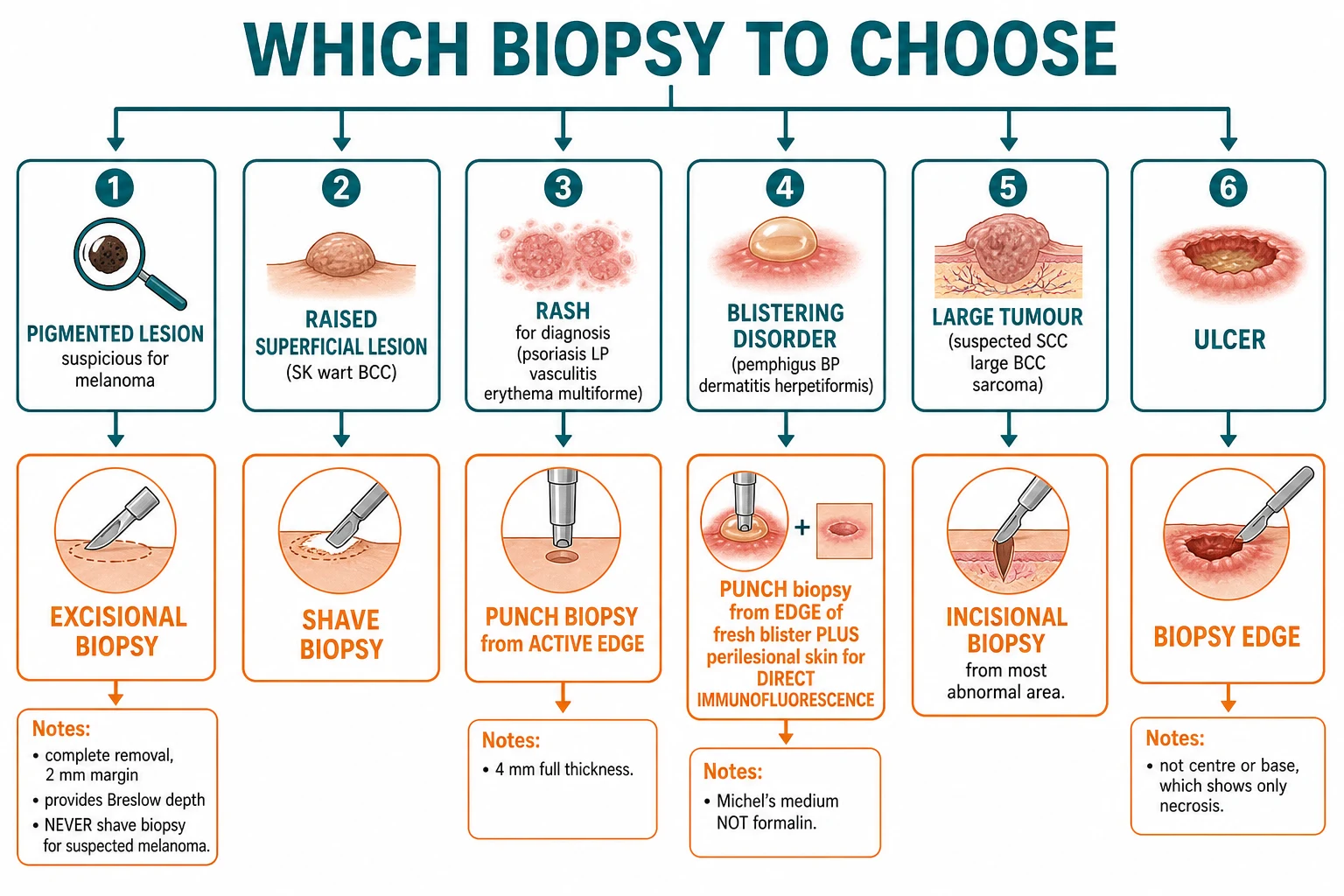
- A 7 mm pigmented lesion on the calf with an irregular border and colour variegation. → C — excisional biopsy (never shave a suspected melanoma).
- A tense bulla on the trunk of an 80-year-old — suspected bullous pemphigoid. → A — punch for H&E + perilesional punch for DIF in Michel's medium.
- A raised pedunculated seborrhoeic keratosis on the cheek. → B — shave (saucerisation).
- Tender erythematous nodules on the shins of a 35-year-old woman with recent streptococcal pharyngitis. → D — deep incisional wedge to capture the septal panniculitis of erythema nodosum.
- A 6 mm well-defined nodular BCC on the back of an 80-year-old who is a poor surgical candidate. → E — curettage + electrodesiccation; small, low-risk, primary, on the trunk.
Epidemiology & Risk Factors
Skin biopsy is one of the most common surgical procedures performed worldwide. In the United States alone, dermatologists perform several million skin biopsies annually; the indication mix is dominated by pigmented lesions (around one third of dermatology biopsies), followed by suspected non-melanoma skin cancer and inflammatory dermatosis. The lifetime probability of undergoing a skin biopsy in a fair-skinned population is approximately 20-30%, driven largely by the rising incidence of non-melanoma skin cancer and melanoma.[1]
The pre-test probability of malignancy shapes the biopsy threshold. In Australia, where melanoma incidence is among the highest in the world (approximately 40-60 cases per 100 000 per year and a lifetime risk approaching 1 in 38), pigmented lesion biopsy is liberal and the threshold for excision biopsy in primary care is low. In the United Kingdom, where the lifetime melanoma risk is closer to 1 in 100, the BAD / NICE NG12 pathway reserves urgent referral for lesions meeting specific dermoscopic or ABCDE criteria. In India and most of South-East Asia, melanoma incidence is far lower, and the dominant biopsy indications are inflammatory dermatoses, leprosy, cutaneous tuberculosis and post-kala-azar dermal leishmaniasis (PKDL); pigmented lesion excision is offered at a lower index, and the broader use of punch biopsy reflects cost-effective practice. [1]
Risk factors for being biopsied cluster by indication: [1]
Pigmented lesion biopsy — fair skin (Fitzpatrick I-II), history of melanoma or dysplastic naevus syndrome, multiple naevi (>100), giant congenital melanocytic naevus, atypical naevi, intermittent intense UV exposure (sunburn history). [1]
Inflammatory dermatosis biopsy — failure of empiric topical therapy; atypical morphology (cutaneous T-cell lymphoma is the great mimicker of chronic eczema); immunocompromise (HIV, transplant recipients); systemic features suggesting vasculitis or connective tissue disease. [1]
NMSC biopsy — cumulative UV exposure; fair skin; outdoor occupation; immunosuppression (solid-organ transplant recipients carry a 65-fold increased risk of cutaneous SCC and the lifetime prevalence of biopsy-confirmed NMSC approaches 50%); previous ionising radiation; genodermatoses (Gorlin, xeroderma pigmentosum). [1]
Patient factors that complicate the biopsy are also populations worth naming explicitly: Fitzpatrick IV-VI (post-inflammatory hyperpigmentation and keloid scarring are higher); bleeding diatheses and anticoagulation; diabetes and other causes of impaired wound healing; immunosuppression; pregnancy (where some biopsies are deferred); and age — neonates (different anatomy and consent), children (behavioural management), and the elderly (anticoagulation, fraility, atypical presentations, amelanotic melanoma). [1]
Pathophysiology
Why depth determines diagnosis
A biopsy is a depth-specific operation. Spongiotic dermatitis is diagnosed by the appearance of intercellular oedema in the epidermis (a punch biopsy reaches it). Bullous pemphigoid is diagnosed by subepidermal cleft formation with eosinophils and linear IgG/C3 at the basement membrane (a punch biopsy reaches it). Nodular melanoma is staged by measuring the distance from the granular layer to the deepest tumour cell, which is by definition full-thickness. Panniculitis is diagnosed by the pattern of inflammation in subcutaneous fat, which a punch biopsy may only sample serendipitously and which a deep incisional ellipse reliably reaches.[1][8]
The cellular events a biopsy captures depend on the age of the lesion at the moment of biopsy. Leukocytoclastic vasculitis has its diagnostic neutrophilic infiltrate and nuclear dust in lesional skin aged 24-48 hours; biopsying a week-old haemorrhagic macule yields only haemosiderin and non-specific necrosis. Dermatitis herpetiformis has neutrophil-rich papillary microabscesses in lesional skin aged 12-48 hours; biopsying established urticarial plaques misses them. Pemphigoid blisters are best characterised in fresh lesions, but for DIF the immunoreactants are degraded in lesional skin — the rule is that DIF samples perilesional skin rather than the blister itself. [1]
Why site selection determines diagnosis
The site chosen is part of the pathology request. For inflammatory disease, the active edge — the expanding front of a plaque — is where early diagnostic features are concentrated (spongiosis, vacuolar interface change, leukocytoclastic debris). For an ulcer, the edge captures the junction between diseased and normal tissue (granulation tissue, neovascularisation, atypical epithelial proliferation). For alopecia, the active edge of recent hair loss is where active inflammation is preserved; the comparison biopsy from clinically uninvolved scalp is the pathologist's control. For a panniculitis, the biopsy must extend into the subcutis (a 4 mm punch often fails here — an incisional ellipse is the rule). [1]
Why the orientation of an elliptical biopsy determines the scar
The skin is not isotropic. Resting skin tension lines (Kraissl's lines, after the anatomist who mapped them) run perpendicular to the underlying facial muscles and reflect the long axis along which a wound closes with least tension. Excisions and incisional biopsies drawn along these lines produce narrow, fine-lined scars; those drawn across them produce wide, depressed or hypertrophic scars. On the face the lines run horizontally on the forehead, obliquely on the cheek, and circumferentially around the eye and mouth; on the trunk they run transversely; on the limbs they run longitudinally on the long axis. The classic teaching — draw the long axis of an excision 3-4 times the width, parallel to the resting skin tension lines — is the single most reproducible manoeuvre to optimise any scar.[2]
Why the layered closure determines the outcome
A layered closure transfers tension from the skin surface to the deeper dermis, where it is borne by buried absorbable sutures that dissolve over weeks. The epidermal layer is then approximated with fine non-absorbable sutures (or adhesive strips for low-tension wounds) that are removed at 5-7 days on the face and 7-10 days elsewhere. This is the standard for any defect that cannot close without tension. A single-layer closure under tension produces a stretched scar; a layered closure under no tension produces a fine-line scar. The suture selection itself follows anatomy: 6-0 or 7-0 on the face (periorbital, perioral), 5-0 elsewhere on the face, 4-0 on the trunk and limbs, 3-0 on the scalp. [1]
Crush artefact, the silent pathology killer
The most common avoidable artefact is crush artefact, produced when forceps clamp the dermal or subcutaneous edge of a punch biopsy cylinder. The cell nuclei shear, elastin and collagen are compressed, and the diagnostic detail in the deepest part of the specimen is lost. The fix is mechanical and uncompromising: lift the cylinder with a 25-G needle or a single-tooth hook; never crush it. If a forceps is used at all, it must grasp only the very edge of the subcutaneous fat, not the dermis. When the specimen is bisected for processing, the cut is made with a fresh scalpel blade, not by tearing. The pathologist reading the slide cannot recover what the operator has crushed.[1][18]
Why fixative choice is unfixable
Three rules govern the fixative table: [1]
- 10% neutral buffered formalin for routine H&E. Formalin cross-links proteins by methylene bridges, preserves cellular architecture, and is the universal fixative for dermatopathology. The tissue-to-formalin ratio should be at least 10:1, and the specimen should be immersed within minutes (no more than 30) of removal to prevent drying artefact.[18]
- Michel's medium (ammonium sulphate-based) for direct immunofluorescence. Michel's preserves IgG, IgA, IgM, C3 and fibrinogen for up to 5 days at room temperature, allowing transport from peripheral clinics to reference laboratories. Formalin destroys these immunoreactants — placing a DIF specimen in formalin is an irrecoverable error and the test must be repeated. A comparative study found Michel's medium equivalent to honey as a transport medium for DIF, and superior to plain saline.[6][7]
- 2.5% glutaraldehyde in cacodylate buffer for electron microscopy. Glutaraldehyde cross-links proteins more rapidly and completely than formalin, preserving ultrastructural detail (basement membrane zone in inherited epidermolysis bullosa, viral inclusions, lysosomal storage diseases).
- Sterile saline (no fixative) for microbiology and PCR. The specimen is transported fresh, or inoculated onto culture media at the bedside. Sabouraud dextrose agar for fungi; Löwenstein-Jensen or BACTEC for mycobacteria; PCR buffer for HSV, VZV, mycobacteria, Leishmania, Treponema.
Fixative and timing facts
Clinical Presentation — what the lesion tells the operator
The biopsy is chosen by reading the lesion, not by route through the textbook. The five patterns that matter at viva: [1]
Pigmented lesion where melanoma cannot be excluded. Asymmetry, border irregularity, colour variegation, diameter >6 mm, evolution. Any pigmented lesion meeting one or more criteria is biopsied by excisional biopsy with 2-3 mm clinical margin, oriented along the skin tension lines, with the long axis 3-4 times the width to allow primary closure.[3][4][5]
Inflammatory dermatosis with diagnostic uncertainty. An atypical, refractory or treatment-resistant rash lasting more than 6 weeks. The 4 mm punch biopsy is taken from the active edge of a fully evolved lesion — neither the freshest papule nor the oldest plaque — and includes subcutaneous fat. The requisition must list the clinical differential (not just "rash") so the pathologist can direct special stains. [1]
Blistering disease. Tense bullae on the trunk and limbs of an elderly patient (bullous pemphigoid); flaccid bullae and mucosal erosions in a younger patient (pemphigus); grouped vesicles on extensor surfaces (dermatitis herpetiformis). Two biopsies are mandatory: a 4 mm punch from the edge of a fresh blister for H&E in formalin, and a separate 4 mm punch from perilesional (1-2 cm away) normal-appearing skin for DIF in Michel's medium. The DIF rule is inviolable: a specimen placed in formalin is a wasted specimen.[6][7]
Panniculitis. Deep, tender, indurated nodules on the legs or trunk. A 4 mm punch biopsy is frequently too superficial and the diagnosis is missed; the procedure of choice is an incisional ellipse extending into subcutis to capture the septal or lobular pattern. The requisition should specify the suspected pattern (erythema nodosum, erythema induratum, pancreatic panniculitis, lupus profundus, subcutaneous panniculitis-like T-cell lymphoma, alpha-1 antitrypsin deficiency panniculitis).[8][9]
Alopecia. Cicatricial (scarring) alopecia where follicular ostia are lost. Biopsy from the active edge of recent hair loss with a 4 mm punch, plus a second 4 mm punch from uninvolved scalp several centimetres away for comparison. The transverse (horizontal) section is the modern standard because it permits accurate terminal-to-vellus hair ratio (normal greater than 7:1), follicular counts and assessment of perifollicular inflammation. The pathology of LPP, FFA, DLE, folliculitis decalvans, dissecting cellulitis and central centrifugal cicatricial alopecia can be distinguished with confidence only on horizontally sectioned biopsies.[10][11]
The atypical presentation in the elderly deserves separate emphasis: an ulcerated or bleeding lesion on sun-damaged skin that fails to heal in 6 weeks is amelanotic melanoma until proven otherwise; biopsy the edge of the ulcer, not the necrotic base. In the immunocompromised host, multiple or atypical lesions warrant biopsy at a lower threshold, with paired specimens for microbiology and PCR added to the dermatopathology jar. [1]
Differential Diagnosis — when the technique must be chosen by the differential
Alopecia. Scarring alopecia — LPP, FFA, DLE, folliculitis decalvans, dissecting cellulitis, central centrifugal cicatricial alopecia — is irreversible and biopsy is urgent. Non-scarring alopecia — androgenetic, alopecia areata, telogen effluvium, anagen effluvium, trichotillomania, traction — is potentially reversible and biopsy is reserved for diagnostic uncertainty. The biopsy interpretation hinges on the transverse section technique which gives follicular density and terminal-to-vellus ratio.[10][11]
The differential cannot-miss: [1]
- Invasive melanoma — any pigmented lesion where the diagnosis cannot be clinically excluded. Ng and colleagues showed that partial (incisional or punch) biopsy of melanocytic lesions upstaged 22% of cases compared with excisional biopsy — a statistic any examiner will accept as the answer to "why not just punch the mole?".[3]
- Subcutaneous panniculitis-like T-cell lymphoma — an atypical panniculitis in an adult or child with B symptoms; biopsy must reach subcutis.
- Dermatofibrosarcoma protuberans — a slow-growing indurated plaque on the trunk of a young adult; standard excision is associated with local recurrence because the slender CD34-positive spindle cells infiltrate fat in a honeycomb pattern with extensive subclinical extension; the biopsy is followed by Mohs (slow-Mohs with CD34 immunostain) which reduces recurrence to under 2-5%.[12][13][16]
- Cutaneous T-cell lymphoma — patch-stage mycosis fungoides mimicking eczema in an adult; biopsy must reach subcutis and include multiple sections from the active edge.
Clinical & Bedside Assessment — site, technique and consent
Pre-biopsy assessment is structured into four steps. Confirm the diagnosis and the indication. The lesion is examined clinically and with dermoscopy where available; clinical photographs are taken before the procedure. Review the patient. Allergies (chlorhexidine, latex, lidocaine, adhesive, antibiotic), anticoagulant and antiplatelet therapy, pacemaker or implantable defibrillator, immunisation status (tetanus for wounds if relevant), pregnancy, breast-feeding, and the ability to consent and lie still for the procedure. Review the indication and consider whether Mohs is more appropriate than standard excision as the definitive procedure after biopsy (H-zone of the face, recurrent tumour, aggressive histology, DFSP). Consent is taken for the procedure, anaesthesia, specimen handling, photography, the possibility of further surgery if margins are positive or histology is unexpected, and the warning signs that warrant return (bleeding, infection, dehiscence). [1]
Site selection is part of the request. For inflammatory disease, the fully evolved active edge of a representative lesion is chosen; for an ulcer, the edge of the lesion; for a tumour, the most indurated or atypical area; for alopecia, the active edge of recent hair loss plus a comparison biopsy from uninvolved scalp; for suspected melanoma, the lesion itself is excised with a 2-3 mm clinical margin. For subungual lesions the biopsy is taken from the nail matrix (longitudinal excision of a pigmented streak) or nail bed, not the nail fold, unless the latter is clinically involved. [1]
Skin tension lines govern the orientation of any elliptical incision. The skin of the periocular region, the perioral region and the preauricular cheek is incised along the radial lines parallel to the lid margin or lip vermilion; the forehead is incised transversely; the cheek is incised along an oblique line from the lateral canthus to the angle of the mandible; the neck is incised transversely; the trunk is incised along the lines of maximal skin tension (often horizontal on the chest and abdomen); the limbs are incised along the long axis. A 3-4:1 length-to-width ellipse allows primary closure without dog-ears; the half-buried mattress suture or a simple running subcuticular is the standard layered closure technique. [1]
Local anaesthetic is administered before incision. The standard regimen is 1% lidocaine with adrenaline 1:100 000 — a vasoconstrictor that extends the duration of action and reduces bleeding. The maximum safe dose is 7 mg/kg with adrenaline and 4.5 mg/kg plain (the addition of adrenaline roughly doubles the safe dose). Allow 5-10 minutes for vasoconstriction before incision. The absolute contraindication to adrenaline-containing solutions is traditionally taught for digits, nose, ear and penis (the "end-arteries"), but contemporary series suggest that low-concentration adrenaline in these sites is generally safe; nevertheless the conservative practice is to use plain lidocaine in these sites, both because the convention is upheld by most examiners and because rare cases of digital tip necrosis leading to amputation have been published.[14][15]
The following pre-procedural details are examinable: [1]
- Anaesthetic onset — 1-2 minutes; maximum effect at 5-10 minutes.
- Maximum lidocaine doses — 7 mg/kg with adrenaline, 4.5 mg/kg plain (for a 70-kg adult this corresponds to roughly 50 mL of 1% lidocaine with adrenaline or 31 mL of 1% plain).
- Topical anaesthetic alternatives — LET (lidocaine 4%, epinephrine 0.1%, tetracaine 0.5%) for paediatric facial lacerations; EMLA (eutectic mixture of lidocaine and prilocaine) cream under occlusion for 45-60 minutes for superficial procedures in children.
- Anticoagulation policy — contemporary practice continues anticoagulation through biopsy because bleeding from a superficial skin procedure is easier to manage than the thromboembolic risk of interruption. The decision is individualised for high-risk patients (mechanical heart valve, recent stent, atrial fibrillation with prior stroke).
- Pacemaker / ICD — bipolar electrocautery is used; battery-operated heat cautery is the universal fallback when monopolar is contraindicated. [1]
Investigations
Routine skin biopsy rarely needs laboratory testing. The investigation list is triggered by history: [1]
- Coagulation screen only if the patient has a bleeding disorder or is on warfarin with an unknown INR. Aspirin, clopidogrel and DOACs do not require pre-biopsy testing; the bleeding time and platelet count are unhelpful.
- Imaging for deep infiltrative tumour or perineural invasion — MRI of the cranial nerves for facial SCC or BCC with histological perineural invasion; ultrasound of regional nodes for palpable nodes in SCC, melanoma or Merkel cell; CT of the chest for suspected pulmonary metastasis in advanced disease.
- Anogenital or mucosal biopsy — a swab for sexually transmitted infection is taken at the same encounter where the differential includes HSV, syphilis or HPV; HIV, hepatitis B and C serology may be indicated by risk.
- Connective tissue disease serology when the differential includes lupus (ANA, anti-dsDNA, anti-Ro, anti-La, ENA panel), dermatomyositis (anti-Jo-1, anti-Mi-2, anti-TIF1γ, anti-NXP2), or vasculitis (ANCA, ANA, complement, cryoglobulins, hepatitis B and C serology). These are sent before the biopsy is interpreted, because the histology is interpreted in the serological context.
- Microbiology of the biopsy itself — when the differential includes an infection (leprosy, cutaneous TB, atypical mycobacteria, leishmaniasis, deep fungal infection, syphilis), the specimen is divided: one portion in formalin for histology with special stains (Fite, Ziehl-Neelsen, Giemsa, Warthin-Starry, PAS), a second portion in sterile saline for culture and PCR. [1]
The investigation that matters at the procedural level is the specimen handling table. The five bins are H&E, DIF, EM, microbiology and PCR; the medium for each is fixed by the technique, and the rule is to put the right tissue in the right medium without ambiguity: [1]
Specimen handling by indication
| Indication | Medium | Notes |
|---|---|---|
| H&E (most biopsies) | 10% neutral buffered formalin | Tissue: formalin 1:10; immersion within 30 minutes |
| Direct immunofluorescence (DIF) | Michel's medium | Perilesional skin within 1-2 cm of blister; never formalin |
| Electron microscopy | 2.5% glutaraldehyde in cacodylate buffer | Reserved for EM units (inherited EB, viral inclusions) |
| Microbiology culture | Sterile saline (no fixative) | Sterile container; culture for bacteria, fungi, mycobacteria |
| PCR (HSV, VZV, mycobacteria, Leishmania, Treponema) | Saline or specific viral transport medium | Sterile collection; refer to laboratory-specific protocol |
| Alopecia biopsy | Formalin for vertical section; separate specimen for horizontal (transverse) section | Two punches recommended — one for routine H&E, one for transverse |
A note on alopecia biopsy technique. The 4 mm punch is taken in the standard fashion, but instead of being bisected longitudinally and submitted in one cassette, the second 4 mm punch is submitted for transverse sectioning: the cylinder is bisected horizontally at the level of the mid-dermis, and both halves are embedded face-down, allowing the microtome to section perpendicular to the long axis of the hair shafts. This generates many more follicles per slide and allows accurate terminal-to-vellus ratio (normal above 7:1) and follicular density per mm². The interpretation distinguishes LPP (lichenoid interface around the infundibulum and isthmus with peri-follicular fibrosis), FFA (lymphocytic infiltrate around vellus follicles with perifollicular erythema and fibrosis), DLE (interface dermatitis with basement membrane thickening, follicular plugging and periadnexal infiltrate), folliculitis decalvans (intrafollicular neutrophilic infiltrate with follicular rupture), and central centrifugal cicatricial alopecia (premature desquamation of the inner root sheath with peri-follicular fibrosis).[10][11]
Management — the procedure as a sterile operation
Universal aseptic precautions
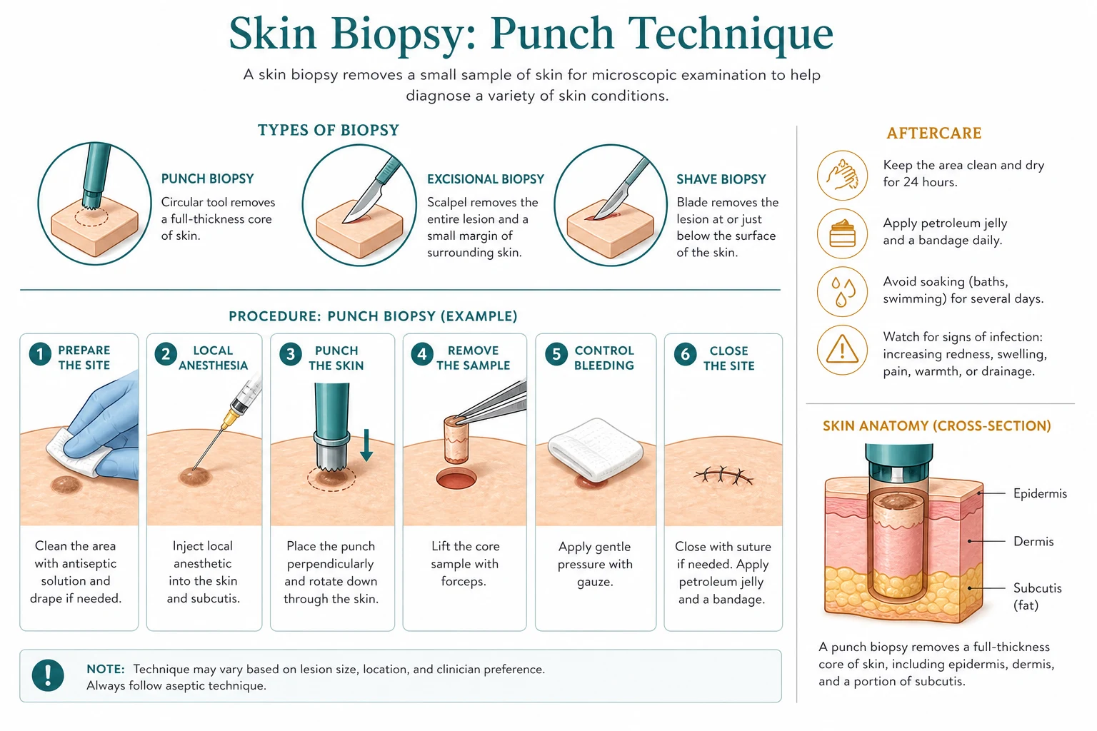
Skin biopsy is an aseptic procedure but not a sterile one. Skin preparation with chlorhexidine 0.5% in alcohol or povidone-iodine is standard; allow the antiseptic to dry before incision (chlorhexidine 30 seconds, iodine 60-120 seconds). Sterile gloves and instruments are used. Single-use disposable punch blades are strongly preferred; reusable punches are autoclaved between uses and inspected for dulling (a dull punch crushes the specimen). [1]
Position and exposure
The patient is positioned for direct lighting and ergonomic operator access. Trunk and limb biopsies are best done supine; face and scalp biopsies sitting; lateral for lateral face. Mark the intended ellipse before injection — local anaesthetic distorts the landmarks. [1]
Procedure — punch biopsy
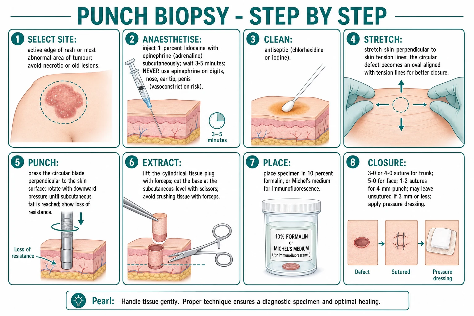
- Select the active edge of a fresh, fully evolved lesion.
- Inject 1% lidocaine with adrenaline subcutaneously (or intradermally for shave); 0.5-1 mL per site is typical for a small punch; larger excisional biopsies require more.
- Clean with chlorhexidine; allow to dry.
- Stretch the skin perpendicular to the resting skin tension line — this converts the circular defect into an oval aligned with the lines and allows easier closure.
- Apply the punch blade perpendicular to the skin surface, rotate clockwise and counter-clockwise with gentle downward pressure until a sudden loss of resistance is felt (the blade has crossed the dermal-subcutaneous junction).
- Lift the cylinder with a needle or single-tooth hook. Never crush with forceps.
- Snip the base with iris scissors at the subcutaneous level.
- Place the specimen directly in the appropriate medium (formalin for H&E; Michel's medium for DIF; sterile saline for microbiology).
- Achieve haemostasis — pressure, figure-of-eight suture for a 4 mm defect, or topical haemostatic agent (absorbable gelatin sponge, thrombin).
- Close with 1-2 interrupted sutures if the defect is 4 mm or larger; ≤3 mm defects heal by secondary intention.
- Apply a non-adherent dressing with petrolatum gauze; the dressing is changed at 24-48 hours. [1]
Procedure — shave biopsy
- Infiltrate local anaesthetic under or adjacent to the lesion.
- Hold the lesion gently between thumb and forefinger (the lesion itself — not the surrounding skin).
- Sweep a No. 15 or No. 22 blade tangentially under the lesion, producing a saucerised disc. The depth is operator-controlled; the goal is removal of the visible lesion with a thin margin of normal-appearing epidermis and a sliver of upper dermis.
- Achieve haemostasis with aluminium chloride 20% or pinpoint electrocautery.
- Send the specimen in formalin.
- Apply a non-adherent dressing. [1]
Procedure — excisional biopsy
- Draw the planned ellipse before anaesthetic infiltration, oriented along the resting skin tension lines, length:width 3:1 to 4:1.
- Infiltrate local anaesthetic along the proposed incision lines.
- Incise with a No. 15 blade along both sides of the ellipse; the incision should be perpendicular to the skin and full-thickness into subcutaneous fat.
- Undermine the wound edges laterally by 0.5-1 cm to relieve tension.
- Excise the ellipse from the fat with curved iris scissors or a scalpel; the deep margin should include a sliver of subcutaneous fat.
- Achieve haemostasis (bipolar or battery-operated cautery, ties for larger vessels).
- Close in layers:
- Deep dermis — buried interrupted sutures of 4-0 or 5-0 absorbable material (polyglactin 910, polydioxanone, poliglecaprone) on a tapered needle. Knots are buried deep to the dermis to prevent spitting.
- Epidermis — running or interrupted sutures of 5-0 or 6-0 non-absorbable monofilament (nylon, polypropylene) on a cutting needle. Adhesive strips or tissue glue as adjuncts.
- Ink the specimen at the margins with multiple colours (or use sutures to orient) so that positive margins can be mapped on re-excision.
- Send in formalin.
- Apply a non-adherent dressing with a pressure pad if a flap has been raised. [1]
Procedure — incisional biopsy
- Choose the most abnormal area (most indurated, most pigmented, edge of ulcer).
- Draw a partial-thickness ellipse along the skin tension lines, length:width 2:1 or 3:1.
- Infiltrate, incise, undermine and excise as for an excisional biopsy, but only a portion of the lesion is removed. The wedge should include full-thickness into subcutaneous fat, particularly for panniculitis.
- Close in layers.
- Document the precise location and the orientation of any further procedure (the rest of the lesion may require complete excision based on the result). [1]
Procedure — curettage and electrodesiccation
- Infiltrate local anaesthetic.
- Use a sharp ring curette (Volkmann or Fox) to scrape away the tumour — the friable tumour offers little resistance and the curette falls off as it reaches normal dermis, which gives a tactile estimate of margin.
- Electrodesiccate the wound base and a 1-2 mm rim with diathermy at low power.
- Repeat the cycle two further times (three cycles is the standard for low-risk BCC).
- The wound heals by secondary intention; no specimen is submitted for histology.[1]
Post-procedure
The wound is reviewed at 1-2 weeks; sutures are removed at 5-7 days on the face and 7-14 days elsewhere. Wound care: keep clean and dry for 24-48 hours, then gentle washing; apply petrolatum to keep moist; protect from sun for 12 months. Safety-net instructions: return for spreading erythema, purulent discharge, dehiscence, or any new symptoms. [1]
Escalation triggers to dermatology / plastic surgery
- Positive or close margins (especially melanoma, SCC, DFSP).
- Histological surprise (incidental melanoma in a shave biopsy).
- Lesion on the face where specialist reconstruction is required.
- Suspected recurrence or persistent disease.
- Complex anatomical site requiring flap or graft reconstruction.
- Patient factors — anticoagulated, immunocompromised, paediatric, pregnancy. [1]
The Mohs vs wide local excision question
Mohs micrographic surgery (MMS) is the dominant technique in the United States for high-risk NMSC on the H-zone of the face, for recurrent tumours, for aggressive histological subtypes (infiltrative, morpheaform, micronodular BCC, poorly differentiated SCC, perineural invasion), for DFSP, and for specific infiltrative tumours (atypical fibroxanthoma, Merkel cell carcinoma, microcystic adnexal carcinoma). Cure rates are the highest of any skin cancer treatment — 98 to 99% for primary BCC, 94 to 96% for recurrent BCC, 96 to 97% for primary SCC. The 100% margin assessment on horizontal en face frozen sections distinguishes Mohs from wide local excision, which samples under 1% of the margin by bread-loafing.[12][13][16]
Wide local excision (WLE) remains the rule in Europe, Australia and India. For low-risk BCC on the trunk, a 4 mm clinical margin is adequate (90-95% cure); for SCC, a 4-6 mm margin is adequate (90-95% cure for low-risk lesions). WLE is widely available, faster, and cheaper than Mohs; the trade-off is a higher rate of positive margins in high-risk lesions and a higher recurrence rate over five years. [1]
The regional delta is the deliberate answer to an examiner who asks why the same lesion is biopsied differently in New York, London and Sydney. In the US, the biopsy is followed by Mohs for high-risk H-zone tumours; in the UK (BAD / NICE NG12) and Australia (Cancer Council), the biopsy is followed by WLE with predetermined margins. The diagnostic biopsy is the same; the choice of definitive excision is the regional delta.[16][17]
Specific Subtypes & Scenarios
Pigmented lesion biopsy
The procedure is an excisional biopsy with 2-3 mm clinical margin along the long axis of the lesion, oriented along the skin tension lines. The incision is a 3-4:1 ellipse. The depth is full thickness into subcutaneous fat. The specimen is inked at margins so positive margins can be mapped. If the lesion is too large or anatomically awkward to excise in primary care (facial lesion >1 cm, acral lesion, lesion crossing an anatomical subunit), a punch biopsy of the most atypical area (darkest, most raised, ulcerated) is acceptable as a diagnostic biopsy but does not give margin information and is a second-line procedure; the patient should be referred for definitive excision regardless. Shave biopsy of a suspected melanoma is an absolute contraindication; partial biopsy of any melanocytic lesion upstaged 22% of cases in the Ng series.[3][4][5]
Lentigo maligna (melanoma in situ on sun-damaged skin)
Lentigo maligna is the one melanocytic lesion where Mohs is being used as a diagnostic biopsy, because subclinical extension on chronically sun-damaged skin is notorious. Standard 5 mm clinical margins clear only about 60-70% of lesions; Mohs with MART-1 immunostaining (or slow-Mohs with permanent paraffin sections) reduces recurrence to under 2%.[17]
Inflammatory dermatosis
The procedure is 4 mm punch biopsy from the active edge, including subcutaneous fat. For vesiculobullous disease, a paired perilesional punch is taken for DIF in Michel's medium. For panniculitis, an incisional wedge into subcutis is taken because the pathology lives in the fat. The pathologist's requisition should list the clinical differential, not just "rash — please section". [1]
Alopecia
The procedure is a paired 4 mm punch biopsy — one from the active edge of recent hair loss and one from uninvolved scalp several centimetres away for comparison. The active-edge biopsy is bisected vertically and submitted in formalin for vertical H&E; the comparison biopsy is submitted for transverse (horizontal) sectioning because the terminal-to-vellus ratio is interpretable only in transverse section.[10][11]
The normal terminal-to-vellus ratio is greater than 7:1. A ratio below 4:1 supports androgenetic alopecia in pattern distribution, alopecia areata (with peribulbar lymphocytic infiltrate), or telogen effluvium (with anagen-to-telogen ratio below the normal 12:1). Follicular density by area is also informative: a count below the 5th percentile for the scalp region supports LPP or FFA when combined with perifollicular fibrosis and a lymphocytic infiltrate. [1]
Nail biopsy
The procedure is performed under digital nerve block with plain lidocaine (no adrenaline — the terminal vessels of the digit are end-arteries, and very rare necrosis has been reported). The nail plate may be removed partially with a nail elevator to access the nail bed. A 3 mm punch through the nail plate into the matrix or bed is performed for a discrete lesion. For longitudinal melanonychia (a pigmented streak) the modern procedure is a longitudinal excision of the matrix that captures the full width of the streak and allows orientation; this can produce permanent nail dystrophy and is reserved for lesions suspicious for subungual melanoma. [1]
Genital and mucosal biopsy
Punch or shave biopsy with topical or infiltrated anaesthetic. For the penis the standard is plain lidocaine (no adrenaline). Mucosal wounds are clean and heal rapidly. [1]
Paediatric biopsy
LET cream (lidocaine, epinephrine, tetracaine) under occlusion for 30-45 minutes before procedure reduces the need for injection. Distraction techniques (bubble-blowing, tablet video, parental presence) are standard. The 3 mm punch is preferred for facial cosmesis; 2 mm punches exist for eyelid or vermilion biopsy. The differential for paediatric pigmented lesions includes congenital melanocytic naevi (where biopsy of changing areas is recommended before puberty), Spitz naevi (where the classic "Spitzoid" lesion is biopsied completely because it is histologically difficult to distinguish from melanoma), and juvenile xanthogranuloma (often a clinical diagnosis). [1]
Pregnancy
Lidocaine is safe in pregnancy; adrenaline is safe in low concentration. The procedure is performed in the second trimester as a default for elective biopsies; melanoma staging is not delayed because early delivery is not justified. [1]
Anticoagulation and bleeding risk
Contemporary practice is to continue anticoagulation through biopsy because the thromboembolic risk of interruption exceeds the bleeding risk from a superficial skin procedure. Warfarin patients do not need INR-driven deferral; aspirin, clopidogrel, and DOACs are continued. Haemostasis is achieved with bipolar or battery-operated cautery, topical haemostatic agents (absorbable gelatin sponge, thrombin), or a figure-of-eight suture for the wound. [1]
Pacemaker and implantable defibrillator
Bipolar electrocautery is used. Monopolar electrosurgery is avoided on the chest wall because the current can be sensed by the device and trigger inappropriate defibrillation or inhibit pacing. Battery-operated heat cautery is the universal fallback. Pre-biopsy documentation of device presence is mandatory. [1]
Skin of colour
Higher Fitzpatrick phototypes (IV-VI) carry a higher risk of post-inflammatory hyperpigmentation and keloid scarring. The incision is placed along the resting skin tension lines, and within relaxed skin tension lines where they differ. Counselling on pigmentary change is part of the consent. The biopsy indication and interpretation are the same as in fair skin. [1]
Complications & Pitfalls
Immediate (intra-procedure)
- **Bleeding** — controlled by pressure, cautery, figure-of-eight suture, topical haemostatic agent
- **Pain** — minimised by slow infiltration of warm anaesthetic, small-gauge needle (27-30 G), pretreatment with topical LET or EMLA
- **Vasovagal syncope** — recumbent positioning, reassurance, ATLS if persistent
- **Lidocaine toxicity** — maximum 7 mg/kg with adrenaline, 4.5 mg/kg plain; perioral tingling, tinnitus, seizures if exceeded
- **Allergy** — true allergy to lidocaine is rare; ester-type anaesthetics (procaine, tetracaine) more often responsible than amide-type
Early (1-7 days)
- **Infection** — under 2% for routine biopsies; higher on lower legs, ears, genitalia, in diabetics
- **Haematoma** — control with pressure and dressing; drain if expanding or painful
- **Dehiscence** — from premature suture removal, infection, haematoma or excessive tension
- **Suture granuloma** — from buried absorbable sutures that take weeks to dissolve; usually self-limiting
- **Wound edge necrosis** — from excessive cautery or tight sutures under tension
Late (weeks-months)
- **Hypopigmentation** — permanent, worst at darker phototypes (Fitzpatrick IV-VI); may be cosmetically limiting
- **Hyperpigmentation** — usually temporary; post-inflammatory; resolves over 6-12 months
- **Hypertrophic scar** — raised, itchy scar within the wound edges; peaks at 3-6 months
- **Keloid scar** — extends beyond wound edges; risk highest on chest, ear lobes, deltoid, presternal area in Fitzpatrick IV-VI
- **Recurrence or persistent disease** — inadequate biopsy or inadequate excision margin
Specific to biopsy type
- **Shave of suspected melanoma** — Breslow depth lost; staging wrong; treatment wrong
- **Formalin-fixed DIF** — immunoreactants destroyed; specimen wasted; biopsy must be repeated
- **Old lesion vasculitis (>72 h)** — non-specific necrosis obscures LCV; reschedule within 24-48 h
- **Crush artefact** — forceps compression destroys diagnostic detail in punch biopsy
- **Epinephrine on digits/nose/ear/penis** — rare but reported necrosis; use plain lidocaine
- **Biopsied centre of ulcer** — necrosis only; biopsy the edge
The four pitfalls that examiners will test repeatedly: [1]
- Shave biopsy of a suspected melanoma — the deepest tumour cell is left behind; AJCC T-stage is wrong; sentinel node biopsy threshold is wrong; definitive wide local excision margins are wrong.
- DIF specimen in formalin — immunoreactants are cross-linked and destroyed; the test cannot be repeated on this specimen; a fresh biopsy is needed.
- Late biopsy of vasculitis — older lesions (>72 h) show non-specific necrosis that obscures the leukocytoclastic pattern; biopsy must be within 24-48 h.
- Biopsy of ulcer base — only necrotic slough; the diagnostic pattern is at the edge. [1]
Two less-tested but viva-worthy pitfalls: [1]
- Partial biopsy of a melanocytic lesion understages the lesion in approximately 22% of cases (Ng series) and underestimates Breslow thickness in approximately one-third of lesions compared with the final wide local excision (Kim 2021). The punch biopsy of the darkest area is acceptable as a fallback but is never a substitute for complete excisional biopsy of a suspected melanoma.[3][4]
- Panniculitis biopsy without subcutis — the diagnosis is missed in approximately half of panniculitis cases when only a superficial 4 mm punch is taken; the procedure of choice is an incisional ellipse into subcutaneous fat.[8][9]
Prognosis & Disposition
Wound healing after biopsy follows a predictable timeline. Epithelialisation of a shave biopsy site occurs within 24-48 hours; of a punch biopsy defect within 5-7 days; of a sutured excision within 7-14 days. Scar maturation continues for 6-12 months. Most scars mature to a fine line; 5-10% become hypertrophic; 1-2% become keloidal, especially on the chest, ear lobes, deltoid and presternal area, and in Fitzpatrick IV-VI skin. Post-inflammatory hyperpigmentation is usually temporary and fades over 6-12 months; hypopigmentation is often permanent. [1]
Follow-up depends on the indication. After excisional biopsy of melanoma, the patient is reviewed in 1-2 weeks for wound check, then definitively staged and re-excised with wide local excision margins based on Breslow depth within 2-4 weeks; subsequent surveillance is per AJCC or NICE guidelines (clinical examination every 3-6 months for 2 years, then every 6-12 months). After incisional biopsy of panniculitis, the result is back in 5-10 working days, the patient is reviewed to correlate the histology with the clinical pattern, and treatment is escalated or de-escalated accordingly. After DIF for autoimmune blistering disease, the DIF result is back in 24-72 hours and a clinic review combines H&E and DIF to establish the diagnosis (pemphigoid, pemphigus, EBA, DH, vasculitis or lupus) before immunosuppressive therapy is started. [1]
The safety-net advice is the same for all biopsies: return for spreading erythema, purulent discharge, dehiscence, or fever; photoprotection of the scar for 12 months; any new pigmented lesion warrants re-evaluation, not reassurance. [1]
Special Populations
Paediatric. Pre-biopsy counselling is essential; the LET (lidocaine, epinephrine, tetracaine) cream under occlusion for 30-45 minutes reduces the need for injection. Distraction techniques (tablet video, bubble-blowing, parental presence) are standard. Spitz naevi of childhood are biopsied completely because the histological distinction from melanoma is difficult. Genodermatoses (Gorlin, xeroderma pigmentosum, epidermolysis bullosa) carry lifelong biopsy indication. [1]
Pregnancy. Lidocaine (Category B) is safe in pregnancy; adrenaline is safe in low concentration. The procedure is performed in the second trimester as a default for elective biopsies; melanoma staging is not delayed because early delivery is not justified. Tetracycline antibiotics for post-procedure infection are contraindicated; cephalexin is the standard. [1]
Elderly. Anticoagulation is continued. The biopsy threshold is lower because amelanotic melanoma and atypical pigmented lesions are more common. Pressure dressings are used liberally on the scalp and temples because elderly skin tears more easily. Capacity and consent are considerations in patients with cognitive impairment. [1]
Immunocompromised. Solid-organ transplant recipients, HIV with low CD4, haematological malignancy, and patients on biologic immunosuppression develop more aggressive, more multiple, and more atypical skin cancers. Biopsy is performed at a lower threshold; multiple lesions may be biopsied at one session; microbiology and PCR are added to the dermatopathology requisition because atypical infections are common. [1]
Anticoagulated. Warfarin, DOACs, aspirin and clopidogrel are continued. INR is documented for warfarin patients but does not defer the procedure. Haemostasis is achieved with figure-of-eight sutures, topical haemostatic agents (absorbable gelatin sponge, thrombin) and electrocautery. Aspirin and DOACs do not require dose adjustment for skin biopsy. [1]
Pacemaker / implantable defibrillator. Bipolar electrocautery is used in preference to monopolar. Battery-operated heat cautery is the universal fallback. The device is identified pre-procedure; the operator is familiar with the device-specific recommendations. [1]
Skin of colour. Post-inflammatory hyperpigmentation and keloid scarring are higher in Fitzpatrick IV-VI. The incision is placed along the resting skin tension lines. Counselling on pigmentary change is part of the consent. The biopsy threshold is the same as in fair skin, but the interpretation threshold is lower — atypical presentations of inflammatory disease are biopsied at a lower index in skin of colour because the clinical patterns are more often atypical. [1]
Evidence, Guidelines & Regional Differences
The evidence base for skin biopsy is largely expert consensus and decades of retrospective series. Few high-quality RCTs exist because biopsy is the universal reference standard — there is no "control" against which to randomise. The most-cited primary literature is: [1]
- Greenwood, Merry, Boswell 2022 — modern review of skin biopsy techniques in primary care.[1]
- Pickett 2011 — review of shave and punch biopsy in primary care.[2]
- Ng, Swain, Dowling 2010 — 22% of melanocytic lesions were upstaged by partial biopsy in a 218-case series.[3]
- Kim 2021 — Breslow thickness changed in one-third of melanomas between initial punch biopsy and wide local excision.[4]
- Jones 2023 — shave biopsy for melanoma is associated with significant inaccuracy.[5]
- Kumudhini 2019 — comparative validation of Michel's medium versus honey as transport for DIF.[6]
- Meijer 2018 — serration pattern of the basement membrane zone in EBA on DIF.[7]
- Rose 2012 — diagnostic difficulties in panniculitis and biopsy technique recommendations.[8]
- Streicher 2014 — panniculitis absent on biopsy in alpha-1 antitrypsin deficiency.[9]
- Knopp 2026 — modern interpretation of alopecia biopsies.[10]
- Rutnin 2022 — quantitative hair follicle counts in scalp biopsy.[11]
- Rahman 2024 — BCC has greater subclinical extension than SCC.[12]
- Greywal 2020 — characteristics of BCC with large subclinical extension.[13]
- Arp 2023 — lidocaine with epinephrine in digital nerve blocks.[14]
- Hong 2021 — case report of digital tip necrosis leading to amputation after adrenaline-containing digital block.[15]
- Veronese 2023 — DFSP recurrence after WLE versus Mohs.[16]
- Elshot 2023 — lentigo maligna systematic review and meta-analysis on surgical technique and sentinel node biopsy.[17]
- Noble 2025 — grossing the biopsy specimen at the bedside, an under-emphasised determinant of pathological quality.[18]
The regional guidelines that examiners cite are: [1]
- BAD / NICE NG12 (UK): urgent suspected-cancer referral for pigmented lesions meeting ABCDE criteria; excisional biopsy with 2 mm clinical margin is the recommended biopsy for melanoma; wide local excision with Breslow-derived margins; histopathological reporting per the RCPath dataset.
- AAD / NCCN (US): excisional biopsy with 1-3 mm margin preferred for pigmented lesions; full-thickness into subcutaneous fat; sentinel node biopsy for lesions ≥0.8 mm or with high-risk features.
- Cancer Council Australia: excisional biopsy for suspected melanoma; wide local excision with Breslow-derived margins; sentinel node biopsy considered for lesions ≥0.8 mm.
- EADV (Europe): excision with 4 mm margin for low-risk BCC; Mohs for high-risk BCC; curettage + electrodesiccation reserved for selected low-risk BCC where surgery is unsuitable.
- IADVL / ICMR (India): punch biopsy from active edge is the workhorse for inflammatory disease; cost of Michel's medium limits routine DIF to academic centres; excisional biopsy for pigmented lesions offered at lower threshold because of demographic risk. [1]
The regional delta that examiners most often test is the Mohs vs wide local excision question. In the US, Mohs is the dominant tissue-sparing technique for facial NMSC and is reimbursed; most facial NMSC excisions are performed by Mohs surgeons. In the UK, Mohs is commissioned through regional skin cancer specialist centres and reserved for H-zone, recurrent, infiltrative and DFSP tumours; wide local excision is the standard elsewhere. In Australia, wide local excision is the rule with Mohs availability rapidly expanding. In India, wide local excision is the rule and Mohs is concentrated in a few academic centres. [1]
The controversies still alive: [1]
- Partial biopsy of melanocytic lesions — whether punch biopsy of the darkest area is acceptable when excisional biopsy is impractical. The literature is clear: 22% of lesions are upstaged, and partial biopsy underestimates Breslow in one-third of cases.[3][4]
- Mohs for lentigo maligna — increasingly accepted with MART-1 immunostaining or slow-Mohs with permanent paraffin; cure rate is high and the 5-year recurrence is reduced.[17]
- Biopsy of pigmented lesion in primary care — excisional biopsy by the GP if trained and competent; otherwise urgent 2-week-wait referral to dermatology.
- Adjunctive imaging — reflectance confocal microscopy and optical coherence tomography for margin definition in poorly defined NMSC; not a replacement for biopsy.
Exam Pearls
Fixative mnemonic
FORMAL
When NOT to use shave biopsy
SHAVE
Quick self-test: defend the choice of biopsy for a pigmented lesion on the calf
The procedure is full-thickness excisional biopsy with 2-3 mm clinical margin, oriented along the skin tension lines with a length:width ellipse of 3:1 to 4:1. The incision extends into subcutaneous fat. Shave biopsy is absolutely contraindicated because it transects the lesion horizontally and the deepest part of the tumour is left behind; Breslow depth — the most important prognostic factor in melanoma — cannot be measured; AJCC T-stage, sentinel node biopsy threshold, definitive wide local excision margin and adjuvant therapy decision all depend on it. A punch biopsy of the darkest area is an acceptable fallback when excisional biopsy is impractical, but the lesion should be referred for complete excision regardless. The specimen is inked at margins and sent in formalin.
Quick self-test: how do you biopsy a suspected panniculitis?
A 4 mm punch biopsy is usually too superficial and the diagnosis is missed. The procedure of choice is an incisional ellipse deep into subcutaneous fat, with the specimen taken from an indurated area at the edge of a recent lesion. The pathology request specifies the suspected pattern (erythema nodosum, erythema induratum, pancreatic panniculitis, alpha-1 antitrypsin deficiency panniculitis, lupus profundus, subcutaneous panniculitis-like T-cell lymphoma). The specimen is submitted in formalin for routine histology; if infection is suspected, a second portion is submitted in sterile saline for culture and PCR.
Definition
The targeted removal of a sample of living skin for histopathological, immunofluorescent, microbiological or molecular analysis. Five core techniques: punch (full-thickness diagnostic), shave / saucerisation (superficial; raised benign lesions only — never melanoma), excisional (full-thickness; melanoma), incisional (partial-thickness; large lesions), curettage + electrodesiccation (destructive therapy; small low-risk BCC).
Mechanism
Select the right lesion (active edge; fresh blister; ulcer rim)Choose the right technique (punch / shave / excisional / incisional / C+E)Anaesthetise with 1% lidocaine ± adrenaline (AVOID adrenaline on digits/nose/ear/penis)Excise / punch along skin tension lines (length:width 3:1 to 4:1)Lift the cylinder — never crush; snip the base at subcutisPlace the specimen in the right medium (H&E → formalin; DIF → Michel's; microbiology → sterile saline; EM → glutaraldehyde)Close in layers — deep dermal buried absorbable + epidermal non-absorbableReview histology; escalate if positive or close margins
Red Flags
Exam application bank (NEET-PG / INICET)
One-line answer
Skin biopsy is the diagnostic removal of a sample of living skin so it can be examined under the microscope. Five techniques cover almost every indication: punch biopsy (3-4 mm cylindrical blade → full-thickness epidermis, dermis and subcutaneous fat — the standard diagnostic procedure for inflammatory disease, alopecia and blistering disorders); shave biopsy / saucerisation (tangential cut with a flat blade → epidermis and upper dermis only — for raised benign lesions like seborrhoeic keratoses, viral warts and superficial BCC — NEVER for suspected melanoma); excisional biopsy (full-thickness elliptical excision along skin tension lines with 1-3 mm clinical margin — the diagnostic and sometimes therapeutic procedure for suspected melanoma and pigmented lesions); incisional biopsy (partial-thickness ellipse — for large lesions where complete excision is impractical, and for deep processe
Worked stems (answer without another resource)
Stem 1 — Classic presentation. Map symptoms to mechanism; name the first investigation and first treatment step with dose/route if drug therapy is standard. [1]
Stem 2 — Unstable / complicated. List red flags that force immediate resuscitation, theatre, ICU, antidote, or reperfusion — and what you do in the first 15 minutes. [1]
Stem 3 — Atypical group. Elderly, pregnancy, child, or immunocompromised: how presentation and thresholds change. [1]
Stem 4 — Differential trap. Name the three closest mimics and one discriminator for each. [1]
Stem 5 — Disposition. Who goes home with safety-netting, who is admitted, who needs HDU/ICU/theatre, and what follow-up is mandatory. [1]
Rapid viva checklist
- Definition + classification
- Pathophysiology chain
- Bedside signs / criteria
- Score with exact components (if any)
- Emergency bundle
- Definitive therapy with doses
- Complications of disease and of treatment
- Special populations
- Guideline/trial name if classic
- Three exam traps
Coverage self-check
If you cannot answer any stem above from this page alone, re-read the matching section — the page is intended to be self-sufficient for final-prof and NEET-PG/INICET questions on Skin biopsy.
[1]References
- [1]Greenwood JD, Merry SP, Boswell CL. Skin Biopsy Techniques Prim Care, 2022.PMID 35125151
- [2]Pickett H. Shave and punch biopsy for skin lesions Am Fam Physician, 2011.PMID 22046939
- [3]Ng JC, Swain S, Dowling JP, et al. The impact of partial biopsy on histopathologic diagnosis of cutaneous melanoma: experience of an Australian tertiary referral service Arch Dermatol, 2010.PMID 20231492
- [4]Kim TH, Shin K, Kim YS, et al. Effect of changes in Breslow thickness between the initial punch biopsy results and final pathology reports in acral lentiginous melanoma patients Sci Rep, 2021.PMID 34615974
- [5]Jones S, Ho J, Fine J, et al. Clinical Impact and Accuracy of Shave Biopsy for Initial Diagnosis of Cutaneous Melanoma J Surg Res, 2023.PMID 36739830
- [6]Kumudhini S, Pai R, Shetty A. A comparative study of Michel's medium versus honey as a transport medium for skin specimens prior to direct immunofluorescence microscopy and antigen mapping J Cutan Pathol, 2019.PMID 31087406
- [7]Meijer JM, Aries JCQ, Diercks GFH, et al. Serration pattern analysis for differentiating epidermolysis bullosa acquisita from other pemphigoid diseases J Am Acad Dermatol, 2018.PMID 29154993
- [8]Rose C, Bröcker EB, Leverkus M. Histopathology of panniculitis--aspects of biopsy techniques and difficulties in diagnosis J Dtsch Dermatol Ges, 2012.PMID 22084866
- [9]Streicher JL, Zullo SW, Hinds BR, et al. Cutaneous manifestation of α₁-antitrypsin deficiency: panniculitis absent on biopsy Cutis, 2014.PMID 24999642
- [10]Knopp EA, Bhatt M, Elston D. Updates in the Interpretation of Alopecia Biopsies Dermatol Clin, 2026.PMID 41951327
- [11]Rutnin S, Somanachan I, Thanomkitti K, et al. Variation of Hair Follicle Counts among Different Scalp Areas: A Quantitative Histopathological Study Skin Appendage Disord, 2022.PMID 35118125
- [12]Rahman SM, Yoneyama K, Hasan R, et al. Basal cell carcinoma has greater subclinical extension than squamous cell carcinoma: a cohort study Arch Dermatol Res, 2024.PMID 38761227
- [13]Greywal T, Adamson AS, Bichakjian CK, et al. Key characteristics of basal cell carcinoma with large subclinical extension J Eur Acad Dermatol Venereol, 2020.PMID 31419354
- [14]Arp AS, Le BR, Prabhu N. The Anesthetic Effects of Lidocaine with Epinephrine in Digital Nerve Blocks: A Systematic Review J Am Podiatr Med Assoc, 2023.PMID 37713411
- [15]Hong IS, Heiner LR. Epinephrine-Containing Digital Nerve Block: A Case of Digital Tip Necrosis Leading to Amputation in a Patient With No Known Vascular, Rheumatologic, or Smoking History J Hand Surg Glob Online, 2021.PMID 35415561
- [16]Mullen JT, Feng L, Xing Y, et al. Dermatofibrosarcoma Protuberans: Wide Local Excision Versus Mohs Micrographic Surgery Surg Oncol Clin N Am, 2016.PMID 27591501
- [17]Elshot YS, Ottevanger RR, Bekkenk MW, et al. Lentigo maligna (melanoma): A systematic review and meta-analysis on surgical techniques and presurgical mapping by reflectance confocal microscopy J Eur Acad Dermatol Venereol, 2023.PMID 36652277
- [18]Noble CA. Clinical-pathologic correlation: The impact of grossing at the bedside Semin Diagn Pathol, 2025.PMID 38336505