Dermatology · Medicine
Skin biopsy techniques (shave, punch, excisional, incisional)
Also known as Punch biopsy technique · Shave biopsy · Saucerisation · Excisional biopsy · Incisional biopsy · DIF specimen handling
Board-level procedural module on choosing and performing skin biopsy: shave/saucerisation, punch, incisional and excisional ellipses; site selection rules (rash edge, ulcer edge, fresh blister, deep fat for panniculitis); melanoma-preferential excisional sampling with narrow clinical margins; specimen handling (formalin H&E vs Michel medium DIF vs culture); stepwise punch technique; complications and sampling pitfalls. Complements the broader skin-biopsy atlas leaf with technique-first depth for OSCE and fellowship viva.
On this page & tools
Your progress
Saved locally on this device.
Exam tags
Red flags
Scope of this leaf
The atlas already contains a broad skin-biopsy topic. This leaf is the technique-first companion: decision rules, stepwise method, specimen logistics, and high-stakes pitfalls for OSCE and fellowship viva. [1]
Classification of techniques
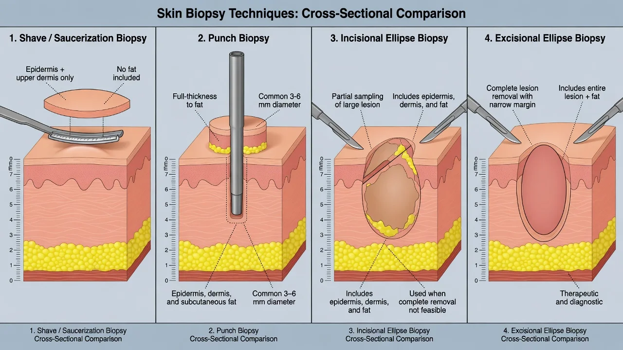
| Technique | Depth | Primary role | Typical sizes |
|---|---|---|---|
| Shave / saucerisation | Epidermis ± upper dermis | Raised benign epidermal lesions; some superficial BCC pathways | Blade-controlled |
| Punch | Full thickness to fat | Inflammatory disease, small tumours, alopecia sampling | Often 3–4 mm (up to 6–8 mm special) |
| Incisional ellipse | Full thickness, partial lesion | Large lesions; panniculitis; when total removal impractical | Wedge to fat |
| Excisional ellipse | Full thickness, entire lesion | Suspected melanoma; complete removal of small tumours | 1–3 mm clinical margin for diagnosis |
| Curettage (± electrodesiccation) | Variable | Often therapeutic more than diagnostic staging | — |
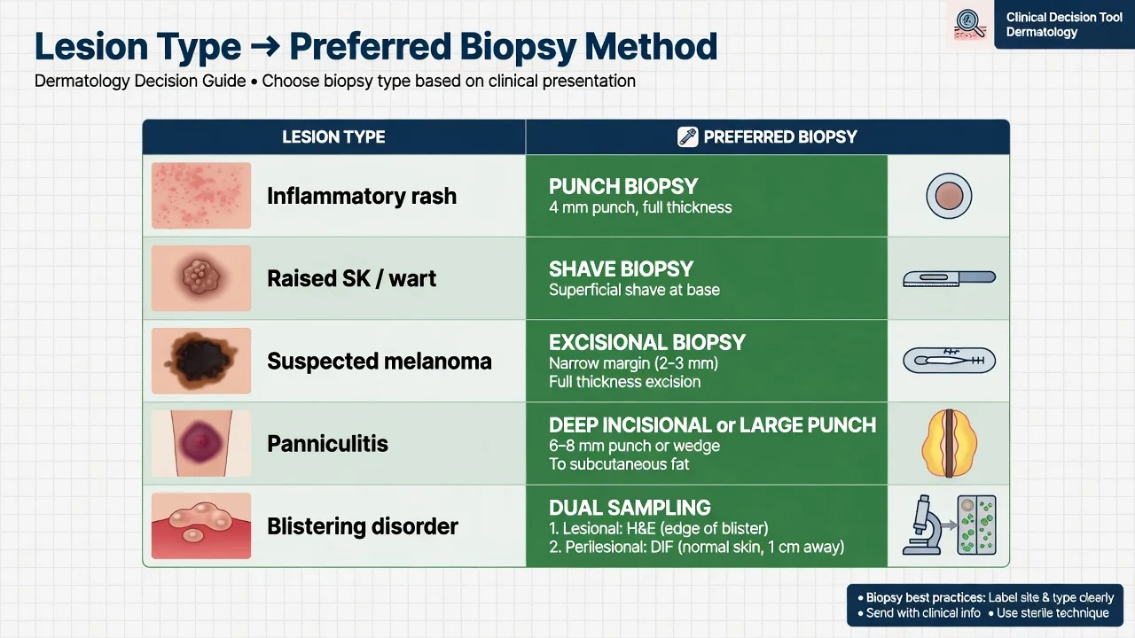
Decision table (exam gold)
| Clinical problem | Preferred technique | Why |
|---|---|---|
| Inflammatory rash / most dermatoses | Punch 3–4 mm | Shows epidermis–dermis–superjacent fat architecture |
| Seborrhoeic keratosis, skin tag, many warts | Shave | Deep dermis not required |
| Suspected melanoma / atypical pigmented lesion | Excisional narrow-margin complete | Preserves architecture and Breslow when invasive |
| Large plaque where total excision impossible | Incisional representative thickest/most atypical area | Partial but adequate depth |
| Panniculitis | Deep incisional or large punch to fat | Disease lives in subcutis |
| Blistering disease | Lesional H&E edge + perilesional DIF | Different media/sites |
| Ulcer | Active edge, not necrotic centre | Viable diagnostic tissue |
| Cicatricial alopecia | Punch(es) with sectioning plan | Horizontal ± vertical strategies |
Why depth and completeness matter
Histologic pattern diagnosis in inflammatory disease needs the full vertical unit. Panniculitis sampled too superficially is a classic false-negative pathway.[7] For melanoma, partial biopsies can misclassify or understage; Australian tertiary data showed diagnostic harm from incomplete sampling strategies, and European diagnostics guidance prioritises complete excision with narrow clinical margins for suspicious pigmented lesions whenever feasible.[3][4] Shave sampling of melanoma remains debated in some service data but is not the default teaching answer when melanoma is in the differential.[5][9]
Pre-procedure setup
- Indication and differential written on the request form — pathologists are not mind-readers.
- Photography and site marking before LA distorts landmarks.
- Consent: scar, infection, bleeding, need for further surgery, nerve injury at high-risk sites.
- Asepsis and appropriate local anaesthesia (see local-anaesthesia-dermatology leaf).
- Orient ellipses along resting skin tension lines when possible for cosmesis.[1][2]
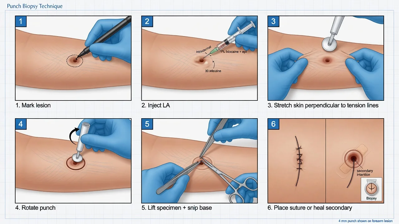
Stepwise: punch biopsy (OSCE core)
- Mark the representative site (active edge of rash; not pure secondary infection if avoidable).
- Infiltrate lidocaine ± epinephrine; wait for blanch/anaesthesia.[2]
- Stretch skin perpendicular to planned closure lines so the defect becomes oval along RSTL.
- Apply punch vertically; rotate with steady pressure to the hub/fat.
- Lift specimen gently (needle or fine forceps on edge — avoid crush); snip base.
- Haemostasis; suture punches typically ≥4 mm on cosmetically sensitive sites; small punches may heal secondarily.
- Place in correct medium immediately.
Stepwise: shave / saucerisation
Stabilise skin; use a flexible blade at a shallow angle for pure shave or a deeper saucer for thicker epidermal tumours. Haemostasis with pressure, topical agents, or light electrodesiccation. Do not use as the planned first move for melanoma-suspicious lesions.[2][3][4]
Stepwise: incisional and excisional ellipses
- Excision: include entire clinically visible lesion plus small diagnostic margin (commonly about 1–3 mm clinical for suspected melanoma diagnostics — definitive wide margins come after histology).[4]
- Incision: full-thickness wedge through the most representative thickest or active area, always including subcutis when panniculitis or deep process is possible.[7]
- Length:width often ~3:1 for tension-free closure; layered closure when depth requires.
Special techniques
Direct immunofluorescence (DIF)
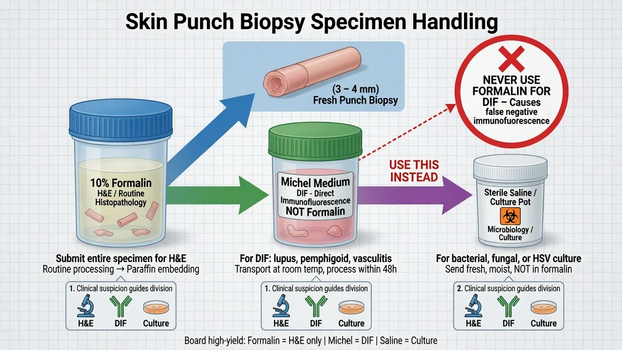
For autoimmune blistering disease and selected lupus/vasculitis work-ups: take perilesional skin (near but not through a fresh blister for many immunobullous protocols) into Michel medium (or validated short-term saline transport) — never formalin.[6][10][8] Separate lesional tissue goes to formalin for H&E. Serration pattern analysis on DIF can refine pemphigoid vs EBA in specialist labs.[8]
Alopecia
Often two punches (e.g. 4 mm) from active areas with a planned horizontal and vertical sectioning strategy; clinical note must state scarring vs non-scarring suspicion. [1]
Panniculitis
Deep sample is non-negotiable; communicate “rule out panniculitis” on the form.[7]
Specimen handling (unforgiving rules)
Label patient, site, and clinical question. Orientation sutures for ellipses when margin or polarity matters. [1]
Complications and pitfalls
Bleeding, infection, dehiscence, hypertrophic scar/keloid, pigment change, incomplete sampling, wrong fixative, missing clinical information, sampling pure crust. Scouting/partial biopsies of melanoma can contribute to upstaging surprises at definitive surgery — plan the first sample as if it must stand alone for diagnosis.[3][9]
Special populations
Anticoagulated patients: most punches proceed with local haemostasis; coordinate high-bleed-risk ellipses with physicians. Children: smallest adequate punch, behavioural LA plan. Cosmetically sensitive face: smaller instruments, meticulous orientation, consider specialist referral for complex pigmented lesions. [1]
Evidence and regional notes
Technique primers (Greenwood; Pickett) cover primary-care and dermatology shared skills.[1][2] Melanoma diagnostics lean on complete narrow-margin excision in European consensus updates; partial biopsy literature quantifies the cost of shortcuts.[4][3][5] DIF transport science supports Michel medium as standard with limited saline alternatives when logistics demand.[6][10]
Clinical pearl
Red flags
Exam application bank (NEET-PG / INICET)
One-line answer
Board-level procedural module on choosing and performing skin biopsy: shave/saucerisation, punch, incisional and excisional ellipses; site selection rules (rash edge, ulcer edge, fresh blister, deep fat for panniculitis); melanoma-preferential excisional sampling with narrow clinical margins; specimen handling (formalin H&E vs Michel medium DIF vs culture); stepwise punch technique; complications and sampling pitfalls. Complements the broader skin-biopsy atlas leaf with technique-first depth for OSCE and fellowship viva.
Worked stems (answer without another resource)
Stem 1 — Classic presentation. Map symptoms to mechanism; name the first investigation and first treatment step with dose/route if drug therapy is standard. [1]
Stem 2 — Unstable / complicated. List red flags that force immediate resuscitation, theatre, ICU, antidote, or reperfusion — and what you do in the first 15 minutes. [1]
Stem 3 — Atypical group. Elderly, pregnancy, child, or immunocompromised: how presentation and thresholds change. [1]
Stem 4 — Differential trap. Name the three closest mimics and one discriminator for each. [1]
Stem 5 — Disposition. Who goes home with safety-netting, who is admitted, who needs HDU/ICU/theatre, and what follow-up is mandatory. [1]
Rapid viva checklist
- Definition + classification
- Pathophysiology chain
- Bedside signs / criteria
- Score with exact components (if any)
- Emergency bundle
- Definitive therapy with doses
- Complications of disease and of treatment
- Special populations
- Guideline/trial name if classic
- Three exam traps
Coverage self-check
If you cannot answer any stem above from this page alone, re-read the matching section — the page is intended to be self-sufficient for final-prof and NEET-PG/INICET questions on Skin biopsy techniques (shave, punch, excisional, incisional).
Expanded exam teaching (depth pass)
Clinical reasoning
For Skin biopsy techniques (shave, punch, excisional, incisional), examiners test whether you can prioritise life threats, choose the right first test, and give specific therapy (agent, dose, route, timing). Generic phrases without numbers score poorly.
Mechanism → feature map
Build a short chain: cause → pathophysiologic intermediate → clinical feature → complication. Every major symptom in the classic vignette should sit on that chain.
Investigation strategy
- Bedside/first-line tests that change immediate management
- Confirmatory or staging tests
- What a normal result does not exclude
- When not to delay treatment for imaging (unstable patient)
Management ladder
- Resuscitation / ABC / sepsis or haemorrhage bundle as relevant
- Specific antidote / procedure / antimicrobial / reperfusion / surgery
- Supportive care and monitoring targets
- Definitive long-term therapy and secondary prevention
- Disposition and safety-net advice
Special populations
Always prepare one line each for children, pregnancy, elderly, renal/hepatic impairment, and immunocompromised patients when the topic allows.
Pitfalls that fail candidates
- Treating the number not the patient
- Missing pregnancy status when relevant
- Imaging before stabilisation
- Wrong empiric cover or wrong antidote timing
- Incomplete counselling on recurrence, adherence, or red-flag return
Board-level procedural module on choosing and performing skin biopsy: shave/saucerisation, punch, incisional and excisional ellipses; site selection rules (rash edge, ulcer edge, fresh blister, deep fat for panniculitis); melanoma-preferential excisional sampling with narrow clinical margins; specimen handling (formalin H&E vs Michel medium DIF vs culture); stepwise punch technique; complications and sampling pitfalls. Complements the broader skin-biopsy atlas leaf with technique-first depth for [1]
Structured revision sheet
Must-know numbers and names
List every score, size threshold, dose, and time window from this topic on a blank page from memory, then check against the sections above.
Three classic MCQ angles
- Most likely diagnosis given a vignette
- Next best step in management
- Most appropriate investigation
Three classic SAQ angles
- Pathophysiology in five steps
- Management algorithm with doses
- Complications and prevention
Clinical station flow
Greet → focused history → targeted exam → investigations → explain diagnosis → emergency care → definitive plan → safety-net / follow-up → answer examiner questions on mechanism and pitfalls.
Exam anchors
Exam triad
ACT
Define the problem and red flags
Highest-yield test or bedside clue
First-line management and follow-up
References
- [1]Greenwood JD, Merry SP, Boswell CL. Skin Biopsy Techniques Prim Care, 2022.PMID 35125151
- [2]Pickett H. Shave and punch biopsy for skin lesions Am Fam Physician, 2011.PMID 22046939
- [3]Ng JC, Swain S, Dowling JP, et al. The impact of partial biopsy on histopathologic diagnosis of cutaneous melanoma: experience of an Australian tertiary referral service Arch Dermatol, 2010.PMID 20231492
- [4]Garbe C, Amaral T, Peris K, et al. European consensus-based interdisciplinary guideline for melanoma. Part 1: Diagnostics: Update 2022 Eur J Cancer, 2022.PMID 35570085
- [5]Jones S, Henry V, Strong E, et al. Clinical Impact and Accuracy of Shave Biopsy for Initial Diagnosis of Cutaneous Melanoma J Surg Res, 2023.PMID 36739830
- [6]Kumudhini S, Pai S, Rao C, et al. A comparative study of Michel's medium versus honey as a transport medium for skin specimens prior to direct immunofluorescence microscopy and antigen mapping J Cutan Pathol, 2019.PMID 31087406
- [7]Rose C, Leverkus M, Fleischer M, et al. Histopathology of panniculitis--aspects of biopsy techniques and difficulties in diagnosis J Dtsch Dermatol Ges, 2012.PMID 22084866
- [8]Meijer JM, Atefi I, Diercks GFH, et al. Serration pattern analysis for differentiating epidermolysis bullosa acquisita from other pemphigoid diseases J Am Acad Dermatol, 2018.PMID 29154993
- [9]Chacko RS, Shah PC, Feng JX, et al. Risk of tumor upstaging following partial biopsy and scouting biopsy in the treatment of melanoma in situ and invasive melanoma with Mohs micrographic surgery: a retrospective cohort study Arch Dermatol Res, 2025.PMID 40167650
- [10]Vodegel RM, de Jong MC, Meijer HJ, et al. Enhanced diagnostic immunofluorescence using biopsies transported in saline BMC Dermatol, 2004.PMID 15333135