Dermatology · Medicine
Skin flaps and grafts (basics)
Also known as Local flaps dermatologic surgery · Full-thickness skin graft · Split-thickness skin graft · Cutaneous reconstructive ladder · Bilobed flap
Basics of cutaneous reconstruction after excision or Mohs surgery: reconstructive ladder; secondary intention and primary closure; random versus axial flaps; advancement, rotation, transposition (including bilobed), and interpolation flaps; full-thickness versus split-thickness skin grafts; graft-take biology; site-specific nasal and facial planning; complications (necrosis, haematoma, trapdoor); and peri-operative risk modification for board-level exams.
On this page & tools
Your progress
Saved locally on this device.
Exam tags
Red flags
Definition & Classification
A skin flap is relocated skin (± subcutaneous tissue) that retains a vascular pedicle (or is microsurgically re-anastomosed in free flaps). A skin graft is completely detached skin transferred to a recipient bed and depends on graft take biology.[1][7]
Reconstructive ladder (concept)
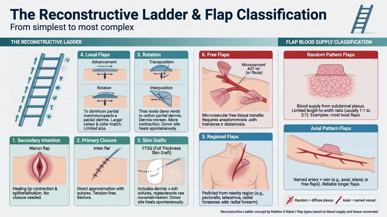
Although real practice is a “reconstructive elevator” (jump to the best option, not always the simplest), exams still expect the ladder logic:[8]
- Secondary intention
- Primary linear closure
- Skin graft (FTSG / STSG)
- Local flap
- Regional / interpolation flap
- Free flap / major reconstruction [1]
Flap classification
| Axis | Categories | Exam examples |
|---|---|---|
| Blood supply | Random (subdermal plexus) vs axial (named vessel) | Most small facial flaps random; forehead flap axial (supratrochlear) |
| Movement | Advancement, rotation, transposition, interpolation | Island/V-Y; O-to-Z rotation; bilobed; paramedian forehead |
| Composition | Cutaneous, fasciocutaneous, myocutaneous | Beyond basic derm surgery often |
Flaps and grafts are core reconstructive tools after dermatologic surgery and Mohs defects.[1]
Graft classification
| Graft | Contents | Typical use |
|---|---|---|
| FTSG | Epidermis + full dermis | Small facial defects needing better texture/colour match |
| STSG | Epidermis + partial dermis | Larger wounds, granulating beds, less cosmetic priority |
| Composite | Skin + cartilage (e.g.) | Selected nasal alar defects (specialist) |
Classification Figures & Planning Map
Epidemiology & Clinical Context
Most dermatology flap/graft volume follows skin cancer excision, especially facial Mohs defects where tissue sparing creates complex shapes near free margins (lid, lip, ala, helix).[1][2][10] Secondary intention remains underused but excellent on selected concave units.[5]
Pathophysiology
Graft take (must recite)
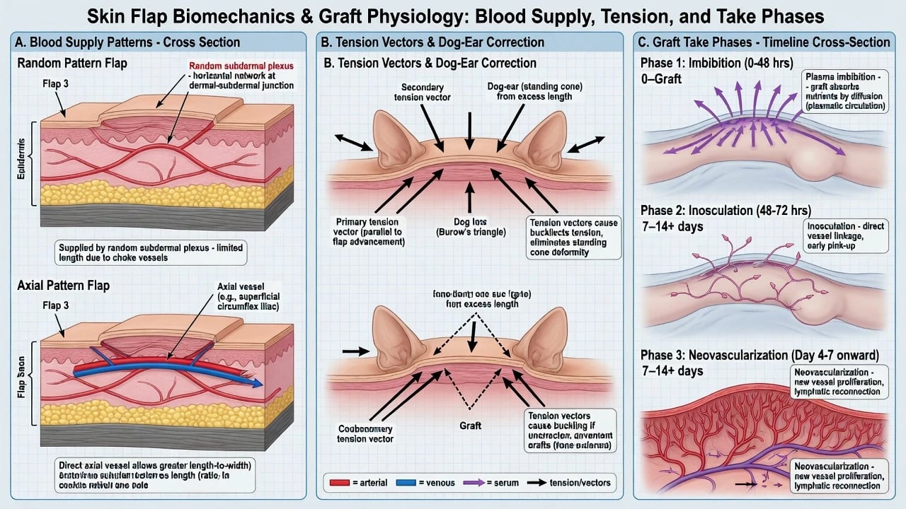
- Imbibition — plasma nutrition first 24–48 h
- Inosculation — vessel link-up
- Neovascularisation — new vessels secure survival [1]
Shear, haematoma, infection, or avascular beds (bone without periosteum, cartilage without perichondrium, heavy radiation) impair take.[7]
Flap survival threats
- Excessive tension (especially random flap tips)
- Pedicle kink/compression
- Smoking, poorly controlled diabetes, infection
- Haematoma elevating the flap off its bed/pedicle inflow [1]
Clinical Presentation — Defect Assessment
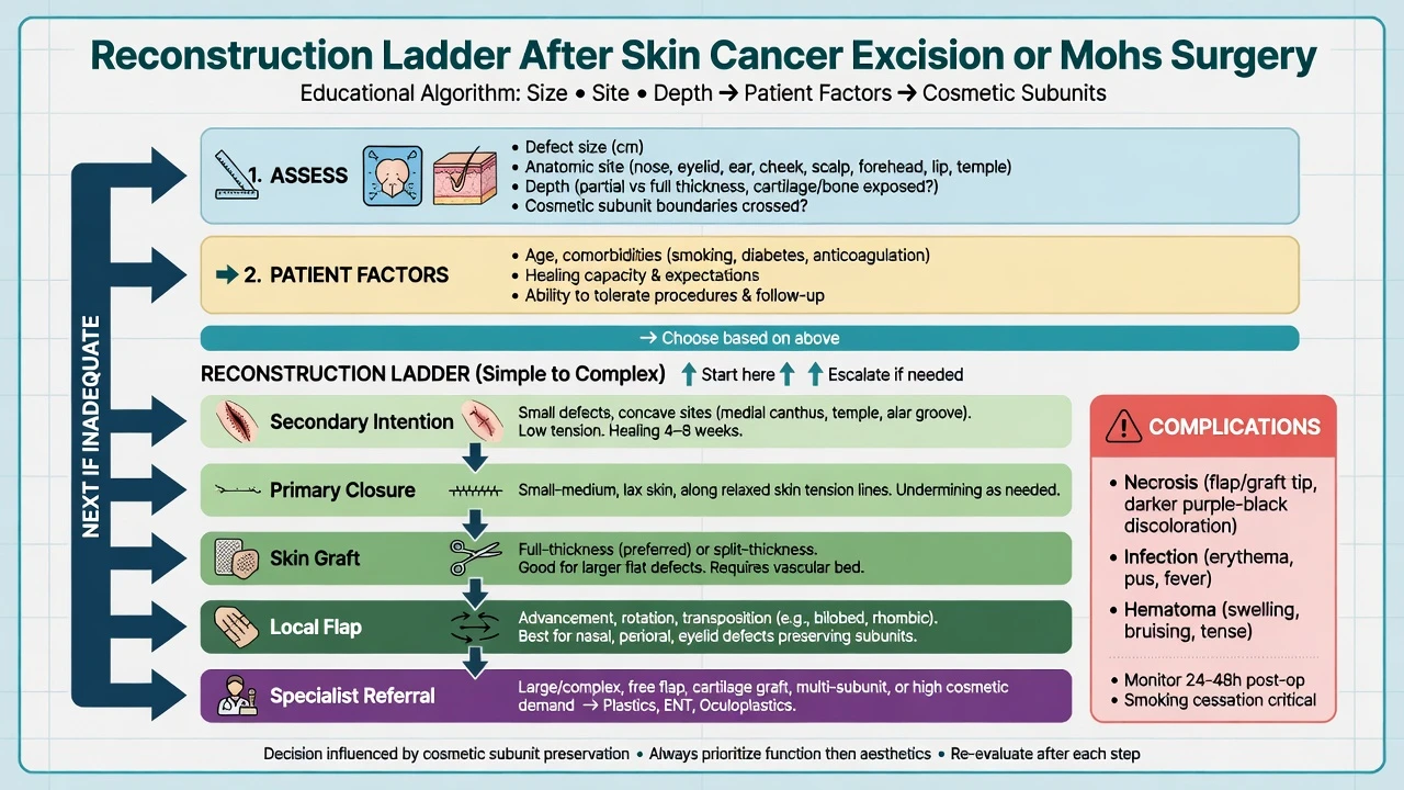
Before designing anything, answer: [1]
- Oncology secure? Margins clear?
- Size / depth / exposed structures (bone, cartilage, mucosa)
- Site: cosmetic subunit + free margin risk (lid ectropion, lip competence, nasal valve)
- Laxity of adjacent tissue and RSTL orientation
- Patient: smoker, anticoagulant, prior radiation, expectations [1]
Nasal reconstruction literature emphasises subunit thinking and lining/support/cover layers for full-thickness defects.[2]
Differential Selection of Repair
| Situation | Prefer |
|---|---|
| Small defect, loose skin, along RSTL | Primary closure |
| Concave temple/medial canthus/selected conchal bowl | Secondary intention (± purse-string) |
| Shallow defect, good vascular bed, limited local tissue | FTSG |
| Large trunk/limb wound | STSG |
| Need tissue bulk / better contour / avoid contraction | Local flap |
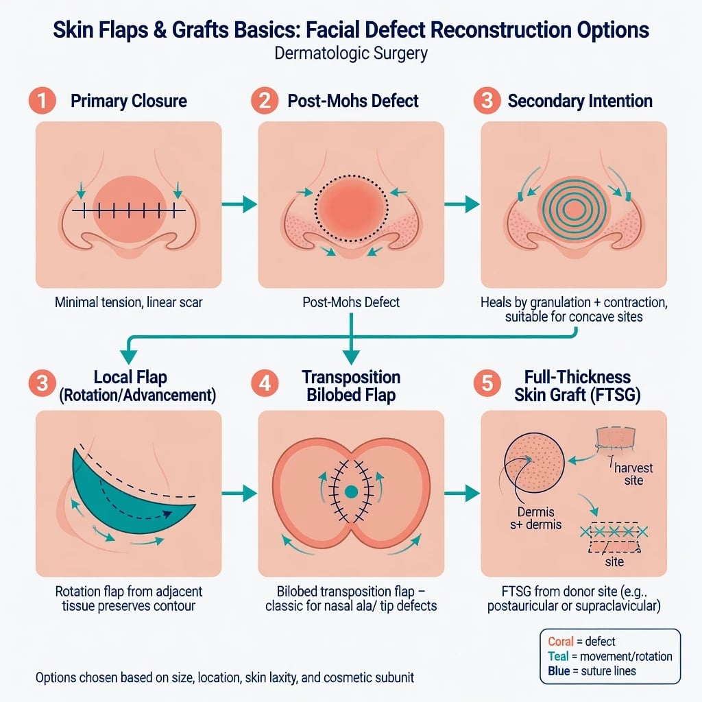
| Distal nasal defect classic exam | Bilobed transposition |
| Large distal nasal/subunit needs | Interpolation (paramedian forehead) |
| Beyond local options | Refer plastics/OMFS |
Secondary intention and purse-string techniques are legitimate, not “giving up.”[5] Bilobed flaps remain a workhorse for distal nasal reconstruction when designed correctly.[3]
Clinical & Bedside Assessment / Consent
- Photograph, measure, mark subunits and RSTLs.
- Anticipate standing cutaneous cones (dog-ears) and plan Burow’s triangles; Burow’s tissue can itself become a graft in selected designs.[9]
- Consent: scar, contour, colour mismatch, partial necrosis, infection, revision, sensory change, temporary pedicle (interpolation) if relevant.
Investigations & Peri-operative Optimisation
- Review anticoagulation/bleeding risk (balance oncologic timing vs haemostasis).
- Optimise glucose; strong smoking cessation counselling.
- Confirm tetanus status when relevant; perioperative antibiotics only when indicated by wound class/centre protocol (not universal for all clean facial repairs).
- For cancer: prefer margin-controlled excision (Mohs) before elaborate reconstruction when indicated.[1]
Management — Urgent Salvage
- Haematoma → evacuate early.
- Infection → drain if needed + antimicrobials + debridement of necrotic tissue.
- Venous congestion vs arterial inflow failure — release sutures/tension, consider leech therapy only in specialist settings for venous issues; arterial failure often needs revision.
- Failed graft → debride and replan (second graft, flap, or secondary intention). [1]
Management — Definitive Techniques (Basics)
Primary closure
- Undermine adequately in safe plane; align with RSTLs; evert edges; layered closure when depth requires. [1]
Secondary intention
- Best on concave units; worse on convex tips where contraction/notching risks rise.[5]
- Daily wound care; expect longer healing but often excellent cosmesis in right sites.
Local flaps
| Flap | Movement idea | Classic teaching use |
|---|---|---|
| Advancement | Slide forward (Burow’s, V-Y) | Forehead/temple, lip, cheek with laxity |
| Rotation | Pivot around arc | Cheek, scalp |
| Transposition | Lift over intervening tissue | Rhombic; bilobed nose |
| Interpolation | Pedicle crosses intact skin; later division | Paramedian forehead to nose |
Bilobed design principles (Zitelli modifications in modern teaching) aim to redistribute tension and fit nasal subunits.[3] Forehead/temple reconstruction can often be simplified with thoughtful local options.[6] Lip reconstruction after Mohs requires functional orbicularis and free-margin respect.[10]
Grafts
- FTSG: thin, defatted appropriately; quilt or bolster to reduce shear; donor sites commonly pre/postauricular, supraclavicular, or adjacent Burow’s tissue.[7][9]
- Tie-over bolsters are traditional; systematic review data show practice variation and uncertain mandatory benefit for all FTSGs — technique individualisation is acceptable when immobilisation is otherwise secured.[4]
- STSG: meshed or sheet; donor heals by secondary re-epithelialisation from adnexae.[7]
Flap
- Keeps blood supply
- Better bulk/colour often
- Design skill critical
- Tip necrosis risk
FTSG
- Full dermis
- Good facial match if thin bed
- Needs vascular bed
- Less bulk than flap
STSG
- Partial dermis
- Covers large areas
- More contraction
- Donor site care needed
Specific Subtypes & Site Scenarios
- Nose: bilobed for many distal defects; forehead flap when larger subunit/full-thickness needs arise; always think lining and support for through-and-through defects.[2][3]
- Ear: cartilage support; exposed cartilage may need perichondrial flaps or grafts carefully.
- Eyelid: anterior lamella vs full-thickness — ectropion risk; low threshold to refer.
- Lip: functional seal more important than perfect scar line.[10]
- Scalp: poor elasticity → often rotation flaps, grafts, or secondary intention on bone with periosteum.
Complications & Pitfalls
- Partial/full necrosis
- Trapdoor / pin-cushion deformity of curved flaps
- Contour step-off and colour mismatch (grafts)
- Ectropion, nasal valve collapse, microstomia if free margins ignored
- Reconstructing before clear margins
- Over-complicating a defect that secondary intention would heal beautifully.[5]
Prognosis & Disposition
- Most well-designed local flaps and small FTSGs heal with good function in weeks; scar maturation months.
- Plan suture removal by site (face earlier than limb).
- Separate cancer follow-up from scar management.
- Revision (dermabrasion, steroid, re-excision of dog-ear) only after maturation unless function is threatened. [1]
Special Populations
- Smokers / vasculopaths: favour lower-tension designs, grafts with excellent beds, or delayed reconstruction strategies.
- Anticoagulated: meticulous haemostasis; haematoma is the enemy of both flaps and grafts.
- Irradiated skin: reduced flap reliability — specialist planning.
- Elderly: often excellent secondary intention candidates on concave facial units.[5]
Evidence, Guidelines & Regional Differences
- Reconstructive ladder remains a teaching scaffold from broader reconstructive surgery.[8]
- Nasal and facial plastic literature underpins subunit-based planning.[2]
- Bilobed flap technique papers guide distal nasal repairs.[3]
- FTSG bolster evidence is mixed; immobilisation principle matters more than one ritual dressing.[4]
- Resource-limited settings: mastery of primary closure, secondary intention, and simple advancement/rotation flaps covers most defects without free-flap infrastructure.
Exam Pearls
FLAPS
FLAPS
Flap keeps blood supply
Simple when sufficient; best option overall
Free margins and cosmetic units
Imbibition → inosculation → neovascularisation
Tension, haematoma, smoking threaten survival
- Flap vs graft one-liner is non-negotiable.[1][7]
- Bilobed = distal nose classic.[3]
- Secondary intention on concave sites is sophisticated, not lazy.[5]
- Clear cancer margins before complex cover.[1]
- Burow’s triangle tissue can be recycled as a graft in selected closures.[9]
Exam application bank (NEET-PG / INICET)
One-line answer
Basics of cutaneous reconstruction after excision or Mohs surgery: reconstructive ladder; secondary intention and primary closure; random versus axial flaps; advancement, rotation, transposition (including bilobed), and interpolation flaps; full-thickness versus split-thickness skin grafts; graft-take biology; site-specific nasal and facial planning; complications (necrosis, haematoma, trapdoor); and peri-operative risk modification for board-level exams.
Worked stems (answer without another resource)
Stem 1 — Classic presentation. Map symptoms to mechanism; name the first investigation and first treatment step with dose/route if drug therapy is standard. [1]
Stem 2 — Unstable / complicated. List red flags that force immediate resuscitation, theatre, ICU, antidote, or reperfusion — and what you do in the first 15 minutes. [1]
Stem 3 — Atypical group. Elderly, pregnancy, child, or immunocompromised: how presentation and thresholds change. [1]
Stem 4 — Differential trap. Name the three closest mimics and one discriminator for each. [1]
Stem 5 — Disposition. Who goes home with safety-netting, who is admitted, who needs HDU/ICU/theatre, and what follow-up is mandatory. [1]
Rapid viva checklist
- Definition + classification
- Pathophysiology chain
- Bedside signs / criteria
- Score with exact components (if any)
- Emergency bundle
- Definitive therapy with doses
- Complications of disease and of treatment
- Special populations
- Guideline/trial name if classic
- Three exam traps
Coverage self-check
If you cannot answer any stem above from this page alone, re-read the matching section — the page is intended to be self-sufficient for final-prof and NEET-PG/INICET questions on Skin flaps and grafts (basics).
Expanded exam teaching (depth pass)
Clinical reasoning
For Skin flaps and grafts (basics), examiners test whether you can prioritise life threats, choose the right first test, and give specific therapy (agent, dose, route, timing). Generic phrases without numbers score poorly.
Mechanism → feature map
Build a short chain: cause → pathophysiologic intermediate → clinical feature → complication. Every major symptom in the classic vignette should sit on that chain.
Investigation strategy
- Bedside/first-line tests that change immediate management
- Confirmatory or staging tests
- What a normal result does not exclude
- When not to delay treatment for imaging (unstable patient)
Management ladder
- Resuscitation / ABC / sepsis or haemorrhage bundle as relevant
- Specific antidote / procedure / antimicrobial / reperfusion / surgery
- Supportive care and monitoring targets
- Definitive long-term therapy and secondary prevention
- Disposition and safety-net advice
Special populations
Always prepare one line each for children, pregnancy, elderly, renal/hepatic impairment, and immunocompromised patients when the topic allows.
Pitfalls that fail candidates
- Treating the number not the patient
- Missing pregnancy status when relevant
- Imaging before stabilisation
- Wrong empiric cover or wrong antidote timing
- Incomplete counselling on recurrence, adherence, or red-flag return
Basics of cutaneous reconstruction after excision or Mohs surgery: reconstructive ladder; secondary intention and primary closure; random versus axial flaps; advancement, rotation, transposition (including bilobed), and interpolation flaps; full-thickness versus split-thickness skin grafts; graft-take biology; site-specific nasal and facial planning; complications (necrosis, haematoma, trapdoor); and peri-operative risk modification for board-level exams. [1]
Structured revision sheet
Must-know numbers and names
List every score, size threshold, dose, and time window from this topic on a blank page from memory, then check against the sections above.
Three classic MCQ angles
- Most likely diagnosis given a vignette
- Next best step in management
- Most appropriate investigation
Three classic SAQ angles
- Pathophysiology in five steps
- Management algorithm with doses
- Complications and prevention
Clinical station flow
Greet → focused history → targeted exam → investigations → explain diagnosis → emergency care → definitive plan → safety-net / follow-up → answer examiner questions on mechanism and pitfalls.
[1] [1]References
- [1]Zhang AY, Meine JG. Flaps and grafts reconstruction Dermatol Clin, 2011.PMID 21421147
- [2]Spataro E, Branham GH. Principles of Nasal Reconstruction Facial Plast Surg, 2017.PMID 28226366
- [3]Okland TS, Lee YJ, Sanan A, et al. The Bilobe Flap for Nasal Reconstruction Facial Plast Surg, 2020.PMID 32512603
- [4]Marsidi N, Boteva K, Vermeulen SAM, et al. To Tie or Not to Tie-Over Full-Thickness Skin Grafts in Dermatologic Surgery: A Systematic Review of the Literature Dermatol Surg, 2021.PMID 32796333
- [5]Lam TK, Lowe C, Johnson R, et al. Secondary Intention Healing and Purse-String Closures Dermatol Surg, 2015.PMID 26418684
- [6]Redondo P. Simplifying Forehead and Temple Reconstruction: A Narrative Review J Clin Med, 2023.PMID 37629442
- [7]Prohaska J, Cook C. Skin Grafting(Archived) 2026.PMID 30422469
- [8]Levin LS. The reconstructive ladder. An orthoplastic approach Orthop Clin North Am, 1993.PMID 8101984
- [9]Zitelli JA. Burow's grafts J Am Acad Dermatol, 1987.PMID 3305605
- [10]Chellappan B, Obanigba G, Nguyen M, et al. Lip Reconstruction After Mohs Micrographic Surgery: A Guide on Flaps Cutis, 2023.PMID 37289682