Dermatology · Medicine
Solar lentigo
Also known as Solar lentigo · Senile lentigo · Age spot · Liver spot · Sun spot
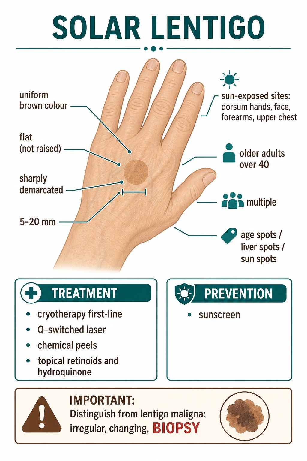
Solar lentigo = benign acquired pigmented macule from chronic UV-induced melanocyte proliferation. Uniform light-brown flat macule on sun-exposed skin (dorsum of hands, face, forearms, upper chest). Older adults (40). Multiple; 'age spots' / 'liver spots' / 'sun spots'. Benign — does NOT transform into melanoma. MUST distinguish from lentigo maligna (irregular borders, colour variation, large, changing — BIOPSY). Treatment: cryotherapy (first-line), Q-switched laser, chemical peels, topical retinoids/hydroquinone. Sunscreen for prevention of new lesions.
On this page & tools
Your progress
Saved locally on this device.
Exam tags
Red flags
Overview & definition
Solar lentigo (synonyms: senile lentigo, "age spot", "liver spot", "sun spot") is a benign acquired pigmented macule on sun-exposed skin arising from chronic ultraviolet-induced melanocyte proliferation in the basal epidermis. It is one of the most common dermatologic findings in fair-skinned adults over 40 and the commonest reason for cosmetic pigment consultation in primary care. The clinical and dermoscopic signature is a uniform, sharply demarcated, flat light-brown to tan macule that does not change over months to years. The diagnostic imperative is to differentiate solar lentigo from lentigo maligna (melanoma in situ) and lentigo maligna melanoma, which can look similar at the bedside in elderly patients.[1][5]
The lesion arises from cumulative UV-induced proliferation of basal melanocytes, not from melanocytic atypia. Histologically the rete ridges are elongated, club-shaped and heavily pigmented; the number of melanocytes is mildly increased but cytologically bland. There is no risk of malignant transformation of a solar lentigo itself, but the surrounding chronically sun-damaged skin is itself a high-risk field for both lentigo maligna and other non-melanoma skin cancers. [1]
High-yield numbers for the examiner
Pathophysiology
Cumulative UV exposure to the basal epidermis, predominantly UVB but also UVA, leads to: [1]
- Melanocyte proliferation at the dermo-epidermal junction (numbers increase modestly — unlike melanocytic naevi there is no nested proliferation).
- Increased melanin synthesis with transfer to surrounding keratinocytes — producing the clinical hyperpigmented macule.
- Elongated, club-shaped rete ridges with a focal increase in melanin granules at the tips of the ridges.
- Solar elastosis in the dermis (chronic UV damage to elastin and collagen) is universally present in the surrounding sun-damaged skin and supports the diagnosis clinically. [1]
The trigger for the chronic melanocyte proliferation is multifactorial: cumulative UV dose is central, but the immune microenvironment, oxidative stress and aging-related epigenetic changes may contribute. Importantly, solar lentigo is NOT a melanocytic naevus — it is a reactive proliferation of normal-appearing melanocytes in chronically photo-damaged skin. Multiple studies demonstrate a normal melanocyte-keratinocyte ratio with no BRAF V600E mutation, distinguishing it from a true lentiginous melanocytic naevus.[1][2]

Epidemiology & risk factors
- Prevalence rises steeply with age: ~20% in 20-30 year-olds, >50% by age 50, and >90% in fair-skinned adults over 65.
- Sex distribution: roughly equal; women may seek cosmetic treatment more often.
- Skin type: most common in Fitzpatrick skin types I-III (fair skin, blue/green eyes, blonde or red hair), moderate in IV, occasional in V-VI.
- Occupational / recreational UV exposure: outdoor workers, sailors, gardeners, walkers, golfers, beach/sun users; cumulative dose is the principal risk.
- History of sunburn doubles the prevalence independent of skin type.
- Tanning bed use is an independent risk factor for both solar lentigo and lentigo maligna.
- Photosensitising drugs (amiodarone, thiazides, tetracyclines) can intensify both the lentigines and the surrounding photodamage.
- Immunosuppression (transplant, HIV) increases both prevalence and the risk of co-existent lentigo maligna or other non-melanoma skin cancer. [1]
Classification
The single clinical entity "solar lentigo" is one of several lentigo variants; precise classification matters for management: [1]
- Solar (senile) lentigo — the standard "age spot" of sun-exposed skin.
- Lentigo simplex — a small (1-5 mm) macule arising in childhood or adolescence, can occur on any skin (sun-exposed and covered), often solitary or in low numbers, with similar histology (elongated rete ridges; lentigo simplex sometimes shows melanocytic hyperplasia).
- PUVA lentigo — pigmented macules arising from long-term psoralen-UVA phototherapy; can persist for years after treatment ends.
- Radiation lentigo — analogous to solar but following ionising radiation exposure.
- Ink-spot lentigo — small, heavily pigmented macule on sun-exposed skin; histology shows a sharply demarcated basal hyperpigmentation; benign.
- Lentigo maligna — atypical melanocytic proliferation (melanoma in situ) on chronically sun-damaged skin; bigger, more irregular, often slowly growing, and the diagnostic trap.
- Lentigo maligna melanoma — invasive lentigo maligna; biopsy-proven, surgical excision required. [1]
Clinical features
Classic solar lentigo is described on the bedside with six identifying features: [1]
- Colour: uniform light tan to medium brown; never two-toned or multicoloured.
- Surface: flat (macular), not palpable; no scale, no atrophy, no ulceration.
- Edge: sharp, well-demarcated; round or oval; 5-20 mm (can be up to several cm).
- Distribution: sun-exposed sites — face (especially forehead, temples, malar), bald scalp, dorsal hands, extensor forearms, upper chest/shoulders, shin.
- Number: often multiple, sometimes dozens, occasionally confluent.
- Time course: stable over months to years; gradual increase in number with cumulative UV exposure; does NOT involute spontaneously. [1]
Asymmetry, border irregularity, colour variation, diameter > 6 mm or change over time are not features of solar lentigo and should drive consideration of biopsy to exclude lentigo maligna.[5]
Differential diagnosis
The "flat brown macule on sun-damaged skin of an older adult" differential includes many diagnoses; correct identification is the bedside skill: [1]
[1]The lentigo maligna dilemma
Lentigo maligna is a melanoma in situ on chronically sun-damaged skin of older adults (typically >65, often on face or bald scalp). It grows slowly and insidiously over months-years; lesions can become >2-3 cm. The clinical challenge is that LM and a large, irregular solar lentigo can look identical at the bedside. The helpful signs: [1]
- Asymmetry: LM is asymmetric in shape.
- Border irregularity: notches, fading, or angular edges on a brown patch.
- Colour variation: LM shows two or more shades of brown, sometimes grey, black, or red.
- Diameter: LM is generally >1 cm at diagnosis.
- Evolution: documented growth over months to years. [1]
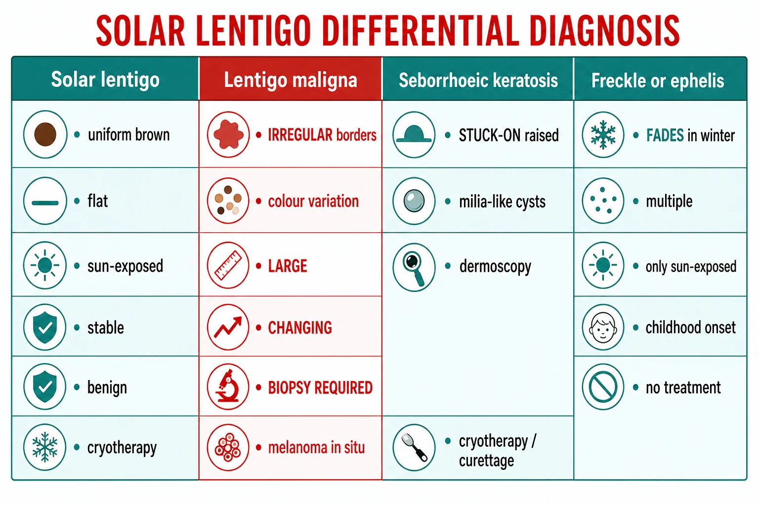
Any of these features triggers a biopsy — the gold standard is a 3-4 mm punch biopsy, ideally from the most atypical area, supplemented by a broad shave biopsy if the lesion is large. Pre-operative dermoscopy-guided biopsy of grey/dark areas (rhomboid structures, annular-granular pattern, grey dots/granules, asymmetric follicular openings) improves diagnostic yield over random biopsy.[5]
The biopsy decision pathway — when to biopsy a flat brown macule on sun-exposed skin
Apply the 7-point dermoscopic checklist
Asymmetry, atypical network, blue-whitish veil, atypical vessels, irregular streaks/dots/globules, regression structures, irregular pigmentation. ANY one feature raises concern and mandates the next step.
Check the ABCDE clinical criteria
Asymmetry, Border irregularity, Colour variation (>=2 shades), Diameter >6 mm, Evolution over months. ANY ABCDE feature in an elderly fair-skinned patient warrants biopsy.
Identify lentigo maligna red flags
Face/scalp of an adult >65; documented slow growth; asymmetric follicular openings; slate-grey or rhomboid structures on dermoscopy; annular-granular pattern. These are the lentigo-maligna-specific triggers.
Choose the right biopsy technique
3-4 mm PUNCH biopsy from the most atypical or darkest area is the gold standard. For large lesions, supplement with a BROAD SHAVE biopsy. SHAVE biopsy alone is INADEQUATE — the architecture of rete ridges and dermo-epidermal junction must be assessable. AVOID destroying the lesion with cryotherapy before histology is obtained.
Mark the biopsy site and refer if positive
Tattoo or photograph the biopsy site. If histology confirms lentigo maligna → 5 mm clinical margins on excision or Mohs micrographic surgery (preferred on the face for tissue-sparing). Topical imiquimod is reserved for inoperable disease. Document the result in the GP record and inform the patient.
Dermoscopy
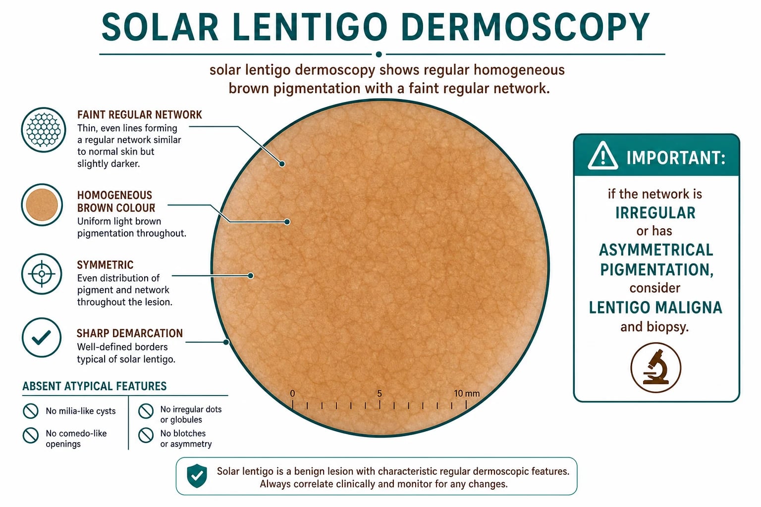
Dermoscopy (polarised handheld, 10-30×) is the single most useful bedside adjunct. Solar lentigo shows: [1]
- Uniform light-brown homogeneous pigmentation with a faint, regular pigment network.
- Sharp, well-defined border with a parallel pattern; no abrupt cutoff.
- No milia-like cysts or comedo-like openings (distinguishes from SK).
- No blue-grey dots/granules that would suggest regression in melanoma.
- No atypical vascular pattern. [1]
If the network is irregular, if there are grey/blue granules, white scar-like areas, or regression structures, or if there is an asymmetric follicular opening pattern, biopsy is mandatory to exclude lentigo maligna.[5]
Investigations
- The diagnosis is clinical in classic lesions.
- Dermoscopy as above is the single most useful adjunct.
- Reflectance confocal microscopy (RCM) is a non-invasive specialist tool that can confirm a uniform epidermal honeycomb pattern and absence of atypical melanocytes — useful for very large facial lesions where biopsy can be cosmetic, or to map the extent of lentigo maligna before excision.
- Wood's lamp examination: solar lentigo typically does NOT enhance (because pigment is epidermal and within keratinocytes), whereas lentigo simplex can show accentuation of borders. Not routinely used.
- Biopsy (3-4 mm punch or broad shave) is reserved for atypical / irregular / changing lesions.
- Histology: elongated club-shaped rete ridges, increased basal melanin, mildly increased melanocyte numbers with no atypia, no pagetoid spread, no dermal invasion. [1]
Management
Treatment is cosmetic, not medical. Patient counselling is the cornerstone: many patients are content with reassurance that the lesion is benign and not pre-cancerous. Active treatment is offered for cosmetic concerns. First-line is cryotherapy for most lesions, laser for selected cases.[3][4]
Cryotherapy
Liquid nitrogen applied by cotton-tipped applicator or spray. A single freeze-thaw cycle of 5-10 seconds is sufficient for most lesions; longer freeze (15+ seconds) gives faster clearing but a higher risk of post-treatment hypopigmentation. Spontaneous crusting and re-epithelialisation over 2-4 weeks. Excellent efficacy (>80% clear at 3 months), low cost, single visit. [1]
Lasers
- Q-switched pigment lasers (ruby 694 nm, alexandrite 755 nm, Nd:YAG 1064 nm) target melanin selectively; a single session often clears individual lesions with very good cosmetic match to surrounding skin.
- Intense pulsed light (IPL) is effective for broad fields of solar lentigines; 2-4 sessions at 3-4 week intervals.
- Fractional non-ablative lasers (1550 nm, 1927 nm thulium) for widespread photodamage; pigment lightening over 3-6 months. [1]
Topical therapies
- Topical retinoids (tretinoin 0.025-0.1% cream nightly) — slow lightening over 6-12 months; useful for multiple lesions.
- Hydroquinone 4% cream (sometimes compounded with tretinoin + corticosteroid in the "Kligman triple combination") — over-the-counter in some regions; lightening over 3-6 months. Caution about ochronosis with prolonged use.
- Cysteamine cream — recent addition with efficacy similar to hydroquinone.
- Vitamin C, niacinamide, azelaic acid — modest brightening effects; commonly used in cosmetic products. Specific drug and protocol choices for the active treatment options, with the doses, routes, monitoring and rationale an examiner expects to see reproduced accurately:[1]
Liquid nitrogen cryotherapy (cotton-tipped applicator or cryospray)
Dose
Single freeze-thaw cycle of 5 to 10 seconds; spray distance 1 cm; ice-ball margin 2 mm beyond lesion edge. Longer freezes (15+ seconds) clear faster but raise post-treatment hypopigmentation risk.
Tretinoin (all-trans retinoic acid) cream
Dose
Start 0.025% cream; escalate to 0.05% or 0.1% as tolerated. Apply a pea-sized amount to the affected area at night; full-face spread if treating photodamage field.
Hydroquinone 4% cream (sometimes compounded with tretinoin 0.05% and hydrocortisone 1% — the Kligman triple combination)
Dose
Apply a thin layer twice daily for 3 to 6 months; pulse therapy (2 months on / 2 months off) reduces ochronosis risk. Maximum 4% strength OTC in US; up to 4% prescription-only in UK; 1 to 2% in EU.
Cysteamine 5% cream (novel depigmenting agent)
Dose
Apply once daily (or twice daily) to pigmented lesions; 15-minute leave-on before rinsing or overnight depending on formulation.
Q-switched pigment laser (ruby 694 nm, alexandrite 755 nm, or Nd:YAG 1064 nm)
Dose
Single session typically sufficient for individual lesions; spot size 3 to 5 mm; fluence titrated to immediate ash-grey whitening (3 to 5 J/cm² for Q-switched ruby 694 nm).
Broad-spectrum sunscreen SPF 50+ (5-UVA PF >=5, critical wavelength >=370 nm)
Dose
Apply ~1 teaspoon for face/ears/neck, ~2 tablespoons for full body. Apply 15 to 30 minutes before sun exposure; reapply every 2 hours, and after swimming, sweating, or towel-drying.
Chemical peels
- Trichloroacetic acid (TCA) 10-35% peel — full-face peel for diffuse photodamage; 7-10 day downtime; medium-depth peel for more dramatic effect.
- Glycolic acid peels 30-70% — repeated every 2-4 weeks; mild-moderate lightening. [1]
Surgery / destructive
- Curettage / electrodesiccation for small lesions; risk of scarring.
- Surgical excision for diagnostic lesions where malignancy cannot be excluded. [1]
Prevention
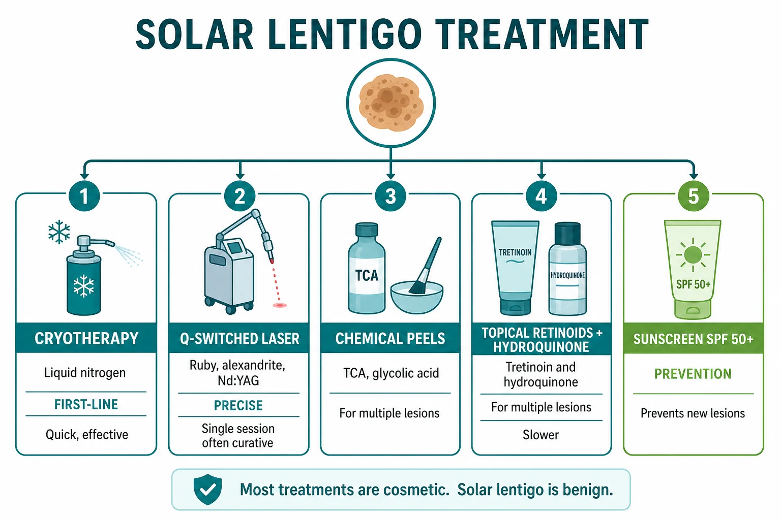
- Broad-spectrum SPF >=30 sunscreen, ideally SPF 50+ with 5-UVA PF >=5; apply generously (a teaspoon for face/ears/neck; two tablespoons per body) 15-30 minutes before sun exposure; reapply every 2 hours and after swimming/sweating.
- Sun-protective clothing (UPF 50+, broad-brimmed hat, sunglasses) prevents new lesions and reduces risk of co-existing malignancy.
- Avoid peak UV hours (10 am-4 pm).
- Avoid indoor tanning beds entirely. [1]
Specific subtypes & scenarios
- Ink-spot lentigo: small (1-3 mm), dark, sharply demarcated, on face or dorsal hands; histology shows elongated rete ridges with a focal basal hyperpigmentation; benign; cryotherapy treats easily.
- PUVA lentigo: pigmented macules persisting after long-term PUVA phototherapy; can be removed with laser or cryotherapy but the surrounding photodamage is the principal concern.
- Lentigo on dark skin (Fitzpatrick V-VI): lesions are often blue-grey rather than brown; dermoscopy reveals a pattern more typical of dermal melanin; biopsy is reasonable if atypical.
- Mosaic / unilateral: very rarely, segmental distribution; biopsy for atypia.
- Large (>1 cm) facial lesion in elderly patient: biopsy unless all features are textbook benign. [1]
Complications & pitfalls
The principal complication of labeling a lesion as solar lentigo is missing a lentigo maligna or lentigo maligna melanoma. Diagnostic pitfalls include: [1]
- Treating without confirming diagnosis: cryotherapy destroys the lesion and any diagnostic histology. Biopsy before destructive treatment if there is any doubt.
- Amalgamating adjacent lesions (multiple lesions coalesce) and missing atypical foci within.
- Patient anxiety: many patients still worry about malignancy; clear documentation in primary care of the benign nature is helpful. [1]
Procedure-related complications: post-cryotherapy hypopigmentation (often permanent and most noticeable in dark skin), post-laser hypopigmentation (transient) or hyperpigmentation (post-inflammatory), post-peel erythema and crusting (expected for 7-10 days), scarring (rare with modern techniques). [1]
Prognosis & disposition
Solar lentigo is entirely benign with no risk of malignant transformation. The lesions persist indefinitely without treatment and new lesions continue to appear with cumulative UV exposure and age. Treatment, when effective, clears the lesion but does NOT prevent new ones. Disposition is outpatient cosmetic dermatology or primary care; no follow-up is needed for classic lesions, although annual full-skin examination is appropriate for high-risk patients (>50, fair skin, history of NMSC or melanoma, immunosuppression). [1]
Special populations
- Children: rare; consider lentigo simplex or ephelis; biopsy if atypical.
- Pregnancy: pigmented macules can darken; observation; treatment deferred until after breastfeeding.
- Immunosuppressed (transplant, HIV): higher prevalence of solar lentigines and a much higher risk of co-existing lentigo maligna and SCC; annual dermatology review with baseline dermoscopic photography for surveillance is recommended.
- Patients with xeroderma pigmentosum: solar lentigo may be the earliest cutaneous sign; aggressive photoprotection and dermatology follow-up are mandatory from childhood.
- Dark-skinned patients (Fitzpatrick V-VI): lentigo can be more difficult to distinguish clinically from post-inflammatory hyperpigmentation; cryotherapy should be applied cautiously given a higher risk of post-treatment hypopigmentation. [1]
Evidence, guidelines & regional differences
- Treatment choices are based on the Mardani 2025 systematic review of clinical trials in solar lentigines, which confirmed efficacy of cryotherapy, Q-switched pigment laser, IPL, and topical lightening agents.[1]
- Lentigo maligna management is documented in the Kallini 2013 review: 5 mm clinical margins on excision; Mohs surgery preferred for tissue-sparing in cosmetically sensitive areas (face); topical imiquimod is reserved for inoperable lesions.[5]
- Cryotherapy in dermatology: classic reviews of the Zouboulis protocol.[3][4]
- Regional deltas: Canada and Australia promote photoprotection campaigns due to high ambient UV. The US FDA regulates hydroquinone 4% as an OTC drug; in the EU hydroquinone is restricted to 1-2% by prescription only.
- Dermoscopic criteria for lentigo maligna are reviewed in Zalaudek 2012 (cited in many subsequent reviews).
Solar lentigo bedside signature (and what is NOT it)
High-yield microcard
[1]The seven teaching points that examiners reward, and the ones that prevent the most common clinical errors. Each is a high-yield one-liner that distils a paragraph above.[1][5]
Exam application bank (NEET-PG / INICET)
One-line answer
Solar lentigo = benign acquired pigmented macule from chronic UV-induced melanocyte proliferation. Uniform light-brown flat macule on sun-exposed skin (dorsum of hands, face, forearms, upper chest). Older adults (>40). Multiple; 'age spots' / 'liver spots' / 'sun spots'. Benign — does NOT transform into melanoma. MUST distinguish from lentigo maligna (irregular borders, colour variation, large, changing — BIOPSY). Treatment: cryotherapy (first-line), Q-switched laser, chemical peels, topical retinoids/hydroquinone. Sunscreen for prevention of new lesions.
Worked stems (answer without another resource)
Stem 1 — Classic presentation. Map symptoms to mechanism; name the first investigation and first treatment step with dose/route if drug therapy is standard. [1]
Stem 2 — Unstable / complicated. List red flags that force immediate resuscitation, theatre, ICU, antidote, or reperfusion — and what you do in the first 15 minutes. [1]
Stem 3 — Atypical group. Elderly, pregnancy, child, or immunocompromised: how presentation and thresholds change. [1]
Stem 4 — Differential trap. Name the three closest mimics and one discriminator for each. [1]
Stem 5 — Disposition. Who goes home with safety-netting, who is admitted, who needs HDU/ICU/theatre, and what follow-up is mandatory. [1]
Rapid viva checklist
- Definition + classification
- Pathophysiology chain
- Bedside signs / criteria
- Score with exact components (if any)
- Emergency bundle
- Definitive therapy with doses
- Complications of disease and of treatment
- Special populations
- Guideline/trial name if classic
- Three exam traps
Coverage self-check
If you cannot answer any stem above from this page alone, re-read the matching section — the page is intended to be self-sufficient for final-prof and NEET-PG/INICET questions on Solar lentigo.
Expanded exam teaching (depth pass)
Clinical reasoning
For Solar lentigo, examiners test whether you can prioritise life threats, choose the right first test, and give specific therapy (agent, dose, route, timing). Generic phrases without numbers score poorly.
Mechanism → feature map
Build a short chain: cause → pathophysiologic intermediate → clinical feature → complication. Every major symptom in the classic vignette should sit on that chain.
Investigation strategy
- Bedside/first-line tests that change immediate management
- Confirmatory or staging tests
- What a normal result does not exclude
- When not to delay treatment for imaging (unstable patient)
Management ladder
- Resuscitation / ABC / sepsis or haemorrhage bundle as relevant
- Specific antidote / procedure / antimicrobial / reperfusion / surgery
- Supportive care and monitoring targets
- Definitive long-term therapy and secondary prevention
- Disposition and safety-net advice
Special populations
Always prepare one line each for children, pregnancy, elderly, renal/hepatic impairment, and immunocompromised patients when the topic allows.
Pitfalls that fail candidates
- Treating the number not the patient
- Missing pregnancy status when relevant
- Imaging before stabilisation
- Wrong empiric cover or wrong antidote timing
- Incomplete counselling on recurrence, adherence, or red-flag return
Solar lentigo = benign acquired pigmented macule from chronic UV-induced melanocyte proliferation. Uniform light-brown flat macule on sun-exposed skin (dorsum of hands, face, forearms, upper chest). Older adults (>40). Multiple; 'age spots' / 'liver spots' / 'sun spots'. Benign — does NOT transform into melanoma. MUST distinguish from lentigo maligna (irregular borders, colour variation, large, changing — BIOPSY). Treatment: cryotherapy (first-line), Q-switched laser, chemical peels, topical ret [1]
Exam pearls
- Classic viva opener: "An 80-year-old retired farmer presents with a 2 cm brown patch on his cheek that has slowly grown over 3 years. Asymmetry is present and dermoscopy shows asymmetric follicular openings. What's the diagnosis and next step?" — answer is lentigo maligna; next step is 3-4 mm punch biopsy from the most atypical area, NOT cryotherapy.
- Viva differential: "Differentiate solar lentigo from seborrhoeic keratosis, lentigo simplex, ephelis, café-au-lait and lentigo maligna" — the bedside signatures are: SK is stuck-on and raised; lentigo simplex is congenital/childhood-onset anywhere; ephelis fades in winter; café-au-lait is congenital and uniform; lentigo maligna is large, multi-coloured and changing.
- Image viva: "Describe this dermoscopic image" — solar lentigo shows uniform light-brown pigmentation with a faint regular network and a moth-eaten border; absence of milia-like cysts rules out SK; absence of grey granules / rhomboid structures rules out lentigo maligna.
- Mechanism viva: "Solar lentigo is BRAF/NRAS-independent. What drives the rete-ridge elongation?" — FGFR3, PIK3CA and STK11 mutations in adjacent keratinocytes drive the epidermal hyperplasia. This distinguishes the lesion biologically from a melanocytic naevus and from most melanomas.
- Treatment viva: "First-line procedural treatment, expected outcome, main complication" — cryotherapy with single 5-10 second freeze-thaw cycle; >80% clearance at 3 months; main complication is post-treatment hypopigmentation (often permanent in Fitzpatrick IV-VI).
- Regional viva: "Compare management in Australia vs UK" — Australia: high ambient UV, daily SPF 50+, more cryotherapy/laser, annual skin check. UK: 2-week-wait referral pathway for atypical lesions, hydroquinone up to 4% on prescription. Both share cryotherapy first-line.
- Pitfall viva: "A patient has cryotherapy for a presumed solar lentigo on the cheek. Histology is impossible. What was the wrong step?" — Treating without first excluding lentigo maligna. Cryotherapy destroys diagnostic tissue. Biopsy first, destroy never first.[5]
References
- [1]Mardani G, Nasiri MJ, Namazi N, et al. Treatment of Solar Lentigines: A Systematic Review of Clinical Trials J Cosmet Dermatol, 2025.PMID 40145274
- [2]Hara Y, Shibata T. Characteristics of dermal vascularity in melasma and solar lentigo Photodermatol Photoimmunol Photomed, 2024.PMID 38353352
- [3]Zouboulis CC. [Cryosurgery in dermatology] Hautarzt, 2015.PMID 26497955
- [4]Zouboulis CC. Cryosurgery in dermatology Eur J Dermatol, 1998.PMID 9854156
- [5]Kallini JR, Jain SK, Khachemoune A. Lentigo maligna: review of salient characteristics and management Am J Clin Dermatol, 2013.PMID 24019181