Dermatology · Medicine
Special stains and immunofluorescence in dermatopathology
Also known as Dermatopathology special stains · Direct immunofluorescence skin · DIF dermatology · Salt-split skin immunofluorescence · Histochemical stains skin biopsy
Practical dermatopathology toolkit for special histochemical stains (PAS, GMS, AFB/Fite, VVG, Congo red, Fontana-Masson, mucin stains) and immunofluorescence (DIF, IIF, salt-split skin) in skin disease. Covers specimen handling (Michel medium vs formalin), biopsy site selection, classic IF patterns for pemphigus/pemphigoid/EBA/DH/LABD, serologic adjuncts, and exam-critical pitfalls that change infection versus autoimmunity pathways.
On this page & tools
Your progress
Saved locally on this device.
Exam tags
Red flags
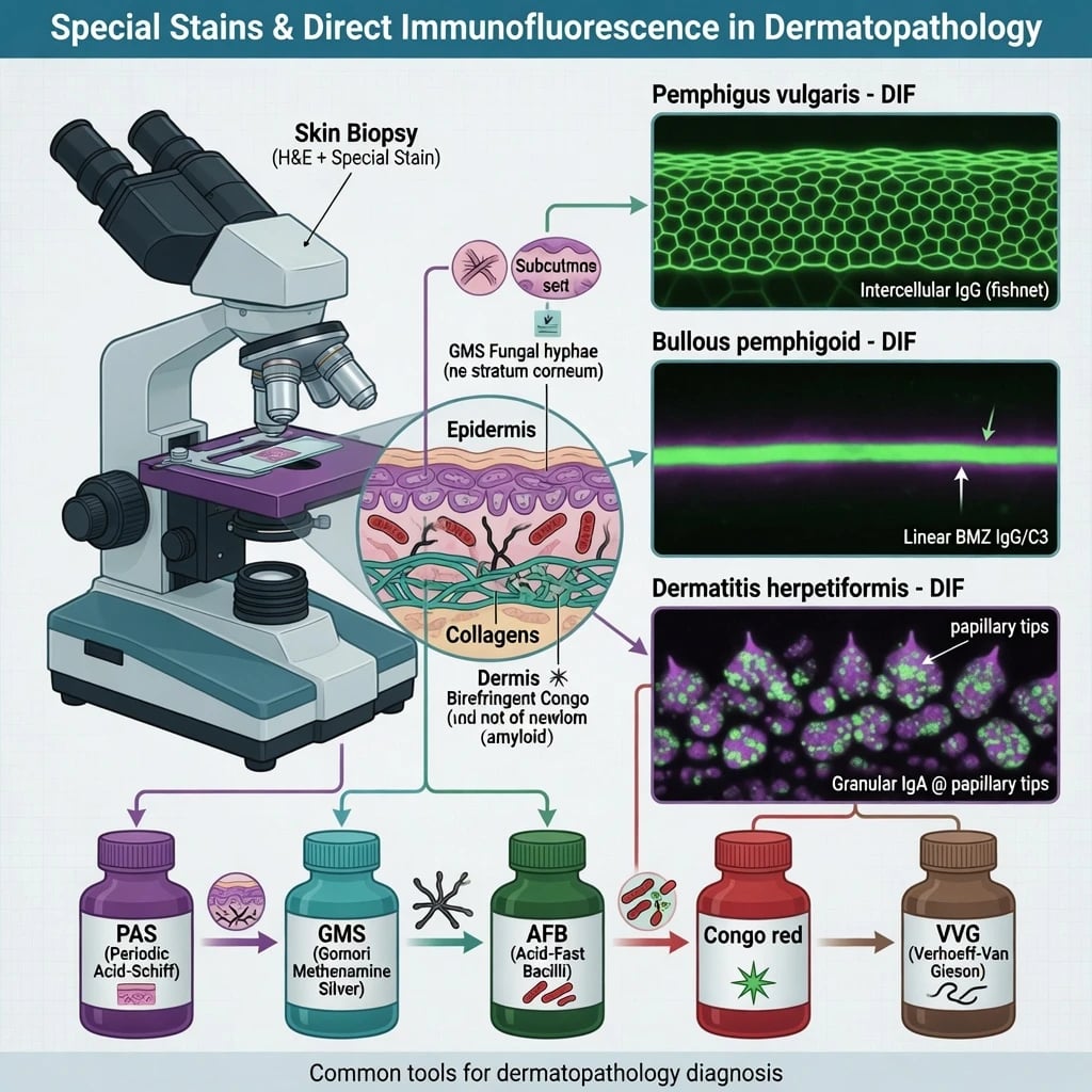
Definition & Classification
Dermatopathology uses three complementary ancillary families: [1]
| Method | What it shows | Typical fixative |
|---|---|---|
| Histochemical special stains | Organisms, amyloid, elastic, collagen, mucin, some pigments | Formalin |
| Immunohistochemistry (IHC) | Protein antigens via antibodies (lineage, pathogens, tumour markers) | Formalin (usually) |
| Immunofluorescence (IF) | In vivo/deposited immunoreactants (IgG, IgA, IgM, C3) | Michel medium (or snap-frozen) for DIF |
This topic focuses on special stains + IF; lineage IHC for lymphomas/histiocytes is covered with lymphoid infiltrates. [1]
Special stains by target
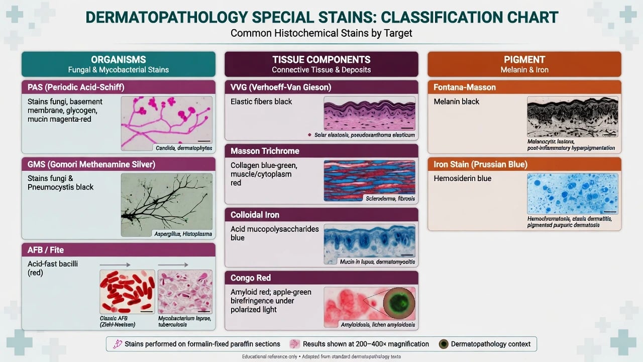
| Target | High-yield stains (teaching set) |
|---|---|
| Fungi / basement membrane | PAS, GMS |
| Mycobacteria | Ziehl–Neelsen AFB; Fite (better for M. leprae teaching) |
| Bacteria (selected) | Gram; silver methods (e.g. Warthin–Starry family) for selected spirochaetes/bacilli |
| Elastic tissue | Verhoeff–Van Gieson (VVG) |
| Collagen | Masson trichrome |
| Acid mucins | Colloidal iron / Alcian blue family |
| Amyloid | Congo red (± polarisation for apple-green birefringence) |
| Melanin | Fontana–Masson |
| Iron/haemosiderin | Perls Prussian blue |
PAS/GMS remain the workhorse pair when fungal infection is in the histologic differential.[6] Elastic stains refine diagnoses where elastic loss or fragmentation is the disease signal.[7]
Immunofluorescence modalities
| Test | Sample | Question answered |
|---|---|---|
| DIF | Patient skin (usually perilesional) | What is deposited in vivo? |
| IIF | Patient serum on substrate | Circulating autoantibodies? |
| Salt-split skin IF | Substrate or patient skin split at lamina lucida | Roof vs floor binding (BP group vs EBA zone teaching) |
| ELISA / bioassays | Serum | Specific antigens (BP180/BP230, desmogleins, etc.) |
Modern diagnosis of autoimmune blistering diseases integrates clinical pattern, histopathology, DIF, and serology rather than any single test.[2][1]
Epidemiology & When Tests Are Highest Yield
- Immunobullous disease (BP in elderly; pemphigus with mucosal risk; DH with gluten-sensitive enteropathy context) drives most DIF volume in dermatology labs.[4][3][9]
- Infectious dermatopathology needs special stains whenever granulomas, neutrophils with “empty” organisms on H&E, or immunosuppressed hosts raise pretest probability.[6]
- False-negative DIF rises with lesional necrotic skin only, delayed transport, or formalin fixation of the IF specimen.[10][1]
Pathophysiology (How the Tests Work)
- PAS oxidises carbohydrates → magenta fungal walls and highlights basement membrane glycoproteins.[6]
- Congo red intercalates amyloid β-pleated sheets → characteristic birefringence under polarised light (classic teaching).
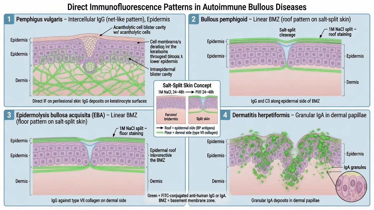
- DIF detects tissue-bound Ig/complement deposited at adhesion structures:
- 1 M NaCl salt-split cleaves through lamina lucida: BP180-region antibodies typically decorate the epidermal roof; type VII collagen (EBA) antibodies decorate the dermal floor on salt-split teaching models.[5][4]
Clinical Presentation — Test Triggers
Order with intent, not as a reflexive panel: [1]
| Clinical clue | Priority ancillary |
|---|---|
| Flaccid blisters, Nikolsky, mucosal erosions | DIF + pemphigus serology pathway |
| Tense bullae, elderly, urticarial plaques | DIF + BP ELISA concepts |
| Pruritic vesicles on elbows/knees/buttocks | DIF for DH + coeliac work-up |
| Chronic oral/ocular scarring blisters | MMP pathway (DIF ± serology/specialist) |
| Granuloma / non-healing ulcer in at-risk host | PAS/GMS/AFB ± culture |
| Suspected elastic tissue disorder patterns | VVG |
| Waxy papules / macroglossia context | Congo red |
Differential Diagnosis by IF Pattern
| DIF pattern | First-line differential |
|---|---|
| Intercellular IgG ± C3 | Pemphigus vulgaris / foliaceus; consider paraneoplastic pemphigus if severe mucocutaneous + internal context |
| Linear BMZ IgG/C3 | Bullous pemphigoid, mucous membrane pemphigoid, EBA, anti-p200 and related subepidermal diseases — refine with salt-split/serology |
| Linear IgA BMZ | Linear IgA bullous dermatosis |
| Granular IgA dermal papillae | Dermatitis herpetiformis |
| Negative DIF | May be early/treated disease, wrong site/medium, or non-autoimmune blistering (infection, TEN, porphyria, etc.) |
Pattern recognition is foundational in DIF diagnosis of autoimmune bullous dermatoses.[1][2]
Clinical & Specimen Handling (Bedside Skills)
- H&E specimen: edge of early blister / representative lesion in formalin.
- DIF specimen: perilesional erythematous skin (not pure necrotic centre) in Michel medium (or frozen protocol per lab).[10][1]
- Label containers unambiguously; never put the only DIF piece in formalin “by habit.”
- For alopecia/elastic questions, ensure the lab knows the clinical question so VVG is considered.[7]
Investigations — Practical Menu
Special stains (high-yield use cases)
- PAS ± GMS: fungi (dermatophytes, Candida, deep fungi), glycogen, BM thickening contexts; literature supports selective rather than blind universal ordering, but threshold should be low when infection is plausible.[6]
- AFB / Fite: mycobacteria; Fite preferred teaching stain when leprosy is suspected.
- VVG: elastic fibre quantity/quality — useful across anetoderma, mid-dermal elastolysis concepts, PXE-like patterns, and selected scarring alopecias/surgical questions.[7]
- Congo red: cutaneous amyloid (macular/lichen/nodular types) and systemic amyloid clues in skin.
- Fontana–Masson / iron stains: separate melanin from haemosiderin when pigment is ambiguous.
IF + serology package for blistering disease
- DIF first-line tissue test for autoimmune blistering diseases.[1][2]
- IIF on appropriate substrates and antigen-specific ELISA (e.g. BP180/BP230; desmoglein 1/3) refine subtype and activity monitoring in contemporary pathways.[2][8][3]
- Salt-split when subepidermal linear IgG disease needs BP-group vs EBA separation.[5]
Management — Resuscitation Priorities
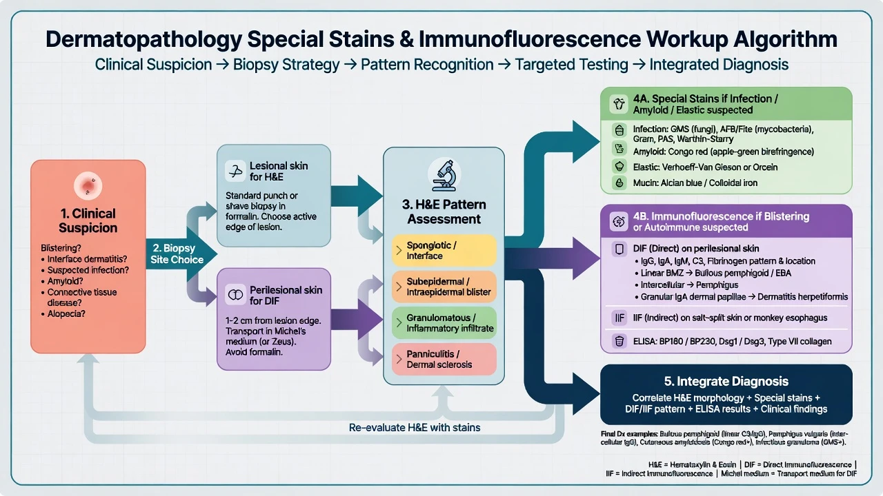
Ancillary tests must not delay life-saving care: [1]
- Airway/mucosal protection and fluid/barrier care in extensive erosions (pemphigus/TEN differentials).
- If infection is plausible, avoid high-dose immunosuppression until stains/cultures are planned.
- Start the correct diagnostic pathway the same day (right medium, right site). [1]
Management — How Results Change Therapy
| Result | Typical therapeutic pivot |
|---|---|
| DIF + fishnet IgG (pemphigus) | Systemic immunosuppression / rituximab-era pathways as indicated; wound care |
| DIF + linear BMZ (BP) | Superpotent topicals ± systemic agents per severity; not antifungals |
| Granular IgA (DH) | Dapsone + gluten-free diet pathway after G6PD/safety checks |
| PAS fungal hyphae in “eczema” | Stop sole steroid strategy; treat infection |
| AFB-positive granuloma | Antimicrobial antimycobacterial pathway, not only steroids |
| Congo red amyloid | Investigate locally restricted vs systemic amyloid as clinically indicated |
State-of-the-art blistering disease diagnosis is multimodal; serology can support when DIF is delayed or equivocal, but tissue DIF remains central.[2]
Special stains
- Formalin OK
- Organisms/amyloid/elastic
- Guide infection therapy
- Do not prove autoimmunity
DIF/IIF
- Michel/frozen for DIF
- Autoantibody deposits
- Pattern = disease class
- Needs correct site
ELISA/serology
- Serum antigen specificity
- Complements DIF
- Monitors activity
- Not a full substitute for tissue
Specific Subtypes & Scenarios
Pemphigus vs pemphigoid (exam core)
- Pemphigus: suprabasal/subcorneal acantholysis + intercellular DIF.[3]
- Pemphigoid group: subepidermal split + linear BMZ DIF.[4][8]
DH vs LABD
- DH: granular IgA in papillae; gluten-sensitive enteropathy association.[9]
- LABD: linear IgA along BMZ; drug triggers (e.g. vancomycin teaching association) in some adults.
EBA vs BP on salt-split
- Teaching rule: floor binding favours EBA (type VII collagen zone); roof favours BP180-region pemphigoid diseases — always integrate clinical/serology because exceptions and technical issues exist.[5][4]
Infection traps
- Steroid-treated tinea can look like eczema — PAS saves the diagnosis.[6]
- Negative stains ≠ no organisms if sampling is poor; culture/PCR remain partners.
Complications & Pitfalls
- DIF specimen in formalin.
- Biopsying only the blister roof or fully necrotic centre for DIF.
- Interpreting weak non-specific fluorescence as disease.
- Ordering every stain on every biopsy without a question (cost/noise) — but also under-ordering PAS when infection is likely.[6]
- Assuming ELISA alone replaces DIF in all centres and all diseases.[2]
Prognosis & Disposition of Testing
- Correct early classification shortens time to disease-directed therapy and reduces iatrogenic steroid injury in occult infection.
- If transport fails, repeat biopsy promptly rather than guessing.
- Complex MMP/pemphigus cases need dermatology–ophthalmology–ENT pathways beyond a single stain result. [1]
Special Populations
- Elderly: high BP pretest probability; still exclude drug and bullous scabies mimics clinically.[8]
- Children: consider LABD and genetic mechano-bullous disease differentials.
- Pregnancy: coordinate diagnostics with obstetric safety; clinical management beyond this topic.
- Resource-limited labs: prioritise Michel DIF + PAS as highest leverage pair when full menus are unavailable.
Evidence, Guidelines & Regional Differences
- Contemporary reviews emphasise combined clinical–histologic–IF–serologic diagnosis for AIBD.[2]
- PAS/GMS utility is supported by dermatopathology literature with nuanced recommendations rather than universal reflex on every specimen.[6]
- Elastic VVG has defined niche utility documented in cutaneous pathology series.[7]
- Michel medium remains the practical transport standard for IF when freezing is not immediate.[10]
- Regional access to advanced ELISA/biochips varies; DIF skill remains globally foundational.[1]
Exam Pearls
STAINS-IF
STAINS
Perilesional for DIF; lesional edge for H&E
Michel for DIF; formalin for stains
Fishnet / linear BMZ / granular IgA
PAS GMS AFB/Fite before more steroids
Roof BP-zone vs floor EBA-zone teaching
ELISA/IIF complete the package
- Fishnet IgG → pemphigus until proven otherwise.[3][1]
- Linear BMZ IgG/C3 → pemphigoid group; use salt-split/serology to refine.[4][5]
- Granular IgA papillae → DH.[9]
- PAS before chronic “eczema” steroids when tinea is possible.[6]
- VVG when elastic pathology is the question.[7]
Exam application bank (NEET-PG / INICET)
One-line answer
Practical dermatopathology toolkit for special histochemical stains (PAS, GMS, AFB/Fite, VVG, Congo red, Fontana-Masson, mucin stains) and immunofluorescence (DIF, IIF, salt-split skin) in skin disease. Covers specimen handling (Michel medium vs formalin), biopsy site selection, classic IF patterns for pemphigus/pemphigoid/EBA/DH/LABD, serologic adjuncts, and exam-critical pitfalls that change infection versus autoimmunity pathways.
Worked stems (answer without another resource)
Stem 1 — Classic presentation. Map symptoms to mechanism; name the first investigation and first treatment step with dose/route if drug therapy is standard. [1]
Stem 2 — Unstable / complicated. List red flags that force immediate resuscitation, theatre, ICU, antidote, or reperfusion — and what you do in the first 15 minutes. [1]
Stem 3 — Atypical group. Elderly, pregnancy, child, or immunocompromised: how presentation and thresholds change. [1]
Stem 4 — Differential trap. Name the three closest mimics and one discriminator for each. [1]
Stem 5 — Disposition. Who goes home with safety-netting, who is admitted, who needs HDU/ICU/theatre, and what follow-up is mandatory. [1]
Rapid viva checklist
- Definition + classification
- Pathophysiology chain
- Bedside signs / criteria
- Score with exact components (if any)
- Emergency bundle
- Definitive therapy with doses
- Complications of disease and of treatment
- Special populations
- Guideline/trial name if classic
- Three exam traps
Coverage self-check
If you cannot answer any stem above from this page alone, re-read the matching section — the page is intended to be self-sufficient for final-prof and NEET-PG/INICET questions on Special stains and immunofluorescence in dermatopathology.
Expanded exam teaching (depth pass)
Clinical reasoning
For Special stains and immunofluorescence in dermatopathology, examiners test whether you can prioritise life threats, choose the right first test, and give specific therapy (agent, dose, route, timing). Generic phrases without numbers score poorly.
Mechanism → feature map
Build a short chain: cause → pathophysiologic intermediate → clinical feature → complication. Every major symptom in the classic vignette should sit on that chain.
Investigation strategy
- Bedside/first-line tests that change immediate management
- Confirmatory or staging tests
- What a normal result does not exclude
- When not to delay treatment for imaging (unstable patient)
Management ladder
- Resuscitation / ABC / sepsis or haemorrhage bundle as relevant
- Specific antidote / procedure / antimicrobial / reperfusion / surgery
- Supportive care and monitoring targets
- Definitive long-term therapy and secondary prevention
- Disposition and safety-net advice
Special populations
Always prepare one line each for children, pregnancy, elderly, renal/hepatic impairment, and immunocompromised patients when the topic allows.
Pitfalls that fail candidates
- Treating the number not the patient
- Missing pregnancy status when relevant
- Imaging before stabilisation
- Wrong empiric cover or wrong antidote timing
- Incomplete counselling on recurrence, adherence, or red-flag return
Practical dermatopathology toolkit for special histochemical stains (PAS, GMS, AFB/Fite, VVG, Congo red, Fontana-Masson, mucin stains) and immunofluorescence (DIF, IIF, salt-split skin) in skin disease. Covers specimen handling (Michel medium vs formalin), biopsy site selection, classic IF patterns for pemphigus/pemphigoid/EBA/DH/LABD, serologic adjuncts, and exam-critical pitfalls that change infection versus autoimmunity pathways. [1]
Structured revision sheet
Must-know numbers and names
List every score, size threshold, dose, and time window from this topic on a blank page from memory, then check against the sections above.
Three classic MCQ angles
- Most likely diagnosis given a vignette
- Next best step in management
- Most appropriate investigation
Three classic SAQ angles
- Pathophysiology in five steps
- Management algorithm with doses
- Complications and prevention
Clinical station flow
Greet → focused history → targeted exam → investigations → explain diagnosis → emergency care → definitive plan → safety-net / follow-up → answer examiner questions on mechanism and pitfalls.
[1] [1]References
- [1]Morrison LH. Direct immunofluorescence microscopy in the diagnosis of autoimmune bullous dermatoses Clin Dermatol, 2001.PMID 11604308
- [2]van Beek N, Holtsche MM, Atefi I, et al. State-of-the-art diagnosis of autoimmune blistering diseases Front Immunol, 2024.PMID 38903493
- [3]Schmidt E, Kasperkiewicz M, Joly P. Pemphigus Lancet, 2019.PMID 31498102
- [4]Schmidt E, Zillikens D. Pemphigoid diseases Lancet, 2013.PMID 23237497
- [5]Woodley DT. Immunofluorescence on salt-split skin for the diagnosis of epidermolysis bullosa acquisita Arch Dermatol, 1990.PMID 2405782
- [6]Shalin SC, Ferringer T, Cassarino DS. PAS and GMS utility in dermatopathology: Review of the current medical literature J Cutan Pathol, 2020.PMID 32515092
- [7]Kazlouskaya V, Malhotra S, Lambe J, et al. The utility of elastic Verhoeff-Van Gieson staining in dermatopathology J Cutan Pathol, 2013.PMID 23216221
- [8]Miyamoto D, Santi CG, Aoki V, et al. Bullous pemphigoid An Bras Dermatol, 2019.PMID 31090818
- [9]Antiga E, Maglie R, Quintarelli L, et al. Dermatitis Herpetiformis: Novel Perspectives Front Immunol, 2019.PMID 31244841
- [10]Vaughn Jones SA, Palmer I, Bhogal BS, et al. The use of Michel's transport medium for immunofluorescence and immunoelectron microscopy in autoimmune bullous diseases J Cutan Pathol, 1995.PMID 7499578