Dermatology · Medicine
Spitz naevus (spindle and epithelioid cell naevus)
Also known as Spitz naevus · Spindle and epithelioid cell naevus · Benign juvenile melanoma (historical, misleading term) · Pigmented spindle cell naevus of Reed · Atypical Spitz tumour (borderline variant)
Spitz naevus is a benign acquired melanocytic naevus of childhood and adolescence composed of large spindled and epithelioid melanocytes, presenting most often as a rapidly growing pink, hairless, dome-shaped papule. Histology shows spindled/epithelioid melanocytes, Kamino bodies, symmetry, and maturation with depth. The central diagnostic challenge is distinguishing benign Spitz naevus from spitzoid melanoma, with the atypical Spitz tumour occupying a borderline zone. Management is complete surgical excision with clear margins.
On this page & tools
Your progress
Saved locally on this device.
Exam tags
Red flags
Overview and definition
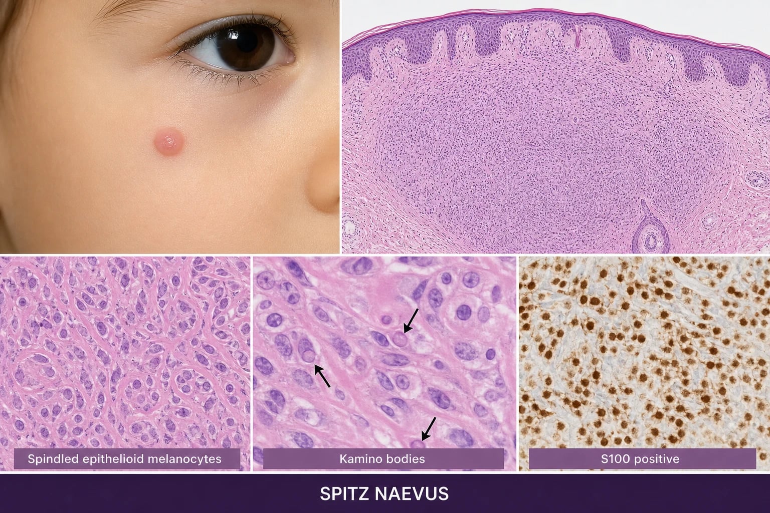
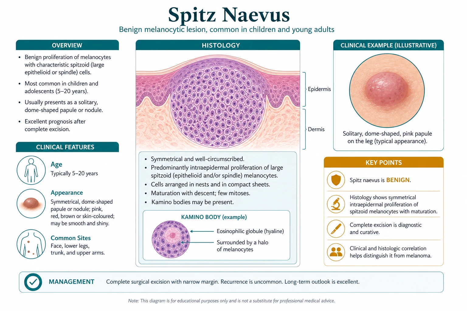
Spitz naevus — also called the spindle and epithelioid cell naevus — is a distinctive, benign, acquired melanocytic naevus that most often appears in children and adolescents as a rapidly growing, pink, red, or skin-coloured, hairless, dome-shaped papule. It was first described by Sophie Spitz in 1948 as "juvenile melanoma," a label that was always misleading and has been abandoned precisely because it captures the topic's central problem: a benign lesion that can look histologically alarming and that must be separated from spitzoid melanoma.[3][4]
The reason Spitz naevus warrants a topic of its own is diagnostic rather than therapeutic. The individual cells are large, with abundant amphophilic cytoplasm and prominent nucleoli; they lie in fascicles and nests; they may even scatter up into the epidermis (pagetoid spread). Each of those features, taken alone, is melanoma-like. What rescues the diagnosis is a set of reassuring architectural cues — symmetry, maturation with depth, sharp lateral demarcation, and Kamino bodies — together with a low proliferative rate and an HMB-45 gradient. A subset of lesions cannot be confidently placed as benign or malignant; these are the atypical Spitz tumours (ASTs), a borderline category that has driven most of the modern controversy over margins, sentinel lymph node biopsy, and molecular testing.[1][2]
Classification — the spitzoid spectrum
Spitz naevi do not sit in isolation. They are best understood as one point on a spectrum of spitzoid melanocytic proliferations that runs from unequivocally benign to unequivocally malignant, with the atypical Spitz tumour in between. The spectrum is increasingly refined by molecular genetics, which has split the old morphologic lump into biologically distinct entities.[2][15] It helps to keep the spectrum and the variants distinct. The spectrum (Spitz naevus — atypical Spitz tumour — spitzoid melanoma) is about biological potential and is graded by integrating architecture, cytology, and increasingly molecular data. The variants (classic pink, pigmented/Reed) are about morphology and dermoscopy, and carry largely benign prognoses when they meet the classic criteria. A lesion can be morphologically pigmented and still biologically borderline, so the two axes are reported independently: a pathologist who writes "atypical Spitz tumour, pigmented variant" is telling the clinician both things at once. The examiner will probe whether the candidate can hold both axes simultaneously, rather than collapsing the whole topic into "Spitz naevus equals benign, everything else equals melanoma".[2][15]
Within the benign end, two morphologic variants dominate clinical practice and deserve to be named explicitly: [1]
- Classic (pink, non-pigmented) Spitz naevus — the vascular, rapidly growing pink papule of childhood, typically on the face. Its dermoscopy is dominated by regularly arranged dotted and glomerular vessels against a background of reticular depigmentation.
- Pigmented Spitz naevus, including the pigmented spindle cell naevus of Reed — a dark brown to blue-black, sharply bordered, often symmetric papule more common in darker phototypes, whose starburst dermoscopy pattern (radial streaks and pseudopods at the periphery) is one of the closest mimics of superficial spreading melanoma.[9]
Molecular classification now recognises at least three biologically defined groups that cut across the older morphology: HRAS-mutant/11p-gain Spitz naevi; BAP1-loss spitzoid naevi/tumours (often combined with BRAF V600E and yielding the dome-shaped epithelioid 'BAPoma' appearance); and a growing family of kinase-fusion spitzoid neoplasms (ALK, RET, NTRK1/3, MET, ROS1, MAP3K8) that tend to behave as low-grade lesions.[6][8][15]
Epidemiology and risk factors
Spitz naevi are uncommon but not rare, and they are over-represented in childhood compared with adult dermatology. They account for roughly 1 to 4 per cent of melanocytic lesions excised in children, with a peak incidence between 3 and 13 years of age.[14] They are distinctly uncommon in adults, and an adult spitzoid lesion carries a higher probability of being an atypical Spitz tumour or a spitzoid melanoma than the same lesion would in a child. Larger contemporary series have refined this picture. In a major Boston paediatric cohort, spitzoid proliferations presented across a wide paediatric age range with a slight female predominance, the classic pink morphology predominated, and outcomes were favourable even for atypical lesions — a pattern that has underpinned the move toward less invasive management in children. Importantly, that same reassuring series documented that genuinely malignant behaviour, when it occurs, is rare but real, and is not reliably predicted by histology alone. The practical implication for the clinician is that paediatric Spitz naevi can usually be managed conservatively once histology is secure, but that no single clinical or demographic feature abolishes the need for expert pathology review of an atypical lesion.[14]
The classic pink lesion predominates in fair-skinned children, whereas the pigmented/Reed variant is relatively more frequent in darker skin phototypes, where it often presents on the lower limbs. The face (especially the cheeks), the lower legs, and the trunk are the dominant sites; agminated (clustered) and eruptive/multiple Spitz naevi are rare but well recognised. There is no convincing link to ultraviolet exposure or family history as a principal driver, and the condition is essentially sporadic.[3]
A risk factor in the practical (rather than aetiological) sense is age: an adult presenting with a lesion that looks like a Spitz naevus is the single most important red flag in this topic, because the prior probability of malignancy rises sharply with age. [1]
Pathophysiology — why a benign lesion looks malignant
The histologic appearance of Spitz naevus is the key to understanding the whole topic, because it explains both the diagnostic difficulty and the molecular biology. The cell of origin is a naevus melanocyte, but an unusually large one. In a classic Spitz naevus the melanocytes are large and epithelioid or spindled, with abundant amphophilic cytoplasm and prominent nucleoli, arranged in fascicles and nests at the dermoepidermal junction and in the dermis. Two architectural principles keep the lesion benign despite these alarming cells: the lesion is symmetrical, and it matures with depth — the cells become smaller, more dispersed, and less pigmented as they descend, the opposite of melanoma.[4][5]
The signature structures are Kamino bodies — pale eosinophilic globules sitting at the dermoepidermal junction. They are composed of basement-membrane material (type IV collagen and laminin), they are characteristic of Spitz naevi, and they are uncommon in melanoma, which makes them one of the most useful single features for separating the two. Epidermal hyperplasia with elongated rete ridges and a background of ectatic capillaries (the dermoscopic correlate of the dotted vessels) complete the picture.[4]
The molecular story has matured considerably and is now central to the management of ambiguous lesions. Three pathways recur: [1]
- HRAS mutation with 11p copy-number gain. Bastian and colleagues showed that a subset of Spitz naevi — perhaps 10 to 20 per cent, characteristically those with a desmoplastic/spindled morphology — carry an activating HRAS mutation together with gains on the short arm of chromosome 11, where HRAS resides. This is essentially specific to Spitz naevi and is not a feature of conventional naevi or of most melanomas.[6]
- MAP-kinase activation with cell-cycle arrest. The same pathway can be driven by BRAF V600E, yet Spitz naevi characteristically show senescence-like cell-cycle arrest rather than the uncontrolled proliferation of melanoma — the molecular explanation for why a BRAF-driven lesion can still be benign.[7]
- Kinase fusions and BAP1 loss. A growing proportion of spitzoid neoplasms, including the atypical Spitz tumour, harbour kinase fusions (ALK, RET, NTRK1/3, MET, ROS1, MAP3K8) that define distinct, generally low-grade entities, while BAP1 loss (often combined with BRAF V600E) produces a characteristic dome-shaped, epithelioid, naevoid morphology — the so-called BAP1-inactivated spitzoid naevus/tumour.[8][15] The thread that unites these findings is oncogene-induced senescence. A Spitz naevus is, in effect, a melanocyte that switched on an oncogenic MAP-kinase signal and then stopped — the same pathway that, left unchecked, produces melanoma. The arrest is enforced by tumour-suppressor machinery, chiefly p16 (encoded by CDKN2A at chromosome 9p21) and the establishment of senescence-associated heterochromatin, which together drive the cell into a stable, non-proliferative state. This explains two clinical observations at once: why a Spitz naevus can harbour a BRAF V600E or HRAS mutation yet remain benign, and why loss of 9p21 — the lesion that disables p16 — is one of the most reliable molecular signals that a spitzoid proliferation has crossed into melanoma. The HMB-45 gradient (loss of deep expression) and the low Ki-67 proliferative index are the immunohistochemical fingerprints of this arrested state.[7][10]
The practical consequence is that molecular testing (FISH, comparative genomic hybridisation, and increasingly next-generation sequencing) and immunohistochemistry (PRAME, the HMB-45 maturation gradient, Ki-67) are no longer research tools: they are part of how an ambiguous spitzoid lesion is triaged in a modern pigmented-lesion service.[10][15]
Clinical presentation
The clinical presentation is defined by a lesion that grows quickly and looks threatening in a child. The classic lesion is a firm, smooth-surfaced, hairless, dome-shaped papule or nodule, 2 mm to 2 cm across, pink, red, or skin-coloured, with a surface that may show fine telangiectasia. Onset is over weeks to a few months — the rapid growth is usually what brings the child to the clinic, and it is exactly what raises melanoma concern. Most lesions are asymptomatic; some are tender, and a few bleed on minor trauma.[3][14] The surface detail matters at the bedside. The hairlessness is a useful sign: a Spitz naevus characteristically sits on skin that has lost its terminal hairs, and the smooth, tense surface distinguishes it from the warty, stuck-on feel of a seborrhoeic keratosis or the scaly surface of a keratoacanthoma. The telangiectatic vessels are fine and regular when the lesion is benign; coarse, irregular vessels, or a friable bleeding surface, point instead to a pyogenic granuloma or an amelanotic melanoma. Colour uniformity is informative too — a Spitz naevus is usually a single shade of pink or red, whereas the multiple shades and the focal blue-black or white regression of a melanoma are absent.[3]
The two clinical variants present differently and are examined separately: [1]
Typical tempo of a classic paediatric Spitz naevus
- Classic (pink) Spitz naevus — the vascular childhood lesion on the face or leg described above. Dermoscopy shows regularly arranged dotted or glomerular (coiled) vessels against a background of reticular (network-like) depigmentation.
- Pigmented Spitz naevus / Reed naevus — a dark brown to blue-black, sharply bordered, often symmetric papule, more common in darker phototypes and on the lower limbs. Its dermoscopy is the starburst pattern: regular radial streaks or pseudopods distributed symmetrically around the lesion's perimeter. This is the variant most often mistaken clinically and dermoscopically for melanoma.[9] A useful clinicopathologic caveat: the dermoscopic pattern of a Spitz naevus evolves. A Reed naevus classically passes through phases — homogeneous, then starburst (with peripheral streaks), then a structureless or reticular phase as it stabilises — so a lesion caught mid-transition can show a mixed, atypical-looking pattern that does not fit the textbook starburst. In children this usually still resolves to a benign lesion; in adults the same indeterminate pattern is treated with suspicion, because the probability that it represents a spitzoid or conventional melanoma is materially higher. The dermoscopist's job is therefore not to name the lesion but to ask whether the pattern is regular and symmetric (reassuring) or irregular and asymmetric (excise), and to integrate that judgement with the patient's age.[3][9]
Atypical presentations that must trigger escalation (rather than reassurance) include: a spitzoid lesion in an adult; ulceration, colour change, or very rapid growth after a stable period; satellite lesions or regional lymphadenopathy; and any lesion whose dermoscopy is frankly atypical or non-specific. These features shift the working diagnosis toward atypical Spitz tumour or spitzoid melanoma.[1]
Differential diagnosis
A rapidly growing pink papule on a child's face has a focused differential, and the task is to rank the benign mimics while never losing sight of the malignant possibility. The table below lists the common contenders with the feature that distinguishes each. [1]
For the dark pigmented papule, the differential shortens to the Reed/pigmented Spitz naevus versus superficial spreading melanoma, thrombosed haemangioma, pigmented basal cell carcinoma, and blue naevus. The discriminating features are symmetry of border and of dermoscopic structures (favouring Reed), versus the asymmetry, multiple colours, and atypical network of melanoma.[3][9]
The rule the examiner rewards is simple: a spitzoid lesion in an adult is a red flag. Spitzoid melanoma cannot be excluded clinically in adults, so the adult lesion is excised with margins and sent for expert pathology review — observation is reserved, at most, for the classic, confident, paediatric pink Spitz naevus under expert surveillance.[1][2]
Clinical and bedside assessment
The focused examination is short but must be deliberate. Inspect the lesion for site, size, shape, surface, and colour uniformity; note hairlessness, telangiectasia, tenderness, and (critically) ulceration; and examine the surrounding skin for satellite lesions and the regional nodes for lymphadenopathy. A lesion on the cheek prompts examination of the preauricular, parotid, and cervical nodes; a leg lesion prompts examination of the groin. [1]
Dermoscopy is the single most useful bedside test. Apply it routinely and describe the pattern rather than the diagnosis. The classic pink Spitz naevus shows regular dotted or glomerular vessels against a reticular depigmentation (a white network). The pigmented/Reed variant shows the starburst pattern — radial streaks and pseudopods arranged symmetrically around the perimeter — together with a central dark structureless area.[9] A genuinely atypical or non-specific dermoscopic pattern, particularly in an adult, is not reassuring and mandates excision.
It is worth being explicit about what dermoscopy cannot do. The Spitz naevus is one of the canonical melanoma mimics precisely because its pattern can overlap with melanoma, and because the morphologically ambiguous atypical Spitz tumour sits on a spectrum with spitzoid melanoma. A confident dermoscopic diagnosis of a classic paediatric Spitz naevus can, in expert hands, justify observation; a lesion that is atypical or in an adult cannot be dismissed on dermoscopy alone.[3]
Investigations
Diagnosis is clinicopathologic: clinical impression plus dermoscopy plus complete-excision histopathology. There is no blood test and no imaging of value for the typical lesion; investigations are reserved for staging once a spitzoid melanoma is confirmed. [1]
Diagnostic pathway for a suspected spitzoid lesion
Clinical assessment: site, size, morphology, ulceration, satellites, regional nodes.
Dermoscopy: describe the pattern (vascular / starburst / atypical).
Complete surgical excision (not shave, not punch) — preserve the entire lesion for the pathologist.
Histopathology: assess symmetry, maturation with depth, Kamino bodies, pagetoid spread, mitoses, margins.
For ambiguous lesions: expert second opinion + ancillary IHC (Ki-67, HMB-45 gradient, PRAME) and/or molecular testing (FISH, CGH, HRAS/BAP1/kinase fusions).
Stage with sentinel lymph node biopsy only if an atypical Spitz tumour or spitzoid melanoma is confirmed and the MDT supports it.
Why a shave or punch biopsy is contraindicated. The histologic diagnosis depends on judging symmetry, maturation with depth, and the deep margin, all of which require the complete lesion en bloc. A shave removes the superficial component and distorts or discards the deep dermis; a punch samples only part of the lesion. Either can convert a diagnosable Spitz naevus into an unsolvable problem, or miss the deepest, most diagnostic element. The correct procedure is complete excision.[3][4]
Histology of the classic Spitz naevus combines reassuring architecture with cytologically large cells: [1]
- Large spindled and epithelioid melanocytes in fascicles and nests, with abundant amphophilic cytoplasm and prominent nucleoli.
- Kamino bodies — pale eosinophilic globules of basement-membrane material at the dermoepidermal junction; characteristic of Spitz naevi and uncommon in melanoma.
- Symmetry and sharp lateral demarcation.
- Maturation with depth — cells diminish in size and pigment as they descend.
- Pagetoid spread that is central and confined, never reaching the lesion's lateral edges.
- Epidermal hyperplasia with elongated rete ridges and ectatic capillaries in the papillary dermis. [1]
Features that instead favour an atypical Spitz tumour or spitzoid melanoma are the mirror image: asymmetry, lack of maturation, deep mitoses (especially atypical forms), high cellularity, expansile growth, ulceration, and pagetoid spread that extends to the edges. When these dominate, the lesion is downgraded from benign Spitz naevus toward the AST/melanoma end of the spectrum.[1][2]
Ancillary testing for ambiguous lesions has moved into routine practice: [1]
- Immunohistochemistry — a low Ki-67 proliferative index and an HMB-45 gradient (positive superficially, negative deeply) both favour a benign Spitz naevus; loss of that gradient and a high Ki-67 favour melanoma. PRAME positivity is emerging as a useful melanoma-favouring marker.
- FISH — a four-probe panel (6p25, 6q23, 11q13, and centromere 6) plus 9p21 (CDKN2A) loss is highly specific for separating malignant from benign melanocytic neoplasms.[10]
- Comparative genomic hybridisation (CGH) and targeted next-generation sequencing — detect the copy-number changes (multiple aberrations in melanoma) and the defining mutations/fusions (HRAS/11p gain, BAP1 loss, kinase fusions) of the spitzoid subgroups.[15] How these tests are read in practice. In a benign Spitz naevus, comparative genomic hybridisation typically shows no copy-number changes, or a single isolated gain (classically 11p in the HRAS group); in melanoma it shows multiple, often complex aberrations (gains of 6p, 7 and 1q; losses of 6q, 9p and 10). The FISH panel flags a lesion as suspicious when any of its probes shows the defined gain or loss pattern, and adding the 9p21 probe increases sensitivity for the spitzoid subgroup specifically. None of these tests is perfect in isolation: a FISH-positive or PRAME-positive lesion is not automatically melanoma, and a FISH-negative atypical lesion is not automatically benign. They are interpreted together with morphology and the clinical context, which is why ambiguous spitzoid lesions are best managed at a centre with integrated pigmented-lesion pathology and molecular diagnostics.[10][15]
Because Spitz naevus diagnosis carries significant inter-observer variability, an expert second opinion from a pigmented-lesion pathologist is recommended for any atypical case before a major management decision is made.[5] A practical point on reading the pathology report: the categories a pigmented-lesion pathologist uses are themselves a spectrum, and the wording matters. A lesion reported simply as a "Spitz naevus" is benign. An "atypical Spitz tumour" (AST), or the older descriptors "Spitz tumour of uncertain malignant potential" (STUMP) and "melanocytic tumour of uncertain malignant potential" (MELTUMP), signals that the pathologist cannot confidently place the lesion as benign or malignant — these are the cases that warrant complete excision, expert review, ancillary testing, and multidisciplinary discussion. A report of "spitzoid melanoma" places the lesion at the malignant end and triggers the melanoma pathway. Reading the report in these terms — rather than treating every Spitz-labelled lesion identically — is what drives an appropriate, risk-stratified management plan.[1][2]
Management — resuscitation and time-critical framing
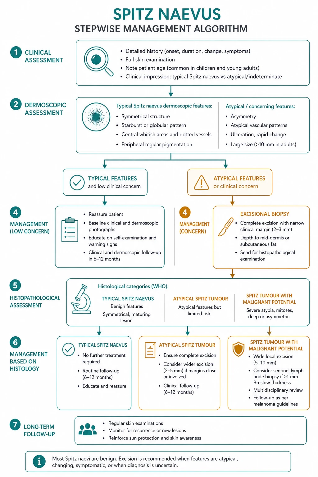
Spitz naevus is not, in itself, a time-critical emergency: a classic paediatric lesion can be scheduled electively. The urgency lies in the atypical or adult spitzoid lesion, which must be treated as suspected melanoma. Two principles frame every decision: [1]
[1]A clinically or dermoscopically atypical spitzoid lesion should be referred via the local fast-track suspected-skin-cancer pathway and excised promptly; an ulcerated or rapidly changing lesion in an adult is managed as suspected melanoma from the outset.[1]
Management — definitive and stepwise
The definitive management of a Spitz naevus is complete surgical excision, and the controversies are about how widely to excise, whether to observe selected paediatric lesions, and what to do with the atypical Spitz tumour. [1]
Typical Spitz naevus (benign, paediatric). Complete surgical excision with a narrow clear margin (commonly 3 to 5 mm) confirms the diagnosis and excludes atypical/malignant variants. Most units excise; an increasingly discussed alternative is observation of a classic, confident, paediatric pink Spitz naevus under expert dermoscopic surveillance, reserving excision for lesions that change or become atypical. This observational approach is defensible only when an experienced dermoscopist is confident of the diagnosis and the family is reliable; it trades the small operative burden of excision against the risk of an unnecessary scar and the (low but real) chance of over-treating a lesion that would have regressed.[3][14] The criteria that make observation defensible are narrow and worth stating explicitly. Observation is reasonable only when all of the following hold: the patient is a child or adolescent; the lesion is clinically typical (a pink, hairless, dome-shaped papule) with a classic, regular dermoscopic pattern (dotted/glomerular vessels with reticular depigmentation, or a symmetric starburst pattern); an experienced dermoscopist is confident of the diagnosis; the lesion is stable or evolving in the expected direction; the family is reliable and engaged with surveillance; and there is a clear safety-net of periodic expert review with a low threshold to excise if the pattern changes. If any criterion is missing — an atypical or non-specific pattern, an adult, a changing lesion, or unreliable follow-up — excise. Even proponents of observation accept that the majority of lesions are still excised, because the downside of missing a spitzoid melanoma dwarfs the cost of a small scar.[3][14]
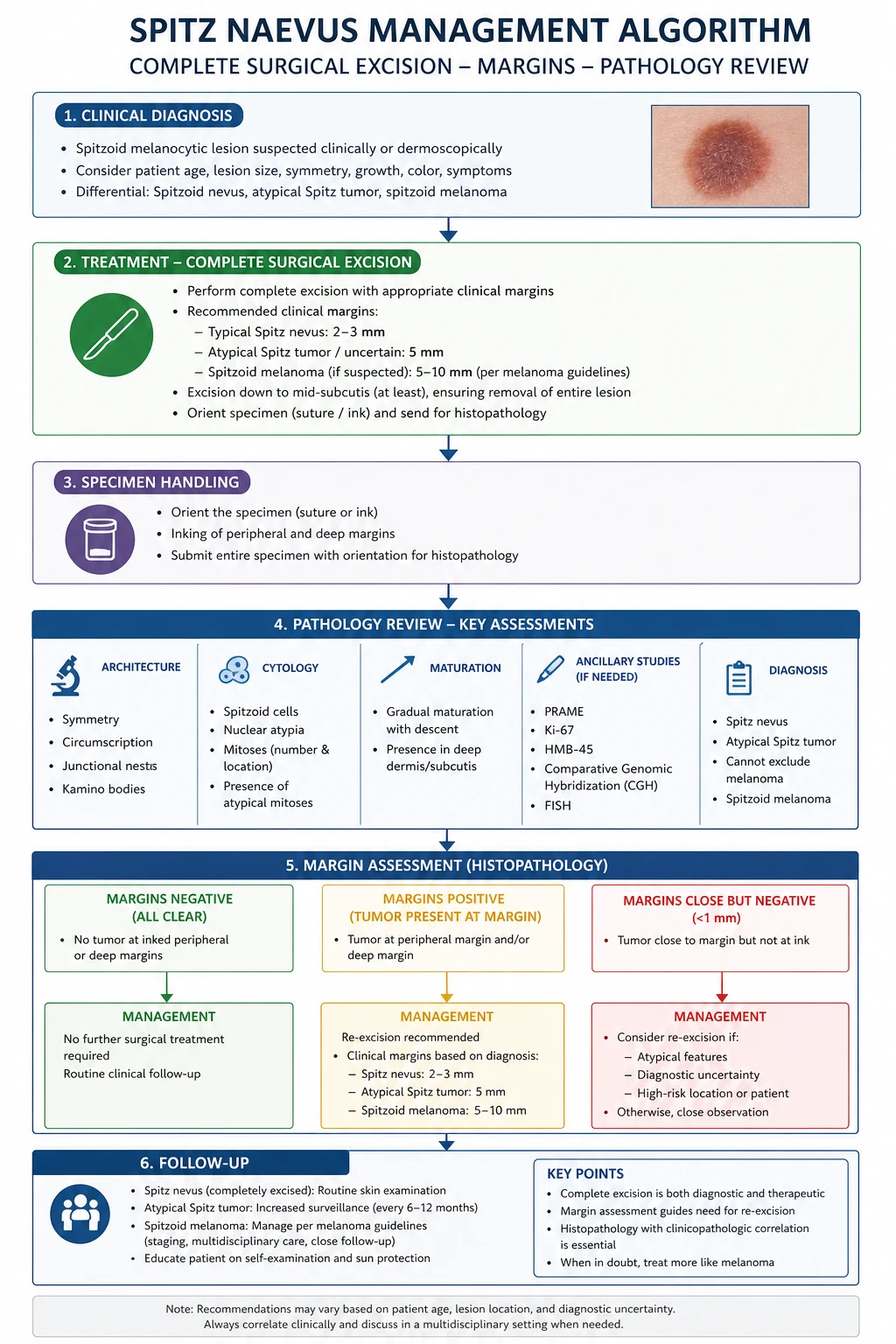
Atypical Spitz tumour. Complete excision with a wider margin (typically 5 mm), expert pathology review, and multidisciplinary discussion are the foundation. Sentinel lymph node biopsy is the contested element: it is technically feasible and yields a positive node in a substantial minority of ASTs, but a positive sentinel node in an AST does not reliably predict systemic disease — many patients with positive nodes never progress. Current systematic reviews therefore support selective, MDT-driven use of SLNB rather than routine biopsy, with most patients managed by complete excision and close surveillance.[11][12][13]
Spitzoid melanoma. Once an overtly malignant spitzoid neoplasm is confirmed, management follows the conventional melanoma pathway: wider excision margin determined by Breslow thickness, sentinel lymph node biopsy where appropriate, and melanoma-team follow-up.[1]
Re-excision. Positive or close margins after any excision warrant re-excision to a clear margin; the width is tailored to the histologic category (narrow for a typical Spitz naevus, wider for an AST or spitzoid melanoma). A diagnosis of atypical Spitz tumour on an incompletely excised specimen should trigger re-excision and MDT review rather than observation.[2]
Follow-up. After complete excision of a typical Spitz naevus, a full-skin examination every 6 to 12 months for at least five years, with monitoring of the excision site for recurrence, is reasonable. Atypical Spitz tumours warrant closer, specialist-led surveillance; spitzoid melanoma is followed per melanoma guidelines.[3][14]
Specific subtypes and scenarios
Pigmented spindle cell naevus of Reed. A dark brown to blue-black, sharply bordered, often symmetric papule, more common in darker phototypes and on the lower limbs. Histologically it is built from heavily pigmented spindle cells in fascicles at the dermoepidermal junction. Dermoscopy is diagnostic when classic — the starburst pattern, with regular radial streaks or pseudopods around the perimeter and a central dark structureless zone. It is benign but a near-perfect melanoma mimic; the reassuring features are symmetry of border and of dermoscopic structures. Excision confirms the diagnosis.[9]
Atypical Spitz tumour (AST). The borderline lesion, with overlapping Spitz naevus and melanoma features. Behaviour is unpredictable but generally favourable: most ASTs behave benignly even when the sentinel node is positive, yet rare cases metastasise, and no single histologic feature reliably predicts outcome. Management is complete excision with a wider margin, expert review, MDT discussion, selective SLNB, and close follow-up.[2][12]
Molecularly defined subtypes. The BAP1-inactivated spitzoid naevus/tumour (often BRAF V600E combined) is a dome-shaped, epithelioid, naevoid lesion with a distinctive loss-of-BAP1 phenotype; it is usually indolent but sits on the spitzoid spectrum. HRAS-mutant/11p-gain Spitz naevi are characteristically desmoplastic/spindled and benign. The kinase-fusion spitzoid neoplasms (ALK, RET, NTRK1/3, MET, ROS1, MAP3K8) define a family of generally low-grade lesions, several of which now have named entities and, in selected cases, targetable kinase activity.[6][8][15]
Agminated and eruptive Spitz naevi. Rare presentations in which Spitz naevi cluster in one region (agminated) or appear in crops (eruptive). Each lesion is assessed on its own merits; biopsy the most atypical lesion first, and manage the remainder conservatively if histology is reassuring. A note on natural history underpins the observational philosophy. Untreated classic Spitz naevi do not grow indefinitely: they characteristically enlarge over weeks to a few months and then stabilise, and over years many flatten, lose their vascularity, and may acquire pigment or even regress partially. This predictable, self-limited course is the biological reason that observation of a confident, classic paediatric lesion is defensible — the lesion is expected to behave in a benign way. The corollary is that a lesion that continues to grow beyond the expected window, or that changes colour or ulcerates after stabilising, is no longer following the benign script and must be excised.[3][14]
Complications and pitfalls
The complications of Spitz naevus are largely diagnostic rather than natural-historical, because the benign lesion itself is well-behaved. They fall into three groups. [1]
Disease-related. Local recurrence after incomplete excision is the main one; a recurrence should prompt re-examination of the original histology, because an incompletely excised atypical Spitz tumour can recur locally. Rare metastasis from an atypical Spitz tumour, and the consequences of an unrecognised spitzoid melanoma, are the serious ends of the spectrum.[2] The spectrum of diagnostic error is the real complication in this topic, and it runs in both directions. Over-diagnosis — calling a benign Spitz naevus a melanoma — exposes a child (or adult) to unnecessary wide excision, sentinel node biopsy, lifetime melanoma follow-up, and the psychological burden of a cancer label, with no survival benefit. Under-diagnosis — calling a spitzoid melanoma a Spitz naevus — risks delayed treatment of a potentially lethal disease. Because the two errors pull in opposite directions, the field has converged on two safeguards: complete excision of any lesion submitted for diagnosis, so that the architecture is preserved; and expert pigmented-lesion pathology review of any case that is not confidently benign. The cost of a second opinion is trivial compared with the cost of either misdiagnosis, and this is the single most defensible habit to carry into practice.[5]
Diagnostic pitfalls are where most of the harm in this topic occurs: [1]
SHAVE
Procedure-related. Bleeding, infection, and scarring are the usual minor surgical complications; a facial lesion in a child warrants careful attention to cosmetic outcome and may justify specialist plastic surgical excision. [1]
Psychosocial pitfall. Mislabelling a child's benign lesion as melanoma subjects the family to unnecessary anxiety, wider excision, and sometimes SLNB; the converse — false reassurance of a lesion that is in fact a spitzoid melanoma — is the more dangerous error. Accurate, expert pathology is the safeguard against both. [1]
Prognosis and disposition
A few practical points sharpen the prognosis discussion. First, recurrence after complete excision of a classic Spitz naevus is genuinely rare; a lesion that recurs at the scar should be re-evaluated, because the original diagnosis may have been an under-called atypical Spitz tumour. Second, the favourable prognosis of an atypical Spitz tumour holds even in the presence of a positive sentinel node, which is the single most counter-intuitive fact in the topic and the one examiners return to. Third, spitzoid melanoma is prognostically identical to conventional melanoma of the same Breslow thickness — there is no "Spitz discount" once the lesion is malignant, and it must be staged and followed on that basis.[1][2]
Classic Spitz naevus has an excellent prognosis: it is benign, complete excision is curative, and local recurrence after complete excision is rare. The paediatric patient returns to routine dermatology follow-up.[3]
Atypical Spitz tumour has an uncertain but generally favourable prognosis. Most ASTs behave benignly even with positive sentinel nodes, but rare cases metastasise, and no single histologic feature reliably predicts behaviour. This irreducible uncertainty is precisely why ASTs are managed with complete excision, expert review, and close follow-up rather than being dismissed.[11][12][13]
Spitzoid melanoma behaves like conventional melanoma, and its prognosis is determined by Breslow thickness, ulceration, and sentinel-node status, exactly as for any cutaneous melanoma. Disposition is to the melanoma pathway.[1]
Special populations
Paediatric (the dominant population). Excision is performed under age-appropriate anaesthesia with attention to the cosmetic result on the face. Observation is reasonable only for a classic, confident paediatric Spitz naevus under expert dermoscopic surveillance with a reliable family; otherwise excise. Counselling should cover the benign nature of the lesion, sun protection, and self-examination.[14]
Adults. The threshold for excision is lower and observation is not appropriate: an adult spitzoid lesion is excised with margins and sent for expert pathology review, because spitzoid melanoma cannot be excluded clinically.[1][2]
Pregnancy and immunosuppression. Spitz naevi in these settings are rare and managed by the standard surgical principles. A changing lesion in a pregnant or immunosuppressed patient should not be attributed to the physiological or immunological state without histology — biopsy the atypical lesion. [1]
Darker phototypes. The pigmented/Reed variant is relatively more common, and the examination should include acral and palmoplantar sites; dermoscopy of acral lesions uses the parallel-pattern approach. [1]
Evidence, guidelines, and regional differences
Three live controversies define the evidence base, and examiners test all three. [1]
Surgical margins. There is no single agreed margin for a typical Spitz naevus; 3 to 5 mm is common practice, with wider margins for atypical Spitz tumour and melanoma-in-situ-equivalent margins for spitzoid melanoma. The trend over time has been toward complete excision for all but the most classic paediatric lesions, on the grounds that histology cannot reliably exclude melanoma in advance.[3]
Sentinel lymph node biopsy in AST. SLNB is technically feasible and positive in a substantial minority of ASTs, but a positive sentinel node in an AST does not reliably predict systemic disease. The systematic-review evidence now supports selective, MDT-driven SLNB rather than routine biopsy, with most patients managed by complete excision and surveillance.[11][12][13] The numbers that anchor the SLNB debate. In systematic reviews, sentinel-node positivity in atypical Spitz tumours runs in the region of a quarter to a third of biopsied patients, yet the proportion that go on to systemic or distant disease is far smaller — single-digit percentages in most series. This disjunction between nodal positivity and clinical progression is the central reason SLNB is not a reliable staging tool in this group, and why completion lymphadenectomy on the strength of a positive node alone is no longer recommended. The corollary, drawn from the paediatric observational series, is that carefully selected atypical Spitz tumours managed without SLNB still do well, lending weight to selective, multidisciplinary-team-driven use. The take-home for an examiner is that a positive sentinel node in an atypical Spitz tumour is a reason for closer follow-up, not an automatic reason for aggressive surgery.[11][12][13]
Atypical Spitz tumours and SLNB — systematic reviews
Key finding
Across systematic reviews, sentinel-node positivity in atypical Spitz tumours is substantially more frequent than systemic progression: many patients with positive nodes never develop distant disease, so a positive node does not upstage management on its own. The reviewers recommend selective, MDT-driven SLNB rather than routine biopsy.
Practice change
Do not auto-upstage an AST to completion-lymphadenectomy on the basis of a positive sentinel node; manage the patient in a specialist MDT and individualise follow-up.
Paediatric spitzoid proliferations — observational series
Key finding
Large paediatric series report favourable outcomes for spitzoid proliferations, including atypical Spitz tumours, even when managed without SLNB — supporting conservative, surveillance-based management in carefully selected children.
Practice change
In a well-selected child with a classic or low-risk atypical lesion, observation and complete excision without SLNB is defensible; reserve SLNB for higher-risk cases after MDT discussion.
Molecular diagnostics (FISH, CGH, PRAME, HRAS/BAP1/kinase-fusion testing) are increasingly used to resolve ambiguous spitzoid lesions, but they are adjuncts to, not replacements for, expert morphology. Their value is highest exactly where morphology is most ambiguous.[10][15]
Australasian (RANZCD/ACD) practice mirrors the UK approach with a somewhat stronger tradition of observation of classic, confident paediatric pink Spitz naevi under expert surveillance, and centralised expert pathology review for atypical lesions.
Exam pearls
[1]Red flags
Exam application bank (NEET-PG / INICET)
One-line answer
Spitz naevus is a benign acquired melanocytic naevus of childhood and adolescence composed of large spindled and epithelioid melanocytes, presenting most often as a rapidly growing pink, hairless, dome-shaped papule. Histology shows spindled/epithelioid melanocytes, Kamino bodies, symmetry, and maturation with depth. The central diagnostic challenge is distinguishing benign Spitz naevus from spitzoid melanoma, with the atypical Spitz tumour occupying a borderline zone. Management is complete surgical excision with clear margins.
Worked stems (answer without another resource)
Stem 1 — Classic presentation. Map symptoms to mechanism; name the first investigation and first treatment step with dose/route if drug therapy is standard. [1]
Stem 2 — Unstable / complicated. List red flags that force immediate resuscitation, theatre, ICU, antidote, or reperfusion — and what you do in the first 15 minutes. [1]
Stem 3 — Atypical group. Elderly, pregnancy, child, or immunocompromised: how presentation and thresholds change. [1]
Stem 4 — Differential trap. Name the three closest mimics and one discriminator for each. [1]
Stem 5 — Disposition. Who goes home with safety-netting, who is admitted, who needs HDU/ICU/theatre, and what follow-up is mandatory. [1]
Rapid viva checklist
- Definition + classification
- Pathophysiology chain
- Bedside signs / criteria
- Score with exact components (if any)
- Emergency bundle
- Definitive therapy with doses
- Complications of disease and of treatment
- Special populations
- Guideline/trial name if classic
- Three exam traps
Coverage self-check
If you cannot answer any stem above from this page alone, re-read the matching section — the page is intended to be self-sufficient for final-prof and NEET-PG/INICET questions on Spitz naevus (spindle and epithelioid cell naevus).
[1]Prevention and patient safety-net
Spitz naevus is essentially sporadic and not UV-driven, so primary prevention is limited to general sun protection and skin awareness. The safety-net is operational: any lesion that is changing, bleeding, or new — and any spitzoid lesion in an adult — must be referred and biopsied rather than observed on clinical grounds. For the paediatric patient excised completely, advise sun protection, teach the family skin self-examination, and schedule structured dermatology follow-up to detect recurrence or new atypical lesions.[3][14]
Self-test: a 6-year-old presents with a 4 mm pink, hairless, dome-shaped papule on the cheek, present for two months and growing. What is the most likely diagnosis, the key dermoscopic finding, and the management?
Most likely diagnosis: classic Spitz naevus. Key dermoscopic finding: regular dotted/glomerular vessels with reticular depigmentation. Management: complete surgical excision with clear margins (3–5 mm) — not shave — with expert pathology review; observation under expert surveillance is an alternative only for a classic, confident paediatric lesion.
Self-test: a 42-year-old presents with a new pink nodule on the leg with a non-specific dermoscopic pattern. What is the principle that governs management?
In an adult, a spitzoid lesion is a red flag — spitzoid melanoma cannot be excluded clinically or dermoscopically. The governing principle is complete excision with margins and expert pathology review, with ancillary IHC and molecular testing for ambiguous histology. Observation is not appropriate in adults.
References
- [1]Battistella M, et al. Spitz melanoma Clin Dermatol, 2025.PMID 39265841
- [2]Massi D, et al. Spitz melanocytic tumours - a review Histopathology, 2022.PMID 34958498
- [3]Ghoreishi A, et al. Spitz Nevus: Review and Update Clin Plast Surg, 2021.PMID 34503728
- [4]Spatz A, et al. The spitz nevus: review and update Clin Plast Surg, 2010.PMID 19914455
- [5]Barnhill RL, et al. Nevus Spitz--everlasting diagnostic difficulties--the review Coll Antropol, 2008.PMID 19140279
- [6]Bastian BC, et al. Mutations and copy number increase of HRAS in Spitz nevi with distinctive histopathological features Am J Pathol, 2000.PMID 10980135
- [7]Maldonado JL, et al. Mechanisms of cell-cycle arrest in Spitz nevi with constitutive activation of the MAP-kinase pathway Am J Pathol, 2004.PMID 15111324
- [8]Requena C, et al. BAP1-deficient and VE1-negative atypical Spitz tumor J Cutan Pathol, 2015.PMID 25953246
- [9]Marchell R, et al. Dermoscopy of pigmented Spitz and Reed nevi: the starburst pattern Arch Dermatol, 2005.PMID 16103350
- [10]Gerami P, et al. A highly specific and discriminatory FISH assay for distinguishing between benign and malignant melanocytic neoplasms Am J Surg Pathol, 2012.PMID 22588064
- [11]Cerrato F, et al. Outcomes in pediatric atypical spitz tumors treated without sentinel lymph node biopsy Pediatr Dermatol, 2012.PMID 22211716
- [12]Lallas A, et al. Atypical Spitz tumours and sentinel lymph node biopsy: a systematic review Lancet Oncol, 2014.PMID 24694641
- [13]Mazza M, et al. Sentinel Lymph Node Biopsy in Atypical Spitz Tumor: A Systematic Review J Clin Med, 2024.PMID 38892943
- [14]Bartenstein DW, et al. Clinical features and outcomes of spitzoid proliferations in children and adolescents Br J Dermatol, 2019.PMID 30467833
- [15]Tetzlaff MT, et al. Toward a Molecular-Genetic Classification of Spitzoid Neoplasms Clin Lab Med, 2017.PMID 28802494