Dermatology · Medicine
Cutaneous squamous cell carcinoma
Also known as Squamous cell carcinoma · SCC · Cutaneous SCC · Epidermoid carcinoma
Cutaneous squamous cell carcinoma (SCC) is a malignant tumour of epidermal keratinocytes arising on a background of chronic ultraviolet damage and field cancerization, evolving through actinic keratosis → SCC in situ (Bowen's disease) → invasive SCC. It is the second commonest skin cancer, with metastatic potential (~3-5% overall, much higher in immunosuppressed and high-risk tumours) driven by UV-induced TP53/NOTCH/CDKN2A loss. Diagnosis is histological; staging uses AJCC 8th edition and the more prognostic Brigham (BWH) system built on high-risk features (diameter ≥2 cm, depth 6 mm, perineural invasion, poor differentiation, ear/lip/scalp/mask-face site, immunosuppression, recurrence). Management is surgical — excision with 4-6 mm margins for low-risk tumours and Mohs micrographic surgery for high-risk, recurrent or functionally/cosmetically critical sites — with radiotherapy, topical field therapy for in-situ/field disease, and anti-PD-1 (cemiplimab) or EGFR inhibitors for advanced disease. Fellowship-level assessment demands mastery of the AK→Bowen's→invasive spectrum, the high-risk feature set, the Brigham/AJCC staging, Mohs indications, transplant-specific SCC behaviour (SCC:BCC ratio inversion, sirolimus), Marjolin ulcer behaviour, and the management of perineural, nodal and metastatic disease with immune-checkpoint and EGFR-directed therapy.
On this page & tools
Your progress
Saved locally on this device.
Exam tags
Red flags
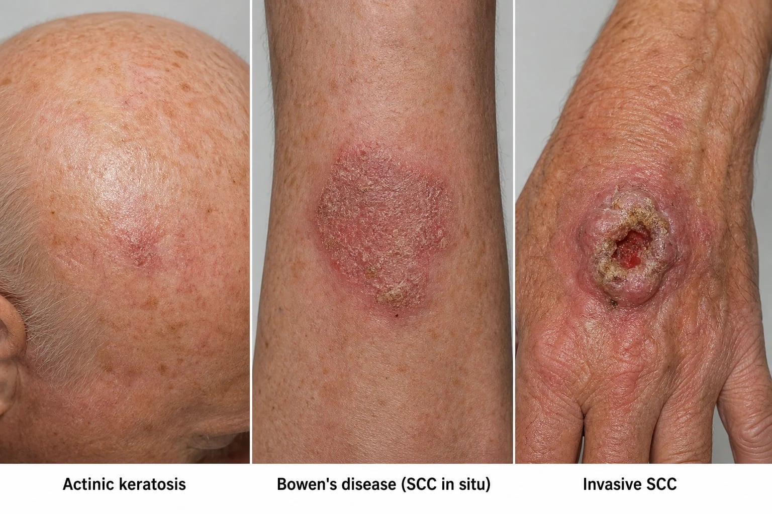
Definition & Classification
Cutaneous squamous cell carcinoma (SCC) is a malignant epithelial tumour arising from epidermal keratinocytes, characterised histologically by atypical keratinocyte proliferation with invasion through the basement membrane. It exists on a disease spectrum: actinic keratosis (AK) → SCC in situ (Bowen's disease) → invasive SCC.[2][6]
Histological subtypes (influence behaviour):[3]
| Subtype | Features / significance |
|---|---|
| Conventional (well-to-moderately differentiated) | Commonest; keratin pearls |
| Verrucous | Warty, slow-growing, locally destructive, rarely metastasises; radiation contraindicated (anaplastic transformation) |
| Acantholytic (adenoacanthoma) | Pseudoglandular spaces; behaves as conventional |
| Desmoplastic / spindle-cell | Scar-like, infiltrative, higher recurrence/metastasis |
| Adenosquamous, pigmented | Rare variants |
| Keratoacanthoma | Rapidly growing crateriform nodule; debated — most dermatopathologists now classify as well-differentiated SCC |
| Lymphoepithelioma-like | Dense lymphoid infiltrate; favourable despite unusual histology |
| Clear-cell | Glycogen-rich; mimics other clear-cell tumours |
The clinical course is dominated by conventional SCC (>80%); verrucous, desmoplastic and acantholytic subtypes carry disproportionate metastatic risk and demand high-risk management even when clinically indolent. [1]
Epidemiology & Risk Factors
Cutaneous SCC is the second commonest skin cancer (after BCC) and the commonest keratinocyte carcinoma with metastatic potential; incidence is rising globally as populations age and accumulate UV exposure.[2][6] In the United States more than one million cases are diagnosed annually and the absolute number of deaths from cutaneous SCC now equals or exceeds that from melanoma and from leukaemia — a public-health burden obscured by high case volume and low per-tumour mortality.[2]
Cumulative ultraviolet (UV) exposure is the dominant cause. The lifetime risk in fair-skinned populations exceeds 15% and the typical patient is an older man with Fitzpatrick phototype I–II, a history of outdoor occupation or recreation, and a background of solar elastosis, telangiectasia and dyspigmentation. Risk increases steeply with intermittent intense exposure (sunburns) and chronic cumulative exposure; PUVA photochemotherapy, indoor tanning, and equatorial/tropical residence all contribute.[5][6]
Immunosuppression is the second great risk amplifier. Solid-organ transplant recipients have a 65- to 250-fold higher incidence than the general population and the SCC:BCC ratio inverts (≈1:4 in immunocompetent patients → ≈4:1 in transplant recipients); SCCs are more numerous, more aggressive, develop at younger ages, and recur more often. Risk rises with duration and intensity of immunosuppression; heart and lung recipients (heavier immunosuppression) are at greatest risk. Chronic lymphocytic leukaemia (CLL), HIV/AIDS (especially CD4 count under 200 cells/µL), iatrogenic immunosuppression for autoimmune disease (azathioprine, ciclosporin, TNF inhibitors, JAK inhibitors), and haematopoietic stem-cell transplantation with chronic GVHD all confer similar but smaller excesses.[1][2][6]
Chemical and physical carcinogens. Arsenic exposure (contaminated groundwater, pesticides, industrial exposure, Fowler's solution) produces arsenical keratoses on palms/soles and a field-cancerisation pattern of Bowenoid and invasive SCC. Ionising radiation (therapeutic — historical radiotherapy for acne, tinea capitis, ankylosing spondylitis, Hodgkin lymphoma; occupational — radiologists, nuclear workers) and UVB/PUVA induce a slowly progressive radiation dermatitis culminating in SCC after 20- to 40-year latency. Chronic thermal injury (kangri-burn cancer in Kashmir, peat-fire cancer, Kang cancers of the lower leg) operates through a similar pathway. [1]
Chronic scarring and chronic inflammation are textbook SCC substrates. Burn scars, osteomyelitis sinuses (post-traumatic or tuberculous), chronic venous/arterial ulcers, vaccination scars, acne conglobata scars, pilonidal sinuses, pressure-sore scars, and chronically draining wounds of any cause are the classic Marjolin ulcer substrates — long latency (often decades), aggressive behaviour, high metastatic potential (up to 30%). Lichen sclerosus (anogenital), lichen planus (oral, vulvovaginal), and discoid lupus erythematosus are chronic scarring inflammatory dermatoses with documented SCC transformation, especially on the vulva, lip and oral mucosa. [1]
Genetic susceptibility amplifies risk dramatically. Xeroderma pigmentosum (defective nucleotide-excision repair of UV photoproducts) produces a 10,000-fold increased NMSC risk in childhood. Epidermodysplasia verruciformis (EVER1/EVER2 — abnormal beta-HPV immunity) leads to life-long HPV-driven Bowenoid and invasive SCC. Oculocutaneous albinism, Rothmund–Thomson, Bloom and Fanconi syndromes (DNA-repair defects) all predispose. Ferguson-Smith self-healing epitheliomata (TGFBR1) and multiple self-healing palmoplantar carcinomas are rarer. [1]
Other risk factors include HPV (β-HPV types 5, 8 — epidermodysplasia and transplant SCC; α-HPV — anogenital, oropharyngeal, periungual SCC), tobacco (lip and oral SCC, with synergistic UV risk), ionising radiation, chronic lymphoedema (Stewart–Treves-related but predominantly angiosarcoma), PUVA (>350 treatments multiplies SCC risk 30-fold), azathioprine (photosensitises via 6-thioguanine incorporation), and chronic arsenic exposure.[1][6]
Pathophysiology
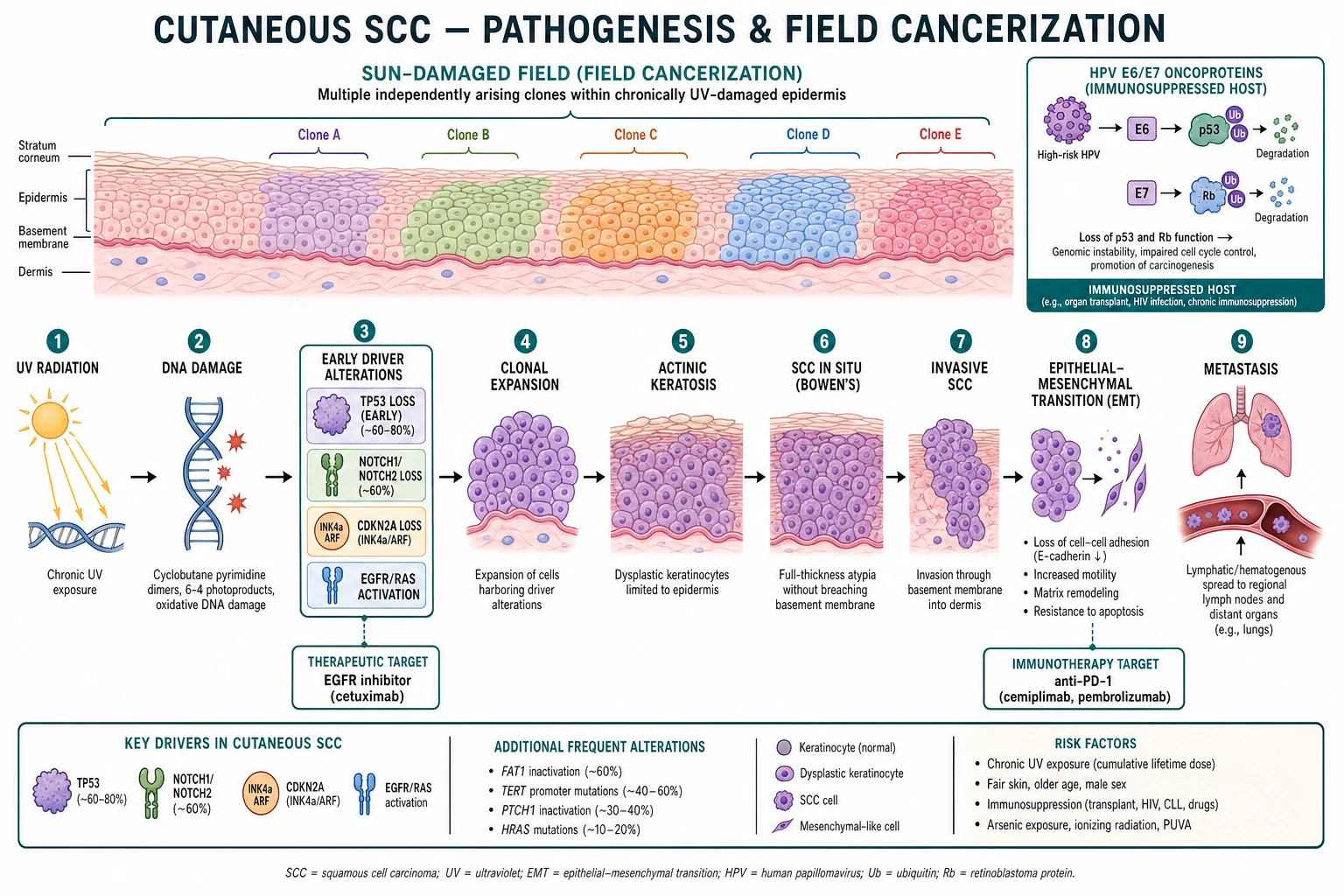
UV-driven mutagenesis is the molecular cornerstone. UVB (290–320 nm) is directly absorbed by DNA and produces cyclobutane pyrimidine dimers and 6-4 photoproducts; the canonical UV-signature mutations are C→T and CC→TT transitions at dipyrimidine sites. UVA (320–400 nm) contributes through reactive oxygen species and dermal-matrix damage. TP53 is mutated in 60–80% of cutaneous SCCs (also in 50–80% of precursor AKs); loss of G1/S checkpoint function and resistance to apoptosis allow damaged keratinocytes to clonally expand.[3][7]
NOTCH1 and NOTCH2 (tumour-suppressor Notch receptors) are inactivated in ~60% of SCCs — a remarkably high frequency that places Notch loss alongside TP53 as a defining early event. CDKN2A (p16/INK4A) loss short-circuits the Rb pathway. EGFR overexpression and RAS-MAPK activation drive proliferation and are rational therapeutic targets (cetuximab, panitumumab). PIK3CA, HRAS, KNSTRN, and FAT1 mutations accumulate with progression. [1]
Field cancerization describes a broad expanse of chronically sun-damaged skin harbouring multiple independent premalignant clones, each arising from separate UV-induced mutational events. AKs and in-situ SCCs are visible markers of the field; invasive SCCs arise from within it. This is why field-directed therapy (5-FU, imiquimod, photodynamic therapy) reduces the incidence of new invasive SCC across the field and not just at treated lesions.[7]
HPV contributes especially in the immunosuppressed. β-HPV (types 5, 8, 17, 20 and others) E6 and E7 oncoproteins degrade p53 and Rb respectively, accelerating the same pathways UV already damages. β-HPV DNA is detectable in >80% of transplant SCCs and in the majority of AKs on sun-exposed skin. α-HPV (types 16, 18) drive anogenital, oropharyngeal and periungual SCC. [1]
Epithelial–mesenchymal transition (EMT), with loss of E-cadherin, gain of vimentin and activation of TWIST/SNAIL, marks the in-situ → invasive transition and is the biological substrate of desmoplastic and spindle-cell SCC. Perineural invasion — tropism of tumour cells for nerve sheaths — is mediated by NGF/TrkA, NCAM and glial-derived neurotrophic factor signalling and underlies the high-risk phenotype. [1]
Premalignant Lesions (precursors to invasive SCC)
Actinic keratosis (AK) — the commonest precursor — is intraepidermal keratinocyte dysplasia on sun-damaged skin; ~0.025–20% per lesion per year progress to invasive SCC, with higher per-lesion risk in thick/hypertrophic lesions and in transplant recipients. SCC in situ (Bowen's disease) — full-thickness epidermal atypia — is the immediate precursor with much higher per-lesion transformation risk and is treated definitively (excision, 5-FU, imiquimod, PDT, curettage+cryotherapy).[3][6]
Arsenical keratoses are punctate, keratotic, often yellow and symmetrically distributed on palms and soles, frequently accompanied by hyperpigmentation, Mee's lines, and a Bowenoid-field pattern on the trunk. Latency from arsenic exposure is 10–40 years; they progress to invasive SCC in a minority but mark a high-risk patient. [1]
Chronic radiation dermatitis — atrophic, hypopigmented/hyperpigmented skin with telangiectasia and poikiloderma, often with keratotic papules within the field — produces SCC after 20–40 years, usually on a background of prior radiotherapy for benign disease (tinea capitis, acne, eczema, ankylosing spondylitis) or malignancy (Hodgkin lymphoma, breast cancer, head and neck cancer). [1]
Lichen sclerosus of the vulva carries a 4–5% lifetime risk of vulvar SCC; lichen planus of the oral mucosa and vulva has a similar risk. Both should be biopsied when nodular, eroded, hyperkeratotic, or unresponsive to standard therapy. Discoid lupus erythematosus (especially long-standing mucosal DLE) and chronic cutaneous lupus rarely transform to SCC. [1]
Porokeratosis (especially the linear and large variants), chronic erosive lichen planus, xeroderma pigmentosum skin, albinism skin, chronic stasis dermatitis with ulceration, and epidermolysis bullosa dystrophic scars are all recognised precursor states. [1]
Clinical Presentation
- Classic lesion: a hyperkeratotic, scaling, crusted, indurated or ulcerated nodule or plaque on a chronically sun-exposed site — scalp (bald), face, pinna, lower lip, dorsal hands, forearms. May be tender or bleed; often arises within field of actinic damage.[2][4]
- Spectrum in situ: AK (rough scaly macule/papule); Bowen's disease (fixed, scaly, sharply demarcated erythematous patch).
- High-risk clinical clues: large size (≥2 cm), rapid growth, bleeding, fixation to deeper tissue, recurrence after treatment.
- Perineural disease presents with pain, numbness, tingling or motor weakness in a cranial nerve distribution — a high-risk feature demanding imaging.[1]
- Atypical / special sites examiners test: lower lip, pinna/ear, genital/perianal, peri/subungual, within a burn scar (Marjolin ulcer), and in a transplant patient (multiple, fast-growing).[6]
Invasive morphological variants are clinical-dermatopathological entities that demand identification because their behaviour diverges from conventional SCC.[3]
- Keratoacanthoma — a rapidly growing (weeks), symmetric, crateriform nodule with a central keratin plug; considered by most contemporary dermatopathologists to be a well-differentiated SCC variant rather than a benign tumour. Most are excised.
- Verrucous carcinoma — warty, exophytic, slowly growing, locally destructive but rarely metastasising; on the sole (epithelioma cuniculatum), genitalia (giant condyloma of Buschke–Löwenstein), oral cavity, and periungual region. Radiotherapy is contraindicated — anaplastic transformation is well documented.
- Desmoplastic SCC — infiltrative, scar-like, ill-defined, sclerotic plaque or nodule, typically on the head/neck; high local recurrence and perineural invasion rates.
- Spindle-cell SCC — atypical spindle cells in a sun-damaged field; immunostains (cytokeratins, p63) confirm epithelial origin and exclude atypical fibroxanthoma/pleomorphic dermal sarcoma.
- Acantholytic (adenoid/pseudoglandular) SCC — pseudoglandular spaces from loss of intercellular bridges; behaves as conventional.
- Marjolin ulcer — SCC arising in a chronic burn scar, osteomyelitis sinus, venous ulcer or any chronic wound, usually after decades; aggressive with high metastatic potential.
- Scar SCC — broader category including post-traumatic scars, vaccination scars, acne scars, and scars from prior procedures.
- Radiation SCC — SCC arising in chronic radiation dermatitis, typically decades after therapeutic irradiation; aggressive, often multifocal, with a field-cancerisation pattern. [1]
Differential Diagnosis
| Mimic | Distinguishing features |
|---|---|
| Basal cell carcinoma | Pearly rolled border, telangiectasia, slower growth; dermoscopy (arborising vessels) |
| Keratoacanthoma | Rapid (weeks) symmetric crateriform nodule with a central keratin plug; histology overlaps SCC |
| Hypertrophic actinic keratosis | Rough, scaly, no dermal invasion; biopsy if indurated |
| Verruca vulgaris | Viral, thrombosed capillaries, younger patients |
| Amelanotic / inflamed melanoma | Variable pigmentation; dermoscopy; biopsy |
| Merkel cell carcinoma | Rapid, painless, red-violaceous nodule on sun-damaged head/neck (CK20, MCPyV) |
| Atypical fibroxanthoma / pleomorphic dermal sarcoma | Spindle-cell sun-damaged tumour; histology |
| Pyogenic granuloma | Friable bleeding red papule; short history |
| Inflamed seborrhoeic keratosis | "Stuck-on", milia-like cysts |
Dermoscopy
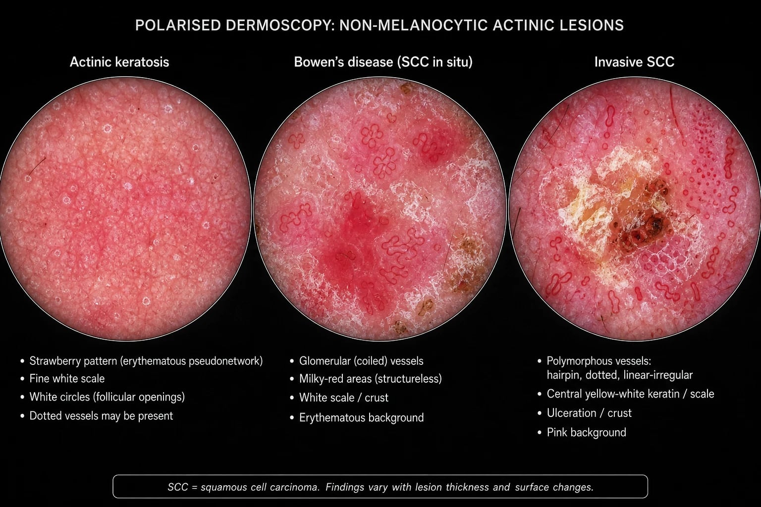
- AK: pink-red background "strawberry" pseudonetwork with surface scale; small white circles ("rosacea-like").[3]
- Bowen's disease (SCC in situ): glomerular (coiled) vessels with milky-red areas and scale.
- Invasive SCC: polymorphous vessels (hairpin, dotted and linear-irregular), central yellow-white keratin/ulceration, white structureless areas and horn cysts — vascular polymorphism is a red flag for invasion.
Histopathology
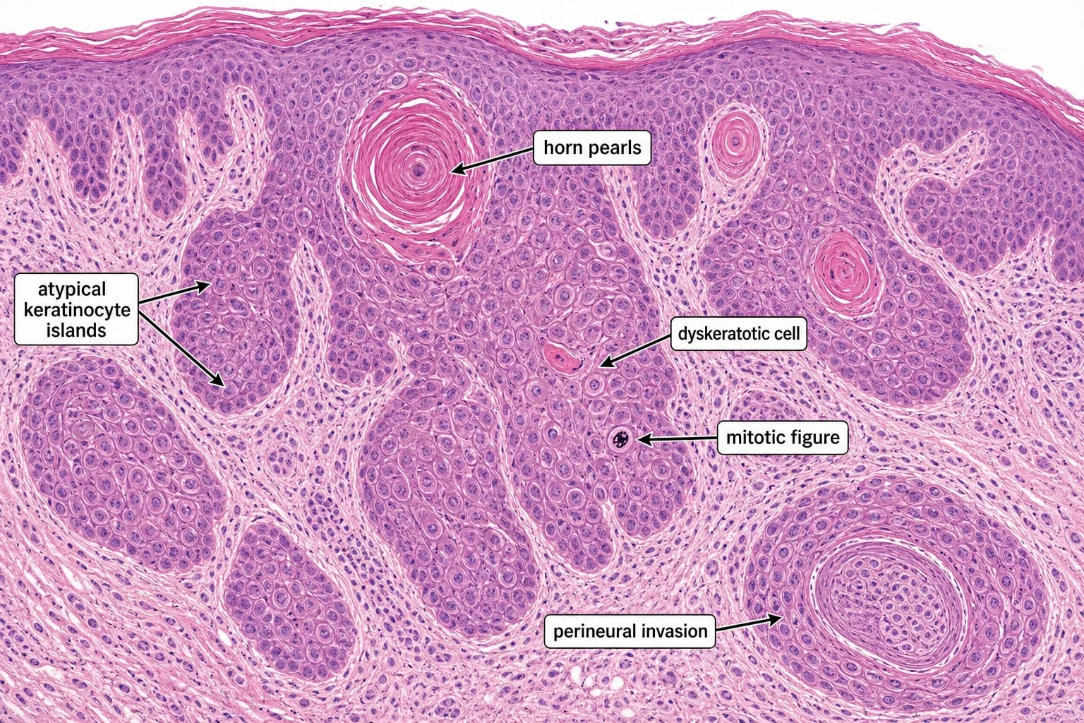
The histology report determines staging and management. Essential elements:[2][3]
- Diagnosis and differentiation (well / moderate / poor) — poor differentiation is a high-risk feature.
- Invasion depth (mm) — >6 mm is a high-risk threshold.
- Perineural invasion — especially named nerves ≥0.1 mm diameter.
- Lymphovascular invasion, desmoplastic/stromal reaction, subtype.
- Margins — involved / close / positive.
- Biopsy (shave, punch or incisional into dermis) suffices for diagnosis; small lesions may be excisional. [1]
High-Risk Features & Staging

HIGH RISK SCC — the eight NCCN/Brigham features (mnemonic: BIG-CRIMP)
BIG-CRIMP
Tumours ≥ 2 cm on low-risk sites and ≥ 1 cm on high-risk (mask-area) sites are categorised high-risk by NCCN.
Depth into deep dermis or subcutis — a Brigham high-risk factor and a key AJCC criterion.
Poor histologic differentiation is an independent predictor of nodal metastasis and disease-specific death.
Named-nerve perineural invasion (≥ 0.1 mm calibre) is a high-risk feature demanding MRI ± Mohs + adjuvant RT.
Local recurrence after prior excision, curettage, or topical therapy doubles the risk of further recurrence and metastasis.
Solid-organ transplant, CLL, HIV, azathioprine/ciclosporin, JAK inhibitors — multiple/aggressive SCC, SCC:BCC inverts to 4:1.
Central face, eyelids, eyebrows, nose, perinasal, perioral, chin, pre-/post-auricular, ear, temple, scalp, non-glabrous lip.
Prior ionising radiation to the same anatomical site — adds second-hit mutagenic load to a field of damage.
High-risk features (NCCN / Brigham):[1][6]
- Diameter ≥2 cm (≥1 cm on mask area of face, ear, scalp, hands, feet, genitalia)
- Invasion depth >6 mm (or Clark level V) — to subcutis fat
- Perineural invasion (named nerve ≥0.1 mm diameter)
- Poor differentiation
- High-risk site: ear (pinna), non-glabrous lip, scalp/temple, "mask area" of face (central face, eyelids, eyebrows, periorbital, nose, perinasal, perioral, mentum, preauricular/postauricular skin, temple)
- Immunosuppression (transplant, CLL, HIV, iatrogenic)
- Prior radiotherapy at the site; recurrent tumour; rapid growth; lymphovascular invasion; desmoplastic subtype [1]
AJCC 8th edition (T): T1 = ≤2 high-risk features; T2 = ≥2 high-risk features (excluding bone); T3 = (not used for cutaneous); T4 = bone invasion. Brigham (BWH) staging is more prognostic: T1 = 0 risk factors; T2 = 1; T3 = 2 risk factors; T4 = bone invasion (all high-risk).[1]
N (regional nodes): parotid and cervical chains for head/neck; regional basin elsewhere. N1 = single ipsilateral node ≤3 cm; N2 = single ipsilateral 3–6 cm or multiple ipsilateral ≤6 cm; N3 = >6 cm or bilateral/contralateral. M1 = distant metastasis (lung, bone, liver, brain, non-regional nodes — the lung is the commonest first site). [1]
Clinical & Bedside Assessment
- Examine the lesion and the entire sun-exposed field (scalp, face, ears, lips, hands); assess high-risk features at the bedside.[6]
- Palpate regional nodes (parotid for face/scalp lesions; cervical, axillary, inguinal).
- Cranial nerve examination for any face/scalp lesion (perineural disease).
- Examine oral mucosa and lips.
Investigations
- Biopsy of the dermal component (shave/punch/incisional) for diagnosis; small lesions excisional.
- Imaging reserved for high-risk tumours and suspected invasion: MRI for perineural/bone/skull-base disease; CT for nodal/parotid disease; PET-CT for advanced/metastatic disease staging.[1]
- Node ultrasound ± FNA for clinically suspicious nodes.
- Sentinel lymph node biopsy (SLNB) is not standard for cutaneous SCC (limited prognostic/therapeutic evidence) outside trial/high-risk protocols. NCCN, EADO and BAD advise imaging surveillance (US ± CT/MRI) for high-risk tumours and therapeutic lymph node dissection for clinically positive disease; SLNB is considered only in selected high-risk patients within multidisciplinary discussion.
- Staging investigations for advanced/metastatic disease: contrast CT chest/abdomen/pelvis (lung is the commonest first metastasis), brain MRI if neurological symptoms, and baseline bloods including FBC, LFTs, renal function and LDH.
Management — Surgery
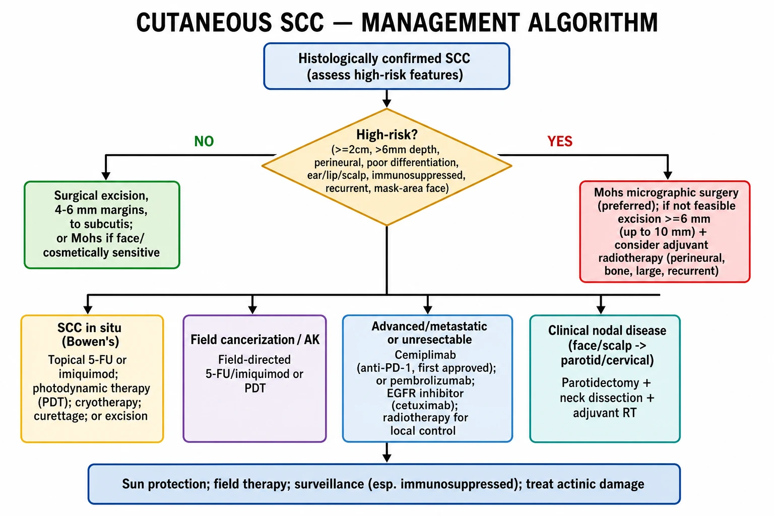
Surgery is first-line for invasive SCC.[2][3]
- Excision margins: 4-6 mm for low-risk tumours (to subcutaneous fat); ≥6 mm (up to 10 mm) for high-risk tumours.
- Mohs micrographic surgery (preferred) for: high-risk tumours, recurrent SCC, the mask area of face and other critical/cosmetic sites (eyelid, nose, lip, ear, genital, perianal, digit), ill-defined clinical margins, tumours >2 cm, and immunosuppressed patients — it achieves the highest cure rate with maximal tissue conservation.[2]
- Curettage & electrocautery only for small, low-risk, non-critical-site lesions in low-risk patients.
Management — In Situ Disease & Field Cancerization
- SCC in situ (Bowen's disease): topical 5-FU or imiquimod; photodynamic therapy (PDT); cryotherapy; curettage; or excision for refractory/large lesions.[3]
- Field cancerization / multiple AK: field-directed 5-FU, imiquimod, or PDT; chemical peel; reduces the burden of premalignant clones and lowers new invasive SCC incidence.[7]
Management — Radiotherapy
- Primary radiotherapy for unresectable tumours or patients unfit for surgery (lower cure rate than surgery; reserve for palliation, very elderly, or surgically unfavourable sites).
- Adjuvant radiotherapy after excision of high-risk tumours — perineural (named nerve), bone invasion, large margins, positive nodes, recurrent disease.[1]
- Verrucous SCC is a contraindication to radiotherapy (risk of anaplastic transformation).
- Standard regimens: orthovoltage or megavoltage photons, 50–70 Gy in 2–3 Gy fractions over 3–6 weeks; electron boost for superficial tumours; brachytherapy for selected sites.
Management — Nodal & Advanced Disease
- Clinically positive nodes (face/scalp → parotid/cervical): parotidectomy + neck dissection + adjuvant radiotherapy.[1]
- Advanced / metastatic / unresectable SCC: cemiplimab (anti-PD-1 — first agent approved for advanced cutaneous SCC; FDA 2018, EMA 2019; durable response rates around 40–50% in the EMPOWER-CSCC-1 trial, with complete responses in ~7%); pembrolizumab (KEYNOTE-629 — anti-PD-1, response rate ~35%); EGFR inhibitor cetuximab (modest activity, often combined with radiotherapy or carboplatin); radiotherapy for local control.[3]
- Hedgehog pathway inhibitors (vismodegib, sonidegib) are used for basal cell carcinoma (locally advanced/metastatic BCC) and are not standard for cutaneous SCC; they may be considered in exceptionally rare BCC/SCC combined or basosquamous tumours, but for pure SCC the dominant molecular target is PD-1 (cemiplimab, pembrolizumab), with EGFR (cetuximab, panitumumab) as a second-line option.
Systemic therapy drug doses (advanced/metastatic SCC)
Specific doses and regimens the examiner expects you to know:[2][3]
- Cemiplimab (anti-PD-1 monoclonal antibody; FDA-approved 2018, EMA 2019 — first systemic agent approved specifically for advanced cutaneous SCC): 350 mg IV over 30 minutes every 3 weeks until disease progression or unacceptable toxicity (alternative weight-based regimen 1 mg/kg q3w previously used during early approval; flat 350 mg dose is now standard). Continue for up to 24 months in responding patients. Premedication not routinely required. Baseline work-up: hepatitis B/C, HIV, latent TB, thyroid function, FBC, LFTs, renal function, cortisol; baseline imaging with CT chest/abdomen/pelvis and brain MRI if indicated. Immune-related adverse events (irAEs) — dermatitis, colitis, hepatitis, pneumonitis, hypophysitis, thyroiditis, type 1 diabetes, myocarditis, encephalitis — require corticosteroid or infliximab management per ESMO/ASCO/SITC guidelines. Response rate ~40–50% with durable responses and complete response in approximately 7% (EMPOWER-CSCC-1 trial).
- Pembrolizumab (anti-PD-1): 200 mg IV over 30 minutes every 3 weeks (alternative 400 mg q6w) until progression, unacceptable toxicity, or up to 24 months (KEYNOTE-629 trial — overall response rate approximately 35% in advanced cutaneous SCC; approved for recurrent/metastatic disease that is not curable by surgery or radiotherapy). Same irAE profile and monitoring as cemiplimab.
- Cetuximab (anti-EGFR chimeric IgG1 monoclonal antibody — second-line after PD-1 failure or contraindication): loading dose 400 mg/m² IV over 2 hours (maximum infusion rate 10 mg/min), followed by weekly maintenance 250 mg/m² IV over 1 hour. Premedicate with H1-antihistamine and corticosteroid to attenuate infusion reactions (acneiform rash, fever, chills, hypotension). Acneiform rash is the hallmark toxicity and correlates with response. Often combined with carboplatin or radiotherapy for locally advanced disease; modest monotherapy activity (~10–15% response). Baseline and periodic electrolytes (Mg, Ca, K) monitoring for hypomagnesaemia.
- Panitumumab (fully human anti-EGFR IgG2 — alternative to cetuximab): 6 mg/kg IV over 1 hour every 2 weeks; no routine premedication (lower infusion-reaction rate than cetuximab). Comparable EGFR-inhibitor class toxicities.
- Hedgehog pathway inhibitors (Smoothened inhibitors — NOT standard for cutaneous SCC but the examiner expects explicit comparison):
- Vismodegib: 150 mg orally once daily, continuously, for advanced/metastatic or locally advanced basal cell carcinoma. Class toxicities: muscle spasms, alopecia, dysgeusia, weight loss, teratogenicity (pregnancy prevention programme mandatory — verify negative pregnancy test before, during and 3 months after; effective contraception during and 24 months after last dose).
- Sonidegib: 200 mg orally once daily, continuously, for locally advanced BCC; same Smoothened-inhibitor class toxicities and pregnancy-prevention programme (verify negative pregnancy test, effective contraception during and 6 months after last dose). Cutaneous SCC has no robust clinical-trial signal for hedgehog inhibition unless basosquamous differentiation is documented; PD-1 / EGFR remain the dominant molecular targets.
- Platinum-based chemotherapy (reserved for PD-1-refractory or rapid cytoreduction): cisplatin 75 mg/m² IV day 1 with 5-FU 1000 mg/m²/day IV continuous infusion days 1–4, q3–4 weeks; or carboplatin AUC 5 IV day 1 q3w; or capecitabine 1000–1250 mg/m² orally twice daily on days 1–14, q3w. Modest response rates (~20–30%), short-lived, with cumulative neuropathy, nephrotoxicity and ototoxicity (cisplatin) or myelosuppression (carboplatin). Best used as a bridge to immunotherapy in fit patients.
- Adjuvant anti-PD-1 after surgery in high-risk disease: cemiplimab is approved by the FDA as adjuvant therapy for high-risk cutaneous SCC following surgery and radiotherapy (C-POST trial); same 350 mg IV q3w dosing, maximum 48 weeks.
- Dose modifications and toxicity thresholds the examiner tests: hold cemiplimab/pembrolizumab for grade 3+ irAEs until resolution to grade 1 or baseline; initiate oral prednisolone 1–2 mg/kg/day (or IV methylprednisolone for severe colitis/hepatitis/pneumonitis/myocarditis); taper over 4–6 weeks; add infliximab 5 mg/kg for steroid-refractory colitis; permanently discontinue for life-threatening events (grade 4 pneumonitis, myocarditis, encephalitis, recurrent grade 3 colitis on taper). Permanent discontinuation of cetuximab for grade 3–4 infusion reactions. [1]
Special Populations
- Solid-organ transplant recipients & the immunosuppressed: SCC is commoner, more aggressive, multiple, and a leading cause of death long-term post-transplant. Management pillars: reduce immunosuppression where possible; consider mTOR inhibitor (sirolimus) conversion (reduces NMSC incidence by ~50% in trial data); intensive dermatology surveillance; aggressive treatment of high-risk tumours; field therapy. Acitretin chemoprevention (low-dose 10–25 mg daily) is used in selected high-risk transplant recipients, with monitoring for hyperlipidaemia, hepatotoxicity and teratogenicity.[1][6]
- Chronic wound / burn scar (Marjolin ulcer): treat as high-risk — wide excision ± RT; high metastatic potential.
- Lichen sclerosus / lichen planus: chronic inflammation predisposes; biopsy any nodule or non-healing erosion. Vulvar SCC arising in lichen sclerosus is managed by gynaecological oncology.
- Lip SCC: higher metastatic potential; nodal surveillance; specialist lip reconstruction.
- HIV/AIDS: more aggressive SCC; coordinate with HIV physician to optimise antiretroviral therapy; otherwise standard management with high-risk thresholds applied.
- Xeroderma pigmentosum: extreme UV avoidance from birth; isotretinoin/acitretin chemoprevention; aggressive excision; dermatology surveillance every 3 months.
- CLL: risk-modifier — incidence 8–10× higher; tumour biology more aggressive; coordinate with haematology to optimise disease control.
Prognosis & Surveillance
- Excellent for low-risk tumours — surgical cure >90-95%.
- Metastasis in ~3-5% overall, but substantially higher with high-risk features (perineural, depth >6 mm, immunosuppression, lip/ear, recurrent, poor differentiation); metastatic cutaneous SCC carries a poor prognosis.[2][6]
- Despite lower per-tumour metastatic rate than melanoma, cutaneous SCC causes substantial mortality because of its high volume.
- Surveillance: lifelong; intensity stage- and risk-stratified; high-risk and immunosuppressed patients every 3-6 months with full skin + node exam; sun protection and field therapy to reduce new primaries.[7]
Evidence, Guidelines & Regional Differences
- Guidelines: NCCN (North America), EADO/EORTC (Europe), BAD (UK); sun-belt regions (Australia) — high incidence and aggressive surveillance. The Brigham (BWH) vs AJCC 8th staging debate continues; Brigham stratifies prognosis more granularly.[1][6]
- Emerging: cemiplimab/anti-PD-1 has transformed advanced SCC; neoadjuvant and perioperative immunotherapy under investigation; gene-expression profiling (not routine).
Prevention
- Sun protection (shade, clothing, broad-spectrum SPF 30+); avoidance of indoor tanning.[5]
- Field therapy of actinic damage; treatment of AKs.
- Transplant / immunosuppressed surveillance clinics; sirolimus conversion where appropriate.[1]
- Tobacco cessation (lip SCC); HPV vaccination (anogenital/oropharyngeal SCC prevention).[5]
Exam Pearls
[1]Red Flags
Exam application bank (NEET-PG / INICET)
One-line answer
Cutaneous squamous cell carcinoma (SCC) is a malignant tumour of epidermal keratinocytes arising on a background of chronic ultraviolet damage and field cancerization, evolving through actinic keratosis → SCC in situ (Bowen's disease) → invasive SCC. It is the second commonest skin cancer, with metastatic potential (~3-5% overall, much higher in immunosuppressed and high-risk tumours) driven by UV-induced TP53/NOTCH/CDKN2A loss. Diagnosis is histological; staging uses AJCC 8th edition and the more prognostic Brigham (BWH) system built on high-risk features (diameter ≥2 cm, depth >6 mm, perineural invasion, poor differentiation, ear/lip/scalp/mask-face site, immunosuppression, recurrence). Management is surgical — excision with 4-6 mm margins for low-risk tumours and Mohs micrographic surgery for high-risk, recurrent or functionally/cosmetically critical sites — with radiotherapy, topical
Worked stems (answer without another resource)
Stem 1 — Classic presentation. Map symptoms to mechanism; name the first investigation and first treatment step with dose/route if drug therapy is standard. [1]
Stem 2 — Unstable / complicated. List red flags that force immediate resuscitation, theatre, ICU, antidote, or reperfusion — and what you do in the first 15 minutes. [1]
Stem 3 — Atypical group. Elderly, pregnancy, child, or immunocompromised: how presentation and thresholds change. [1]
Stem 4 — Differential trap. Name the three closest mimics and one discriminator for each. [1]
Stem 5 — Disposition. Who goes home with safety-netting, who is admitted, who needs HDU/ICU/theatre, and what follow-up is mandatory. [1]
Rapid viva checklist
- Definition + classification
- Pathophysiology chain
- Bedside signs / criteria
- Score with exact components (if any)
- Emergency bundle
- Definitive therapy with doses
- Complications of disease and of treatment
- Special populations
- Guideline/trial name if classic
- Three exam traps
Coverage self-check
If you cannot answer any stem above from this page alone, re-read the matching section — the page is intended to be self-sufficient for final-prof and NEET-PG/INICET questions on Cutaneous squamous cell carcinoma.
[1]References
- [1]Waldman A, Schmults C. Cutaneous Squamous Cell Carcinoma Hematol Oncol Clin North Am, 2019.PMID 30497667
- [2]Wysong A. Squamous-Cell Carcinoma of the Skin N Engl J Med, 2023.PMID 37314707
- [3]Jiang R, Fritz M, Que SKT. Cutaneous Squamous Cell Carcinoma: An Updated Review Cancers (Basel), 2024.PMID 38791879
- [4]Firnhaber J. Skin Cancer Prim Care, 2025.PMID 40835287
- [5]Hyeraci M, Papanikolau ES, Grimaldi M, et al. Systemic Photoprotection in Melanoma and Non-Melanoma Skin Cancer Biomolecules, 2023.PMID 37509103
- [6]Que SKT, Zwald FO, Schmults CD. Cutaneous squamous cell carcinoma: Incidence, risk factors, diagnosis, and staging J Am Acad Dermatol, 2018.PMID 29332704
- [7]Willenbrink TJ, Ruiz ES, Cornejo CM, et al. Field cancerization: Definition, epidemiology, risk factors, and outcomes J Am Acad Dermatol, 2020.PMID 32387665