Dermatology · Medicine
Sturge-Weber syndrome
Also known as Sturge-Weber syndrome (SWS) · Encephalotrigeminal angiomatosis · Sturge-Weber-Dimitri syndrome
Sturge-Weber syndrome (SWS; encephalotrigeminal angiomatosis) is a sporadic mosaic neurocutaneous vascular disorder defined by facial capillary malformation (port-wine stain), ipsilateral leptomeningeal angiomatosis, and ocular vascular anomalies (glaucoma, choroidal haemangioma). It is caused by a post-zygotic somatic activating mutation in GNAQ (p.R183Q). V1 facial port-wine stain mandates ophthalmology and neuroimaging screening. Management is multidisciplinary: seizure control, glaucoma care, pulsed-dye laser for skin, and consideration of low-dose aspirin for stroke-like episodes.
On this page & tools
Your progress
Saved locally on this device.
Exam tags
Red flags
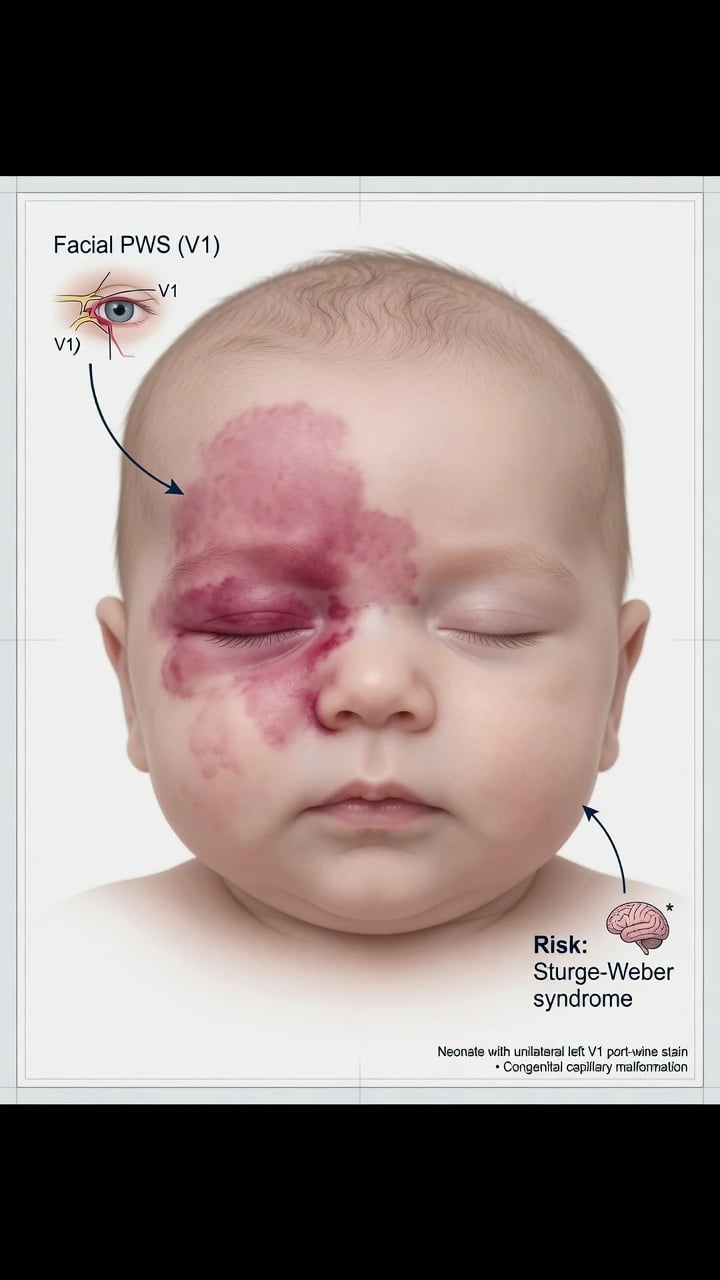
Definition and Classification
Sturge-Weber syndrome (SWS), also called encephalotrigeminal angiomatosis, is a rare sporadic neurocutaneous vascular disorder in which abnormal capillary–venous vessels affect the facial skin, the leptomeninges of the brain, and the eye in a metameric pattern. It is not a Mendelian inherited syndrome: the causal mutation arises post-zygotically, producing somatic mosaicism confined to affected tissues.[1][4][9]
The classic clinical triad is:
[5]- Facial capillary malformation — port-wine stain (PWS; naevus flammeus), typically unilateral and in a trigeminal distribution
- Ipsilateral leptomeningeal angiomatosis — pial vascular malformation overlying the cerebral cortex
- Ocular vascular anomalies — glaucoma and/or choroidal haemangioma
Not every patient expresses all three elements; classification systems capture incomplete forms.[2][6]
Roach classification
| Type | Features | Clinical note |
|---|---|---|
| Type I | Facial PWS + leptomeningeal angiomatosis ± glaucoma | Classic SWS |
| Type II | Facial PWS + glaucoma without intracranial involvement | Skin–eye form |
| Type III | Leptomeningeal angiomatosis without cutaneous PWS | Diagnostic trap — presents as epilepsy/calcification |
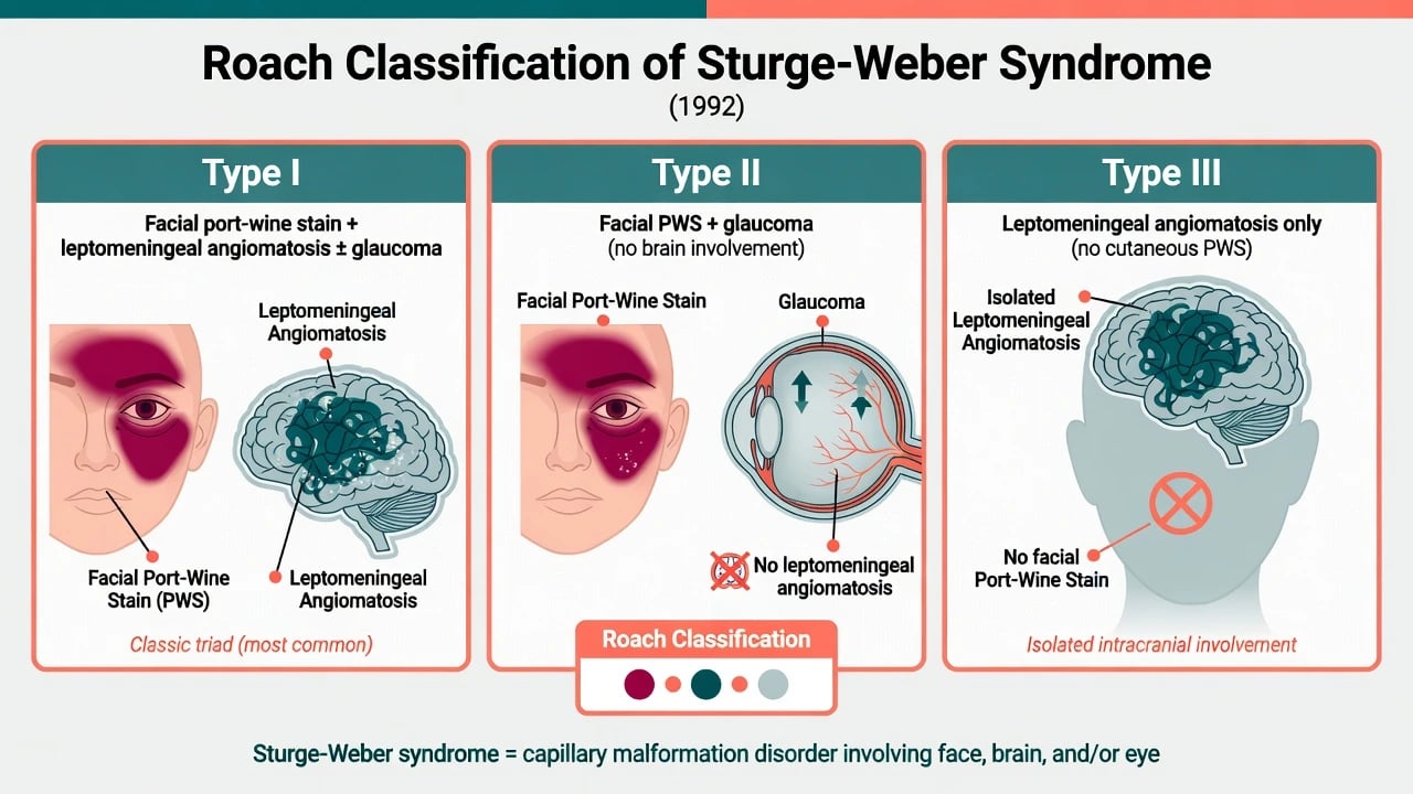
Within ISSVA, the facial lesion is a simple slow-flow capillary malformation; SWS is the syndromic association of that malformation with CNS and ocular vascular anomalies rather than a separate tumour biology.[7]
Epidemiology and Risk Factors
Facial PWS occurs in roughly 0.3% of live births. Only a minority of facial PWS are associated with SWS; overall risk estimates for SWS among facial PWS are on the order of ~8–15%, but risk is strongly distribution-dependent. Involvement of the forehead and upper eyelid (V1 / frontonasal embryologic territory) confers the highest risk of leptomeningeal and ocular disease; isolated cheek (V2) or mandibular (V3) stains without forehead involvement carry substantially lower risk.[2][7][10]
There is no consistent sex or ethnic predilection. Family history is usually negative because the mutation is somatic mosaic, not germline. Recurrence risk to siblings is not elevated above the general population for classic mosaic SWS; genetic counselling focuses on explaining mosaicism rather than Mendelian transmission.[1][4]
Pathophysiology
Molecular genetics — GNAQ
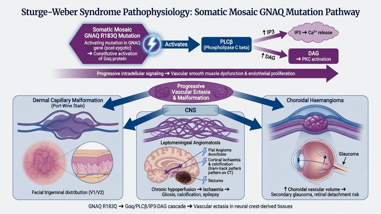
In 2013, Shirley and colleagues demonstrated that both isolated PWS and SWS are associated with a somatic mosaic activating mutation in GNAQ (most commonly p.Arg183Gln / R183Q), encoding the G-protein alpha subunit Gαq. The mutation is present in affected skin, brain, and/or eye tissue and absent from blood in most patients, confirming post-zygotic mosaicism.[1]
Constitutive Gαq signalling drives PLCβ → IP3/DAG → calcium cascades, producing progressive vascular ectasia of capillaries and post-capillary venules without true neoplastic endothelial proliferation. This explains why the cutaneous lesion is a malformation (present at birth, grows with the child, never involutes) rather than a haemangioma.[1][4]
Leptomeningeal and cerebral injury
Leptomeningeal angiomatosis impairs venous drainage of the underlying cortex. Chronic ischaemia, gliosis, and gyral calcification follow. Calcification classically produces the tram-track (parallel cortical) pattern on skull radiograph or CT — a high-yield exam sign, though contrast-enhanced MRI is the modern first-line imaging modality for diagnosis and extent mapping.[2][3][5]
Impaired cortical perfusion and epileptogenic cortex produce focal seizures, often starting in infancy, with risk of epileptic encephalopathy, hemiparesis, and developmental delay if seizures are poorly controlled. Stroke-like episodes reflect transient or progressive hypoperfusion rather than classic arterial thromboembolism alone.[3][6]
Ocular disease
Abnormal vessels in the anterior chamber angle and elevated episcleral venous pressure impair aqueous outflow, causing glaucoma (congenital, infantile, or later childhood/adult onset). Choroidal haemangioma (often diffuse) can cause refractive error, retinal detachment risk, and visual field loss. Periocular PWS — especially upper eyelid — is the key cutaneous predictor of ocular involvement.[8][5]
Clinical Presentation
Cutaneous
The facial PWS is a flat pink-to-red patch at birth that darkens toward purple with age and may thicken or cobblestone in adulthood. Distribution follows trigeminal or frontonasal embryologic segments; midline-crossing can occur. Soft-tissue and bony hypertrophy of the face, gingival staining, and mucosal extension are common with extensive lesions.[2][7]
Neurological
- Seizures — often focal motor, beginning in the first year of life in many Type I patients; may become refractory
- Hemiparesis / hemiatrophy ipsilateral to the brain lesion
- Stroke-like episodes, headaches/migraine
- Developmental delay, intellectual disability, behavioural disorders — severity correlates with seizure burden and extent of angiomatosis
- Visual field defects from occipital involvement
Ocular
- Congenital or childhood glaucoma (buphthalmos, corneal clouding, photophobia, epiphora)
- Choroidal haemangioma
- Amblyopia, refractive error, strabismus
Atypical presentations
Roach Type III (no cutaneous PWS) may present as childhood epilepsy with cortical calcification — examiners love this trap. Bilateral facial PWS and bilateral brain disease predict more severe neurological morbidity.[2][9]
Differential Diagnosis
| Mimic | Distinguishing feature from SWS |
|---|---|
| Isolated facial PWS | No leptomeningeal angiomatosis or glaucoma after appropriate screening |
| Infantile haemangioma | Appears after birth, proliferates, then involutes; GLUT1+ tumour biology |
| Salmon patch (naevus simplex) | Midline nape/glabella; usually fades; not progressive purple thickening |
| PHACES | Segmental infantile haemangioma (not PWS) + posterior fossa/arterial/cardiac/eye/sternal defects |
| Klippel–Trenaunay | Limb PWS + venous varicosities + overgrowth (often PIK3CA spectrum) |
| Tuberous sclerosis | Ash-leaf macules, angiofibromas, cortical tubers — not PWS + leptomeningeal angioma |
| NF1 | Café-au-lait macules, neurofibromas — different genetics and CNS tumours |
SWS / PWS
Infantile haemangioma
Salmon patch
Clinical and Bedside Assessment
- Map the PWS — photograph, document forehead, upper/lower eyelid, cheek, lip, mucosa; note laterality.
- Ophthalmic urgency — corneal diameter, haze, photophobia, parental report of eye rubbing; same-week ophthalmology for periocular V1 lesions.
- Neurological screen — seizures (including subtle focal twitches), developmental milestones, head circumference, focal motor signs.
- Diascopy — PWS blanches incompletely; helps separate from purpura.
- Family counselling — explain mosaic (not usually inherited) while arranging MDT pathways.[5][7]
Investigations
- MRI brain with gadolinium (and vascular sequences as per local protocol): leptomeningeal enhancement, cortical atrophy, accelerated myelination patterns, venous anomalies. Optimal timing in asymptomatic infants is guided by consensus and local sedation risk; any neurological symptom warrants prompt imaging.[5]
- CT may show tram-track calcification but is not first-line in infants (radiation).
- EEG if seizures suspected or for surgical planning.
- Ophthalmology: serial intraocular pressure, gonioscopy, dilated fundoscopy for choroidal haemangioma.[8]
- Molecular testing for GNAQ in tissue is confirmatory in research/atypical cases but not required for classic clinical diagnosis.
- Developmental and educational assessments longitudinally.
Management
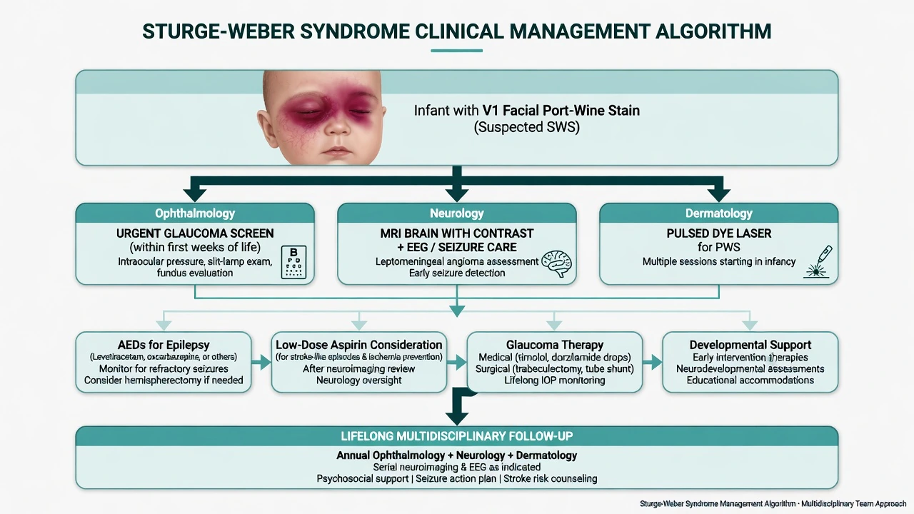
Multidisciplinary framework
Care is coordinated across dermatology, paediatric neurology, ophthalmology, and often psychology/education support. Consensus recommendations (Sabeti et al., 2021) emphasise structured neuroimaging, seizure vigilance, and lifelong eye surveillance.[5][10]
Neurological therapy
- Antiseizure medications early after first seizures; aggressive control improves developmental outcome.
- Refractory epilepsy may need ketogenic diet, neuromodulation, or resective/hemispherectomy surgery in selected candidates.
- Low-dose aspirin is used in many centres to reduce stroke-like episodes and is supported by observational and expert consensus data, though randomised evidence remains limited; discuss bleeding risk individually.[3][11][5]
- Migraine management and stroke-like episode protocols (hydration, avoid hypotension, urgent imaging when atypical).
Ophthalmic therapy
- Topical pressure-lowering agents as first line for glaucoma.
- Surgical options (trabeculectomy, tube shunts, cyclodestructive procedures) when medical therapy fails.
- Monitor and manage choroidal haemangioma complications with retina specialists as needed.[8]
Cutaneous therapy
- Pulsed-dye laser (PDL 585/595 nm) is gold-standard for lightening facial PWS; multiple sessions; earlier treatment in infancy generally yields better lightening and may reduce later hypertrophy.
- Complete clearance is uncommon; counsel realistic expectations.
- Camouflage cosmetics; adult nodular lesions may need alternative lasers or lesion-directed care.[7]
Supportive care
Developmental therapies, educational plans, psychological support for facial difference, and transition to adult neurology/ophthalmology services.
[5]Complications, Emergencies and Pitfalls
Exam application bank (NEET-PG / INICET)
One-line answer
Sturge-Weber syndrome (SWS; encephalotrigeminal angiomatosis) is a sporadic mosaic neurocutaneous vascular disorder defined by facial capillary malformation (port-wine stain), ipsilateral leptomeningeal angiomatosis, and ocular vascular anomalies (glaucoma, choroidal haemangioma). It is caused by a post-zygotic somatic activating mutation in GNAQ (p.R183Q). V1 facial port-wine stain mandates ophthalmology and neuroimaging screening. Management is multidisciplinary: seizure control, glaucoma care, pulsed-dye laser for skin, and consideration of low-dose aspirin for stroke-like episodes.
Worked stems (answer without another resource)
Stem 1 — Classic presentation. Map symptoms to mechanism; name the first investigation and first treatment step with dose/route if drug therapy is standard. [1]
Stem 2 — Unstable / complicated. List red flags that force immediate resuscitation, theatre, ICU, antidote, or reperfusion — and what you do in the first 15 minutes. [1]
Stem 3 — Atypical group. Elderly, pregnancy, child, or immunocompromised: how presentation and thresholds change. [1]
Stem 4 — Differential trap. Name the three closest mimics and one discriminator for each. [1]
Stem 5 — Disposition. Who goes home with safety-netting, who is admitted, who needs HDU/ICU/theatre, and what follow-up is mandatory. [1]
Rapid viva checklist
- Definition + classification
- Pathophysiology chain
- Bedside signs / criteria
- Score with exact components (if any)
- Emergency bundle
- Definitive therapy with doses
- Complications of disease and of treatment
- Special populations
- Guideline/trial name if classic
- Three exam traps
Coverage self-check
If you cannot answer any stem above from this page alone, re-read the matching section — the page is intended to be self-sufficient for final-prof and NEET-PG/INICET questions on Sturge-Weber syndrome.
Expanded exam teaching (depth pass)
Clinical reasoning
For Sturge-Weber syndrome, examiners test whether you can prioritise life threats, choose the right first test, and give specific therapy (agent, dose, route, timing). Generic phrases without numbers score poorly.
Mechanism → feature map
Build a short chain: cause → pathophysiologic intermediate → clinical feature → complication. Every major symptom in the classic vignette should sit on that chain.
Investigation strategy
- Bedside/first-line tests that change immediate management
- Confirmatory or staging tests
- What a normal result does not exclude
- When not to delay treatment for imaging (unstable patient)
Management ladder
- Resuscitation / ABC / sepsis or haemorrhage bundle as relevant
- Specific antidote / procedure / antimicrobial / reperfusion / surgery
- Supportive care and monitoring targets
- Definitive long-term therapy and secondary prevention
- Disposition and safety-net advice
Special populations
Always prepare one line each for children, pregnancy, elderly, renal/hepatic impairment, and immunocompromised patients when the topic allows.
Pitfalls that fail candidates
- Treating the number not the patient
- Missing pregnancy status when relevant
- Imaging before stabilisation
- Wrong empiric cover or wrong antidote timing
- Incomplete counselling on recurrence, adherence, or red-flag return
Sturge-Weber syndrome (SWS; encephalotrigeminal angiomatosis) is a sporadic mosaic neurocutaneous vascular disorder defined by facial capillary malformation (port-wine stain), ipsilateral leptomeningeal angiomatosis, and ocular vascular anomalies (glaucoma, choroidal haemangioma). It is caused by a post-zygotic somatic activating mutation in GNAQ (p.R183Q). V1 facial port-wine stain mandates ophthalmology and neuroimaging screening. Management is multidisciplinary: seizure control, glaucoma care [1]
Structured revision sheet
Must-know numbers and names
List every score, size threshold, dose, and time window from this topic on a blank page from memory, then check against the sections above.
Three classic MCQ angles
- Most likely diagnosis given a vignette
- Next best step in management
- Most appropriate investigation
Three classic SAQ angles
- Pathophysiology in five steps
- Management algorithm with doses
- Complications and prevention
Clinical station flow
Greet → focused history → targeted exam → investigations → explain diagnosis → emergency care → definitive plan → safety-net / follow-up → answer examiner questions on mechanism and pitfalls.
[5]Other complications: progressive hemiparesis, intellectual disability, visual field loss, facial soft-tissue hypertrophy, bleeding from nodular adult PWS, and psychosocial morbidity.
[5]Special Populations
- Neonate with V1 PWS: photograph, urgent ophthalmology, neurology/MRI pathway per consensus, parental mosaic counselling.[5][7]
- Pregnancy: seizure and glaucoma plans; medication teratogenicity review.
- Adults: late glaucoma surveillance; laser for cosmetic and psychosocial benefit; new neurological symptoms still warrant imaging.
Prognosis and Follow-Up
Prognosis is highly variable. Early seizure control and limited leptomeningeal extent predict better neurodevelopmental outcomes. Glaucoma requires lifelong pressure monitoring. Cutaneous PWS persists lifelong without treatment and progressively darkens. Transition clinics improve continuity of eye and seizure care into adulthood.[3][6][10]
Evidence, Guidelines and Regional Differences
| Source | Contribution |
|---|---|
| Shirley et al., NEJM 2013 | GNAQ R183Q mosaic discovery linking PWS and SWS |
| Sabeti et al., Pediatr Neurol 2021 | Consensus neurology, neuroimaging, ophthalmology pathways |
| Yeom & Comi, Stroke 2022 | Stroke-like pathophysiology and management updates |
| Day et al., 2019 | Hypothesis on presymptomatic aspirin/AED strategies |
Regional differences include MRI sedation policies for infants, access to paediatric PDL under anaesthesia, and thresholds for starting aspirin. Examiners expect candidates to state uncertainty around aspirin while still describing its widespread expert use.[1][5][3][11]
Exam Pearls
[1]SWS screen (V1 baby)
Key Takeaways
- SWS is a sporadic mosaic neurocutaneous vascular disorder driven by somatic GNAQ activation, not a classical Mendelian genodermatosis.
- The cutaneous lesion is a port-wine stain (capillary malformation) — present at birth, never involuting.
- V1 distribution is the practical screening trigger for brain and eye disease.
- Management is multidisciplinary: seizure control, glaucoma care, PDL for skin, and individualised aspirin discussion.
- Roach Type III and confusion with infantile haemangioma are classic exam traps.[1][2][4][5]
References
- [1]Shirley MD, Tang H, Gallione CJ, et al. Sturge-Weber syndrome and port-wine stains caused by somatic mutation in GNAQ N Engl J Med, 2013.PMID 23656586
- [2]Higueros E, Roe E, Granell E, et al. Sturge-Weber Syndrome: A Review Actas Dermosifiliogr, 2017.PMID 28126187
- [3]Yeom S, Comi AM. Updates on Sturge-Weber Syndrome Stroke, 2022.PMID 36263782
- [4]Sánchez-Espino LF, Ivars M, Antoñanzas J, et al. Sturge-Weber Syndrome: A Review of Pathophysiology, Genetics, Clinical Features, and Current Management Approache Appl Clin Genet, 2023.PMID 37124240
- [5]Sabeti S, Ball KL, Bhattacharya SK, et al. Consensus Statement for the Management and Treatment of Sturge-Weber Syndrome: Neurology, Neuroimaging, and Ophthalmology Recommendations Pediatr Neurol, 2021.PMID 34153815
- [6]Comi AM. Sturge-Weber syndrome Handb Clin Neurol, 2015.PMID 26564078
- [7]Poliner A, Fernandez Faith E, Blieden L, et al. Port-wine Birthmarks: Update on Diagnosis, Risk Assessment for Sturge-Weber Syndrome, and Management Pediatr Rev, 2022.PMID 36045161
- [8]Silverstein M, Salvin J. Ocular manifestations of Sturge-Weber syndrome Curr Opin Ophthalmol, 2019.PMID 31313748
- [9]Ramirez EL, Jülich K. Sturge-Weber syndrome: an overview of history, genetics, clinical manifestations, and management Semin Pediatr Neurol, 2024.PMID 39389653
- [10]Shah AD, Alexieff P, Tatachar P. Sturge-Weber Syndrome: A Narrative Review of Clinical Presentation and Updates on Management J Clin Med, 2025.PMID 40217631
- [11]Day AM, Hammill AM, Juhász C, et al. Hypothesis: Presymptomatic treatment of Sturge-Weber Syndrome With Aspirin and Antiepileptic Drugs May Delay Seizure Onset Pediatr Neurol, 2019.PMID 30482419