Dermatology · Medicine
Subcorneal pustular dermatosis (Sneddon-Wilkinson disease)
Also known as Subcorneal pustular dermatosis (SPD) · Sneddon-Wilkinson disease
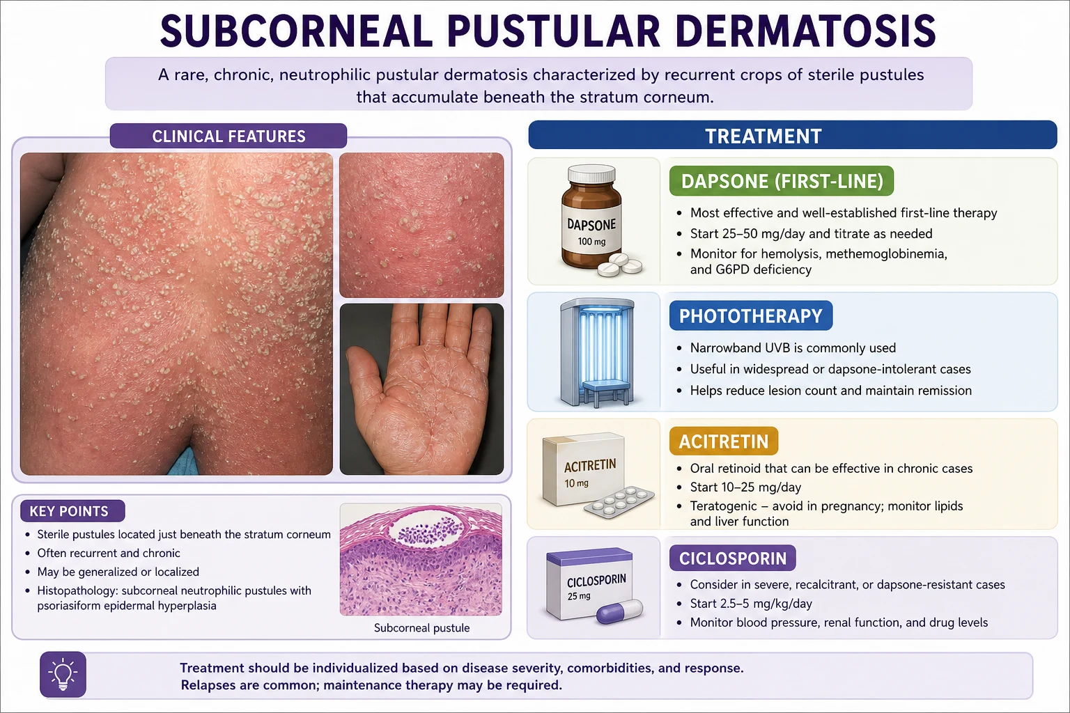
Subcorneal pustular dermatosis (Sneddon-Wilkinson disease) is a chronic, relapsing neutrophilic dermatosis presenting with flaccid pustules in annular/polycyclic patterns on intertriginous skin (groin, axillae, submammary). Histology shows a subcorneal pustule filled with neutrophils with no acantholysis. Direct immunofluorescence is NEGATIVE — the key feature distinguishing classical SPD from IgA pemphigus (identical clinicopathology but DIF-positive for intercellular IgA). Associated with IgG monoclonal gammopathy. First-line treatment is dapsone 50 to 150 mg daily (check G6PD first); alternatives include acitretin, phototherapy, colchicine, ciclosporin and biologics.
On this page & tools
Your progress
Saved locally on this device.
Exam tags
Red flags
Definition & overview
Subcorneal pustular dermatosis (SPD) — Sneddon-Wilkinson disease — is a chronic, relapsing, sterile neutrophilic dermatosis defined by superficial, flaccid pustules that sit immediately beneath the stratum corneum (subcorneal) and are arranged in annular or polycyclic (ring-like, serpiginous) configurations on intertriginous and flexural skin.[1][5]
Three features together make the diagnosis, and the absence of any one should make you reconsider:[1]
- A subcorneal split filled with neutrophils on histology (a true subcorneal pustule, with no acantholysis and no eosinophils).
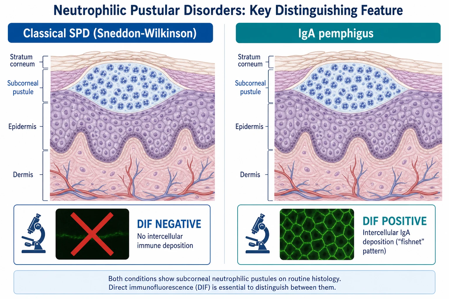
- Negative direct immunofluorescence (DIF) — no intercellular or basement-membrane immunoreactants. This is the keystone that separates SPD from IgA pemphigus, which is clinicopathologically identical but DIF-positive for intercellular IgA.
- A chronic, relapsing-remitting course with a characteristic response to dapsone. [1]
SPD sits within the wider family of neutrophilic dermatoses — a spectrum that also includes pyoderma gangrenosum, Sweet syndrome, erythema elevatum diutinum, Behçet disease and the bowel-associated dermatosis-arthritis syndrome.[2] All share a common final pathway: sterile recruitment and activation of neutrophils in the skin, often driven by an underlying systemic inflammatory or haematological process. The unifying concept is that these are reactive patterns of neutrophilic inflammation, not infections and (in the classical case) not autoantibody-mediated blistering diseases.[2][6]
Eponym and history
Ian Sneddon and Darrell Wilkinson described the disease in 1956 as a distinctive pustular eruption of middle-aged women with a subcorneal pustule on histology and a relapsing course. Their contribution was the separation of SPD from generalised pustular psoriasis, with which it had previously been conflated. The eponym Sneddon-Wilkinson disease is retained for the classical, DIF-negative form; some modern texts reserve "subcorneal pustular dermatosis" as a purely descriptive histological pattern that includes the IgA pemphigus subtype, which is why specifying DIF status is essential in every case.[1][5]
Classification
SPD is best understood along two axes: (a) where it sits within the neutrophilic dermatoses, and (b) the critical immunofluorescence-based split between classical SPD and the SPD-pattern of IgA pemphigus.[6]
Axis 1 — the neutrophilic dermatosis spectrum. SPD belongs to the sterile neutrophilic dermatoses, alongside pyoderma gangrenosum, Sweet syndrome and erythema elevatum diutinum. These entities overlap, may coexist, and frequently signal an underlying systemic inflammatory disease (inflammatory bowel disease, rheumatoid arthritis, haematological malignancy) or a monoclonal gammopathy.[2][7]
Axis 2 — DIF-positive versus DIF-negative. This is the classification that matters at the bedside and in the exam: [1]
Classical SPD (Sneddon-Wilkinson)
- DIF NEGATIVE — no immunoreactants
- No detectable autoantibody; considered reactive neutrophilic pustulosis
- Associates with IgG monoclonal gammopathy, IBD, malignancy
- Responds to dapsone (first-line)
SPD-type IgA pemphigus
- DIF POSITIVE — intercellular (fishnet) IgA in the upper epidermis
- Pathogenic IgA autoantibody against desmocollin 1 (a desmosomal cadherin)
- May associate with IgA gammopathy / lymphoproliferative disease
- Treated with dapsone, retinoids, phototherapy, or rituximab for refractory disease
Intraepidermal neutrophilic IgA pemphigus
- A broader IgA pemphigus pattern; pustules throughout the epidermis (not just subcorneal)
- Often anti-desmocollin 2/3 or less defined antigen
- DIF positive for intercellular IgA — distinguishes from classical SPD
- Histology shows intraepidermal pustule with acantholysis variable
Some authorities argue that all "SPD" with a positive DIF should simply be reclassified as IgA pemphigus and the term Sneddon-Wilkinson reserved exclusively for the DIF-negative form — this is the dominant modern position.[1][6] For exams, state both: the descriptive histological pattern is "subcorneal pustular dermatosis"; the disease Sneddon-Wilkinson is DIF-negative.
Epidemiology & risk factors
SPD is rare. No population incidence is firmly established, but it is encountered far less often than psoriasis or pemphigus. The epidemiology is dominated by three observations:[1][3]
- Sex and age: a striking female predominance (roughly four women for every man) with onset typically in the fifth and sixth decades (median age in the fifties). Presentation before forty or after seventy is well described but uncommon.
- Children: SPD is rare but recognised in childhood, including infancy; paediatric cases are frequently mistaken for impetigo or acrodermatitis enteropathica before biopsy.[4]
- No clear racial predilection, though most reported series are European and South Asian.
SPD — the numbers that orient the diagnosis
Systemic associations (the reason to investigate every patient). SPD is a reactive neutrophilic dermatosis in a substantial minority of cases, so a directed search for an underlying driver is mandatory:[1][7]
| Association | What to do |
|---|---|
| IgG (less often IgA) monoclonal gammopathy / MGUS / multiple myeloma | SPEP, serum free light chain ratio, urine Bence-Jones, CBC, calcium, renal function; bone marrow biopsy if a paraprotein is confirmed |
| Inflammatory bowel disease (ulcerative colitis more than Crohn disease) | History for bowel symptoms; faecal calprotectin; colonoscopy if indicated |
| Pyoderma gangrenosum / Sweet syndrome (the neutrophilic dermatosis spectrum) | Examine for painful nodules, ulcers with undermined edges, or inflammatory plaques |
| Thyroid disease | TSH |
| Solid or haematological malignancy | Age-appropriate screening; maintain a low threshold for paraprotein surveillance |
| Infections (streptococcal, viral, HIV) rarely reported as triggers | Throat swab, ASOT, HIV test if risk factors |
Pathophysiology
The histological lesion — a subcorneal pustule packed with neutrophils — is the end-result of sterile neutrophilic recruitment to the most superficial epidermis. Two questions follow: why do neutrophils collect just beneath the stratum corneum, and is there an autoantibody?[2][5]
The classical, DIF-negative form. No pathogenic autoantibody is detectable. SPD is therefore considered a reactive neutrophilic dermatosis analogous to pyoderma gangrenosum and Sweet syndrome: a systemic trigger (a paraprotein, an inflammatory bowel disease, an infection) generates cytokines that drive neutrophils into the skin. The key mediators are interleukin-8 (IL-8 / CXCL8), a potent neutrophil chemoattractant expressed by keratinocytes in SPD lesions, and the IL-17 / IL-23 axis and tumour necrosis factor-alpha (TNF-alpha), which amplify neutrophil recruitment and survival.[2] The anatomical specificity — neutrophils lodging just under the stratum corneum — is incompletely explained but mirrors the subcorneal localisation seen in IgA pemphigus, where the target antigen (desmocollin 1) is concentrated in the uppermost epidermis.
Proposed role of the paraprotein. In patients with an IgG monoclonal gammopathy, the paraprotein may act as a neutrophil-activating immune complex, generating cytokine release and sterile pustules — a mechanism analogous to the way paraproteins drive lesions in Schnitzler syndrome (urticaria + IgM gammopathy) and some POEMS syndrome vasculitides. Treating the underlying myeloma can clear the skin, supporting a pathogenic link.[7]
The IgA pemphigus (DIF-positive) form. Here the mechanism is genuinely autoimmune. A pathogenic IgA autoantibody binds the extracellular domain of desmocollin 1 (a desmosomal cadherin expressed preferentially in the uppermost epidermis), triggering loss of keratinocyte adhesion and neutrophil attraction to the subcorneal compartment.[6] The result is clinicopathologically indistinguishable from classical SPD — only DIF (and ELISA for anti-desmocollin antibodies) reveals the difference. The same logic explains why some IgA pemphigus responds to dapsone (it calms neutrophils) while also requiring immunosuppression directed at the autoantibody in resistant cases.
Why dapsone works. Dapsone inhibits neutrophil myeloperoxidase and the respiratory burst, and interferes with neutrophil chemotaxis and adherence — precisely the effector cell driving the pustule. This is why a rapid clinical response to dapsone (within days) is both a useful diagnostic clue and the rationale for first-line use, in contrast to the slow response of pustular psoriasis.[1][5]
Clinical presentation
Individual lesion. The pustule is flaccid, superficial and 1 to 5 mm (occasionally coalescing to over 1 cm). Because it is so superficial it ruptures easily, leaving a collarette of scale — a thin rim of stratum corneum at the edge of the deflated pustule that is a useful bedside clue. The pustule fluid is initially clear-yellow, later cloudy; it is sterile.[1][3]
Arrangement and distribution. Pustules cluster and extend peripherally into annular, polycyclic or serpiginous (figure-of-eight) patterns, with new pustules appearing at the advancing edge as older central lesions dry and desquamate. The intertriginous and flexural sites are the classical territory: [1]
- Groin, inguinal and genitocrural folds
- Axillae
- Submammary and abdominal folds
- Flexor forearms, lower abdomen, and proximal thighs
- Occasionally generalises to involve the trunk; may localise to one region for years [1]
Characteristically spared: the mucous membranes, palms, soles, and face — a distribution that helps separate SPD from pustular psoriasis (which favours palms/soles in the localised form and generalises in von Zumbusch) and from AGEP (which is genuinely generalised with facial involvement).[6]
Symptoms. Most patients are asymptomatic or have only mild pruritus and a burning sensation. Systemic symptoms (fever, malaise) are absent in classical SPD; their presence should prompt reconsideration of pustular psoriasis, AGEP, or an infective process, and a search for an associated systemic disease.[1]
Course. The disease runs a chronic, relapsing-remitting course over years to decades. Crops of pustules erupt, extend in rings over days to weeks, settle spontaneously, and recur at the same or new sites. Spontaneous permanent remission is uncommon but reported; most patients require long-term suppressive therapy. [1]
Atypical and special presentations.[3][4]
- Pregnancy: SPD may present or flare in pregnancy; management is constrained by teratogenic and haematological drug risks (see Special Populations).
- Children: rare; lesions may be more acral and are frequently misdiagnosed as impetigo.
- Immunocompromised: secondary infection of pustules is more likely; the neutrophilic phenotype may be blunted.
- Elderly with an underlying myeloma: more extensive disease, possible association with systemic symptoms; the skin may be the presenting feature of the haematological disorder. [1]
Differential diagnosis
The differential is the differential of a sterile subcorneal or superficial pustular eruption, and the exam wants the discriminating features for each.[1][6]
| Condition | Morphology | Histology | DIF | Key discriminator |
|---|---|---|---|---|
| IgA pemphigus (SPD type) | Identical to SPD — annular flaccid pustules | Subcorneal pustule, neutrophils | POSITIVE — intercellular IgA | DIF is the only reliable separator; ELISA anti-desmocollin 1 |
| Generalised pustular psoriasis (von Zumbusch) | Erythematous lakes of pus on erythroderma; systemic toxicity; history of psoriasis | Munro microabscesses, acanthosis, parakeratosis, regular psoriasiform hyperplasia | Negative | Systemic upset, nail/skin psoriasis, IL-36RN mutations |
| Pemphigus foliaceus | Scaly, crusted, superficial erosions (not tense pustules) | Subcorneal acantholysis (not a neutrophilic pustule) | Positive — intercellular IgG/C3 upper epidermis | Acantholysis on H and E; anti-desmoglein 1 |
| Acute generalised exanthematous pustulosis (AGEP) | Acute, drug-triggered, hundreds of small pustules on erythema; fever | Subcorneal/intraepidermal pustules with eosinophils and necrotic keratinocytes | Negative | Drug exposure under four days; self-limiting on withdrawal |
| Bullous impetigo | Flaccid pustules/bullae, honey-coloured crust; children | Subcorneal split with cocci (Gram-positive) | Negative | Culture positive for Staph aureus; contagious |
| Linear IgA bullous dermatosis | Tense bullae in string-of-pearls (cluster-of-jewels); drug-triggered in adults | Subepidermal blister with neutrophils | Linear IgA along BMZ | Linear (not intercellular) IgA on DIF |
| Dermatitis herpetiformis | Intensely pruritic herpetiform vesicles on elbows/knees/buttocks | Subepidermal blister, neutrophilic papillary microabscesses | Granular IgA in dermal papillae | Coeliac association; anti-tissue transglutaminase |
Classical SPD
- Women over 50; annular/polycyclic flaccid pustules on flexures
- Subcorneal neutrophilic pustule, NO acantholysis
- DIF NEGATIVE — sterile, no paraprotein-driven autoantibody
- Responds to dapsone within days
IgA pemphigus
- Clinically identical annular pustules
- Subcorneal pustule, identical histology
- DIF POSITIVE — intercellular (fishnet) IgA, anti-desmocollin 1
- Needs dapsone plus or minus immunosuppression / rituximab
Generalised pustular psoriasis
- Systemic toxicity, fever; pre-existing psoriasis
- Munro microabscesses, psoriasiform hyperplasia
- DIF negative but systemic phenotype
- Treated with acitretin, ciclosporin, anti-IL-36 / biologics, NOT dapsone-first
AGEP
- Drug-triggered (beta-lactams); onset under four days
- Subcorneal pustules with eosinophils and necrotic keratinocytes
- DIF negative; resolves on drug withdrawal
- Self-limiting; supportive care
Clinical & bedside assessment
SPD is a clinicopathological diagnosis confirmed in the laboratory, but the bedside examination establishes the pattern.[1]
- Distribution and arrangement. Map the sites (intertriginous predominance?) and the configuration (annular, polycyclic, serpiginous advancing edge with central clearing?). Photograph the pattern to document progression.
- Individual lesion morphology. Look for the flaccid pustule that ruptures to leave a collarette of scale — the latter is a strong clue to a subcorneal process.
- Mucous membrane, palm, sole and face examination to document sparing (favours SPD) versus involvement (favours AGEP, pustular psoriasis).
- Nail and plaque psoriasis assessment — its presence redirects towards pustular psoriasis.
- Systemic examination for associated disease: lymphadenopathy and hepatosplenomegaly (lymphoproliferative disease), abdominal examination and joint review (inflammatory bowel disease, arthritis of the neutrophilic-dermatosis spectrum), thyroid enlargement, and a search for ulcers with undermined edges or inflammatory nodules (pyoderma gangrenosum / Sweet syndrome overlap).[2]
- Confirm sterility by swabbing an intact pustule for bacterial (and if indicated fungal and viral) culture before starting antibiotics.
Investigations
The diagnosis rests on histopathology and immunofluorescence of a skin biopsy, supported by a screen for systemic associations. There is no pathognomonic blood test.[1][3]
Skin biopsy — two samples from the same site. Biopsy an early, fresh pustule (ideally under 24 hours old). Send: [1]
- One sample in formalin for H and E — the diagnostic histology.
- One sample in Michel medium or fresh-frozen for DIF — the discriminator from IgA pemphigus. [1]
Histopathology (H and E). The cardinal finding is a subcorneal pustule — a blister cavity immediately beneath the stratum corneum, filled with neutrophils, with a minimally inflamed underlying epidermis. Critically: no acantholysis (excludes pemphigus foliaceus), no eosinophils (excludes bullous pemphigoid and AGEP), and no organisms on Gram/PAS stain (excludes impetigo).[5]
Direct immunofluorescence (DIF). The keystone investigation. In classical SPD it is negative — no intercellular IgG, IgA, IgM or C3, and no basement-membrane-zone deposition. In IgA pemphigus it is positive for intercellular IgA in a fishnet/lattice pattern in the upper epidermis.[6] A positive DIF must change the label from "SPD" to "IgA pemphigus."
Indirect immunofluorescence (IIF) and ELISA. IIF (on monkey oesophagus or human skin) and anti-desmocollin 1 ELISA are positive in IgA pemphigus and negative in classical SPD — useful when DIF is equivocal or to monitor disease activity.[6]
The workup of a sterile pustular eruption — BIDS-H
BIDSH
two samples from a fresh pustule — H and E and DIF (Michel medium)
DIF is the test that splits SPD (negative) from IgA pemphigus (intercellular IgA)
AGEP is drug-triggered and self-limiting — a careful drug timeline can avoid an invasive workup
confirm the pustule is sterile — impetigo is culture-positive for Staph aureus
SPEP, serum free light chains, urine Bence-Jones — screen for the IgG monoclonal gammopathy
Mandatory haematological and systemic screen.[1][7]
- Serum protein electrophoresis (SPEP) and serum free light chain (sFLC) ratio — screen for monoclonal gammopathy / myeloma.
- Urine for Bence-Jones protein (urine free light chains).
- Full blood count with differential — baseline for dapsone monitoring and to detect anaemia of myeloma.
- Calcium, urea and electrolytes, creatinine — the CRAB features of myeloma (hypercalcaemia, renal impairment).
- Liver function tests and hepatitis B/C screen — required before dapsone and some immunosuppressants.
- TSH — autoimmune thyroid association.
- HIV test if risk factors present. [1]
When to request bone marrow biopsy. If SPEP or sFLC reveals a paraprotein, refer to haematology; bone marrow biopsy, imaging for lytic lesions (low-dose whole-body CT or skeletal survey) and serum beta-2 microglobulin staging follow haematology protocols. Treating an underlying myeloma can control the SPD.[7]
Management — resuscitation
Classical SPD is not a dermatological emergency in the uncomplicated case — the patient is systemically well and the lesion is sterile. Resuscitative concerns arise only in specific situations:[1]
- Erythroderma or extensive skin loss: rare in SPD; manage as for any erythrodermic patient — fluid and temperature regulation, monitoring for high-output cardiac failure, and skin sepsis prophylaxis.
- Secondary bacterial infection of pustules: take swabs and treat with empirical oral flucloxacillin 500 mg four times daily for 5 to 7 days (cefalexin if penicillin-allergic without anaphylaxis; clindamycin or a macrolide if anaphylaxis), de-escalating to culture. Consider MRSA cover (co-trimoxazole, doxycycline) if risk factors.
- Pre-dapsone workup (the true "resuscitation" step): before the first dose of dapsone, obtain G6PD level, full blood count with reticulocytes, methaemoglobin, liver function, hepatitis B and C serology, and renal function. A G6PD-deficient patient must not receive dapsone — choose an alternative.[8]
Management — definitive & stepwise
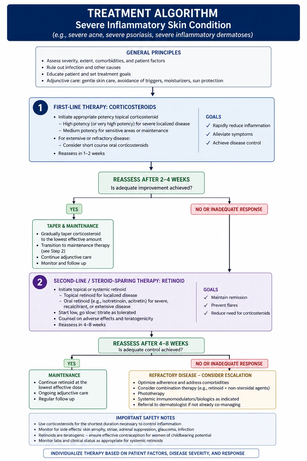
Treatment aims to suppress pustule formation and maintain remission — there is no curative therapy. The ladder below reflects consensus from case series and reviews; there are no randomised trials (the disease is too rare).[1][3]
First-line: dapsone
Dapsone 50 to 150 mg orally once daily (start at 50 mg, titrate by 25 to 50 mg every one to two weeks to response, usual maintenance 100 mg daily) is the drug of choice.[1] It inhibits neutrophil myeloperoxidase and chemotaxis, producing a clinical response within days — a feature that itself helps confirm the diagnosis (psoriasis responds far more slowly to systemic therapy). Continue for several months, then taper to the lowest effective dose.
Dapsone monitoring — the detail the exam wants.[1][8]
- G6PD before the first dose. G6PD deficiency (X-linked, common in Mediterranean, African, South-East Asian and Middle Eastern populations) causes severe haemolysis. If deficient, do not use dapsone.
- Full blood count weekly for the first 4 weeks, then monthly. Watch for haemolytic anaemia (especially in mild G6PD variants or at higher doses) and agranulocytosis — an idiosyncratic, potentially fatal adverse effect most common in the first three months.
- Reticulocyte count — rises as the marrow compensates for dapsone-induced haemolysis.
- Methaemoglobin level if symptomatic (dyspnoea, headache, cyanosis, chocolate-brown blood). Dapsone oxidises haemoglobin iron; symptomatic methaemoglobinaemia (methaemoglobin over 20 to 30 per cent) is treated with methylene blue 1 to 2 mg/kg intravenously (avoid in G6PD deficiency — it is itself an oxidant and can precipitate severe haemolysis) and oxygen.[8]
- Liver function periodically — drug-induced hepatitis.
- Lifespan counselling: warn about the haemolysis (fatigue, pallor, jaundice), the cyanosis of methaemoglobinaemia, and the rash/fever of dapsone hypersensitivity.
Dapsone — what to watch
Second-line and combination therapy
Use these when dapsone is contraindicated, not tolerated, or insufficient:[1][5]
- Acitretin 25 to 50 mg orally daily — a systemic retinoid that normalises epidermal differentiation and dampens neutrophil chemotaxis. Effective as monotherapy or combined with phototherapy. Teratogenic — absolutely contraindicated in pregnancy (and for two to three years afterwards; acitretin can re-esterify to etretinate). Monitor lipids and LFTs.
- Phototherapy — NB-UVB (narrowband UVB, 311 to 313 nm) or PUVA (psoralen + UVA). Given two to three times weekly; useful in widespread disease and in patients who cannot take dapsone. PUVA may be more effective but carries higher long-term skin-cancer risk.
- Colchicine 0.5 to 1.5 mg orally daily — inhibits neutrophil chemotaxis and microtubule function; useful in milder disease and as a steroid-sparing adjunct. Monitor for gastrointestinal intolerance (diarrhoea) and cytopenias.
- Ciclosporin 3 to 5 mg/kg/day orally — rapid-acting; reserve for severe refractory disease. Monitor blood pressure, renal function and trough levels.
- Oral corticosteroids (prednisolone 0.5 to 1 mg/kg/day, short courses) — effective for acute flares but a poor long-term option given the chronicity of SPD; use sparingly and wean quickly.
- Topical corticosteroids and topical tacrolimus — adjuncts for localised or intertriginous disease; rarely sufficient alone. [1]
Refractory disease — biologics
For disease resistant to dapsone, retinoids and phototherapy:[2][9]
- Anti-TNF agents — infliximab (5 mg/kg IV at weeks 0, 2, 6, then 8-weekly) or adalimumab (80 mg subcutaneously then 40 mg alternate weeks). Rationale: TNF-alpha drives neutrophil recruitment. Supported by case reports and the response of related neutrophilic dermatoses.
- Rituximab (anti-CD20, depletes B cells; 1 g IV at days 0 and 14, or 375 mg/m² weekly for four doses) — particularly relevant where there is an IgA-pemphigus phenotype or an associated lymphoproliferative disorder.[9]
- Intravenous immunoglobulin (IVIG) and anakinra (IL-1 receptor antagonist) — isolated reports of success in refractory SPD; considered when other options fail.
Treat the underlying driver
If a monoclonal gammopathy or myeloma is confirmed, joint management with haematology is essential — treating the paraprotein (chemotherapy, bortezomib, or stem-cell transplant as indicated) can control the skin.[7] Likewise, controlling inflammatory bowel disease or thyroid dysfunction may settle the eruption.
Specific subtypes & scenarios
Classical SPD (Sneddon-Wilkinson disease). DIF-negative, older women, flexural annular pustules, sterile subcorneal neutrophilic pustule, responsive to dapsone, associated with IgG paraprotein. The prototypical entity.[1]
SPD-type IgA pemphigus. Identical clinicopathology but DIF positive for intercellular IgA, driven by anti-desmocollin-1 antibodies. May accompany an IgA gammopathy or lymphoproliferative disease. Management combines dapsone (for the neutrophilic component) with immunosuppression; rituximab is increasingly used for refractory disease.[6][9]
The neutrophilic dermatosis spectrum. SPD may coexist or alternate with pyoderma gangrenosum and Sweet syndrome, and may herald inflammatory bowel disease (especially ulcerative colitis) or a haematological malignancy. Recognising the spectrum avoids treating each lesion in isolation; the unifying strategy is neutrophil-targeted therapy (dapsone, colchicine, corticosteroid) plus control of the underlying systemic disease.[2][9]
SPD in pregnancy. Rare; described in case reports. Dapsone carries a risk of neonatal haemolysis and methaemoglobinaemia (especially near term and in G6PD-deficient mothers) and is used cautiously if at all; retinoids are absolutely contraindicated, and tetracyclines (used in some pustular disorders) are avoided. First-line in pregnancy is topical corticosteroids and phototherapy (NB-UVB), with systemic therapy reserved for severe disease under obstetric-medicine liaison.[3]
Paediatric SPD. Very rare; the differential from impetigo, acrodermatitis enteropathica (zinc deficiency) and infantile acropustulosis is critical. Weight-adjusted dapsone (1 to 2 mg/kg/day) after G6PD check is first-line; phototherapy and topical steroids are alternatives.[4]
Complications & pitfalls
Dapsone toxicity — the dominant iatrogenic risk.[8]
- Haemolytic anaemia — dose-dependent oxidative haemolysis; worse in G6PD deficiency. Monitor Hb; transfuse if severe.
- Methaemoglobinaemia — functional "anaemia" from oxidised haemoglobin; presents with cyanosis, dyspnoea, headache, and chocolate-coloured blood that does not brighten on oxygen. Treat symptomatic disease (methaemoglobin over 20 to 30 per cent) with methylene blue 1 to 2 mg/kg IV (contraindicated in G6PD deficiency) plus oxygen.[8]
- Agranulocytosis — idiosyncratic, usually within the first 12 weeks; potentially fatal sepsis. Weekly FBC for the first month is designed to catch this.
- Dapsone hypersensitivity syndrome (DRESS) — fever, rash, lymphadenopathy, eosinophilia and systemic organ involvement (hepatitis, interstitial nephritis, pneumonitis) two to eight weeks after starting. Stop dapsone immediately; treat with systemic corticosteroid and supportive care; avoid rechallenge.
- Mislabelling IgA pemphigus as SPD because DIF was not performed or not interpreted. The two have different natural histories and long-term immunosuppression needs; always insist on DIF.
- Missing an underlying myeloma by not screening with SPEP/sFLC — the paraprotein may precede overt myeloma by years.
- Treating as impetigo in children — repeated courses of antibiotics without biopsy. Culture-positivity excludes SPD.
- Confusing with pustular psoriasis — the latter carries systemic toxicity, responds differently (acitretin/ciclosporin/biologics, not dapsone-first), and has a worse acute prognosis in the von Zumbusch form. [1]
Disease-related complications. Secondary bacterial infection of pustules; rarely cellulitis. The chronic, visible, relapsing nature on flexural and genital skin carries a significant psychosocial burden — address quality of life explicitly.[3]
Prognosis & disposition
SPD is a chronic, relapsing-remitting disease with no cure. The realistic goals of treatment are clearance or substantial reduction of pustules, prevention of flares, and avoidance of treatment toxicity.[1][3]
- Response: most patients respond to dapsone within days to weeks and can be maintained on a low dose for years. Dapsone suppresses, it does not cure — relapse is the rule on cessation.
- Course: flares and remissions over decades; spontaneous permanent remission is uncommon.
- Scarring: lesions heal without scarring (postinflammatory hyperpigmentation may persist, especially in darker skin types).
- Mortality: SPD itself is not fatal, but an underlying myeloma determines prognosis in the paraprotein-associated subgroup.
- Disposition: outpatient dermatology management with long-term haematological surveillance for any confirmed paraprotein (annual SPEP/sFLC at minimum; earlier if counts change). [1]
Special populations
Pregnancy.[3]
- Dapsone: crosses the placenta; risk of neonatal haemolysis and methaemoglobinaemia, particularly near term and if the mother is G6PD-deficient. Use only if essential and under specialist supervision; avoid late third trimester where possible.
- Retinoids (acitretin, isotretinoin): absolutely contraindicated — severe teratogenicity. Acitretin requires contraception for two to three years after stopping (re-esterification to etretinate).
- Tetracyclines: avoided (fetal bone and tooth effects).
- Preferred in pregnancy: topical corticosteroids and NB-UVB phototherapy (no systemic absorption of teratogenic concern). Short, obstetrician-supervised courses of oral corticosteroid for severe flares. [1]
G6PD-deficient patients. Dapsone is contraindicated (severe haemolysis). Use phototherapy, acitretin (non-pregnant), colchicine or ciclosporin instead. Confirm G6PD in all patients before the first dose, with particular attention in Mediterranean, African, South-East Asian and Middle Eastern populations.[8]
Children. Rare; weight-based dosing. Dapsone 1 to 2 mg/kg/day after G6PD check, or phototherapy and topical corticosteroids. Avoid tetracyclines under the age of eight (tooth discolouration) and retinoids in girls of childbearing potential without robust contraception.[4]
Elderly. Watch for renal impairment (reduce dapsone dose, monitor methaemoglobin more closely), polypharmacy interactions (dapsone and trimethoprim, probenecid, rifampicin), and comorbidity-driven risk of agranulocytosis and infection. A newly diagnosed older patient is the one in whom myeloma screening is most urgent.[7]
Evidence, guidelines & regional differences
The evidence base is limited. Because SPD is rare, there are no randomised controlled trials. Treatment guidance rests on large case series, narrative reviews and expert consensus:[1][3]
- The 2016 Watts review (30 years of progress) and the 2008 Cheng review ("50 years on") codify dapsone as first-line and catalogue the response to phototherapy, retinoids and immunosuppressants.[1][5]
- The 2020 Bhargava review (including a pregnancy case) and the 2026 Hong JAAD review of pustular dermatoses consolidate the SPD-versus-IgA-pemphigus split and the place of biologics.[3][6]
- The 2026 Mir JAAD review of monoclonal gammopathies of cutaneous significance formalises the paraprotein–SPD relationship and the surveillance strategy.[7]
- The 2026 Mathews review of dapsone-induced methaemoglobinaemia codifies the monitoring and rescue thresholds.[8]
Controversies. (1) Whether all DIF-positive "SPD" should be reclassified as IgA pemphigus (the dominant modern view) — leaving "Sneddon-Wilkinson" exclusively for the DIF-negative form.[6] (2) The exact pathogenic role of the IgG paraprotein. (3) The optimal sequencing of biologics, given the absence of trial data.
Exam pearls
Subcorneal pustules — the differential
PIMPAGE
subcorneal ACANTHOLYSIS, DIF positive intercellular IgG
identical to SPD but DIF POSITIVE intercellular IgA — anti-desmocollin 1
satellite pustules on erythematous base; KOH positive
systemic toxicity, Munro microabscesses, psoriasis elsewhere
drug-triggered, acute, self-limiting, eosinophils on histology
the IgG paraprotein driver — screen with SPEP
culture POSITIVE for Staph aureus or Strep pyogenes
Red flags
Exam application bank (NEET-PG / INICET)
One-line answer
Subcorneal pustular dermatosis (Sneddon-Wilkinson disease) is a chronic, relapsing neutrophilic dermatosis presenting with flaccid pustules in annular/polycyclic patterns on intertriginous skin (groin, axillae, submammary). Histology shows a subcorneal pustule filled with neutrophils with no acantholysis. Direct immunofluorescence is NEGATIVE — the key feature distinguishing classical SPD from IgA pemphigus (identical clinicopathology but DIF-positive for intercellular IgA). Associated with IgG monoclonal gammopathy. First-line treatment is dapsone 50 to 150 mg daily (check G6PD first); alternatives include acitretin, phototherapy, colchicine, ciclosporin and biologics. [1]
Worked stems (answer without another resource)
Stem 1 — Classic presentation. Map symptoms to mechanism; name the first investigation and first treatment step with dose/route if drug therapy is standard. [1]
Stem 2 — Unstable / complicated. List red flags that force immediate resuscitation, theatre, ICU, antidote, or reperfusion — and what you do in the first 15 minutes. [1]
Stem 3 — Atypical group. Elderly, pregnancy, child, or immunocompromised: how presentation and thresholds change. [1]
Stem 4 — Differential trap. Name the three closest mimics and one discriminator for each. [1]
Stem 5 — Disposition. Who goes home with safety-netting, who is admitted, who needs HDU/ICU/theatre, and what follow-up is mandatory. [1]
Rapid viva checklist
- Definition + classification
- Pathophysiology chain
- Bedside signs / criteria
- Score with exact components (if any)
- Emergency bundle
- Definitive therapy with doses
- Complications of disease and of treatment
- Special populations
- Guideline/trial name if classic
- Three exam traps
Coverage self-check
If you cannot answer any stem above from this page alone, re-read the matching section — the page is intended to be self-sufficient for final-prof and NEET-PG/INICET questions on Subcorneal pustular dermatosis (Sneddon-Wilkinson disease).
References
- [1]Watts PJ, Khachemoune A, Hsia T, Guo Y Subcorneal Pustular Dermatosis: A Review of 30 Years of Progress Am J Clin Dermatol, 2016.PMID 27349653
- [2]Delaleu J, Delluc S, Alioux C, et al. Neutrophilic dermatoses Rev Med Interne, 2022.PMID 35870984
- [3]Bhargava S, Disha, Bhardwaj A Subcorneal pustular dermatosis: Comprehensive review and report of a case presenting during pregnancy Int J Womens Dermatol, 2020.PMID 32637535
- [4]Alhafi MA, Alharbi AA, Alharbi SA, Alruwaili AA Subcorneal Pustular Dermatosis in Paediatrics: A Case Report and Review of the Literature Cureus, 2021.PMID 35004040
- [5]Cheng S, Edmonds E, Ben-Gashir M, Yu RC Subcorneal pustular dermatosis: 50 years on Clin Exp Dermatol, 2008.PMID 18355359
- [6]Hong S, Wang J, Thaqi D, et al. Pustular Dermatoses. Part I. Inflammatory and Autoimmune J Am Acad Dermatol, 2026.PMID 42176709
- [7]Mir TH, Kulchania M, Wolf M, et al. Monoclonal gammopathies of cutaneous significance: A nomenclature and pathophysiology-based classification J Am Acad Dermatol, 2026.PMID 41740896
- [8]Mathews J, Paul M, Miller N, et al. Clinical Indicators Suggesting Dapsone-Induced Methemoglobinemia in Dermatology Outpatients: A Pilot Retrospective Cohort Study Cureus, 2026.PMID 42333280
- [9]Xie Y, Liu Y, Wu R, et al. Case Report: A rare co-occurrence of IgA pemphigus and pyoderma gangrenosum associated with IgA-κ type monoclonal gammopathy of undetermined significance: a 19-year diagnostic and therapeutic journey Front Immunol, 2026.PMID 42245644