Dermatology · Medicine
Sweet syndrome
Also known as Sweet syndrome · Acute febrile neutrophilic dermatosis · Gomm-Button disease
Sweet syndrome (acute febrile neutrophilic dermatosis) = abrupt onset of tender, red-violet papules/plaques/nodules + fever + neutrophilia + dense dermal neutrophilic infiltrate without vasculitis on histology. Su & Liu (1986) diagnostic criteria — 2 MAJOR + 2 of 4 MINOR (von den Driesch modification): major = abrupt tender erythematous plaques + dermal neutrophilic infiltrate without vasculitis; minor = fever/infection, malignancy (AML/MDS)/IBD/drug (G-CSF), response to corticosteroids. Classification: classical/idiopathic ~70%, malignancy-associated ~20% (AML/MDS most common), drug-induced ~10% (G-CSF 1). Treatment: oral corticosteroids (prednisolone 0.5-1 mg/kg/day → dramatic response 24-72h). Alternatives: colchicine, dapsone, ciclosporin, potassium iodide, anakinra. Red flag: atypical/recurrent Sweet syndrome in an adult → screen for haematological malignancy (FBC + film).
On this page & tools
Your progress
Saved locally on this device.
Exam tags
Red flags
Overview & Definition
Sweet syndrome — properly acute febrile neutrophilic dermatosis — is a reactive, sterile, autoinflammatory skin disorder defined by the abrupt eruption of tender, erythematous to violaceous papules, plaques, and nodules, accompanied by fever, neutrophilia, and a characteristic dense dermal neutrophilic infiltrate without vasculitis on histology.[1][3] First described by the British dermatologist Robert Douglas Sweet in 1964 under the name "acute febrile neutrophilic dermatosis", the condition is now recognised as one of the prototypical neutrophilic dermatoses — a family of autoinflammatory skin diseases sharing a final common pathway of sterile neutrophilic infiltration of the skin, alongside pyoderma gangrenosum, subcorneal pustular dermatosis (Sneddon-Wilkinson), erythema elevatum diutinum, bowel-associated dermatitis-arthritis syndrome (BADAS), and the cutaneous lesions of Behçet disease.[4]
Sweet syndrome is, at its core, a hypersensitivity reaction — a cytokine-driven over-exuberant neutrophilic response to a distant trigger (infection, malignancy, drug, autoimmune disease) that manifests in the skin. Crucially, it is not infectious (cultures are sterile, antibiotics alone do not resolve it) and not a vasculitis (vessel walls remain intact on histology — the single most tested fact in fellowship exams).[4][6]
Although most patients present with skin-limited disease, Sweet syndrome is a systemic inflammatory process: extracutaneous neutrophilic infiltration can involve the bones (sterile osteomyelitis), lungs (neutrophilic pneumonitis), eyes (conjunctivitis, episcleritis), kidneys, liver, spleen, gut, and rarely the central nervous system. The condition is highly responsive to systemic corticosteroids — and that dramatic response is itself one of the diagnostic criteria.[1][3]
Classification
Sweet syndrome is classified into three clinical subtypes by trigger. Recognition of the subtype is not academic — it dictates the workup (especially the malignancy screen) and the management (treat the trigger, not just the skin). [1]
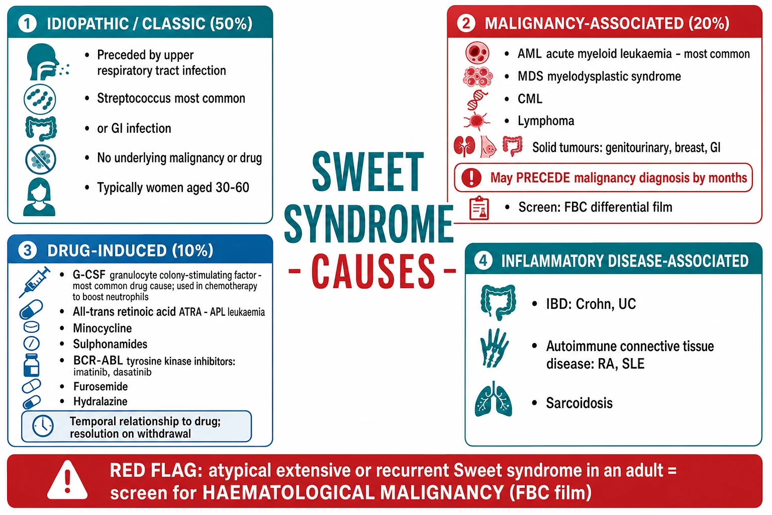
Two morphological-histological variants warrant separate mention because they alter the differential and the workup: [1]
- Histiocytoid Sweet syndrome — histology shows immature myeloid cells that mimic histiocytes but are in fact immature neutrophilic granulocytes (positive for MPO, CD15, CD68; negative for CD34 in mature forms). It is strongly associated with myelodysplastic syndrome and other myeloid neoplasms, and is the variant most easily confused with leukaemia cutis on biopsy — a critical distinction because management diverges completely.[7][8]
- Subcutaneous Sweet syndrome (neutrophilic panniculitis) — the neutrophilic infiltrate extends into the subcutis, presenting as tender subcutaneous nodules most often on the lower limbs, thighs, and arms. It is strongly associated with inflammatory bowel disease and must be distinguished histologically from erythema nodosum (septal pannulitis without neutrophilic predominance) and from Weber-Christian disease.[4]
Since 2020, VEXAS syndrome (vacuoles, E1 enzyme, X-linked, autoinflammatory, somatic) — caused by a somatic mutation in the UBA1 gene — is increasingly recognised as a unifying diagnosis in older men presenting with refractory or recurrent Sweet-like neutrophilic dermatosis, relapsing polychondritis, cytopenias, fever, and pulmonary infiltrates. Send UBA1 sequencing in this phenotype; treatment is IL-1 blockade (anakinra, canakinumab) or JAK inhibitors.[2][9]
Epidemiology & Risk Factors
Sweet syndrome is rare. No robust population incidence figures exist, but case series report a moderate female predominance overall (driven by the classical subtype) and a peak age of 30-60 years.[1][6]
The recognised triggers and risk factors by subtype are: [1]
| Category | Key triggers / associations |
|---|---|
| Infection | Upper respiratory tract infection (Streptococcus pyogenes — classical), GI infection (Yersinia, Salmonella, Campylobacter), hepatitis, tuberculosis, atypical mycobacteria, HIV seroconversion |
| Drugs | G-CSF (number one), all-trans retinoic acid (ATRA), minocycline, sulphonamides, BCR-ABL TKIs (imatinib, dasatinib), BCG vaccine, carbamazepine, azathioprine, furosemide, hydralazine, lenalidomide, bortezomib |
| Haematological malignancy | AML (most common), MDS (especially chronic histiocytoid Sweet), CML, lymphoma, hairy cell leukaemia, multiple myeloma |
| Solid malignancy | Genitourinary (prostate, renal, bladder), breast, gastrointestinal, ovarian, lung — much less common than haematological |
| Inflammatory disease | IBD (ulcerative colitis more than Crohn), rheumatoid arthritis, SLE, Sjögren syndrome, sarcoidosis, Behçet disease, relapsing polychondritis |
| Other | Pregnancy (rare; resolves postpartum), solid-organ transplant, sun exposure (photo-exacerbated variant) |
Pathophysiology
Sweet syndrome is best understood as a cytokine-driven hypersensitivity reaction in which aberrant innate immune signalling produces a runaway neutrophilic infiltrate in otherwise normal skin.[4]
The cytokine cascade. A distant trigger — infection, an IBD flare, malignant cells secreting cytokines, or an exogenous drug — activates skin-resident innate immune cells (keratinocytes, dendritic cells, macrophages) and bone-marrow myeloid precursors. They release a coordinated cytokine cocktail that includes interleukin-1 (IL-1), IL-3, IL-6, IL-8 (CXCL8), granulocyte colony-stimulating factor (G-CSF), GM-CSF, and TNF-α.[4][3] These cytokines have two effects: they expand the circulating neutrophil pool (G-CSF, GM-CSF) and they recruit mature neutrophils into the skin (IL-8 is the most potent neutrophil chemoattractant known). The result is a dense dermal infiltrate of mature neutrophils that — critically — does not destroy vessel walls.
Trigger
Infection (Streptococcus), IBD flare, malignancy-derived G-CSF, exogenous G-CSF, drug (ATRA), autoimmune inflammation
Cytokine storm
IL-1, IL-3, IL-6, IL-8/CXCL8, G-CSF, GM-CSF, TNF-α released by keratinocytes, dendritic cells, macrophages, myeloid precursors
Neutrophil expansion + activation
G-CSF expands marrow neutrophil pool; IL-8 recruits mature neutrophils into skin; IL-1 sustains activation
Dermal infiltration
Dense dermal neutrophilic infiltrate; papillary dermal oedema → pseudovesicular clinical appearance; vessel walls INTACT (no vasculitis)
Resolution
Trigger removed (drug stopped, infection resolves, malignancy treated) ± corticosteroid → neutrophils clear; lesions fade; usually no scarring
Why G-CSF is the #1 drug cause. Granulocyte colony-stimulating factor does exactly what Sweet syndrome does — it expands and activates the neutrophil pool. Exogenous G-CSF (filgrastim, pegfilgrastim, lenograstim) given for chemotherapy-induced neutropenia or stem-cell mobilisation can therefore directly precipitate Sweet syndrome by driving the effector arm of the cascade. Endogenous G-CSF is also elevated in classical disease, supporting its pathogenic role.[3]
Why IL-1 is central. IL-1 is upstream of the entire cascade — it drives the release of IL-6, IL-8, and other neutrophil-recruiting chemokines. This is the mechanistic basis for the efficacy of IL-1 blockade (anakinra, canakinumab) in refractory Sweet syndrome and in VEXAS syndrome.[2][3]
The paraneoplastic mechanism. In malignancy-associated Sweet syndrome, the tumour itself is the source of the cytokines — AML and MDS blasts can secrete G-CSF and IL-1, which activate neutrophils and drive the skin infiltrate. This explains why successful chemotherapy (which removes the cytokine source) often resolves the skin lesions even without corticosteroids.[8]
Pathergy. Skin trauma — venepuncture, biopsy, sunburn, BCG vaccination — can trigger a Sweet lesion at that site, mirroring the pathergy of pyoderma gangrenosum and Behçet disease. The mechanism is local neutrophil recruitment in a primed, hyper-reactive innate immune system.[4]
Why NO vasculitis. Although neutrophils dominate the infiltrate, they localise to the dermal interstitium and papillary dermis rather than to vessel walls. The endothelium remains intact; there is no fibrinoid necrosis of vessel walls and no destructive leukocytoclastic vasculitis. Nuclear dust (karyorrhexis) may be present, but this reflects normal neutrophil turnover, not vessel-wall destruction. This single histological fact — vessel walls intact — is the bedrock distinction from cutaneous small-vessel vasculitis and is the most-tested fact on this topic.[4]
VEXAS syndrome — a genetic paradigm. Since 2020, VEXAS syndrome is recognised as a unifying diagnosis in older men (somatic mutation — by definition acquired, in a single X chromosome) presenting with a refractory Sweet-like neutrophilic dermatosis, relapsing polychondritis, cytopenias (macrocytic anaemia, thrombocytopenia), fever, and pulmonary infiltrates. The mutation is in UBA1, encoding the E1 ubiquitin-activating enzyme; loss of cytoplasmic ubiquitination triggers constitutive innate immune activation. Treatment is IL-1 blockade or JAK inhibition.[2][9]
Clinical Presentation
The clinical signature is a sudden onset of painful, erythematous to violaceous skin lesions in a systemically unwell, febrile patient. The lesions evolve over hours to days, not weeks.[1][4]
Morphology. The lesions are tender papules, plaques, and nodules with a smooth, often pseudovesicular surface — they look as if they might be vesicles or bullae, but on palpation are solid (the appearance comes from marked papillary dermal oedema). True vesiculation and bullae occur in bullous/atypical Sweet, which is more often malignancy-associated. The colour is bright red to deep violaceous (purple), and lesions can coalesce into polycyclic plaques. [1]
Distribution. The classical distribution is upper extremities (arms, dorsa of hands, fingers), face, neck, and upper trunk. The lower legs are involved in the subcutaneous variant (neutrophilic panniculitis). Mucosal surfaces are usually spared — though oral aphthous ulcers occur in a minority; severe mucosal involvement should prompt consideration of Stevens-Johnson syndrome or Behçet disease instead.[1]
Pathergy. New lesions appear at sites of trauma — venepuncture, intravenous cannulae, biopsy sites, insect bites, sunburn, or BCG vaccination. A pathergic reaction at the BCG site is a classical exam stem.[4]
Systemic features. Fever (low- to high-grade) precedes or accompanies the rash in ~80%. Malaise, myalgia, arthralgia, and headache are common. Ocular involvement occurs in ~one-third — conjunctivitis, episcleritis, scleritis, iridocyclitis, and peripheral keratitis. Articular symptoms (arthralgia or true arthritis) are common and can precede the rash.[4][6]
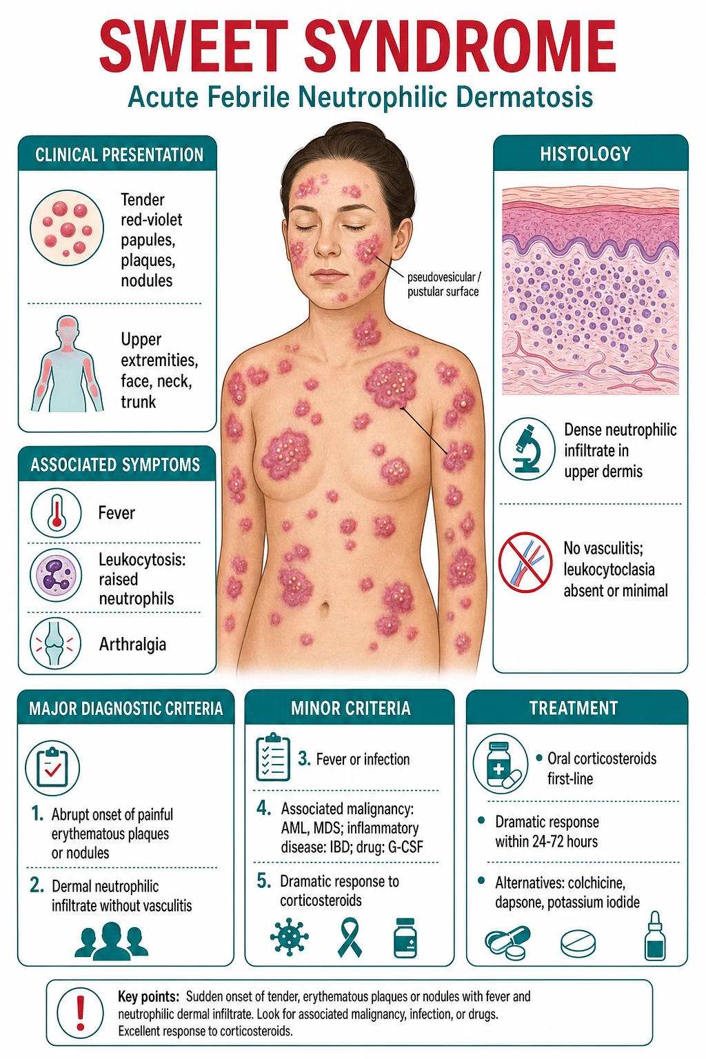
Extracutaneous internal organ involvement is increasingly recognised and can affect almost any organ: [1]
| Organ system | Manifestation |
|---|---|
| Bones | Sterile osteomyelitis / neutrophilic osteitis (typically long bones); positive bone scan, negative cultures |
| Lungs | Neutrophilic pneumonitis — bilateral pulmonary infiltrates, pleural effusion; biopsy shows neutrophilic infiltrate |
| Kidneys | Sterile neutrophilic interstitial nephritis; may cause acute kidney injury |
| Liver / spleen | Hepatosplenomegaly; neutrophilic hepatic infiltrate |
| GI tract | Neutrophilic enterocolitis; overlaps with active IBD |
| CNS | Sterile meningitis, encephalitis, cerebellar ataxia (rare) |
| Eyes | Conjunctivitis, episcleritis, scleritis, iridocyclitis, peripheral ulcerative keratitis |
| Heart | Rare — aortitis, neutrophilic myocarditis |
Morphology in malignancy-associated disease. Lesions may be bullous, ulcerative, or atypical, may appear in unusual sites, or may recur despite corticosteroid — each should escalate the malignancy workup.[8]
Sweet syndrome in children. Rare but well described; more often classical and post-infectious (after viral illness or vaccination). The histology and criteria are identical; weight-based dosing applies. Always screen for malignancy even in children, as paediatric AML can present with Sweet syndrome.[6]
Differential Diagnosis
Because the differential includes true emergencies (Stevens-Johnson syndrome, leukaemia cutis, necrotising fasciitis) and conditions whose management is the opposite of Sweet's (cellulitis treated with antibiotics; vasculitis treated with immunosuppression AFTER infection excluded), the differential is the highest-yield part of the viva. [1]
Clinical & Bedside Assessment
The bedside assessment of a patient with suspected Sweet syndrome has two parallel goals: confirm the diagnosis (apply Su & Liu criteria; biopsy) and find the trigger (infection, drug, malignancy, IBD, autoimmune disease). Both must be done before starting corticosteroids. [1]
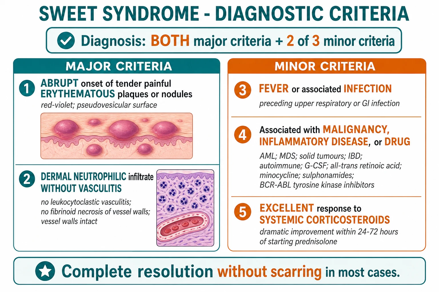
Su & Liu (1986) diagnostic criteria, modified by von den Driesch (1991) — the international diagnostic standard. Diagnosis requires BOTH major criteria AND at least 2 of the 4 minor criteria.[1][3]
| Criterion | Detail |
|---|---|
| **MAJOR 1** | Abrupt onset of **tender or painful erythematous plaques or nodules** |
| **MAJOR 2** | **Dense dermal neutrophilic infiltrate WITHOUT primary vasculitis** (no leukocytoclastic vasculitis; no fibrinoid necrosis of vessel walls) |
| **MINOR 1** | **Fever** or associated **infection** (preceding upper respiratory or GI infection) |
| **MINOR 2** | Associated with **malignancy** (AML, MDS), **inflammatory disease** (IBD, autoimmune), or **drug** (G-CSF, ATRA, minocycline, BCG) |
| **MINOR 3** | Excellent response to **systemic corticosteroids** (dramatic improvement within 24-72 hours) OR to **potassium iodide** |
| **MINOR 4 (von den Driesch)** | **Neutrophilia** on full blood count (absolute neutrophil count above the upper limit of normal) |
Bedside approach: [1]
- Confirm the morphology — abrupt tender red-violet papules/plaques/nodules; pseudovesicular surface; classical distribution.
- Document pathergy — ask about new lesions at sites of venepuncture, cannulae, biopsy, insect bites, sunburn, vaccination; a biopsy-site lesion strongly supports Sweet.
- Screen for the trigger — recent URTI, GI illness, IBD flare, autoimmune disease, pregnancy, drug history (G-CSF, ATRA, minocycline, sulphonamides, BCG, BCR-ABL TKIs), vaccination.
- Screen for malignancy — FBC with differential, peripheral film; consider bone marrow biopsy in any atypical, recurrent, or malignancy-suspicious case.
- Exclude mimics — biopsy to rule out vasculitis, leukaemia cutis, infection; cultures to rule out cellulitis.
- Document extracutaneous disease — eye exam, joint exam, chest exam (pulmonary infiltrates), abdominal exam (hepatosplenomegaly). [1]
Investigations
Investigations serve three purposes: confirm the diagnosis (biopsy), exclude the mimics (biopsy, cultures, immunohistochemistry), and identify the trigger (especially malignancy). [1]
Skin biopsy (the single most important investigation). A punch biopsy of a representative lesion is essential. The histology of classical Sweet syndrome shows:[4][6]
- Dense diffuse dermal neutrophilic infiltrate, predominantly in the upper and mid-dermis
- Marked papillary dermal oedema — gives the pseudovesicular clinical appearance
- Karyorrhexis (nuclear dust) — present but reflects normal neutrophil turnover, NOT vessel destruction
- Vessel walls INTACT — no fibrinoid necrosis, no leukocytoclastic vasculitis (the cardinal distinguishing feature)
- Negative special stains for organisms (PAS, Grocott, Ziehl-Neelsen, Fite) — exclude infection
- In histiocytoid Sweet: immature myeloid cells mimicking histiocytes; MPO+, CD15+, CD68+, CD34 variable; the cells are immature neutrophils, not true histiocytes, and not leukaemic blasts — but the differential with leukaemia cutis requires careful immunophenotyping and bone marrow biopsy.[7][8]
Blood tests. Full blood count with differential typically shows neutrophilia (one of the minor criteria). In malignancy-associated disease the count may be normal, low, or show cytopenias/blasts. ESR and CRP are elevated. U&E, LFTs screen for renal and hepatic involvement.[1]
Microbiology. Skin and blood cultures are STERILE — this is expected and helps exclude infection. Swab any pustular lesions to exclude secondary infection. Throat swab and ASO titre if Streptococcus is suspected; stool culture and serology if Yersinia, Salmonella, Campylobacter suspected.[4]
Autoimmune/serological tests (largely to exclude mimics). ANA, ENA, ANCA (typically negative — distinguishes from ANCA-associated vasculitis), RF, complement; consider hepatitis B/C and HIV serology. SPEP and serum free light chains if a monoclonal gammopathy is suspected (overlap with PG).[4]
Malignancy workup — mandatory in adults. In any adult with atypical, recurrent, or untriggered Sweet syndrome, screen for haematological malignancy:[4][8]
- FBC with differential + peripheral blood film — first-line
- Bone marrow aspirate and trephine — if cytopenias, blasts, dysplasia, or strong clinical suspicion
- LDH, beta-2 microglobulin — non-specific markers of myeloproliferative activity
- Age-appropriate cancer screening — CXR, mammography, colonoscopy, prostate assessment as indicated
- UBA1 sequencing — if VEXAS phenotype (older male, refractory/recurrent neutrophilic dermatosis, chondritis, cytopenias)[2][9]
IBD workup. If GI symptoms or subcutaneous variant — stool calprotectin, faecal culture, colonoscopy with biopsies (ulcerative colitis and Crohn's disease are the strongest IBD associations). [1]
Imaging for extracutaneous disease. CXR or CT chest if pulmonary infiltrates suspected; MRI or bone scan for suspected sterile osteomyelitis; CT abdomen for hepatosplenomegaly. [1]
Management — Resuscitation
Sweet syndrome is not a dermatological emergency in the same sense as Stevens-Johnson syndrome or toxic epidermal necrolysis, but several principles govern the immediate management: [1]
Exclude infection FIRST
Skin and blood cultures, bacterial/viral/fungal/atypical mycobacterial stains on biopsy. Do NOT start corticosteroids while sepsis is unaddressed — masking infection is catastrophic.
Exclude mimics
Biopsy before steroids if possible. Confirm no leukaemia cutis (immunohistochemistry) and no vasculitis (vessel walls intact).
Baseline investigations
FBC, U&E, LFTs, glucose, blood pressure, weight; G6PD if dapsone contemplated; pregnancy test in women of childbearing age; bone density if long steroid course likely.
Identify and address the trigger
Stop the causative drug (G-CSF, ATRA, minocycline). Treat infection. Refer to haematology if malignancy suspected. Treat IBD flare.
Decide setting
Outpatient for localised classical disease; inpatient for extensive disease, diagnostic uncertainty, systemic unwellness, IV therapy, or malignancy workup.
Management — Definitive & Stepwise
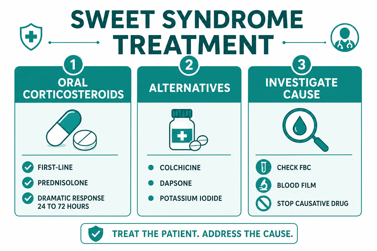
The treatment ladder for Sweet syndrome reflects the disease's dramatic corticosteroid responsiveness and the importance of treating the underlying cause. Therapy is stratified by subtype.[1][3]
First-line: systemic corticosteroids
Oral prednisolone 0.5-1 mg/kg/day is first-line for classical Sweet syndrome. The response is dramatic and characteristic — fever settles within 24-72 hours, lesions flatten and fade, no new lesions appear. Because this dramatic response is one of the diagnostic minor criteria, observing it both confirms the diagnosis and treats the disease. [1]
Taper over 2-6 weeks (some authorities recommend tapering over 4-6 weeks to reduce the ~30% recurrence rate). Tapering too quickly is the most common cause of recurrence.[1]
For severe, extensive, or rapidly progressive disease, intravenous methylprednisolone 0.5-1 g daily for 3-5 days is an option, followed by oral prednisolone.[3]
Prednisolone (oral)
Dose
0.5-1 mg/kg/day (typically 30-60 mg daily in an adult)
Steroid-sparing and alternative agents
For patients who cannot take corticosteroids (diabetes, osteoporosis, active infection, pregnancy), who relapse on taper, or who have recurrent disease, several alternatives are effective. The choice is often regional (EADV favours colchicine and potassium iodide; AAD literature favours early IL-1 blockade).[3]
| Agent | Dose | Mechanism | Cautions |
|---|---|---|---|
| **Colchicine** | 0.5 mg BD-TDS oral | Inhibits neutrophil chemotaxis and microtubule function | GI upset; myelosuppression; avoid in pregnancy and renal impairment |
| **Dapsone** | 50-150 mg daily oral | Inhibits neutrophil myeloperoxidase and ROS | **Check G6PD first**; haemolysis, methaemoglobinaemia; monitor FBC monthly |
| **Ciclosporin** | 3-5 mg/kg/day oral in 2 divided doses | Inhibits T-cell activation and downstream neutrophil recruitment | Nephrotoxicity, hypertension; monitor U&E, BP, ciclosporin levels |
| **Potassium iodide** | 300-900 mg TDS oral | Inhibits neutrophil chemotaxis; suppresses the hypersensitivity reaction | Hypothyroidism; Wolff-Chaikoff effect; avoid in hyperkalaemia |
| **Anakinra** | 100 mg SC daily | **IL-1 receptor antagonist** — blocks the central upstream cytokine | Injection-site reaction; infection (especially TB); IL-1 blockade is first-line for VEXAS |
| **Canakinumab** | 150 mg SC every 8 weeks (weight-based in children) | Monoclonal anti-IL-1β | Infection; cost; reserved for refractory disease and VEXAS |
| **Indomethacin** | 25-50 mg TDS oral | Non-steroidal anti-inflammatory; inhibits prostaglandin-mediated neutrophil activation | GI bleed; renal impairment; asthma |
| **IVIG** | 2 g/kg per cycle over 2-5 days, every 3-4 weeks | Immunomodulatory; multiple mechanisms | Headache, infusion reaction, thrombosis; preferred in pregnancy |
Topical and intralesional corticosteroids
For localised, mild disease, super-potent topical corticosteroid (clobetasol 0.05% BD for 2-4 weeks) or intralesional triamcinolone may suffice as monotherapy or as adjunctive therapy to systemic treatment. Topical therapy is the preferred first agent in pregnancy.[1]
Management by subtype
Classical/idiopathic — oral prednisolone 0.5-1 mg/kg/day, taper over 4-6 weeks; colchicine or potassium iodide as a steroid-sparing adjunct in recurrent disease. Treat any identified trigger (Streptococcus — penicillin V; IBD flare — gastroenterology).[1]
Malignancy-associated — treat the underlying malignancy with the oncology team; successful chemotherapy frequently resolves the skin lesions without corticosteroids. Use systemic corticosteroids for symptomatic skin disease, but cautiously in marrow-failure patients (sepsis risk). Recurrence of Sweet may herald malignancy relapse.[8]
Drug-induced — stop the causative drug (most often G-CSF); lesions resolve on withdrawal over days to weeks. A short course of oral prednisolone (e.g. 0.5 mg/kg/day tapering over 2 weeks) accelerates resolution. Re-exposure should be avoided where possible.[1]
Histiocytoid Sweet — same corticosteroid ladder, but mandatory haematology workup for underlying myelodysplasia; IL-1 blockade is increasingly reported as effective.[7]
VEXAS syndrome — anakinra (IL-1 blockade) or JAK inhibitors (ruxolitinib) are now considered the treatment of choice, given the constitutive innate immune activation; corticosteroids alone are usually insufficient and high-dose steroids carry high morbidity in older patients.[2][9]
Specific Subtypes & Scenarios
Classical/idiopathic Sweet syndrome (~70%). Predominantly affects women aged 30-60, often 1-3 weeks after an upper respiratory or GI infection. Streptococcus is the classical organism; the disease is exquisitely corticosteroid-responsive. Recurrence in ~30%. The workup is largely to exclude malignancy, drug, and IBD.[1][6]
Malignancy-associated Sweet syndrome (~20%). AML is the single most common underlying malignancy, followed by MDS (especially in the histiocytoid variant). Less commonly CML, lymphoma, hairy cell leukaemia, multiple myeloma. Solid tumours (prostate, renal, bladder, breast, GI, ovarian, lung) account for ~15% of the malignancy-associated group. Sweet syndrome may precede the malignancy by months (paraneoplastic herald), co-occur, or relapse with tumour relapse. The morphology is often bullous or atypical. Mandatory workup: FBC + film + bone marrow biopsy if cytopenias/blasts/dysplasia. No female predominance (1:1).[8]
Drug-induced Sweet syndrome (~10%). G-CSF is the number one culprit — by a wide margin. Others include all-trans retinoic acid (ATRA) for acute promyelocytic leukaemia, minocycline, sulphonamides, BCR-ABL tyrosine kinase inhibitors (imatinib, dasatinib), BCG vaccine, carbamazepine, azathioprine, furosemide, hydralazine, lenalidomide, bortezomib. Diagnosis rests on temporal association and resolution on drug withdrawal.[1]
Histiocytoid Sweet syndrome. The histology shows immature myeloid cells with histiocyte-like morphology but a myeloid immunophenotype (MPO+, CD15+, CD68+). It is strongly associated with myelodysplastic syndrome and other myeloid neoplasms and is the variant most easily confused with leukaemia cutis on biopsy. Distinction rests on immunohistochemistry (CD34, CD117/KIT, blast markers) and bone marrow examination.[7][8]
Subcutaneous Sweet syndrome (neutrophilic panniculitis). Presents as tender subcutaneous nodules on the thighs, arms, and lower legs (more than shins). Histology shows a neutrophilic lobular panniculitis. Strongly associated with inflammatory bowel disease — distinguish from erythema nodosum (septal panniculitis with lymphohistiocytic infiltrate, predominantly shins).[4]
Sweet syndrome in pregnancy. Rare; typically in the second or third trimester; resolves postpartum but may recur in subsequent pregnancies. Management: super-potent topical corticosteroid as first-line; if systemic therapy needed, IVIG (2 g/kg) is the safest systemic agent, with low-dose prednisolone as second-line. Avoid colchicine (teratogenicity concerns), ciclosporin in first trimester, and dapsone (haemolysis in glucose-6-phosphate dehydrogenase deficiency and fetal risk).[6]
VEXAS syndrome (UBA1). A somatic mutation in UBA1 (the E1 ubiquitin-activating enzyme) in haematopoietic cells of older men produces a syndrome of refractory neutrophilic dermatosis (Sweet-like or PG-like), relapsing polychondritis, cytopenias (macrocytic anaemia, thrombocytopenia), fever, and pulmonary infiltrates. Skin manifestations are the most common presenting feature. Send UBA1 sequencing in any older man with refractory neutrophilic dermatosis + cytopenias + chondritis. Treatment: anakinra (IL-1 blockade), canakinumab, or JAK inhibitors (ruxolitinib) — corticosteroids alone are typically inadequate.[2][9]
Complications & Pitfalls
- Cutaneous complications — secondary bacterial infection of broken skin; postinflammatory hyperpigmentation (especially in darker skin types); scarring is uncommon in classical disease but may occur in bullous/atypical or malignancy-associated variants.
- Recurrence (~30%) — the most common complication; predicted by rapid corticosteroid taper, untreated underlying cause (especially malignancy relapse or ongoing drug exposure), and possibly a more severe initial phenotype.
- Corticosteroid morbidity — for the 4-6 week typical course, gastritis, glucose dysregulation, mood change, insomnia; for repeated courses, osteoporosis, hypertension, cataract, adrenal suppression, infection. Provide bone protection for courses over 3 months.
- Misdiagnosis of leukaemia cutis as histiocytoid Sweet — a critical pitfall. Both present with papules/plaques/nodules; both can show immature myeloid cells on biopsy. The distinction rests on immunophenotyping (CD34, CD117/KIT) and bone marrow examination. Treating leukaemia cutis with corticosteroids delays oncology referral.[7][8]
- Treating presumed cellulitis with antibiotics — delays corticosteroid initiation; Sweet is culture-negative and worsens without corticosteroids. Biopsy and cultures settle the issue.
- Starting immunosuppression without excluding malignancy — masks haematological malignancy; risks sepsis in marrow-failure patients; misses the opportunity to treat the underlying cause.
- Systemic complications — sterile osteomyelitis, neutrophilic pneumonitis, ocular scarring, neutrophilic interstitial nephritis, splenic infiltration, rare CNS involvement (sterile meningitis, encephalitis).
Prognosis & Disposition
Classical Sweet syndrome has an excellent prognosis: corticosteroids produce a dramatic response within 24-72 hours, and most cases resolve completely over 4-6 weeks without scarring. Recurrence occurs in ~30%, typically within months to a few years; recurrent disease should always prompt reassessment for an underlying cause.[1][3]
Malignancy-associated Sweet syndrome has a prognosis tied to the underlying malignancy: Sweet often resolves with successful cancer treatment and may relapse with tumour relapse (a useful clinical marker). Skin involvement per se does not alter the malignancy prognosis.[8]
Drug-induced Sweet syndrome has the best prognosis of all — lesions resolve on drug withdrawal, typically within days to weeks; a short steroid course accelerates resolution.[1]
VEXAS syndrome has a more guarded prognosis — it is a chronic relapsing autoinflammatory disease with significant morbidity and a reported 1-year mortality of ~10-20% in early cohorts, driven by pulmonary, haematological, and cardiovascular complications. Early IL-1/JAK blockade improves outcomes.[2]
Disposition. Stable Sweet syndrome is managed in the outpatient dermatology clinic with multidisciplinary input from haematology (malignancy workup), gastroenterology (IBD), rheumatology (autoimmune, VEXAS), and ophthalmology (ocular involvement). A documented flare action plan — early presentation, photography, biopsy of any new lesion, swab and culture, and escalation pathway — reduces avoidable admissions. Inpatient admission is reserved for extensive or rapidly progressive disease, diagnostic uncertainty, intravenous therapy, and acute systemic involvement. [1]
Special Populations
Children. Sweet syndrome is rare in children but well described; more often classical and post-infectious (after viral illness or vaccination, including BCG). The histology and diagnostic criteria are identical to adults. Malignancy must still be excluded — paediatric AML can present with Sweet syndrome. Use weight-based dosing for prednisolone (0.5-1 mg/kg/day); topical corticosteroids for localised disease. Colchicine and dapsone (after G6PD check) are useful adjuncts.[6]
Pregnancy. Rare in pregnancy; typically presents in the second or third trimester. Resolves postpartum but may recur in subsequent pregnancies. Management: super-potent topical corticosteroid (clobetasol 0.05%) is first-line. If systemic therapy is needed, IVIG (2 g/kg per cycle) is the safest systemic agent in pregnancy. Low-dose oral prednisolone (after the first trimester) is acceptable second-line. Avoid colchicine (teratogenicity concerns), ciclosporin in the first trimester, dapsone (haemolysis risk and fetal considerations), and high-dose systemic corticosteroids in the first trimester (oral cleft concern).[6]
Elderly. More often malignancy-associated (AML/MDS), more often histiocytoid variant, and more likely to have the VEXAS phenotype (older man with cytopenias and chondritis). Steroid-sparing agents are particularly valuable in the elderly given the cumulative morbidity of corticosteroids (osteoporosis, diabetes, hypertension, infection).[8][9]
Immunocompromised. Sweet syndrome in the setting of chemotherapy-induced neutropenia, post-transplant immunosuppression, or HIV is more often malignancy-associated or drug-induced (G-CSF). Corticosteroid use must be cautious; biopsy to exclude infection is even more critical. [1]
Evidence, Guidelines & Regional Differences
Landmark papers and what they changed: [1]
Su WPD, Liu HNH. Diagnostic criteria for Sweet's syndrome. Cutis 1986;37:167-169.
Key finding
Established the **2 major + 2 of 3 minor criteria** that remain the international diagnostic standard. The most-cited diagnostic framework for Sweet syndrome.
von den Driesch P. Sweet's syndrome (acute febrile neutrophilic dermatosis). J Am Acad Dermatol 1991;27:227-232.
Key finding
Modified Su & Liu to add **neutrophilia as a fourth minor criterion**; reorganised the clinical subtypes (classical, malignancy-associated, drug-induced) that we still use today.
Requena L, et al. Histiocytoid Sweet syndrome. Arch Dermatol 2005;141:833-839. (PMID 16027297)
Key finding
Defined **histiocytoid Sweet syndrome** as a dermal infiltrate of immature neutrophilic granulocytes with histiocyte-like morphology (MPO+, CD15+, CD68+); strongly associated with myelodysplasia.
Beck DB, et al. Somatic mutations in UBA1 and adult-onset inflammatory disease (VEXAS syndrome). NEJM 2020;383:2628-2638.
Key finding
Identified somatic **UBA1** mutations as the cause of VEXAS syndrome — a unifying diagnosis for older men with refractory neutrophilic dermatosis, relapsing polychondritis, cytopenias, and pulmonary infiltrates.
Regional guideline differences: [1]
[1] [1] [1]Indian and South-Asian practice — dapsone (after G6PD check), colchicine, and ciclosporin are cheap, widely available steroid-sparers. Anakinra access is limited to tertiary centres. Skin biopsy with proper stains to exclude infection (leprosy, cutaneous tuberculosis) is particularly important given regional infection prevalence.
Controversies:
- The optimal steroid-sparing agent is not established; head-to-head trials do not exist.
- Whether IL-1 blockade should be first-line in classical Sweet syndrome (rather than a steroid-sparing option) is debated — IL-1 is mechanistically central, but cost and infection risk keep corticosteroids first-line for most authorities.
- Duration of maintenance therapy for recurrent disease is undefined; pragmatic approaches use 3-6 months of a steroid-sparer with taper.
- The role of anakinra in VEXAS syndrome is now fairly well established, but the role of JAK inhibitors (ruxolitinib) is evolving. [1]
Exam Pearls
SWEET
GAMPS
Red Flags
Exam application bank (NEET-PG / INICET)
One-line answer
Sweet syndrome (acute febrile neutrophilic dermatosis) = abrupt onset of tender, red-violet papules/plaques/nodules + fever + neutrophilia + dense dermal neutrophilic infiltrate without vasculitis on histology. Su & Liu (1986) diagnostic criteria — 2 MAJOR + 2 of 4 MINOR (von den Driesch modification): major = abrupt tender erythematous plaques + dermal neutrophilic infiltrate without vasculitis; minor = fever/infection, malignancy (AML/MDS)/IBD/drug (G-CSF), response to corticosteroids. Classification: classical/idiopathic ~70%, malignancy-associated ~20% (AML/MDS most common), drug-induced ~10% (G-CSF #1). Treatment: oral corticosteroids (prednisolone 0.5-1 mg/kg/day → dramatic response 24-72h). Alternatives: colchicine, dapsone, ciclosporin, potassium iodide, anakinra. Red flag: atypical/recurrent Sweet syndrome in an adult → screen for haematological malignancy (FBC + film). [1]
Worked stems (answer without another resource)
Stem 1 — Classic presentation. Map symptoms to mechanism; name the first investigation and first treatment step with dose/route if drug therapy is standard. [1]
Stem 2 — Unstable / complicated. List red flags that force immediate resuscitation, theatre, ICU, antidote, or reperfusion — and what you do in the first 15 minutes. [1]
Stem 3 — Atypical group. Elderly, pregnancy, child, or immunocompromised: how presentation and thresholds change. [1]
Stem 4 — Differential trap. Name the three closest mimics and one discriminator for each. [1]
Stem 5 — Disposition. Who goes home with safety-netting, who is admitted, who needs HDU/ICU/theatre, and what follow-up is mandatory. [1]
Rapid viva checklist
- Definition + classification
- Pathophysiology chain
- Bedside signs / criteria
- Score with exact components (if any)
- Emergency bundle
- Definitive therapy with doses
- Complications of disease and of treatment
- Special populations
- Guideline/trial name if classic
- Three exam traps
Coverage self-check
If you cannot answer any stem above from this page alone, re-read the matching section — the page is intended to be self-sufficient for final-prof and NEET-PG/INICET questions on Sweet syndrome.
References
- [1]Villarreal-Villarreal CD, Ocampo-Candiani J, Villarreal-Martínez A. Sweet Syndrome: A Review and Update Actas Dermosifiliogr, 2016.PMID 26826881
- [2]Loeza-Uribe MP, Hinojosa-Azaola A, Sánchez-Hernández BE, et al. VEXAS syndrome: Clinical manifestations, diagnosis, and treatment Reumatol Clin (Engl Ed), 2024.PMID 38160120
- [3]Calabrese L, Satoh TK, Aoki R, et al. Sweet syndrome: an update on clinical aspects, pathophysiology, and treatment Ital J Dermatol Venerol, 2024.PMID 39560338
- [4]Nelson CA, Stephen S, Ashchyan HJ, et al. Neutrophilic dermatoses: Pathogenesis, Sweet syndrome, neutrophilic eccrine hidradenitis, and Behçet disease J Am Acad Dermatol, 2018.PMID 29653210
- [5]Delaleu J, Lepelletier C, Calugareanu A, et al. Neutrophilic dermatoses Rev Med Interne, 2022.PMID 35870984
- [6]Cohen PR. Sweet's syndrome--a comprehensive review of an acute febrile neutrophilic dermatosis Orphanet J Rare Dis, 2007.PMID 17655751
- [7]Requena L, Kutzner H, Palmedo G. Histiocytoid Sweet syndrome: a dermal infiltration of immature neutrophilic granulocytes Arch Dermatol, 2005.PMID 16027297
- [8]Imoto T, Marzano AV, Borghi A, et al. Neutrophilic and eosinophilic dermatoses associated with hematological malignancy Front Med (Lausanne), 2023.PMID 38249974
- [9]Puléyro-Pérez CA, Hebart C, Moura B, et al. Skin Manifestations of VEXAS Syndrome and Associated Genotypes JAMA Dermatol, 2024.PMID 38865133