Dermatology · Medicine
Syphilis (cutaneous manifestations)
Also known as Syphilis · Lues · The great imitator · Treponema pallidum infection · Condylomata lata · Chancre · Gummatous syphilis
Syphilis is a chronic systemic sexually transmitted infection caused by Treponema pallidum subsp. pallidum (a spirochaete), characterised by distinct clinical stages: primary (painless chancre with clean base and regional lymphadenopathy), secondary (polymorphic rash on palms and soles, condylomata lata, mucous patches, moth-eaten alopecia, generalised lymphadenopathy), latent (asymptomatic with positive serology), and tertiary (gummas, cardiovascular and neurosyphilis). Cutaneous manifestations are the hallmark of secondary and tertiary disease and earned syphilis the epithet 'the great imitator'. Serology: non-treponemal tests (RPR/VDRL) for screening and treatment monitoring; treponemal tests (FTA-ABS, TPPA, treponemal EIA) for confirmation. Treatment: benzathine penicillin G 2.4 MU IM for early disease; IV penicillin for neurosyphilis. Jarisch-Herxheimer reaction within 6-12 hours of treatment.
On this page & tools
Your progress
Saved locally on this device.
Exam tags
Red flags
Definition and overview
Syphilis is a chronic, systemic, sexually transmitted infection caused by Treponema pallidum subsp. pallidum, a thin spirochaete (spiral-shaped bacterium, 6–20 µm long × 0.1–0.2 µm wide) that cannot be cultured on artificial media. The disease progresses through well-defined clinical stages — primary, secondary, latent, and tertiary — each with characteristic mucocutaneous manifestations. Cutaneous involvement is most prominent in secondary and tertiary syphilis and is so polymorphic that syphilis has earned the epithet "the great imitator."[1][2]
Epidemiology
- Resurgent globally since the early 2000s, particularly in MSM (men who have sex with men), persons living with HIV, and young heterosexual adults.[1]
- Congenital syphilis has surged dramatically (10× increase in some regions) due to inadequate maternal screening and treatment.[4]
- Transmission: sexual contact (vaginal, anal, oral) via mucous membranes or abraded skin; transplacental (congenital syphilis); rarely by blood transfusion or needlestick.
- Incubation period: 9–90 days (average 21 days) for primary chancre.
Pathophysiology [1]
T. pallidum penetrates intact mucous membranes or abraded skin and disseminates via the lymphatic system and bloodstream within hours to days, even before the primary chancre appears. The organism has a predilection for small blood vessels (arterioles and venules), producing an obliterative endarteritis and periarteritis — the histopathological hallmark of syphilis at all stages. The organism triggers a Th1 cellular immune response (delayed-type hypersensitivity) followed by a Th2 humoral response (antibody production), but T. pallidum evades complete clearance through antigenic variation of its surface proteins, allowing persistence and progression to later stages.[1]
Primary syphilis
- Chancre: a single, painless, indurated ulcer (the hallmark lesion) appearing at the site of inoculation (genital, anal, or oral) after an incubation of 9–90 days (mean 21 days).
- Appearance: clean-based, punched-out, indurated (button-like) border; 0.5–2 cm diameter; exudes clear serum rich in treponemes.
- Painlessness is a key distinguishing feature from painful genital ulcers (herpes, chancroid, Behçet's).
- Accompanied by regional lymphadenopathy: firm, rubbery, non-tender, bilateral (inguinal for genital chancre; cervical for oral).[1]
- Heals spontaneously in 3–6 weeks without treatment (but infection progresses systemically).
- Diagnosis: dark-field microscopy of chancre exudate (motile spirochaetes); PCR of lesion swab (more sensitive).[5] Serology may be negative early in primary syphilis.
Secondary syphilis
Occurs 2–8 weeks after the chancre heals (or may overlap with it), representing systemic dissemination of treponemes. The cutaneous manifestations are polymorphic — earning syphilis the name "the great imitator".[1][2]
Cutaneous manifestations
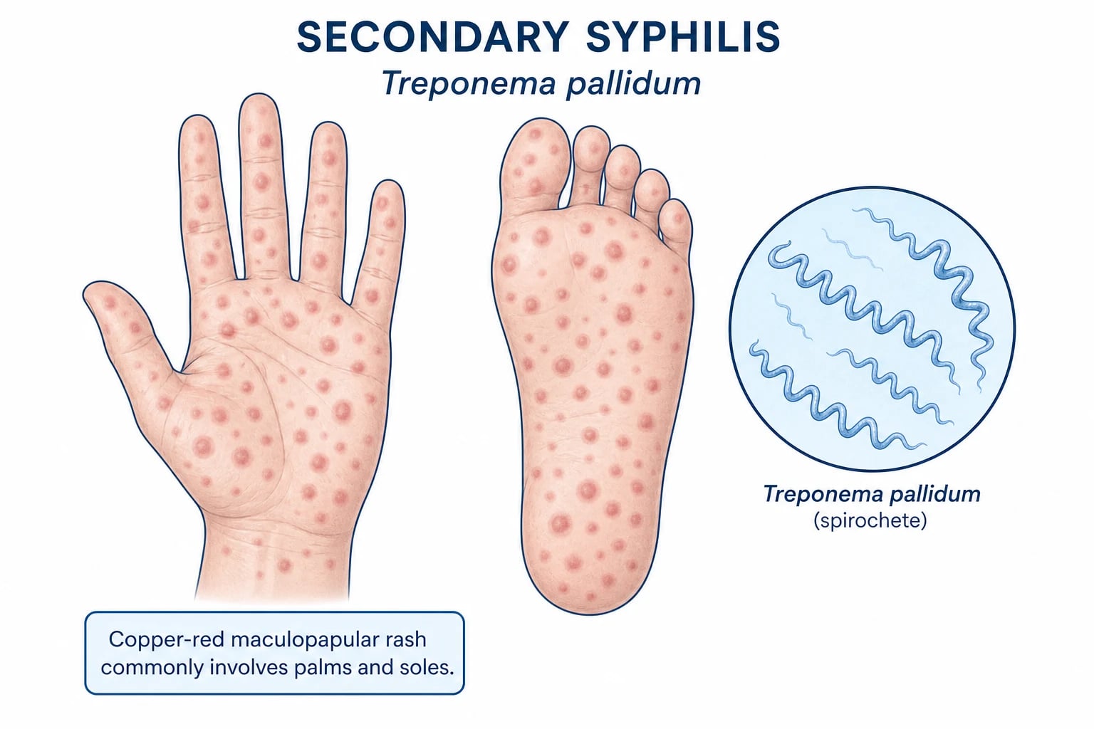
- Maculopapular rash (the classic sign): copper-red (also described as "ham-coloured") macules and papules that begin on the trunk and spread to the extremities, including the palms and soles — palmoplantar involvement is the hallmark of secondary syphilis and should always prompt serological testing.[1][2]
- Condylomata lata: broad, moist, fleshy, grey-white, warty papules/plaques in warm, moist intertriginous areas — perianal, vulval, scrotal, and inguinal folds. Highly infectious (teeming with spirochaetes). Distinct from condylomata acuminata (viral warts, which are dry and keratotic).[7]
- Mucous patches: painless, grey-white, slightly raised plaques on the oral mucosa, tongue, and lips; described as "snail-track" ulcers. Highly infectious.
- Alopecia: moth-eaten (patchy, non-scarring) alopecia of the scalp and beard — a characteristic pattern; eyebrows may also be affected.
- Other cutaneous features: papulosquamous lesions mimicking psoriasis or lichen planus; pustular lesions (malignant syphilis in HIV); annular lesions; split papules at the angles of the mouth; linear hyperkeratotic plaques (clavi syphilitici).
- Generalised lymphadenopathy: firm, non-tender, rubbery nodes.
SECONDARY SKIN signs of syphilis
SECONDARY
Oral mucous patches — shallow grey-white erosions on lips, tongue, buccal mucosa
Copper-red maculopapular rash — the hallmark of secondary syphilis
Moist fleshy anogenital plaques — teeming with treponemes
Mouth-angle fissuring, paronychia, syphilitic dactylitis
Rubbery, non-tender lymphadenopathy is the rule
Patchy non-scarring scalp and eyebrow hair loss
Psoriasiform plaques on the trunk and limbs mimic psoriasis, pityriasis rosea, eczema
Glomerulonephritis, hepatitis, anterior uveitis as systemic features
Low-grade fever, malaise, weight loss, headache, myalgia
Latent syphilis
- Asymptomatic phase with positive serology but no clinical signs; divided into:
- Early latent (<1 year from infection): infectious (relapse possible).
- Late latent (>1 year from infection): non-infectious but treponemes persist.
- Detected only by screening serology. [1]
Tertiary syphilis
Occurs in ~30% of untreated patients after 3–30 years (late latency). Characterised by gummas (destructive granulomatous lesions) and cardiovascular/neurological disease. [1]
- Cutaneous gummas: noduloulcerative lesions — single or multiple destructive granulomatous nodules that ulcerate with a "gummatous" (sticky, necrotic) discharge; heal with scarring ("tissue paper" scars). May involve skin, mucosa, bones (saddle nose), and viscera.
- Cardiovascular syphilis: aortitis, aortic aneurysm, aortic regurgitation.
- Neurosyphilis: can occur at any stage. Meningovascular (stroke, cranial nerve palsies), general paresis (dementia, Argyll Robertson pupil), tabes dorsalis (sensory ataxia, lightning pains, loss of reflexes), or asymptomatic (CSF abnormalities only).
- Late benign (gummatous) syphilis: nodular or noduloulcerative granulomatous lesions of skin, mucosa, bones, and viscera. Cutaneous gummas begin as indolent subcutaneous nodules that become fluctuant, discharge a gummy (sticky) necrotic material, and heal with characteristic "tissue-paper" scars. Common sites include the lower legs, face, scalp, and palate. Over the long term, multiple lesions may coalesce into serpiginous scars.
- Paroxysmal cold haemoglobinuria (Donath–Landsteiner) may complicate active syphilis at any stage (most often described in early congenital syphilis) and is mediated by a biphasic IgG autoantibody that fixes complement on warming; see the Investigations section for the diagnostic work-up.[1]

Congenital syphilis
Transplacental transmission (any stage, highest risk in primary/secondary maternal syphilis).[4]
Early congenital syphilis (onset <2 years)
- Mucocutaneous: vesiculobullous or maculopapular rash (palms, soles, periorificial), snuffles (blood-tinged nasal discharge, highly infectious), mucous patches, condylomata lata.
- Systemic: hepatosplenomegaly, jaundice, anaemia, thrombocytopenia, osteochondritis, periostitis, lymphadenopathy. [1]
Late congenital syphilis (onset >2 years)
- Hutchinson's triad: (1) Hutchinson's teeth (notched, barrel-shaped incisors), (2) interstitial keratitis (bilateral corneal inflammation → blindness), (3) eighth nerve deafness (sensorineural).
- Other: saddle nose deformity, saber shins (anterior bowing of tibia), mulberry molars, rhagades (perioral linear scars). [1]
Investigations
Serological tests (the diagnostic mainstay)
| Test type | Examples | Purpose |
|---|---|---|
| Non-treponemal (reagin) | RPR (Rapid Plasma Reagin), VDRL (Venereal Disease Research Laboratory) | Screening and monitoring treatment response (titres fall with treatment); may become non-reactive after treatment (seroreversion); can be falsely positive (biological false positive — pregnancy, autoimmune disease, HIV, malaria, vaccination). |
| Treponemal | FTA-ABS (Fluorescent Treponemal Antibody Absorption), TPPA (T. pallidum Particle Agglutination), treponemal EIA/CLIA | Confirmatory — highly specific; remain positive for life (serological scar); cannot distinguish past from current infection. |
Diagnostic algorithm
- Screening: treponemal EIA/CLIA (most labs use this first) → if positive, confirm with RPR/VDRL (to determine activity).
- OR: RPR first → if positive, confirm with TPPA/FTA-ABS.
- CSF examination (for suspected neurosyphilis): CSF protein, cells (lymphocytic pleocytosis), CSF-VDRL (specific but insensitive), CSF-FTA-ABS (sensitive but less specific). [1]
Other diagnostics
- Dark-field microscopy: motile spirochaetes in chancre exudate or condyloma lata scraping (diagnostic in primary/secondary lesions).
- PCR for T. pallidum DNA: from mucocutaneous ulcer swab or blood; more sensitive than dark-field.[5]
- Biopsy (secondary/tertiary lesions): shows plasma cell-rich infiltrate and obliterative endarteritis (psoriasiform hyperplasia, band-like lymphoplasmacytic infiltrate in the dermis; Warthin-Starry silver stain highlights spirochaetes).[8]
CSF examination — when and how
Cerebrospinal fluid analysis is required when there is any clinical evidence of neurological, ophthalmic, or otic involvement, when tertiary syphilis is suspected, when there is treatment failure, in HIV co-infection with RPR ≥1:32 or CD4 ≤350/µL, and in infants of seropositive mothers. The diagnostic criteria for neurosyphilis require:[1]
- CSF-VDRL positive (highly specific, but insensitive — a negative result does not exclude neurosyphilis).
- CSF pleocytosis (>5 WBC/µL, predominantly lymphocytes).
- Elevated CSF protein (>45 mg/dL).
- CSF-FTA-ABS is highly sensitive — a negative result largely excludes neurosyphilis. CSF-TPPA with an index >70 is a useful adjunct. [1]
Paroxysmal cold haemoglobinuria (Donath-Landsteiner antibody)
A rare but high-yield paraneoplastic-equivalent syndrome specific to syphilis: cold-reacting IgG autoantibody (Donath-Landsteiner) binds the P antigen on red cells at 4 °C and fixes complement on warming, causing intravascular haemolysis. Presentation: dark urine and back/abdominal pain on cold exposure, anaemia, haemoglobinuria, and often a positive direct Coombs test. It is classically described as a manifestation of early congenital syphilis but can occur at any stage of active treponemal infection. Treatment is penicillin for the underlying syphilis; cold avoidance and supportive transfusion may be needed acutely.[1]
Treatment
| Stage | Treatment |
|---|---|
| Primary, secondary, early latent (<1 year) | Benzathine penicillin G 2.4 million units IM single dose[3] |
| Late latent (>1 year), gummatous, cardiovascular (non-neuro) | Benzathine penicillin G 2.4 MU IM weekly × 3 doses (total 7.2 MU) |
| Neurosyphilis | Aqueous crystalline penicillin G 18–24 MU/day IV (3–4 MU q4h) for 10–14 days |
| Penicillin allergy (non-pregnant) | Doxycycline 100 mg BD for 14 days (early) or 28 days (late); tetracycline 500 mg QDS; ceftriaxone 1–2 g daily IM/IV for 8–10 days (second-line) |
| Pregnancy | Penicillin is the ONLY recommended treatment (no effective alternative); desensitise if allergic |
| Congenital syphilis | Aqueous crystalline penicillin G 100,000–150,000 U/kg/day IV for 10–14 days OR procaine penicillin G 50,000 U/kg/day IM for 10 days |
Jarisch-Herxheimer reaction
- Acute systemic reaction occurring within 6–12 hours of antibiotic treatment (most common in early syphilis), caused by massive release of treponemal lipopolysaccharide from killed organisms.
- Symptoms: fever, chills, myalgia, headache, exacerbation of the rash, tachycardia, hypotension.
- Management: supportive (antipyretics, fluids); NOT a contraindication to completing treatment; self-resolves within 24 hours.
- Especially important to recognise in pregnancy (may cause fetal distress/premature labour) and in neurosyphilis.[6][9]
Special populations — HIV, pregnancy, congenital, and a high-yield dose card
The four scenarios below are where mis-management of syphilis most often damages patients in dermatology, STI and genitourinary medicine practice. The numbers are reproducible in fellowship vivas and are equally useful at the bedside. [1]
HIV co-infection — the dermatology “high-alert” scenario
Persons living with HIV have an estimated 2- to 5-fold higher risk of acquiring syphilis at every CD4 stratum, and the cutaneous picture is often atypical and more aggressive.[1] The characteristic but uncommon “malignant syphilis” (lues maligna) presents as ulceronecrotic, rupioid, crusted, or nodular lesions with a prodrome of fever, headache and myalgia — lesions that may mimic ecthyma, deep mycoses, cutaneous lymphoma, vasculitis, leishmaniasis, or pyoderma gangrenosum. Dark-field microscopy of lesion exudate is frequently positive (organisms are numerous in HIV) and the RPR is usually very high-titre (≥1:32); the prozone phenomenon (false-negative RPR from antibody excess) is also more common in HIV and must be ruled out by laboratory dilution. HIV drives a 3-fold higher risk of progression to neurosyphilis even after apparently adequate benzathine penicillin, and a lower likelihood of an adequate serological response (≥4-fold RPR fall) at 12 months. Current guidance: (1) treat early syphilis with the standard benzathine penicillin G 2.4 MU IM single dose (intensification is not proven to improve outcomes), (2) perform lumbar puncture if RPR ≥1:32, CD4 ≤350/µL, or neurological / ophthalmic / otic symptoms are present, (3) follow RPR at 3, 6, 9, 12 and 24 months, (4) counsel on condom use, PrEP/PEP for partners, screen for other STIs (chlamydia, gonorrhoea, hepatitis B/C, Mycoplasma genitalium) at the same attendance, and (5) document the HIV partner services referral. Re-infection rates exceed 20% in HIV-positive MSM in some cohorts — re-test every three months for the first year.
Congenital syphilis — recognise the neonatal and childhood face
T. pallidum crosses the placenta from ~9 weeks gestation; risk of transmission correlates with maternal stage (≈70–100% in primary/secondary, ≈40% in early latent, ≈10% in late latent). Two-thirds of affected infants are asymptomatic at birth, so the diagnosis rests on universal maternal serology at the first antenatal visit, repeated at 28 weeks and delivery in high-risk mothers, plus direct testing of the neonate (placenta, umbilical cord, serum RPR, CSF examination, long-bone X-rays, ocular and auditory assessment). Early congenital syphilis (presentation before age 2 years) presents with rash (vesiculobullous, especially palms and soles, often desquamating), snuffles (blood-stained nasal discharge — teeming with treponemes), hepatosplenomegaly, jaundice, anaemia, thrombocytopenia, periostitis, osteochondritis, lymphadenopathy, and "pseudo-paralysis" of the affected limb. Late congenital (presentation after age 2 years) is recognised by Hutchinson's triad (notched barrel-shaped incisors, interstitial keratitis, eighth nerve deafness), saber shins, saddle nose, mulberry molars, frontal bossing, Clutton joints (painless knee effusions) and rhagades.[4] Treatment is aqueous crystalline penicillin G 100,000–150,000 U/kg/day IV in divided doses for 10 days, OR procaine penicillin G 50,000 U/kg/day IM for 10 days — penicillin is the only option; ceftriaxone and doxycycline are not adequate. Isolated "low-risk" infants of adequately-treated mothers can sometimes be managed with a single IM benzathine dose, but only after paediatric assessment and RPR titration.
Jarisch–Herxheimer reaction — recognition, physiology, and management
The Jarisch–Herxheimer reaction (JHR) is an acute, self-limiting, cytokine-mediated reaction occurring in ~50–90% of patients with early syphilis within 6–12 hours of the first dose of any effective treponemal antibiotic (less common in late disease). Triggered by release of lipoproteins and endotoxin-like fragments from killed treponemes, it produces a brisk rise in TNF-α, IL-6, IL-8 and complement, manifesting as rigors, fever 38–40 °C, headache, myalgia, tachycardia, hypotension, and a transient flare or first appearance of the secondary rash that resolves within 12–24 hours. JHR is most severe in early syphilis with high RPR titres, in pregnancy (risk of fetal distress, preterm labour and stillbirth — intrapartum monitoring is mandatory), in neurosyphilis (meningismus and cranial-nerve signs may transiently worsen — observe in a monitored bed for 24 hours), and in ocular syphilis.[6][9] Management is supportive: oral or IV paracetamol or ibuprofen, IV fluids, antiemetics, and warmth. Steroids and antihistamines do not prevent or treat JHR; penicillin must never be stopped because JHR is not an allergy. Pretreatment counselling is the single most effective intervention — every patient, every pregnant woman, every parent of a neonate on the day of treatment should be warned explicitly about the JHR.
High-yield drug doses — the fellowship viva card
| Drug | Indication | Dose | Duration / frequency |
|---|---|---|---|
| Benzathine penicillin G | Primary, secondary, early latent syphilis (non-pregnant adult) | 2.4 million units IM, single dose | One injection only |
| Benzathine penicillin G | Late latent (>1 year), tertiary (gummas, cardiovascular, non-neurological), HIV co-infection without neurosyphilis | 2.4 MU IM weekly × 3 doses (total 7.2 MU) | Days 0, 7, 14 |
| Aqueous crystalline penicillin G | Neurosyphilis, ocular syphilis, otic syphilis | 18–24 million units/day IV delivered as 3–4 MU every 4 hours | 10–14 days |
| Procaine penicillin G + probenecid | Alternative late-latent regimen where benzathine is unavailable | Procaine penicillin 1.2 MU IM daily + probenecid 500 mg oral QDS | 17–21 days |
| Procaine penicillin G | Congenital syphilis (alternative to aqueous crystalline) | 50,000 U/kg/day IM | 10 days |
| Aqueous crystalline penicillin G | Congenital syphilis (preferred) | 100,000–150,000 U/kg/day IV in divided doses | 10 days |
| Doxycycline | Penicillin allergy in non-pregnant adults — early disease | 100 mg oral BD | 14 days |
| Doxycycline | Penicillin allergy in non-pregnant adults — late disease | 100 mg oral BD | 28 days |
| Ceftriaxone | Penicillin allergy, second-line, also useful in pregnancy after desensitisation discussion | 1–2 g daily IV or IM | 8–14 days |
| Tetracycline | Penicillin allergy, alternative to doxycycline | 500 mg QDS oral | 14 days (early) / 28 days (late) |
Two extra rules worth memorising: (1) penicillin desensitisation is mandatory in pregnancy when there is a credible IgE-mediated history — doxycycline and tetracycline cause fetal harm (bone and tooth discoloration), and ceftriaxone does not adequately treat the fetus; (2) any child aged younger than 7 years should never receive tetracyclines or doxycycline. [1]
Penicillin allergy and desensitisation protocol
Penicillin remains the only agent proven to prevent vertical transmission of syphilis, so any pregnant patient with a credible IgE-mediated history (urticaria, angio-oedema, bronchospasm, anaphylaxis within minutes-to-hours of a dose) must either be skin-tested to exclude IgE sensitisation or, if skin testing is positive or unavailable, formally desensitised before the first therapeutic dose.[11] Skin testing uses penicilloyl-polylysine (major determinant), minor-determinant mixture, and amoxicillin (the latter detects side-chain sensitisation relevant to aminopenicillins); a negative skin test allows cautious graded challenge under observation, while a positive skin test mandates the desensitisation ladder below.
Wendel oral desensitisation protocol (4–6 hours, ICU / obstetric HDU setting, anaphylaxis trolley at the bedside):[11]
| Step | Penicillin V dose (oral) | Cumulative dose |
|---|---|---|
| 1 | 100 units | 100 units |
| 2 | 200 units | 300 units |
| 3 | 400 units | 700 units |
| 4 | 800 units | 1,500 units |
| 5 | 1,600 units | 3,100 units |
| 6 | 3,200 units | 6,300 units |
| 7 | 6,400 units | 12,700 units |
| 8 | 12,800 units | 25,500 units |
| 9 | 25,000 units | 50,500 units |
| 10 | 50,000 units | 100,500 units |
| 11 | 100,000 units (0.1 MU) | 200,500 units |
| 12 | 200,000 units (0.2 MU) | 400,500 units |
| 13 | 400,000 units (0.4 MU) | 800,500 units |
| 14 | 800,000 units (0.8 MU) — final therapeutic dose | 1.6 MU |
Doses are doubled every 15 minutes. Once the top dose is tolerated, give the full therapeutic parenteral regimen (benzathine penicillin G 2.4 MU IM, or aqueous crystalline penicillin G for neurosyphilis) within 30 minutes — desensitisation is transient, and missing doses can re-establish sensitisation. The original Wendel cohort (15 pregnant women, 13 with syphilis) had no life-threatening reactions, and only one-third had mild cutaneous reactions that did not stop therapy.[11]
Practical rules:
- Premedication with antihistamines or corticosteroids does not prevent IgE-mediated reactions and is not a substitute for the protocol.
- Once desensitised, complete the entire treatment course without interruption; if a dose is missed >24 hours, re-desensitise.
- In non-pregnant adults with true penicillin allergy, doxycycline or tetracycline remains acceptable — desensitisation is reserved for pregnancy, severe neurosyphilis/ocular syphilis where alternatives are inadequate, or tertiary syphilis in young patients.
- Document the skin-test result, the protocol used, and any reactions in the allergy record.
- Reassure the patient that Jarisch-Herxheimer reactions are not allergy — many patients (and clinicians) confuse the two, and inappropriate cessation of penicillin mid-treatment is a common cause of preventable treatment failure.[6][9]
Follow-up
- RPR/VDRL titres at 6 and 12 months (early syphilis) or 6, 12, and 24 months (late syphilis) to confirm response (≥4-fold titre decline).
- HIV co-infection: more frequent follow-up at 3, 6, 9, 12, and 24 months; consider CSF examination if serological failure (failure of RPR titre to fall 4-fold within 12 months), or if RPR ≥1:32 or CD4 ≤350/µL. [1]
Management of sexual contacts and partner notification
- All sexual contacts within the past 90 days of a patient with primary, secondary, or early latent syphilis should be evaluated clinically and serologically, and treated presumptively with benzathine penicillin G 2.4 MU IM single dose, even if their initial serology is negative (they may still be in the pre-seroconversion window).
- For contacts of more than 90 days ago, full clinical examination and serology are mandatory; treatment depends on findings.
- Long-term partners of patients with late latent or tertiary syphilis should have a clinical and serological assessment and, if seropositive, be staged and treated accordingly.
- Mother-to-infant: any infant born to a seropositive mother who has been inadequately treated, or whose mother was treated with a non-penicillin regimen, requires paediatric assessment and treatment.[3]
- Outbreak and HIV partner services: in MSM and HIV co-infected clusters, expedited partner therapy and on-site treatment may be offered.[3]
SYPHILIS penicillin ladder
SYPHILIS
Primary/secondary/early latent: benzathine penicillin G 2.4 MU IM once
Late latent or unknown duration >1 y: benzathine penicillin 2.4 MU IM weekly × 3 weeks
Procaine penicillin 1.2 MU IM daily + probenecid 500 mg QDS × 17 days (alternative late-latent regimen)
Aqueous crystalline penicillin G 18–24 MU/day IV (3–4 MU q4h) × 10–14 days
Aqueous crystalline penicillin G 100,000–150,000 U/kg/day IV × 10 d OR procaine penicillin 50,000 U/kg/day IM × 10 d
Same weekly × 3 benzathine regimen; IV only for neurosyphilis or otic/ocular syphilis
No doxycycline, no ceftriaxone; desensitise if true penicillin allergy
Confirm ≥4-fold RPR fall by 6–12 months; HIV and pregnancy need closer follow-up
- HIV co-infection: more frequent follow-up; consider CSF examination if serological failure. [1]
Differential diagnosis (the great imitator)
- Pityriasis rosea (collarette scaling, herald patch, Christmas-tree distribution).
- Drug eruption (morbilliform; temporal relationship to drug).
- Psoriasis (well-demarcated, silvery scale; nail changes).
- Lichen planus (violaceous, polygonal, Wickham striae).
- Viral exanthem (measles, rubella; prodromal symptoms).
- Scabies (burrows, nocturnal pruritus, family contacts).
- Herpes simplex (painful grouped vesicles; Tzanck positive).
- Chancroid (Haemophilus ducreyi; painful ulcer, ragged edges, tender lymphadenopathy).
- Granuloma inguinale (Klebsiella granulomatis; painless, beefy-red, granulomatous ulcer). [1]
Genital ulcer differential — painless vs painful
The ulcer morphology and the character of regional lymphadenopathy localise the diagnosis in a sexually active adult: [1]
PAINFUL vs PAINLESS genital ulcers
PAINS
Multiple grouped vesicles → ulcers, painful inguinal nodes, Tzanck-positive
Ragged undermined edge, unilateral tender bubo — 'you do cry with ducreyi'
Painless ulcer that heals, followed by tender inguinal bubo and proctocolitis
Single painless indurated clean-based ulcer with non-tender rubbery lymphadenopathy
Beefy-red granulomatous ulcer, Donovan bodies on smear, no lymphadenopathy ('pseudo-bubo' only)
Top differentials by morphology
- Rash with palms and soles: Rocky Mountain spotted fever, meningococcaemia, hand-foot-and-mouth disease, atypical pityriasis rosea, guttate psoriasis, drug reaction, hepatitis B prodrome, ARV-related eruption, secondary syphilis.
- Moth-eaten alopecia: alopecia areata (exclamation-mark hairs, smooth scalp), trichotillomania, tinea capitis (scaling, broken hairs), telogen effluvium (diffuse, no patches), traction alopecia.
- Condylomata lata / intertriginous plaques: condylomata acuminata (HPV, dry keratotic), intertriginous candidiasis (satellite pustules), inverse psoriasis (silvery scale on removal), extramammary Paget disease (well-demarcated eczematous plaque in elderly), Hailey-Hailey (family history, macerated erosions).
- Mucous patches / snail-track ulcers: aphthous ulcers (painful, recurrent), Behçet disease (genital ulcers, uveitis, pathergy), erosive lichen planus (Wickham striae), HSV (vesicles preceding ulcers), pemphigus vulgaris (positive Nikolsky, oral involvement), systemic lupus erythematosus.
- Gummas: deep mycotic infections (sporotrichosis, blastomycosis, chromoblastomycosis), cutaneous TB (scrofuloderma, lupus vulgaris), atypical mycobacteria, vasculitic ulcer (ANCA-positive), pyoderma gangrenosum (violaceous undermined edge, pathergy), cutaneous lymphoma.
Exam pearls [1]
[1]HUTCHINSON'S congenital triad
HUTCH
Notched, barrel-shaped, widely-spaced permanent incisors
Linear perioral scars from childhood snuffles and fissures
Anterior bowing of the tibia from periostitis
Bilateral corneal stromal inflammation → photophobia and blindness
Sensorineural eighth-nerve deafness, often unilateral then bilateral
Red flags
Exam application bank (NEET-PG / INICET)
One-line answer
Syphilis is a chronic systemic sexually transmitted infection caused by Treponema pallidum subsp. pallidum (a spirochaete), characterised by distinct clinical stages: primary (painless chancre with clean base and regional lymphadenopathy), secondary (polymorphic rash on palms and soles, condylomata lata, mucous patches, moth-eaten alopecia, generalised lymphadenopathy), latent (asymptomatic with positive serology), and tertiary (gummas, cardiovascular and neurosyphilis). Cutaneous manifestations are the hallmark of secondary and tertiary disease and earned syphilis the epithet 'the great imitator'. Serology: non-treponemal tests (RPR/VDRL) for screening and treatment monitoring; treponemal tests (FTA-ABS, TPPA, treponemal EIA) for confirmation. Treatment: benzathine penicillin G 2.4 MU IM for early disease; IV penicillin for neurosyphilis. Jarisch-Herxheimer reaction within 6-12 hours of
Worked stems (answer without another resource)
Stem 1 — Classic presentation. Map symptoms to mechanism; name the first investigation and first treatment step with dose/route if drug therapy is standard. [1]
Stem 2 — Unstable / complicated. List red flags that force immediate resuscitation, theatre, ICU, antidote, or reperfusion — and what you do in the first 15 minutes. [1]
Stem 3 — Atypical group. Elderly, pregnancy, child, or immunocompromised: how presentation and thresholds change. [1]
Stem 4 — Differential trap. Name the three closest mimics and one discriminator for each. [1]
Stem 5 — Disposition. Who goes home with safety-netting, who is admitted, who needs HDU/ICU/theatre, and what follow-up is mandatory. [1]
Rapid viva checklist
- Definition + classification
- Pathophysiology chain
- Bedside signs / criteria
- Score with exact components (if any)
- Emergency bundle
- Definitive therapy with doses
- Complications of disease and of treatment
- Special populations
- Guideline/trial name if classic
- Three exam traps
Coverage self-check
If you cannot answer any stem above from this page alone, re-read the matching section — the page is intended to be self-sufficient for final-prof and NEET-PG/INICET questions on Syphilis (cutaneous manifestations).
[1]References
- [1]Lautenschlager S. Cutaneous manifestations of syphilis : recognition and management Am J Clin Dermatol, 2006.PMID 17007540
- [2]Balagula Y, Mattei PL, Wisco OJ, et al. The great imitator revisited: the spectrum of atypical cutaneous manifestations of secondary syphilis Int J Dermatol, 2014.PMID 25312512
- [3]Bond SM, Ramsey SM, Luque AE. Diagnosis and Treatment of Secondary Syphilis in Women J Midwifery Womens Health, 2021.PMID 34101969
- [4]Micle O, Mihai C, Pop OV, et al. Mucocutaneous manifestations of congenital syphilis in the neonate: A review of a surging disease Pediatr Dermatol, 2023.PMID 36583308
- [5]Cole MJ, Perry KR, Parry JV, et al. Treponema pallidum PCR testing for diagnosis of mucocutaneous ulcers suspicious for syphilis Sex Transm Infect, 2022.PMID 34785619
- [6]Nair PA, Tirumalae R, Kumar P. Jarisch-Herxheimer reaction in syphilis Indian J Sex Transm Dis AIDS, 2022.PMID 36743098
- [7]Barei A, Arisi M, Bornì M, et al. Secondary syphilis with extra-genital condyloma lata: A case report and review of the literature Int J STD AIDS, 2022.PMID 36113077
- [8]Lapenda L, Vilela-Pereira B, Torres-Laboy A, et al. Histologic Features of Secondary Syphilis: A Systematic Review and Meta-Analysis Am J Dermatopathol, 2026.PMID 41849752
- [9]Marinella MA. Jarisch-Herxheimer reaction West J Med, 1996.PMID 8909178
- [10]Wu MY, Gong HZ, Hu KR, Zheng HY, Wan X, Li J. Effect of syphilis infection on HIV acquisition: a systematic review and meta-analysis Sex Transm Infect, 2021.PMID 33219164
- [11]Wendel GD Jr, Stark BJ, Jamison RB, Molina RD, Sullivan TJ. Penicillin allergy and desensitization in serious infections during pregnancy N Engl J Med, 1985.PMID 3921835
- [12]Taylor M, Newman L, Ishikawa N, et al. Implementation Approaches for Introducing and Overcoming Barriers to Hepatitis B Birth-Dose Vaccine in sub-Saharan Africa Glob Health Sci Pract, 2022.PMID 35294378