Dermatology · Medicine
Systemic sclerosis
Also known as Systemic sclerosis · Scleroderma · SSc · Progressive systemic sclerosis
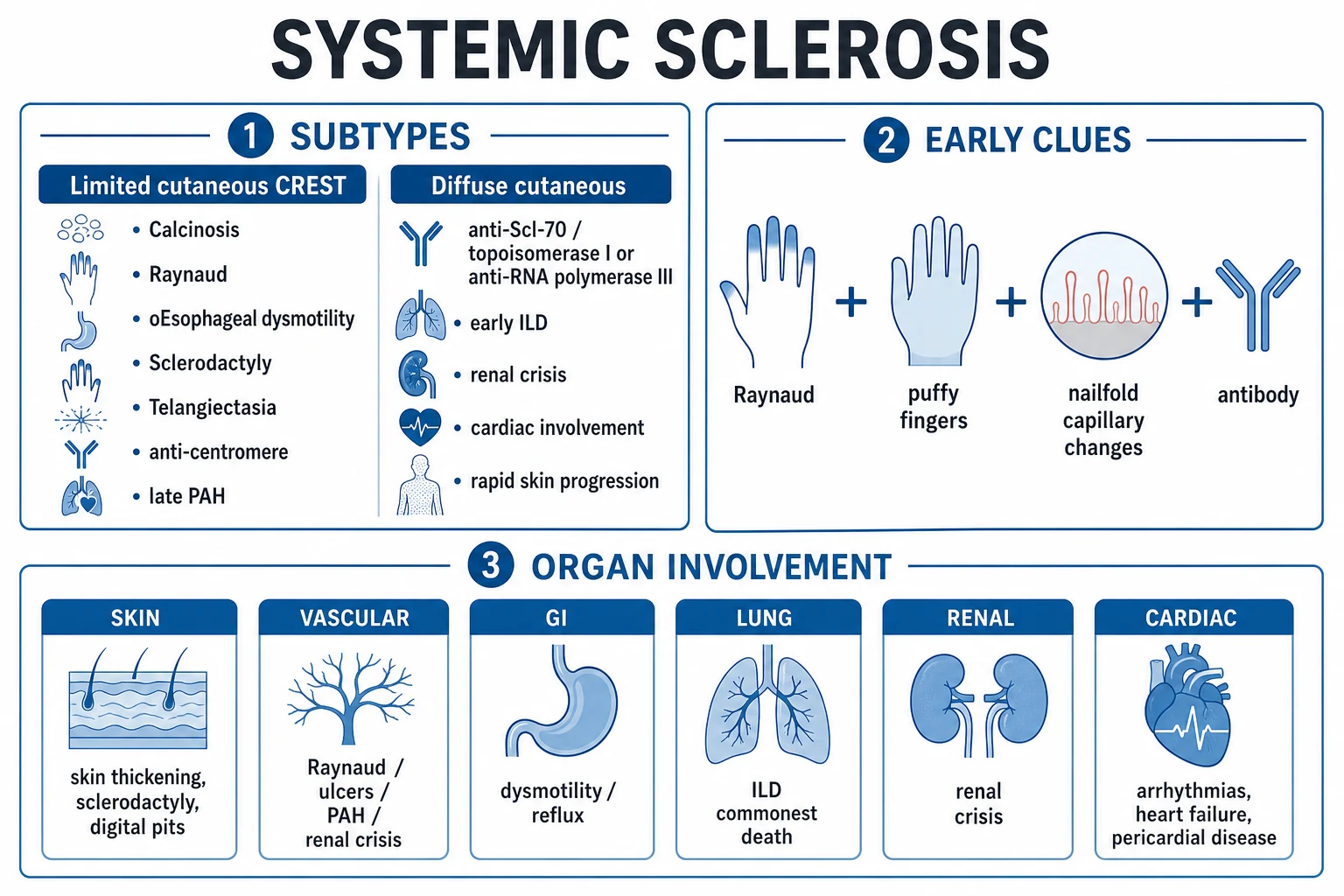
Systemic sclerosis (SSc, scleroderma) is a complex autoimmune connective tissue disease characterised by a pathophysiological triad of microvascular vasculopathy (Raynaud's phenomenon, PAH, renal crisis), tissue fibrosis (skin thickening, ILD, GI dysmotility), and autoimmunity (specific autoantibodies — anticentromere, anti-Scl-70/topoisomerase I, anti-RNA polymerase III). Classified as limited cutaneous (lcSSc, skin distal to elbows/knees; anticentromere; CREST — calcinosis, Raynaud's, oesophageal dysmotility, sclerodactyly, telangiectasia; late PAH) or diffuse cutaneous (dcSSc, proximal skin involvement; anti-Scl-70 with ILD; anti-RNA Pol III with renal crisis). Management is organ-specific: ACE inhibitors for renal crisis (life-saving, do not stop for rising creatinine); mycophenolate/nintedanib for ILD; ERA/PDE-5i/prostacyclin for PAH; CCB/PDE-5i/bosentan/IV iloprost for Raynaud's. Fellowship-level assessment demands mastery of the limited vs diffuse classification with antibody correlation, nailfold capillaroscopy patterns, renal crisis management (ACE-i life-saving, corticosteroids precipitate), annual PAH screening (echo + PFT; DLCO is the earliest marker), and the organ-specific treatment algorithm.
On this page & tools
Your progress
Saved locally on this device.
Practise this topic
Exam tags
Red flags
Definition & Classification
Systemic sclerosis (SSc, scleroderma) is a complex autoimmune connective tissue disease characterised by a pathophysiological triad: (1) microvascular vasculopathy, (2) tissue fibrosis (skin and internal organs), and (3) autoimmunity (specific autoantibodies).[2][3]
Classification by extent of skin involvement (drives antibody profile, organ risk and prognosis):[2][5]
| Subset | Skin extent | Typical antibody | Key organ risk |
|---|---|---|---|
| Limited cutaneous SSc (lcSSc) | Distal to elbows/knees (face, hands, feet) | Anticentromere (ACA) | Late PAH, biliary cirrhosis (PBC overlap) |
| Diffuse cutaneous SSc (dcSSc) | Proximal to elbows/knees (trunk, proximal limbs) | Anti-Scl-70 (topoisomerase I) | Early ILD, renal crisis |
| dcSSc (alternative) | As above | Anti-RNA polymerase III | Renal crisis, malignancy |
CREST syndrome (Calcinosis, Raynaud's, Esophageal dysmotility, Sclerodactyly, Telangiectasia) describes the limited phenotype — though modern classification prefers lcSSc.[2]
CREST — the limited cutaneous SSc syndrome (each letter, with the bedside finding)
CREST
Subcutaneous calcium deposits, classically over the finger pads, elbows and extensor surfaces; may ulcerate through the skin and discharge a chalky white paste. Distinguish from ectopic calcification in dermatomyositis.
Epidemic digital vasospasm (triphasic pallor → cyanosis → rubor) in response to cold or emotional stress; the FIRST manifestation in over 90% of SSc and may precede skin disease by years. Secondary Raynaud = abnormal nailfold capillaries and ANA positive.
Aperistalsis of the lower two-thirds (smooth muscle) → refractory GERD, dysphagia, peptic strictures, Barrett's oesophagus; treat with high-dose PPI ± endoscopic dilation. Most common GI manifestation.
Skin thickening of the fingers DISTAL to the MCP joints (literally 'CREST's' S); with time digits become hidebound, shiny, flexion-contracted, and develop pitting scars from small-vessel ischaemia.
Mat-like, well-demarcated macular telangiectasia on face, palms, lips and oral mucosa (sometimes GAVE = 'watermelon stomach' in the antrum); diagnostically helpful — distinguishes lcSSc from primary Raynaud's or mimic states.
ACR/EULAR 2013 classification criteria — skin thickening of fingers proximal to MCP joints = sufficient for SSc; if not, a point-based score from skin thickening, fingertip lesions, abnormal nailfold capillaries, PAH/ILD, and SSc-specific autoantibodies. Score ≥9 classifies SSc.[5]
Epidemiology
- Prevalence ~50-300/million; female:male 4-8:1; age of onset 30-50.[2]
- African heritage — earlier onset, more diffuse disease, worse prognosis.
- Environmental triggers: silica exposure (mining, stonemasonry), organic solvents (vinyl chloride — sixestomia), certain drugs (bleomycin), and possibly viral (CMV).[3]
- Mortality has improved with ACE inhibitors for renal crisis and PAH therapies, but SSc still carries the highest mortality among rheumatic diseases.[1]
Pathophysiology
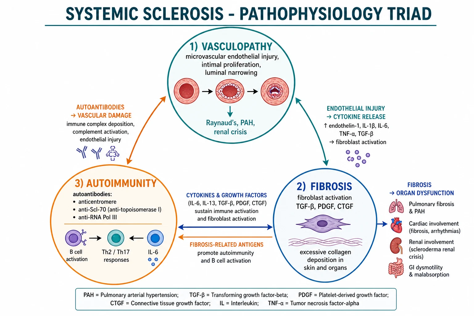
The three arms of the triad are interconnected — microvascular injury releases cytokines (TGF-β, PDGF) that activate fibroblasts; autoantibodies damage endothelium; fibrosis causes organ dysfunction:[3]
- Vasculopathy: endothelial injury (the initiating event) → intimal proliferation → luminal narrowing → tissue ischaemia. Clinically: Raynaud's, PAH, renal crisis, digital ulcers.
- Fibrosis: fibroblast activation (driven by TGF-β, PDGF, CTGF, IL-6) → excessive collagen (type I and III) deposition in skin and organs (lung, GI, heart, kidney).
- Autoimmunity: SSc-specific autoantibodies (anticentromere, anti-Scl-70, anti-RNA Pol III), Th2/Th17 polarisation, B-cell activation, type I interferon signature. [1]
Clinical Presentation
Skin and vascular (the dermatologist's gateway)
[1]Quick numbers for the examiner
- Raynaud's phenomenon often precedes skin disease by years. Secondary Raynaud's (abnormal nailfold capillaries, ANA positive, later onset, or atypical features) distinguishes from primary (benign, young women, normal nailfold capillaries, ANA negative).[4]
- Puffy fingers → skin thickening: early phase is oedematous ("puffy"); then indurated. Sclerodactyly = thickening distal to MCP joints (limited); proximal involvement = diffuse. Modified Rodnan Skin Score (mRSS) quantifies skin thickness at 17 sites (0-3 each).
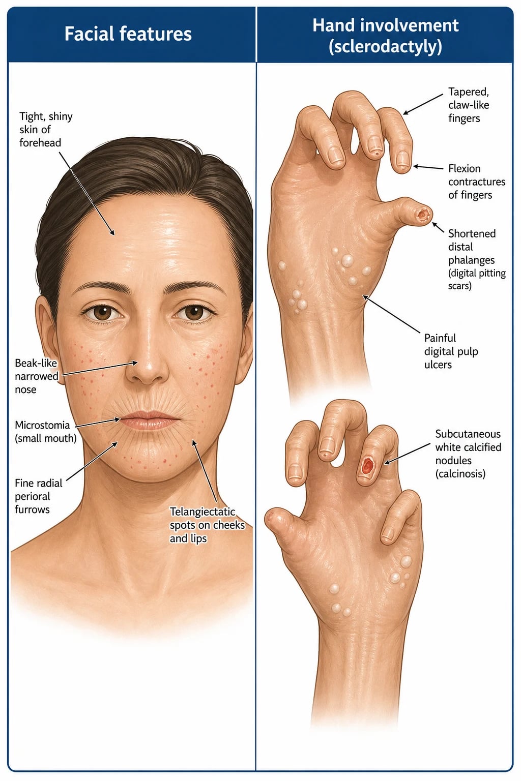
- Digital: pitting scars (distal pulp loss), painful digital ulcers (ischaemic), calcinosis cutis (subcutaneous calcium deposits, especially in CREST), telangiectasia (mat-like, face, hands, lips).
- Facial: microstomia (small mouth), beak-like nose (pinched), perioral radial furrowing, mask-like facies, reduced oral aperture.[2]
Nailfold capillaroscopy
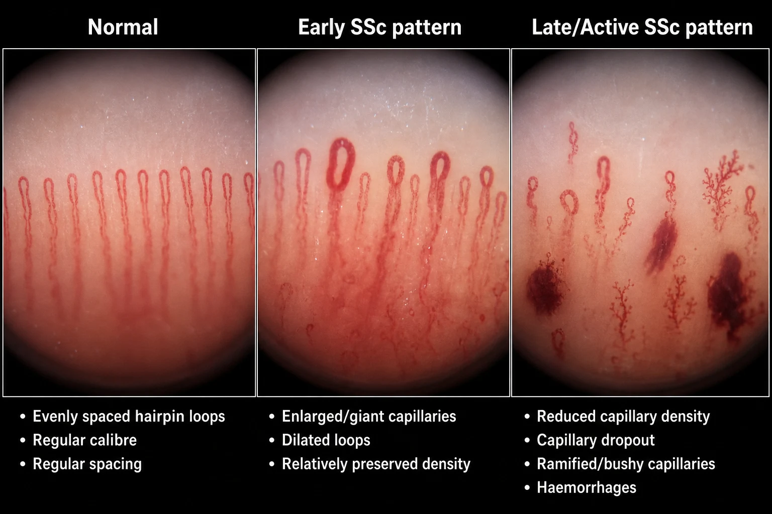
Nailfold capillaroscopy distinguishes primary from secondary Raynaud's and monitors SSc progression:[6]
- Early SSc: enlarged/giant capillaries, relatively preserved density.
- Active SSc: giant capillaries + haemorrhages.
- Late SSc: capillary dropout (reduced density), ramified/bushy capillaries. [1]
Internal organ involvement
| Organ | Features | Screening |
|---|---|---|
| Lung — ILD | Dyspnoea, dry cough; NSIP pattern on HRCT; commonest cause of SSc mortality | Annual PFT (FVC, DLCO); HRCT if abnormal |
| Lung — PAH | Exertional dyspnoea, fatigue; isolated PAH (no ILD) commoner in lcSSc | Annual echo (TRV) + PFT; DLCO is the earliest marker; RHC for confirmation |
| Kidney — renal crisis | Malignant hypertension, AKI, microangiopathic haemolysis | Regular BP monitoring; avoid high-dose steroids (precipitate) |
| GI tract | GERD, oesophageal dysmotility, gastroparesis, intestinal dysmotility, SIBO, malabsorption, faecal incontinence | Endoscopy, manometry; nutritional assessment |
| Heart | Myocardial fibrosis, arrhythmias, pericardial effusion, diastolic dysfunction | ECG, echo; cardiac MRI for myocarditis |
Differential Diagnosis
| Mimic | Distinguishing features |
|---|---|
| Morphea / localised scleroderma | Skin only (plaque, linear, generalised); no internal organ; no Raynaud's; no nailfold changes |
| Primary Raynaud's | Young, symmetric, normal nailfold capillaries, ANA negative |
| Nephrogenic systemic fibrosis | Renal failure + gadolinium exposure; skin induration but no Raynaud's or autoantibodies |
| Eosinophilic fasciitis (Shulman) | Groove sign, peripheral eosinophilia, deep fascia biopsy; no Raynaud's |
| Graft-versus-host disease | Transplant context |
| Diabetic cheiroarthropathy | Long-standing DM; prayer sign; no Raynaud's or nailfold changes |
| Amyloidosis | Periorbital purpura, macroglossia; biopsy |
| Scleromyxoedema (Arndt-Gottron) | Waxy papules in a sclerodermiform distribution; paraproteinaemia (IgG-λ) on SPEP; no Raynaud's, no nailfold changes |
| Scleroedema of Buschke | Post-streptococcal in children, or associated with long-standing diabetes in adults; woody non-pitting induration of neck, shoulders and upper back; no Raynaud's, no nailfold changes, no SSc antibodies |
| Drug-induced (bleomycin, taxanes, vinyl chloride) | Exposure history; occupational or chemotherapy context; may mimic dcSSc but usually improves on withdrawal |
| Chronic graft-versus-host disease | Prior allogeneic haematopoietic stem cell transplant; lichenoid → sclerotic skin; no Raynaud's, no SSc antibodies |
| Secondary causes of Raynaud's (SLE, Sjögren, cryoglobulinaemia, Buerger) | Other connective tissue features or digital ischaemia in a young male smoker; scleroderma-specific antibodies negative; vasospasm or thrombosis |
Quick bedside clues that argue AGAINST systemic sclerosis (favour a mimic instead): [1]
- No Raynaud's phenomenon in a patient with skin thickening → think morphea, scleromyxoedema, eosinophilic fasciitis, NSF or chronic GVHD.
- No abnormal nailfold capillaries → morphea or scleromyxoedema (NSF may also have normal capillaroscopy).
- No SSc-specific antibodies (ACA, Scl-70, RNA Pol III) → consider scleroderma-mimics.
- Deep-fascial involvement sparing the hands and face (eosinophilic fasciitis, 'groove sign') rather than the usual skin-only pattern.
- Recent gadolinium exposure in stage 4-5 CKD → nephrogenic systemic fibrosis. [1]
Clinical & Bedside Assessment
- Full skin examination with mRSS scoring (17 body sites, grade 0-3 thickening at each, maximum total 51); examine hands, face, nailfolds (capillaroscopy at bedside with a drop of immersion oil + dermoscope or USB videocapillaroscope).[6]
- Cardiopulmonary: auscultate (fine bibasal inspiratory crackles of ILD; loud P2, tricuspid regurgitation murmur and right ventricular heave of PAH), assess for signs of right heart failure (elevated JVP, peripheral oedema, tender hepatomegaly, ascites), and palpate the praecordium.
- GI: weight loss, dysphagia, reflux, early satiety (gastroparesis), bloating and diarrhoea (SIBO), constipation or faecal incontinence (anal sphincter fibrosis); inspect the mouth for microstomia, perioral radial furrowing, and tongue tie.
- Musculoskeletal: palpate for tendon friction rubs (palpable or audible crepitus over extensor tendons of fingers, wrists and ankles — a poor-prognosis marker in early dcSSc); measure the inter-incisal distance (oral aperture); check for flexion contractures at MCPs, PIPs and elbows; passive and active range of motion.
- Renal: regular blood pressure monitoring at every visit (scleroderma renal crisis presents with new malignant hypertension); check urinalysis for proteinuria and microscopic haematuria; immediate review of any patient with new BP >180/110 mmHg or rising creatinine.[2]
Complications & Pitfalls
[1]Investigations
- ANA — positive in >90% (fine-speckled, nucleolar, centromere patterns).[2]
- SSc-specific antibodies:[2]
- Anticentromere (ACA) — limited cutaneous; PAH risk.
- Anti-Scl-70 (topoisomerase I) — diffuse cutaneous; ILD risk.
- Anti-RNA polymerase III — diffuse cutaneous; renal crisis + malignancy risk.
- Nailfold capillaroscopy — distinguishes primary from secondary Raynaud's.[6]
- PFT — FVC (restriction in ILD), DLCO (isolated reduction suggests PAH; earliest marker). Annual.
- HRCT — ground-glass, reticulation, traction bronchiectasis, honeycombing (NSIP pattern).[8]
- Echo — annual; tricuspid regurgitant velocity (TRV) and RV assessment screen for PAH. Right heart catheter (RHC) confirms.[2]
- Renal: regular BP, creatinine, urinalysis.
- Cardiac screening: baseline ECG and consider cardiac MRI for arrhythmia and myocardial fibrosis; serum troponin and NT-proBNP when symptomatic. A focused cardiac history (dyspnoea, ankle swelling, chest pain, palpitations) should be re-checked at every visit. Risk factors for SSc-PAH (age, female sex, long-standing lcSSc, anti-centromere) inform screening frequency.
- GI: endoscopy, oesophageal manometry; consider hydrogen breath test for SIBO.
Management — Organ-Specific
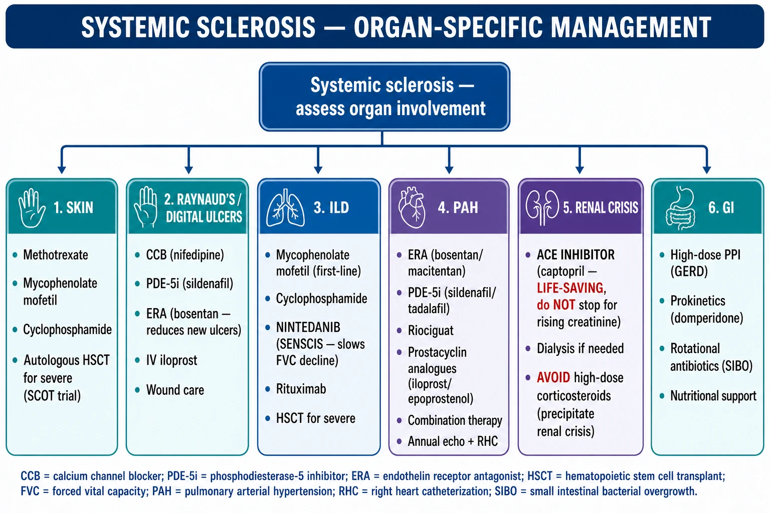
Skin
- Immunosuppression: methotrexate, mycophenolate mofetil, cyclophosphamide for early dcSSc skin involvement.[7]
- Autologous HSCT for severe refractory dcSSc (SCOT, ASTORIA trials — improves survival and skin but carries transplant mortality).[7]
Raynaud's / Digital ulcers
- First-line: calcium channel blockers (nifedipine 10-30 mg modified-release BD, amlodipine 5-20 mg OD).[4]
- Second-line: PDE-5 inhibitors (sildenafil 25-50 mg TDS, tadalafil 20 mg OD); add bosentan 62.5 mg BD for 4 weeks then titrated to 125 mg BD for prevention of NEW digital ulcers (RAPIDS-1/2 trials).[7]
- Severe/critical ischaemia: IV iloprost 0.5-2 ng/kg/min over 6 hours daily for 3-5 days; digital sympathectomy; botulinum toxin; wound care.[4]
Interstitial lung disease (ILD)
- Mycophenolate mofetil 2-3 g/day (first-line) or cyclophosphamide 500-750 mg/m² IV monthly for 6-12 months (Scleroderma Lung Study I/II — both stabilise FVC).[7][8]
- Nintedanib 150 mg BD (anti-fibrotic; SENSCIS trial — slows FVC decline in SSc-ILD).[8]
- Rituximab 1 g IV days 0 and 14 for refractory disease; HSCT for severe progressive ILD.[7]
Pulmonary arterial hypertension (PAH)
- Endothelin receptor antagonists (bosentan, ambrisentan, macitentan).
- PDE-5 inhibitors (sildenafil, tadalafil).
- Riociguat (soluble guanylate cyclase stimulator).
- Prostacyclin analogues (inhaled iloprost, IV epoprostenol).
- Combination therapy (dual/triple); lung transplant for refractory.[7]
Renal crisis (a dermatological/rheumatological emergency)
- ACE inhibitors — LIFE-SAVING. Start captopril 6.25-12.5 mg PO initially, titrate rapidly to 25-50 mg TDS; do NOT stop for rising creatinine (the creatinine will rise before it falls as renal perfusion recovers).[2][7]
- Dialysis if needed; many patients recover renal function after weeks-months.
- AVOID high-dose corticosteroids (>15 mg/day prednisolone) — they precipitate renal crisis in SSc.[2]
GI tract
- High-dose PPI (omeprazole/esomeprazole) for GERD and stricture prevention.
- Prokinetics (domperidone, metoclopramide) for dysmotility/gastroparesis.
- Rotational antibiotics (rifaximin, ciprofloxacin, metronidazole, doxycycline) for SIBO and malabsorption.
- Nutritional support — oral supplements, nasojejunal feeding, PEG if severe.[7]
Diffuse vs Limited SSc — A Deep Dive for the Fellowship Examiner
The extent of skin involvement at disease onset is not merely descriptive — it is the single most powerful predictor of antibody profile, organ-complication pattern and prognosis. LeRoy and Medsger's 1988 distinction (still used, refined by ACR/EULAR 2013) splits SSc into two biologically distinct diseases that happen to share a label. [1]
Diffuse cutaneous SSc (dcSSc) is defined by skin thickening that extends proximal to the elbows or knees, or involves the trunk (chest, abdomen, back), and typically evolves within a recognisable clinical sequence: a brief oedematous phase (puffy hands, "sausage" fingers, tenosynovitis), then rapid induration that peaks at 12-24 months and either plateaus or slowly regresses over years. The "1-to-3-year rule" matters: skin progression, ILD and renal crisis cluster in the first 36 months of disease — that window is when aggressive immunosuppression (mycophenolate, cyclophosphamide, rituximab, consider HSCT) and ACE-inhibitor prophylaxis reduce mortality most. The antibody profile is anti-Scl-70 (topoisomerase I) in 30-60% and anti-RNA polymerase III in 10-20%; both carry substantial visceral risk. Patients with dcSSc should be enrolled in a tight screening pathway — PFTs and HRCT at baseline and every 6-12 months in early disease, monthly BP checks, and a low threshold for cancer imaging if anti-RNA Pol III positive. [1]
Limited cutaneous SSc (lcSSc) is skin thickening confined to areas distal to the elbows and knees (face, neck, hands, feet); the term replaces "acroscleroderma" and the old "CREST" label. Skin thickening is often subtle and slowly progressive, but the disease is anything but benign — PAH is the dominant killer and presents late (often 10-15 years into disease), anti-centromere (ACA) positivity forecasts it, and the slow-tempo skin disease does not protect from catastrophic digital, GI, or pulmonary vascular events. The bonus for lcSSc patients is that severe ILD, renal crisis and acute skin crisis are uncommon. [1]
Scleroderma sine scleroderma is the rare third category — visceral disease with no skin thickening; antibody profile and Raynaud's still point the way, but diagnosis requires high clinical suspicion. [1]
Antibody Correlation — Scl-70 vs ACA vs RNA Polymerase III
The three SSc-specific antibodies are mutually almost exclusive (rare patients have two) and each predicts a distinct clinical trajectory. They anchor antibody-guided management in modern SSc care. [1]
Anti-centromere antibody (ACA) — immunoglobulin-G against the kinetochore protein CENP-B. Found in 20-40% of all SSc patients and up to 70-80% of lcSSc. It is the PAH/CREST antibody — associations include limited cutaneous disease, calcinosis, late-onset PAH (10-15% at 10-15 years), primary biliary cholangitis overlap, digital ulcers, and sicca symptoms. Patients are largely spared renal crisis and severe ILD; prognosis is the best of the three antibody groups. ACA also confers a low risk of malignancy and a slow, often decades-long, disease course. Serology remains positive throughout life; titre does not track disease activity — re-testing once positive is unhelpful. [1]
Anti-Scl-70 (anti-topoisomerase I) — antibody against the nuclear enzyme DNA topoisomerase I; present in 20-30% of all SSc and up to 60-70% of dcSSc. It is the ILD antibody — and ILD develops in 70-80% of Scl-70-positive patients (often early, sometimes subclinical, and progressive). Smoking dramatically worsens the ILD risk; anti-Scl-70 positivity is the single most clinically actionable antibody — it mandates baseline HRCT, prompt institution of mycophenolate (or cyclophosphamide then MMF in severe disease) and consideration of nintedanib. Scl-70 is also associated with diffuse skin disease, joint involvement, myositis and digital ulcers. Prognosis is intermediate, driven by the speed and severity of ILD. [1]
Anti-RNA polymerase III — antibody against the catalytic subunit of RNA polymerase III. Present in 10-20% of SSc but up to 25% of dcSSc. It is the renal-crisis + cancer antibody and marks the most dramatic dcSSc phenotype — rapidly progressive skin thickening, scleroderma renal crisis in 25-30% (often within weeks of SSc onset, frequently in winter), gastric antral vascular ectasia (GAVE), tendon friction rubs, and a 5-10% synchronous solid malignancy rate (breast, lung, ovary, stomach, colon, tongue) that clusters in the 2-5 years around SSc onset. Older age at onset, male sex and abrupt skin progression heighten malignancy risk. Patients with this antibody need age-appropriate cancer screening at diagnosis and annually for the first 5 years, plus extra vigilance for new renal-crisis symptoms. [1]
Other clinically relevant antibodies (mutually not exclusive with the three above but less specific): anti-U1-RNP (overlap with SLE/myositis — mixed connective tissue disease), anti-PM/Scl (polymyositis overlap), anti-Th/To (lcSSc with PAH and ILD), anti-U3-RNP / fibrillarin (dcSSc with cardiac and muscle involvement), anti-Ku (overlap with myositis), anti-NOR-90, anti-RuvBL1/2 (cancer-associated SSc). [1]
Pulmonary Arterial Hypertension in SSc — Diagnosis and Treatment Algorithm
SSc-associated PAH (Group 1 pulmonary hypertension) is the second leading cause of death in SSc overall and the dominant cause of death in lcSSc. It is vasculopathic (intimal proliferation, plexiform lesions of small pulmonary arteries) rather than embolic or hypoxic, and the DLCO falls before the FVC falls — a falling DLCO with a stable FVC should trigger urgent evaluation. [1]
Screening protocol in any patient with SSc ≥3 years (annual thereafter, more often if symptomatic or anti-centromere / anti-Th/To positive): symptom screen (exertional dyspnoea, near-syncope, oedema), DLCO (a fall in DLCO >20% from baseline, or DLCO less than 60% predicted with normal FVC, is suggestive), transthoracic echo (TRV >2.9 m/s, RV dilatation, septal flattening, pericardial effusion), NT-proBNP (elevated in right-heart strain). The DETECT algorithm combines FVC/DLCO ratio, telangiectasia, anti-centromere, NT-proBNP, serum urate and right-axis deviation on ECG to risk-stratify; positive triggers right heart catheterisation. Diagnosis of PAH requires right heart catheterisation with mean pulmonary arterial pressure ≥20 mmHg at rest, pulmonary capillary wedge pressure ≤15 mmHg and pulmonary vascular resistance ≥2 Wood units. [1]
Treatment — risk-stratified (low/intermediate/high): [1]
- Endothelin receptor antagonists (ERA): bosentan 62.5 mg BD for 4 weeks, then 125 mg BD; alternative once-daily agents are ambrisentan 5-10 mg OD and macitentan 10 mg OD. All three require monthly LFTs (bosentan/ambrisentan), avoidance in pregnancy and reliable contraception (teratogenic).
- PDE-5 inhibitors: sildenafil 20 mg TDS (note: PAH dose is TDS, NOT the ED dose) or tadalafil 40 mg OD. Avoid concomitant nitrates; caution with alpha-blockers; may cause headache, flushing, dyspepsia (sildenafil), and rare non-arteritic anterior ischaemic optic neuropathy.
- Soluble guanylate cyclase stimulator: riociguat 1 mg TDS titrated up to 2.5 mg TDS — used if PDE-5i fails or is contraindicated; avoid combining with PDE-5i (hypotension).
- Prostacyclin analogues: iloprost 10-20 µg inhaled 6-9 times daily, IV iloprost, subcutaneous or IV treprostinil, or IV epoprostenol for high-risk or refractory disease. Selexipag 200-1,600 µg BD orally is an oral prostacyclin-receptor agonist for stepwise escalation.
- Initial combination therapy (upfront ambrisentan + tadalafil in AMBITION) is now preferred for many intermediate- and high-risk patients; triple therapy with selexipag on top is established for progressive disease.
- Supportive: oxygen to maintain SaO₂ >90%, supervised exercise, diuretics for RV failure, endothelin-blocking contraception (oestrogen-containing contraception contraindicated — barrier, levonorgestrel-IUS, or Depo-Provera), and lung transplantation for refractory disease. [1]
Scleroderma Renal Crisis — Diagnosis and Treatment Algorithm
Scleroderma renal crisis (SRC) is a life-threatening emergency that occurs in 5-15% of SSc patients, almost always within the first 4 years of dcSSc onset (median 1-2 years), often in winter. The pathophysiology is renal vasculopathy: intimal proliferation and "onion-skinning" of interlobular renal arteries, fibrinoid necrosis, microangiopathic haemolytic anaemia and uncontrolled activation of the renin-angiotensin system → malignant hypertension, AKI and (if untreated) end-organ damage and death. [1]
Risk factors — anti-RNA polymerase III positivity, rapidly progressive skin disease, recent high-dose corticosteroids (>15 mg/day prednisolone or pulsed methylprednisolone), congestive cardiac failure, tendon friction rubs, male sex, and abrupt onset of disease. [1]
Diagnostic criteria (Steen/Medsger): new-onset malignant hypertension (BP ≥180/110 mmHg, often with hypertensive retinopathy grade III/IV, encephalopathy, seizures, papilloedema) in a patient with SSc ± AKI ± microangiopathic haemolytic anaemia. Note: a normotensive variant is described in 5-10% — AKI without hypertension still warrants ACE-inhibitor therapy. [1]
Treatment — every minute matters: [1]
- Stop any nephrotoxic drug (NSAIDs, contrast) but DO NOT stop the ACE inhibitor if already on one — converting to captopril and titrating up aggressively is correct even if creatinine continues to rise.
- Captopril 6.25-12.5 mg PO initially, repeated every 4-8 hours if needed, then titrated rapidly to 25-50 mg TDS (or as high as 150 mg/day in divided doses). Captopril is preferred because of its short half-life — dosing can be adjusted rapidly in unstable patients.
- If oral therapy is not feasible (vomiting, perioperative): IV enalaprilat, though this is rarely needed if an NG tube is available.
- Add a second-line vasodilator if BP remains uncontrolled — IV labetalol, IV nitroprusside (with caution — can raise intracranial pressure) or IV nicardipine.
- Avoid beta-blockers (worsen Raynaud's, reduce renal perfusion).
- Avoid plasmapheresis as routine — no proven survival benefit; reserve for refractory or anti-GBM overlap disease.
- Dialysis (haemodialysis or peritoneal) for uraemia, acidosis, fluid overload or refractory hypertension — about 50-60% of dialysed patients recover renal function and come off dialysis within 12-24 months; CAPD is reasonable while awaiting recovery.
- Prophylactic ACE inhibitors (e.g., lisinopril) are NOT recommended in normotensive dcSSc — the evidence for prophylactic ACE-i preventing SRC is unconvincing; over-diuresis and hypotension may even worsen renal perfusion. ACE inhibitors are reserved for confirmed SRC. [1]
Specific Drug Doses — Reference Table
| Drug | Indication | Dose | Notes |
|---|---|---|---|
| Mycophenolate mofetil | Skin / ILD (dcSSc) | 1-1.5 g BD (2-3 g/day) | First-line ILD (SLS II); monitor FBC, LFTs; teratogenic — reliable contraception |
| Cyclophosphamide | ILD (induction) | 500-750 mg/m² IV monthly × 6-12 | Reserve for severe/progressive ILD; +MESNA, hydration |
| Nintedanib | SSc-ILD | 150 mg BD | Anti-fibrotic; GI side-effects (diarrhoea); LFTs |
| Rituximab | Refractory skin / ILD | 1 g IV days 0 and 14, repeat every 6-9 months | Anti-CD20; monitor immunoglobulins, infection; vaccine response |
| Nifedipine (modified-release) | Raynaud's | 10-30 mg TDS or 30-90 mg OD MR | First-line CCB; titrate to BP/tolerance |
| Amlodipine | Raynaud's | 5-20 mg OD | Alternative first-line CCB |
| Sildenafil | PAH / refractory Raynaud's | 20 mg TDS (PAH dose — NOT the ED dose) | PDE-5i; avoid nitrates |
| Tadalafil | PAH | 40 mg OD | Once-daily alternative to sildenafil |
| Bosentan | PAH / recurrent digital ulcers | 62.5 mg BD × 4 weeks, then 125 mg BD | ERA; monthly LFTs; teratogenic; RAPIDS-1/2 evidence for ulcers |
| Ambrisentan | PAH | 5-10 mg OD | ERA; once-daily; fewer LFT issues than bosentan |
| Macitentan | PAH | 10 mg OD | ERA; SERAPHIN trial |
| Riociguat | PAH | 1 mg TDS up to 2.5 mg TDS | sGC stimulator; contraindicated with PDE-5i |
| Iloprost IV | Severe Raynaud's / critical ischaemia | 0.5-2 ng/kg/min IV over 6 h daily × 3-5 days | Prostacyclin analogue; watch BP, flushing, jaw claudication |
| Inhaled iloprost | PAH | 10-20 µg 6-9×/day | Outpatient prostacyclin |
| Selexipag | PAH | 200-1,600 µg BD orally (titrate) | Oral prostacyclin-receptor agonist |
| Captopril | Scleroderma renal crisis | 6.25-12.5 mg PO, repeat every 4-8 h, titrate to 25-50 mg TDS | LIFE-SAVING; do NOT stop for rising creatinine |
| Methylprednisolone (pulsed) | Severe ILD overlap / myositis | 500-1,000 mg IV daily × 3 days | BUT — avoid high-dose steroids in early dcSSc (precipitates SRC) |
| Prednisolone (maintenance) | Skin / overlap features | ≤5-10 mg/day (≤15 mg/day ceiling) | KEEP LOW in SSc — higher doses precipitate renal crisis |
| N-Acetylcysteine + IVIG | Severe diffuse skin (case-by-case) | NAC 600 mg BD + IVIG 2 g/kg/month | Some centres use cyclically |
| Methotrexate | Skin / joint | 15-25 mg weekly SC/IM | With folic acid; avoid in pregnancy |
Special Populations [1]
- Pregnancy: fertility is preserved but pregnancy in SSc is high-risk — renal crisis risk increases (especially post-partum), PAH is a contraindication to pregnancy, and Raynaud's may improve during pregnancy (vasodilation). Close obstetric + rheumatology co-management.[2]
- Children (juvenile SSc): rarer; overlaps with localised scleroderma; generally better prognosis but ILD and PAH still occur.[1]
Prognosis
- 5-year survival ~70-80% overall; improving with ACE-i and PAH therapy.[1]
- Leading causes of death: ILD, PAH, cardiac, renal crisis (less common with ACE-i), GI (malnutrition, cancer).[2]
- Limited cutaneous generally better prognosis (except late-onset PAH which is the dominant mortality in lcSSc).
- Diffuse cutaneous worse — early ILD, renal crisis (first 3-4 years), more severe skin and GI disease.
- Anti-RNA Pol III positivity carries the highest renal crisis risk; anti-Scl-70 the highest ILD risk.[2]
Evidence, Guidelines & Regional Differences
- ACR/EULAR 2013 classification criteria — the universally adopted diagnostic framework.[5]
- EULAR 2023 treatment update — the principal management guideline; organ-specific recommendations.[7]
- Key trials: SCOT (autologous HSCT), Scleroderma Lung Study I/II (CYC vs MMF; nintedanib SENSCIS), RAPIDS-1/2 (bosentan for digital ulcers).[7]
- EUSTAR (EULAR Scleroderma Trials and Research) database — the largest registry.
Prevention
- No primary prevention — systemic sclerosis is an autoimmune disease with no proven pre-clinical intervention.
- Secondary prevention is the cornerstone of long-term outcome. Every clinic visit is a screening opportunity:
- Annual screening protocol (every SSc patient, every 12 months): PFTs (FVC + DLCO), transthoracic echocardiogram (TRV, RV size, pericardial effusion), renal function (creatinine, urinalysis, BP every visit), and skin assessment (mRSS progression or regression as a proxy for disease activity).
- Cardiac screening — ECG at baseline and when symptomatic; cardiac MRI with gadolinium if arrhythmia or chest pain; serum troponin and NT-proBNP for suspected myocarditis or scleroderma cardiac crisis; risk factors (age, sex, antibody profile, lcSSc duration) guide frequency.
- Smoking cessation is mandatory — nicotine worsens Raynaud's vasospasm and accelerates vascular injury in SSc; it also interferes with calcium channel blocker efficacy.
- Vaccinations: annual influenza, pneumococcal (PCV13 + PPSV23), COVID-19 boosters, and recombinant zoster vaccine for patients on immunosuppression; live vaccines are contraindicated on biologics and high-dose immunosuppression.
- Cold avoidance and warming measures for Raynaud's — insulated gloves, hand warmers, avoidance of vasoconstrictors (β-blockers, sympathomimetic decongestants) and not handling cold items without protection.
- Patient education on renal crisis warning signs: any new severe headache, visual disturbance, BP >180/110 mmHg, or rapid weight gain requires URGENT medical review — early captopril saves lives and ACE inhibitors halve mortality.
- Cancer screening in anti-RNA polymerase III-positive dcSSc — age-appropriate cancer surveillance at diagnosis and annually for 2-5 years (breast, lung, GI, ovary); anti-RNA Pol III carries a 5-10% synchronous malignancy rate.
- PPI prophylaxis for all SSc patients with reflux to prevent oesophageal strictures and Barrett's oesophagus; PPI reduces the rate of progression to Barrett's.
- Bone health — vitamin D and calcium supplementation, DEXA scanning in patients on long-term corticosteroids or with malabsorption; treat osteoporosis when identified.
- Infection prevention — prompt treatment of infected digital ulcers; annual dental review; consider prophylactic rotating antibiotics for recurrent SIBO.
- Pulmonary rehabilitation for ILD patients; supervised exercise programmes improve 6-minute-walk distance and quality of life.
- Vaccinate household contacts to protect immunosuppressed SSc patients from varicella, measles and influenza. [1]
Exam Pearls
[1]Red Flags
Exam application bank (NEET-PG / INICET)
One-line answer
Systemic sclerosis (SSc, scleroderma) is a complex autoimmune connective tissue disease characterised by a pathophysiological triad of microvascular vasculopathy (Raynaud's phenomenon, PAH, renal crisis), tissue fibrosis (skin thickening, ILD, GI dysmotility), and autoimmunity (specific autoantibodies — anticentromere, anti-Scl-70/topoisomerase I, anti-RNA polymerase III). Classified as limited cutaneous (lcSSc, skin distal to elbows/knees; anticentromere; CREST — calcinosis, Raynaud's, oesophageal dysmotility, sclerodactyly, telangiectasia; late PAH) or diffuse cutaneous (dcSSc, proximal skin involvement; anti-Scl-70 with ILD; anti-RNA Pol III with renal crisis). Management is organ-specific: ACE inhibitors for renal crisis (life-saving, do not stop for rising creatinine); mycophenolate/nintedanib for ILD; ERA/PDE-5i/prostacyclin for PAH; CCB/PDE-5i/bosentan/IV iloprost for Raynaud's. F
Worked stems (answer without another resource)
Stem 1 — Classic presentation. Map symptoms to mechanism; name the first investigation and first treatment step with dose/route if drug therapy is standard. [1]
Stem 2 — Unstable / complicated. List red flags that force immediate resuscitation, theatre, ICU, antidote, or reperfusion — and what you do in the first 15 minutes. [1]
Stem 3 — Atypical group. Elderly, pregnancy, child, or immunocompromised: how presentation and thresholds change. [1]
Stem 4 — Differential trap. Name the three closest mimics and one discriminator for each. [1]
Stem 5 — Disposition. Who goes home with safety-netting, who is admitted, who needs HDU/ICU/theatre, and what follow-up is mandatory. [1]
Rapid viva checklist
- Definition + classification
- Pathophysiology chain
- Bedside signs / criteria
- Score with exact components (if any)
- Emergency bundle
- Definitive therapy with doses
- Complications of disease and of treatment
- Special populations
- Guideline/trial name if classic
- Three exam traps
Coverage self-check
If you cannot answer any stem above from this page alone, re-read the matching section — the page is intended to be self-sufficient for final-prof and NEET-PG/INICET questions on Systemic sclerosis.
[1]References
- [1]Di Battista M, Lepri G, Codullo V, et al. Systemic sclerosis: one year in review 2025 Clin Exp Rheumatol, 2025.PMID 40737082
- [2]Jerjen R, Nikpour M, Krieg T, et al. Systemic sclerosis in adults. Part I: Clinical features and pathogenesis J Am Acad Dermatol, 2022.PMID 35131402
- [3]Rosendahl AH, Schönborn K, Krieg T. Pathophysiology of systemic sclerosis (scleroderma) Kaohsiung J Med Sci, 2022.PMID 35234358
- [4]Herrick AL, Wigley FM. Raynaud's phenomenon Best Pract Res Clin Rheumatol, 2020.PMID 32007400
- [5]van den Hoogen F, Khanna D, Fransen J, et al. 2013 classification criteria for systemic sclerosis: an American college of rheumatology/European league against rheumatism collaborative initiative Ann Rheum Dis, 2013.PMID 24092682
- [6]Smith V, Ickinger C, Hysa E, et al. Nailfold capillaroscopy Best Pract Res Clin Rheumatol, 2023.PMID 37419757
- [7]Del Galdo F, Lescoat A, Conaghan PG, et al. EULAR recommendations for the treatment of systemic sclerosis: 2023 update Ann Rheum Dis, 2025.PMID 39874231
- [8]Maher TM. Interstitial Lung Disease: A Review JAMA, 2024.PMID 38648021