Dermatology · Medicine
Telogen effluvium
Also known as Telogen effluvium (TE) · Acute telogen effluvium · Chronic telogen effluvium (CTE) · Postpartum telogen effluvium · Post-febrile telogen effluvium · Diffuse hair shedding
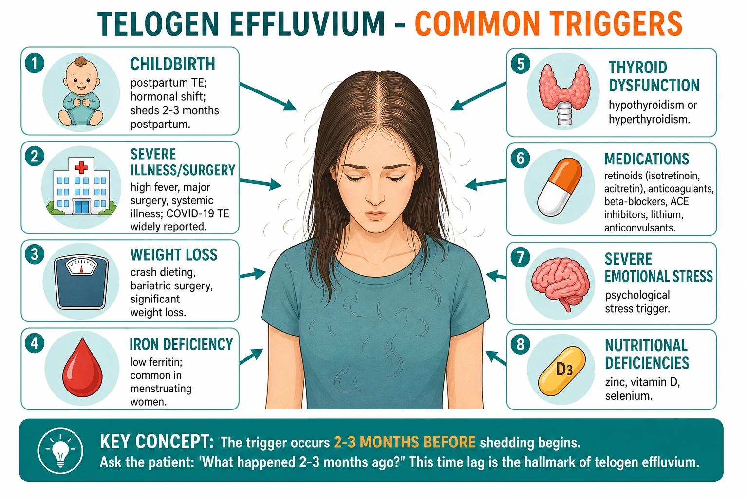
Telogen effluvium (TE) is a non-scarring, diffuse hair loss caused by a premature shift of anagen follicles into the telogen (resting) phase, producing synchronous shedding 2-3 months after a triggering event. Headington classified five functional types (immediate anagen release, delayed anagen release, short anagen, immediate telogen release, delayed telogen release) and the Whiting entity of chronic telogen effluvium (CTE) is a distinct middle-aged female phenotype that fluctuates for years. Common triggers include childbirth, severe illness or fever (including COVID-19), iron deficiency, thyroid dysfunction, crash dieting or bariatric surgery, retinoids, anticoagulants, beta-blockers, lithium, valproate, interferon and severe emotional stress. The hallmark bedside signs are diffuse thinning (not patterned, not patchy), a positive hair pull test (5-6 of 50-60 hairs), trichoscopic empty follicles and upright regrowing hairs, and ABSENT follicular diameter diversity. Investigation is targeted: ferritin (target 70 microg/L), TSH, vitamin D, zinc and CBC. Management is identify-and-treat-the-trigger plus reassurance; acute TE is self-limiting within 6-12 months. Topical or oral minoxidil is reserved for chronic TE or TE overlapping with female pattern hair loss (FPHL).
On this page & tools
Your progress
Saved locally on this device.
Exam tags
Red flags
Overview & Definition
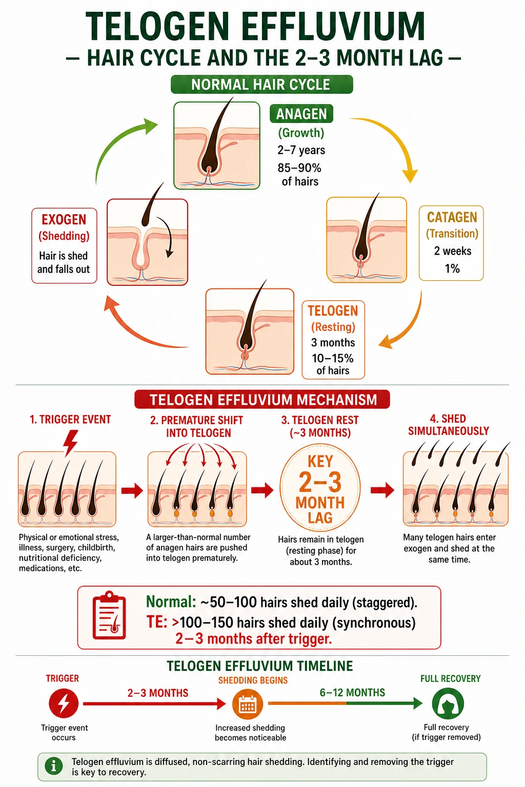
Telogen effluvium (TE) is the most common cause of diffuse hair loss in women and a leading cause in men, accounting for a substantial proportion of new referrals to dermatology and trichology clinics worldwide. It is a non-scarring, reversible, diffuse alopecia caused by a premature and synchronous shift of anagen (growth-phase) hair follicles into the telogen (resting) phase, leading to a noticeable increase in daily hair shedding 2-3 months later.[1][2][3]
The condition was first formalised in 1961 by Kligman as "telogen effluvium" but the modern framework was established by Headington in 1993, who described five functional types based on the part of the hair cycle that the trigger disrupts.[4] Two clinical forms are recognised in routine practice: acute TE (less than 6 months) and chronic telogen effluvium (CTE), the latter described by Whiting in 1996 as a distinct, predominantly middle-aged female phenotype that fluctuates for months to years.[5][7]
The pathophysiology is best understood in relation to the normal hair cycle. At any one time, around 85-90 percent of scalp hairs are in anagen (lasting 2-7 years), 1-2 percent in catagen (2-3 weeks) and 10-15 percent in telogen (~3 months). In TE, a systemic or local trigger forces a much larger cohort of anagen follicles into catagen and then telogen. Because telogen has a fixed duration of about three months, the actual shedding (exogen) is delayed by approximately 8-12 weeks from the trigger — the 2-3 month lag that is the diagnostic hallmark of TE.[1][2][4]
[1]Why some triggers shed in weeks, others in months
The 2-3 month lag is the default of immediate anagen release, but the clinical latency varies with the Headington subtype. Postpartum TE (delayed anagen release) sheds at 1-3 months postpartum because the oestrogen withdrawal in late pregnancy releases a cohort of prolonged-anagen hairs that all enter telogen together. Drug-induced immediate telogen release (interferon, retinoids) sheds in days to weeks because the trigger acts on hairs already in telogen, releasing them prematurely. Short anagen syndrome in children produces persistent mild shedding because anagen never reaches full length. Understanding the subtype explains why two patients with apparently similar triggers can present at very different intervals after the trigger, and is the basis for the "2-3 month" rule being a guideline rather than an absolute.
The five Headington types of TE
AIDDS
Trigger (febrile illness, drug, stress) abruptly stops anagen; hairs enter telogen within weeks. Most common mechanism; sheds at 2-3 months.
Postpartum TE — oestrogen withdrawal in late pregnancy releases prolonged anagen hairs that all enter telogen together; shed at 1-3 months postpartum.
Idiopathic short anagen syndrome in children; persistent mild shedding because anagen never reaches full length.
Prolonged telogen retention (post-travel, seasonal); cluster shedding when telogen finally ends.
Hairs already in telogen are released prematurely by drugs (interferon, retinoids) — fast onset (days-weeks).
Classification & Subtypes
Acute versus chronic TE
The most clinically useful split is duration-based. [1]
[1]Key prevalence numbers
A simpler Rebora classification
Rebora proposed a binary split that is useful in clinic and on the exam: TE with predominant shedding (telogen defluvium) versus TE with predominant hair thinning (telogen dystrophy). The shedding form represents increased hair loss with normal regrowth; the dystrophic form represents follicles that re-enter anagen with thinner, shorter shafts, producing visible thinning despite stable shed counts.[6] This binary view anticipates the chronic-TE / early-FPHL overlap and helps counsel patients about the difference between "hair in the brush" and "less hair on the head".
Epidemiology & Risk Factors
Acute TE is extremely common and is reported in a substantial minority of women after childbirth, after severe febrile illness (including COVID-19), and after major surgery.[1][11] The lifetime risk for a clinically relevant episode in adult women is estimated at around 25-40 percent; the lifetime risk in men is lower (5-10 percent) but still significant. CTE is rarer, accounting for perhaps 5-10 percent of trichology referrals, but its chronicity and quality-of-life impact outweigh its prevalence.[5][7]
The major risk factors cluster into six groups: [1]
- Endocrine and reproductive: postpartum, oral contraceptive withdrawal, hypothyroidism, hyperthyroidism, postpartum thyroiditis.
- Nutritional: iron deficiency (with or without anaemia), low ferritin stores even with normal haemoglobin, vitamin D deficiency, zinc deficiency, biotin deficiency, severe protein-energy malnutrition, crash diets and bariatric surgery.
- Drugs: retinoids (isotretinoin, acitretin), anticoagulants (heparin, warfarin, direct oral anticoagulants), beta-blockers (propranolol, metoprolol), ACE inhibitors, lithium, valproate, carbamazepine, interferon-alpha, all-trans retinoic acid, propylthiouracil, amiodarone.
- Systemic illness and stress: high fever (COVID-19, malaria, sepsis), major surgery, severe trauma, emotional stress (bereavement, divorce, job loss), and post-acute SARS-CoV-2 infection.[11]
- Inflammatory and autoimmune: systemic lupus, secondary syphilis, severe erythroderma.
- Dermatological: severe seborrhoeic dermatitis, severe psoriasis, contact dermatitis of the scalp, post-cicatricial-alopecia recovery.
Pathophysiology
The hair follicle is a mini-organ with autonomous cycling. Around 100 000 follicles on the scalp cycle independently, with each follicle passing through anagen (active growth, 2-7 years), catagen (regression, 2-3 weeks), telogen (resting, ~3 months) and exogen (release of the club hair, often overlapping with early anagen of the next cycle).[2][3]
In TE, a trigger synchronously terminates anagen in a large cohort of follicles. The molecular mediators are still being mapped, but the key players include: [1]
- FGF5 and TGF-beta2, which drive catagen entry.
- Pro-inflammatory cytokines (IL-1, IL-6, TNF-alpha) released during systemic illness or stress.
- Hormonal fluctuations: oestrogen withdrawal (postpartum), cortisol surges (stress), thyroid hormone dysregulation.
- Iron-dependent enzymes: ribonucleotide reductase and several hair-matrix keratinocyte proliferation pathways are iron-dependent; low ferritin shortens anagen.
- Vitamin D and the VDR: the vitamin D receptor is expressed in hair matrix keratinocytes; deficiency correlates with TE and alopecia areata in observational studies.
- Zinc and metalloenzymes: zinc deficiency shortens anagen and induces telogen. [1]
Once a critical proportion of anagen follicles has been pushed into telogen simultaneously, the cluster of club hairs is shed 2-3 months later during exogen. The patient notices the increased shedding only AFTER the trigger is already in the past — a major source of diagnostic confusion.[1][4]

Clinical Presentation
The patient is almost always a woman aged 20-60 who has noticed an alarming increase in hair on the pillow, in the shower drain, on the brush or on clothing. The shedding is often described as coming out "in handfuls". The patient may present 4-8 weeks into the episode, but the trigger sits in the 2-3 month window before. Men present less often, but the same mechanism applies.[1][2]
The history is the most important diagnostic tool. Specific questions should cover: [1]
- 2-3 months before onset: childbirth, miscarriage, termination, change in oral contraceptive, febrile illness (COVID-19, malaria, dengue, typhoid, viral exanthem), surgery, hospitalisation, accident, vaccination, bereavement or major life stress.
- Drugs: any new prescription in the last 6 months; over-the-counter supplements, herbal remedies, weight-loss products.
- Diet and weight: vegetarian or vegan diet, bariatric surgery, crash diet, eating disorder.
- Bleeding and menses: heavy menstrual bleeding, GI bleeding, melaena, haematuria, frequent blood donation.
- Thyroid: weight change, cold or heat intolerance, constipation or diarrhoea, palpitations, tremor, goitre.
- Hair-care practices: chemical relaxers, bleaching, hot combs, tight braiding or weaves (to identify traction overlap).
- Family history of hair loss: to detect androgenetic overlap.
- Systemic symptoms: fatigue, weight loss, night sweats, fever, joint pains (lupus, lymphoma, syphilis, occult malignancy). [1]
Examination confirms the diagnosis. The scalp shows diffuse thinning with a normal part width centrally and preserved follicular ostia; the hair may feel less dense but does not look patchy. The bitemporal areas may be the most noticeable because contrast is greatest. There is no erythema, scale, pustule or scarring. Eyebrows, eyelashes and body hair are usually preserved (a useful discriminator from alopecia areata and from frontal fibrosing alopecia). [1]
The hair pull test is the bedside pivot. The examiner gathers 50-60 hairs between thumb and forefinger, holds them close to the scalp and pulls steadily along the shaft. More than 10 percent extraction (i.e., >5-6 hairs) is positive. The pulled hairs should be inspected: telogen club hairs are short, depigmented at the bulb end, with a small white bulb. Pull test should be performed after 24 hours without shampooing (the "day 5 wash test" variant counts hairs shed during a standardised shampoo).[2][3]
A small number of patients report trichodynia (scalp tenderness, burning, "hair pain") which may coexist with TE and is thought to reflect peri-follicular inflammation; it is not specific to TE. [1]
Diagnosis & Trichoscopy
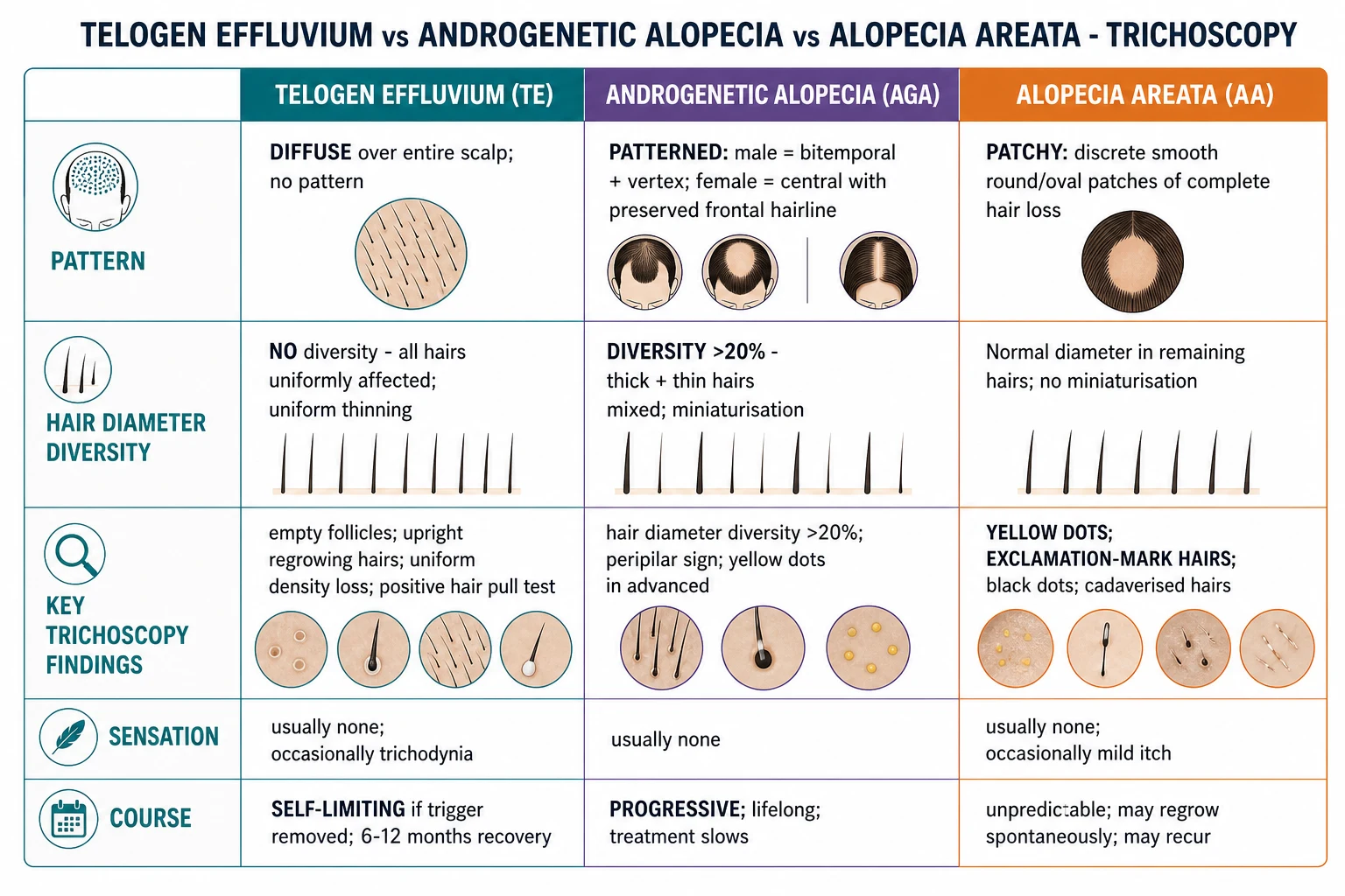
Trichoscopy (dermoscopy of the scalp at 10-70x) is the first-line investigation. It is non-invasive, performed in the clinic, and excludes the main mimics. The trichoscopic features of TE are: [1]
- Empty follicular openings (yellow dots become white dots as the follicular ostia are preserved but empty).
- Upright regrowing hairs — short, vertical, tapered hairs representing new anagen growth; a positive prognostic sign.
- Uniform reduction in hair density without focal bare patches.
- NO hair diameter diversity >20 percent — all terminal hairs remain of similar thickness; AGA has thick + thin (vellus) hairs mixed.
- NO exclamation-mark hairs (broken hairs narrower at the proximal end) — these indicate alopecia areata.
- NO yellow dots (which suggest alopecia areata) and NO black dots (broken hairs in trichotillomania or tinea capitis).
- Peri-pilar sign is usually absent (a brown halo around the follicle seen in AGA and FPHL).[2][3]
Standard first-line investigations
- Ferritin: target >70 microg/L in women with TE (some experts use 100 microg/L). Ferritin is an acute phase reactant; check CRP if the result is unexpectedly high.
- TSH and free T4: to detect overt and subclinical thyroid disease.
- CBC: to detect iron deficiency anaemia (low MCV, low MCH) and to screen for lymphopenia (lupus, lymphoma).
- Vitamin D (25-OH): target >30 ng/mL.
- Zinc: if deficiency is suspected (vegetarian diet, alcohol, malabsorption).
- CRP / ESR: if systemic inflammation is suspected.
- Fasting glucose or HbA1c: if PCOS, metabolic syndrome or crash dieting is suspected. [1]
Second-line investigations
- Hair pluck trichogram: a small bundle of hairs is plucked and examined under microscopy; more than 25 percent telogen hairs is suggestive of TE (normal less than 15 percent).
- Wash test (Rebora): 5-day no-shampoo period followed by a standardised shampoo through a gauze; >100 hairs collected is suggestive of TE.
- 4 mm punch biopsy from the active margin: only if there is diagnostic doubt, suspected scarring alopecia or persistent undiagnosed shedding. Histology shows a normal follicle density with an increased number of telogen follicles and occasional peri-follicular lymphocytes, but no interface change, no fibrosis and no destruction of the bulge.
- ANA, anti-dsDNA, RPR/VDRL: if there are systemic features (lupus, syphilis) or a moth-eaten hair-loss pattern. [1]
Differential Diagnosis
The differential of diffuse hair loss is the most-tested concept in any hair exam. The first branching point is diffuse vs patterned vs patchy; the second is scarring vs non-scarring; the third is reversible vs irreversible.[2][3][5]
[1]Anagen effluvium (the chemotherapy trap)
Anagen effluvium is often listed as a "type of TE" in textbooks but is mechanistically distinct. It is caused by abrupt cessation of anagen (e.g., by antimetabolite chemotherapy, heavy-metal poisoning, or radiation), and hairs are shed within days to 2-4 weeks of the insult (not 2-3 months). The shed hairs are dystrophic anagen hairs: broken, tapered, pigmented, with a pointed or "pencil-tip" proximal end, rather than a club-shaped telogen bulb. Examples: hair loss 1-3 weeks after starting doxorubicin, cyclophosphamide or vincristine. The hair regrows after chemotherapy ends, sometimes with a change in colour or texture.[1][2]
Chronic telogen effluvium versus early FPHL
CTE predominantly affects women aged 30-60, with a fluctuating course, increased daily shedding, and stable or only mildly reduced part width. Early FPHL shows progressive widening of the central part, with a measurable increase in vellus-like (miniaturised) hairs. The two conditions overlap frequently: serial photographs and trichoscopy showing progressive diameter diversity (an increase over time) suggest FPHL evolving out of CTE. The clinical relevance is that FPHL responds to anti-androgens (spironolactone, bicalutamide, finasteride) and to minoxidil, whereas pure CTE is managed by trigger correction and reassurance.[5][7]
Other mimics to exclude
The bedside differential of diffuse hair loss extends well beyond the three main mimics (TE, AGA, AA). A practical approach is to list the diagnosis being considered alongside TE, then apply a single discriminant test for each: [1]
- Diffuse alopecia areata: may mimic TE acutely; trichoscopy shows exclamation-mark hairs and yellow dots; biopsy is sometimes required. Acute loss of more than half of scalp hair with no regrowth within 6 months should raise suspicion of diffuse AA, especially in adolescents. Diffuse AA may be the presenting form of alopecia totalis or universalis.
- Trichotillomania and traction alopecia: history of pulling, braiding, weaves or chemical processing; irregular patches with broken hairs of varying lengths; trichoscopy shows trichoptilosis (split ends), V-sign, flame hairs and hair powder. Traction typically affects the marginal, frontal and temporal hairline; chronic traction produces a scarring pattern with loss of follicular ostia.
- Tinea capitis: Wood's lamp (Microsporum species fluoresce), KOH of broken hairs showing spores within the shaft, fungal culture. Common in school-age children; occipital lymphadenopathy is the clinical clue. Kerion (severe boggy mass) can cause permanent scarring if untreated.
- Secondary syphilis: "moth-eaten" patchy hair loss; positive RPR / VDRL; other stigmata (palmar-plantar rash, mucous patches, condylomata lata). Treat with benzathine penicillin.
- Systemic lupus erythematosus: may present with diffuse non-scarring TE before frank DLE; check ANA, anti-dsDNA, complements, urinalysis. Persistent TE with photosensitivity, arthralgia or cytopenias warrants SLE workup. A scalp biopsy may show hyperkeratotic follicular plugging and interface dermatitis at the dermo-epidermal junction.
- Drug-induced alopecia: careful drug history; retinoids, anticoagulants, beta-blockers, lithium, valproate, interferon, OCP withdrawal. Chemotherapy produces anagen effluvium (rapid, dystrophic hairs in days to weeks) rather than TE. Thallium, arsenic, selenium and mercury produce anagen effluvium with Mees lines.
- Endocrine: hypothyroidism (fatigue, cold intolerance, weight gain, constipation, bradycardia, dry skin) and hyperthyroidism (anxiety, weight loss, palpitations, heat intolerance); hypopituitarism (postpartum Sheehan syndrome with agalactia, amenorrhoea, hypothyroidism, adrenal insufficiency), hypervitaminosis A. Always check TSH and free T4.
- Nutritional deficiency states: iron, vitamin D, zinc, biotin, severe protein-energy malnutrition; consider in bariatric, post-bariatric, vegan and crash-diet populations. Kwashiorkor and marasmus produce flag-sign hair (alternating bands of colour) as well as diffuse TE.
- Loose anagen syndrome: children, predominantly girls; hairs can be pulled out painlessly without a telogen bulb; trichoscopy shows misshapen anagen bulbs with a "floppy sock" appearance. Distinguished from TE by the absence of a telogen bulb and the painless extraction.
- Pressure alopecia: localised to the occiput after prolonged supine pressure (surgical, ICU, comatose patients). Patchy, post-ischaemic; usually reversible within 4-6 months if pressure is relieved.
- Atypical presentations that mimic TE: frontal fibrosing alopecia in early stages can be predominantly non-scarring with perifollicular erythema; trichoscopy shows the lonely hair sign and early loss of follicular ostia. Central centrifugal cicatricial alopecia in women of African ancestry can present as a diffuse thinning of the vertex before scarring is obvious. Both are easily missed as "TE" without careful trichoscopy. [1]
Management
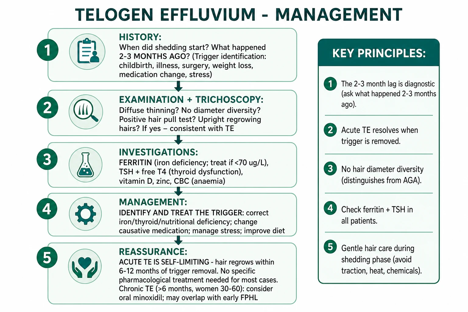
Management of TE is built on three pillars: identify the trigger, correct the trigger, and reassure the patient. Acute TE is self-limiting; chronic TE may need additional treatment.[1][2][3]
Step 1 — Identify and treat the trigger
- Correct iron deficiency: oral ferrous sulfate 200 mg daily (or alternate-day dosing to improve absorption) if ferritin is under 70 microg/L; re-check at 3 months. Vitamin C co-administration and avoidance of tea/coffee around dosing improve absorption. IV iron may be needed if oral is not tolerated or if there is malabsorption.
- Normalise thyroid function: levothyroxine for hypothyroidism; antithyroid drugs for hyperthyroidism; re-check at 6-8 weeks.
- Review medications: if a likely culprit (retinoid, anticoagulant, beta-blocker, lithium, valproate, interferon) is identified, discuss alternatives with the prescribing team. Do NOT stop essential drugs (e.g., anticoagulants) without consulting haematology / cardiology.
- Optimise nutrition: correct vitamin D deficiency with cholecalciferol, zinc supplementation if low, protein-energy repletion after bariatric surgery, avoidance of further crash diets.
- Address stress and sleep: psychological support, sleep hygiene, mindfulness-based interventions; data are limited but clinical experience supports this.
- Manage underlying disease: treat scalp psoriasis, seborrhoeic dermatitis, eczema; control SLE, IBD, hyperthyroidism. [1]
Step 2 — Reassure and educate
- Explain the 2-3 month lag so the patient understands that the trigger is already past.
- Show the patient the trichoscopy images to demonstrate that follicles are intact and regrowing (upright hairs).
- Provide written information about self-limiting course and the 6-12 month regrowth timeline.
- Validate the psychological impact; consider psychology or peer-support referral if QoL is severely affected. [1]
Step 3 — Pharmacological treatment (selected cases)
- No specific treatment is needed for most acute TE. Once the trigger is corrected, the hair regrows.
- Topical minoxidil 2 percent or 5 percent — may be added in chronic TE or when FPHL overlap is suspected. The rationale is to prolong anagen and increase hair diameter, but the evidence base in pure TE is limited. Counsel about the initial 2-6 week "shedding" that can occur when starting minoxidil and that this is reassuring (synchronised transition to anagen).[9]
- Oral minoxidil 0.25-1.25 mg daily — increasingly used in chronic TE and FPHL overlap; effective in a retrospective series of CTE at doses of 0.25-1.25 mg/day. Side-effects: hypertrichosis (15-20 percent), transient fluid retention, occasional hypotension, rare pericardial effusion. Strict pregnancy prevention is mandatory in women of childbearing age.[8]
- Nutritional supplementation — iron, zinc, vitamin D, biotin and marine protein supplements should be prescribed only if a deficiency is documented. Routine supplementation in replete patients is not supported by evidence.
- Avoid in pure TE: finasteride, dutasteride, systemic steroids, PRP, microneedling, low-level laser — these target AGA / FPHL and have no role in pure TE.
Step 4 — Supportive measures and gentle hair care
During the shedding phase the scalp is structurally normal but psychologically vulnerable. Conservative measures reduce secondary injury, support regrowth and improve patient comfort. [1]
- Avoid traction and friction: no tight ponytails, braids, weaves, buns, hair extensions, headbands, helmet straps or hair clips that pull on the frontal and temporal margins. Sleeping on a silk or satin pillowcase reduces friction.
- Avoid thermal and chemical injury: no high-heat straighteners, curling irons or blow-dryers above 60 degrees C; no bleaching, perming, chemical relaxing or colouring for at least 3-6 months after the trigger has resolved. When colouring is unavoidable, use semi-permanent, ammonia-free products and extend the interval between sessions.
- Use a gentle shampoo and conditioner: sulphate-free, fragrance-free shampoos with a near-neutral pH; silicone-free or mild conditioner on the mid-shaft and ends; wash every 2-3 days. Daily washing is acceptable if a gentle shampoo is used.
- Use a wide-tooth comb or soft brush: detangle gently from ends to roots; do not pull through knots. Wet hair is more fragile; comb before washing or after applying conditioner.
- Photographic documentation: standardised global photographs and part-width photos at each visit (frontal, vertex, temporal, occipital) allow objective assessment of progression and response to treatment. Patients often perceive continued shedding as worsening; serial photographs reverse this misperception.
- Cosmetic camouflage: fibre sprays, root-concealing powders, hair-thickening conditioners and styling techniques (blow-dry upside down, side part) can provide immediate cosmetic benefit. Wigs, hairpieces and scalp micropigmentation are options for severe cases.
- Activity and lifestyle: regular aerobic exercise supports hair growth through improved microcirculation and stress reduction; smoking cessation is essential because smoking accelerates follicular miniaturisation and impairs wound healing.
- Diet: an adequate-protein, iron-rich, varied diet with adequate calories; avoid further crash diets; consider referral to a registered dietitian for post-bariatric or restrictive-eating patients.
- Sleep and stress: aim for 7-9 hours of sleep; mindfulness, CBT, yoga and similar interventions have modest evidence for reducing perceived shedding and improving coping.
- Realistic expectations: hair grows approximately 1 cm per month, so even after the trigger is removed the visible recovery to the patient's previous length takes 6-12 months. Reassure the patient that the follicles are intact and that the regrowth will come. [1]
Quality of Life & Psychological Impact
Hair loss is one of the most psychologically distressing dermatological symptoms, with quality-of-life impact comparable to psoriasis or eczema. Even self-limiting TE generates significant anxiety, depressive symptoms, social withdrawal, body-image disturbance and reduced self-esteem, particularly in women. Studies have shown that around 40-60 percent of women with TE report moderate-to-severe distress, and approximately 10-20 percent screen positive for clinical anxiety or depression that warrants referral. [1]
The clinical implication is that psychological assessment and support are part of good TE care, not optional extras. Validated tools such as the Dermatology Life Quality Index (DLQI), Hairdex and the Sinclair hair shedding visual scale can be used to track impact over time. Practical steps include: [1]
- Acknowledge the impact explicitly at the first consultation ("This is real and it's affecting you; let's work on it together").
- Provide written information, a clear timeline and a contact for questions.
- Offer referral for psychological support, CBT or peer support groups when DLQI is high or distress is moderate-severe.
- Screen for disordered eating in patients who crash-dieted and developed TE.
- Address body-image concerns in adolescents and in women from cultures where hair is a strong symbol of femininity and identity.
- Reassess QoL at follow-up; chronic TE in particular requires ongoing psychological support, not just pharmacotherapy. [1]
When to Refer to Dermatology
Most patients with TE can be managed in primary care or by the generalist. The threshold for dermatology referral is determined by diagnostic uncertainty, suspicion of scarring, treatment failure, or psychological morbidity. Specific indications include: [1]
- Diagnostic doubt: trichoscopy does not confirm TE, or features of AGA, AA or cicatricial alopecia are seen.
- Suspected scarring: loss of follicular ostia, perifollicular erythema, scale, pustules or tufted hairs.
- Biopsy needed: persistent diagnostic uncertainty, suspicion of diffuse AA, lupus or secondary syphilis.
- Treatment failure: no improvement after 6-12 months of trigger correction and supportive care, or progressive worsening.
- Chronic TE: for consideration of minoxidil (especially oral), anti-androgen therapy in women with FPHL overlap, or clinical trial enrolment.
- Severe psychological morbidity: DLQI high, depression, anxiety, social withdrawal, suicidal ideation.
- Paediatric or pregnancy cases with diagnostic uncertainty.
- Systemic features suggesting SLE, lymphoma, syphilis or other systemic disease.
- Refractory postpartum TE unmasking FPHL or other latent alopecia.[10]
Special Scenarios
Postpartum TE
The most common acute TE in women. During pregnancy, high oestrogen prolongs anagen, producing thick, lush hair. After delivery, oestrogen falls and a large cohort of anagen hairs is released into telogen. Shedding typically begins 6-12 weeks postpartum, peaks at 3-4 months, and resolves by 6-12 months postpartum. The mechanism is Headington's "delayed anagen release" — a paradoxical effect of the very state that produced thick hair during pregnancy. Postpartum TE is occasionally the event that unmasks underlying FPHL in genetically susceptible women, so follow-up beyond 12 months is sensible in those whose shedding does not fully resolve.[10] Breastfeeding does not worsen TE; iron deficiency from menstrual resumption and pregnancy-related depletion is the main correctable factor.
Post-COVID-19 TE
Severe acute respiratory syndrome coronavirus 2 (SARS-CoV-2) infection is now a well-recognised trigger for TE. The mechanism combines immediate anagen release from the cytokine storm, telogen release from severe systemic illness and nutritional insult, and possibly microvascular / microthrombic effects on the follicular vasculature. Shedding begins 6-12 weeks after the acute infection and is usually self-limiting within 6 months. Post-vaccination TE is also reported, with a similar time course and a milder course. The dominant message is reassurance: full regrowth is expected in the vast majority of patients.[11]
Drug-induced TE
Common offenders are retinoids (isotretinoin, acitretin — usually anagen effluvium with dystrophic hairs), anticoagulants (heparin, warfarin, DOACs), beta-blockers, lithium, valproate, interferon-alpha, all-trans retinoic acid, propylthiouracil, and OCP withdrawal (the pill-induced prolonged anagen is released when the pill is stopped, mimicking postpartum TE). Management is drug review with the prescribing team; if the drug is essential, supportive management and trigger correction (ferritin, vitamin D) usually suffice. Heparin-induced TE is dramatic and often missed. [1]
Nutritional TE
Iron deficiency is the most common nutritional cause. The exact ferritin threshold for intervention is debated (40 vs 70 vs 100 microg/L), but most hair specialists use 70 microg/L as a practical target in symptomatic TE. Vitamin D deficiency (25-OH below 20 ng/mL) is increasingly recognised as a cofactor; cholecalciferol replacement is widely used despite limited RCT data. Zinc deficiency (vegetarian diet, malabsorption, alcohol excess) responds to zinc sulfate 220 mg once daily. Biotin deficiency is rare but should be considered in patients on long-term antiepileptics, in pregnancy, and in inherited biotinidase deficiency. Protein-energy malnutrition (after bariatric surgery, anorexia, severe illness) is a major trigger and may need nutritional rehabilitation in addition to supplementation. [1]
TE in children
Acute TE in children is uncommon and should always prompt a search for an underlying cause: febrile illness, drug (especially isotretinoin in adolescents for acne), nutritional deficiency (iron, zinc, vitamin D, especially in vegetarian or vegan diets), severe stress, and endocrine disease (hypothyroidism, hypoparathyroidism). Trichotillomania is a key mimic. Tinea capitis (with or without kerion) is the most important alternative diagnosis in school-age children; fungal culture is essential. [1]
TE in patients with skin of colour
Trichoscopy is the most reliable diagnostic tool, but the inflammatory pigment changes (peri-pilar hyperpigmentation in darkly pigmented skin, hypopigmentation in resolving TE) can be misinterpreted as scarring. Hair-care practices — chemical relaxers, hot combs, tight braiding, weaves — can superimpose traction alopecia on TE, and a careful, culturally sensitive history of grooming is essential. Treatment is identical in principle, with emphasis on discontinuation of the offending grooming practice.[2]
Complications & Prognosis
The dominant "complication" of TE is the psychosocial impact. Hair is central to identity in most cultures, and even short-lived TE can trigger anxiety, depression, social withdrawal, disordered eating (in patients prone to restrictive dieting), and relationship difficulties. Early acknowledgement of the impact and referral for psychological support are part of good care, not a luxury. [1]
Physical complications are rare. Severe prolonged TE has been associated with secondary trichotillomania (a compulsive response to the visible shedding), with traction alopecia from compensatory tight hairstyles, and with chemical injury from repeated at-home treatments. There is no risk of baldness from TE alone — the follicles are preserved and re-enter anagen once the trigger is removed. [1]
The prognosis of acute TE is excellent: full regrowth to pre-trigger density within 6-12 months in the vast majority. The prognosis of chronic TE is more variable; many patients fluctuate for years, with partial recovery and a tendency to overlap with FPHL over time. Predictors of poor prognosis include: [1]
- Persistent, unidentifiable trigger.
- Concurrent early FPHL.
- Co-existing iron deficiency that does not correct.
- Polypharmacy with multiple TE-inducing drugs.
- Severe psychosocial comorbidity.
- Delayed presentation (often because of missed trigger). [1]
Follow-up is at 3-6 months for acute TE and at 6-12 monthly for chronic TE. Re-referral to dermatology is indicated if there is any suspicion of scarring, persistent diagnostic doubt, or development of patterned loss suggesting FPHL. [1]
Exam Pearls
[1]Red Flags
Exam application bank (NEET-PG / INICET)
One-line answer
Telogen effluvium (TE) is a non-scarring, diffuse hair loss caused by a premature shift of anagen follicles into the telogen (resting) phase, producing synchronous shedding 2-3 months after a triggering event. Headington classified five functional types (immediate anagen release, delayed anagen release, short anagen, immediate telogen release, delayed telogen release) and the Whiting entity of chronic telogen effluvium (CTE) is a distinct middle-aged female phenotype that fluctuates for years. Common triggers include childbirth, severe illness or fever (including COVID-19), iron deficiency, thyroid dysfunction, crash dieting or bariatric surgery, retinoids, anticoagulants, beta-blockers, lithium, valproate, interferon and severe emotional stress. The hallmark bedside signs are diffuse thinning (not patterned, not patchy), a positive hair pull test (>5-6 of 50-60 hairs), trichoscopic empt
Worked stems (answer without another resource)
Stem 1 — Classic presentation. Map symptoms to mechanism; name the first investigation and first treatment step with dose/route if drug therapy is standard. [1]
Stem 2 — Unstable / complicated. List red flags that force immediate resuscitation, theatre, ICU, antidote, or reperfusion — and what you do in the first 15 minutes. [1]
Stem 3 — Atypical group. Elderly, pregnancy, child, or immunocompromised: how presentation and thresholds change. [1]
Stem 4 — Differential trap. Name the three closest mimics and one discriminator for each. [1]
Stem 5 — Disposition. Who goes home with safety-netting, who is admitted, who needs HDU/ICU/theatre, and what follow-up is mandatory. [1]
Rapid viva checklist
- Definition + classification
- Pathophysiology chain
- Bedside signs / criteria
- Score with exact components (if any)
- Emergency bundle
- Definitive therapy with doses
- Complications of disease and of treatment
- Special populations
- Guideline/trial name if classic
- Three exam traps
Coverage self-check
If you cannot answer any stem above from this page alone, re-read the matching section — the page is intended to be self-sufficient for final-prof and NEET-PG/INICET questions on Telogen effluvium.
[1]References
- [1]Asghar F, Shamim N, Farooque U, Sheikh H, Aqeel R. Telogen Effluvium: A Review of the Literature Cureus, 2020.PMID 32607303
- [2]Rebora A. Telogen effluvium: a comprehensive review Clin Cosmet Investig Dermatol, 2019.PMID 31686886
- [3]Chien Yin GO, Siong-See JL, Wang ECE. Telogen Effluvium - a review of the science and current obstacles J Dermatol Sci, 2021.PMID 33541773
- [4]Headington JT. Telogen effluvium. New concepts and review Arch Dermatol, 1993.PMID 8447677
- [5]Whiting DA. Chronic telogen effluvium: increased scalp hair shedding in middle-aged women J Am Acad Dermatol, 1996.PMID 8959948
- [6]Rebora A. Proposing a Simpler Classification of Telogen Effluvium Skin Appendage Disord, 2016.PMID 27843920
- [7]Upton M, Tosti A. Chronic Telogen Effluvium: Is it a Distinct Condition? A Systematic Review Am J Clin Dermatol, 2023.PMID 37052778
- [8]Randolph M, Tosti A. Oral minoxidil treatment for hair loss: A review of efficacy and safety J Am Acad Dermatol, 2021.PMID 32622136
- [9]Gupta AK, Talukder M, Venkataraman M, Bamimore MA. Minoxidil: a comprehensive review J Dermatolog Treat, 2022.PMID 34159872
- [10]Cohen S, Krajden S, Glick BP, Ko J. Postpartum Telogen Effluvium Unmasking Additional Latent Hair Loss Disorders J Clin Aesthet Dermatol, 2024.PMID 38779373
- [11]Bowe S, Bui D, Trovato F, et al. SARS-CoV-2 Infection and COVID-19 Vaccination Associated with Post-Acute Alopecia: Prevalence, Clinical Patterns, and Determinants Among Saudi Adults Viruses, 2026.PMID 42357623